báo cáo hóa học:" Implantation of neural stem cells embedded in hyaluronic acid and collagen composite conduit promotes regeneration in a rabbit facial nerve injury model" doc
Bạn đang xem bản rút gọn của tài liệu. Xem và tải ngay bản đầy đủ của tài liệu tại đây (766.55 KB, 11 trang )
BioMed Central
Page 1 of 11
(page number not for citation purposes)
Journal of Translational Medicine
Open Access
Research
Implantation of neural stem cells embedded in hyaluronic acid and
collagen composite conduit promotes regeneration in a rabbit facial
nerve injury model
Han Zhang
1
, Yue Teng Wei
2
, Kam Sze Tsang
3,4
, Chong Ran Sun
1,5
, Jin Li
1,3,4
,
Hua Huang
1
, Fu Zhai Cui
2
and Yi Hua An*
1
Address:
1
Beijing Neurosurgical Institute, Capital Medical University, Beijing, PR China,
2
Department of Materials Science and Engineering,
Tsinghua University, Beijing, PR China,
3
Department of Anatomical and Cellular Pathology, Chinese University of Hong Kong, Hong Kong, PR
China,
4
Li Ka Shing Institute of Health Sciences, Chinese University of Hong Kong, Hong Kong, PR China and
5
Department of Neurosurgery,
Second Affiliated Hospital of Zhejiang University Medical College, Hangzhou, PR China
Email: Han Zhang - ; Yue Teng Wei - ; Kam Sze Tsang - ;
Chong Ran Sun - ; Jin Li - ; Hua Huang - ;
Fu Zhai Cui - ; Yi Hua An* -
* Corresponding author
Abstract
The implantation of neural stem cells (NSCs) in artificial scaffolds for peripheral nerve injuries
draws much attention. NSCs were ex-vivo expanded in hyaluronic acid (HA)-collagen composite
with neurotrophin-3, and BrdU-labeled NSCs conduit was implanted onto the ends of the
transected facial nerve of rabbits. Electromyography demonstrated a progressive decrease of
current threshold and increase of voltage amplitude in de-innervated rabbits after implantation for
one, four, eight and 12 weeks compared to readouts derived from animals prior to nerve
transection. The most remarkable improvement, observed using Electrophysiology, was of de-
innervated rabbits implanted with NSCs conduit as opposed to de-innervated counterparts with
and without the implantation of HA-collagen, NSCs and HA-collagen, and HA-collagen and
neurotrophin-3. Histological examination displayed no nerve fiber in tissue sections of de-
innervated rabbits. The arrangement and S-100 immunoreactivity of nerve fibers in the tissue
sections of normal rabbits and injured rabbits after implantation of NSCs scaffold for 12 weeks
were similar, whereas disorderly arranged minifascicles of various sizes were noted in the other
three arms. BrdU
+
cells were detected at 12 weeks post-implantation. Data suggested that NSCs
embedded in HA-collagen biomaterial could facilitate re-innervations of damaged facial nerve and
the artificial conduit of NSCs might offer a potential treatment modality to peripheral nerve
injuries.
Background
With the advent of surgical techniques and instruments,
micro-sutures have considerably improved the manage-
ment of peripheral nerve injuries. Autograft of the epineu-
rium of an intact nerve remains to be the gold standard to
bridge a nerve gap defect for the peripheral nerve lesion
[23]. However, there are some limitations of the autolo-
gous nerve grafting technique including the limited
Published: 5 November 2008
Journal of Translational Medicine 2008, 6:67 doi:10.1186/1479-5876-6-67
Received: 18 May 2008
Accepted: 5 November 2008
This article is available from: />© 2008 Zhang et al; licensee BioMed Central Ltd.
This is an Open Access article distributed under the terms of the Creative Commons Attribution License ( />),
which permits unrestricted use, distribution, and reproduction in any medium, provided the original work is properly cited.
Journal of Translational Medicine 2008, 6:67 />Page 2 of 11
(page number not for citation purposes)
number of donor nerves available, unaesthetic scaring,
wound infection, wound pain, relatively long surgical
time and learning curve for the success of nerve grafts, and
poor regeneration. Controversial results were also
reported on multiple anastomoses and acellular muscle
grafts for cable grafting of large nerve defects [6,7,10].
Recent pre-clinical and clinical studies showed that allo-
graft could be an alternative nerve graft [2,7,21]. Nerve
allograft may act as a temporary scaffold across which host
axons regenerate.
Natural or synthetic nerve guides were being developed
and employed as alternatives to autografts in bridging
nerve gap defects [9,22,24]. It was suggested that these
scaffolds help direct axonal sprouting from the injured
nerve and provide a conduit for diffusion of neurotrophic
and neuroprotective factors produced by the lesioned
nerve stumps [14]. An ideal scaffold should be biodegrad-
able, biocompatible, non-toxic and mediate no immune
response. In general, these biomaterials yielded poor
results in the regeneration process of peripheral nerve
injury [9,22]. Severe scarring and fibrosis are the most fre-
quent problems.
Hyaluronic acid (HA) and collagen are ubiquitous and are
major components of extracellular matrix (ECM) in the
mammalian body. HA has a high capacity for holding
water and possesses a high visco-elasticity. It adheres
poorly to cells and prevents scarring. HA was noted to
elicit positive biological effects on cells ex-vivo. Collagen is
the main structural protein of connective tissues, and has
great tensile strength and elasticity, and is employed in the
construction of artificial skin substitutes. Components of
ECM in tissue engineering have been actively studied. HA-
collagen composite scaffolds were widely investigated
recently for possible use as a biomaterial in tissue engi-
neering scaffolds [26].
Stem cells are unspecified cells that can replicate, and
under specific conditions, differentiate into various spe-
cialized cell types. NSCs transplantation was noted to pro-
mote functional recovery in animal models [4,15,17]. A
recent study showed that in vitro culture of NSCs in three-
dimensional HA-collagen matrix enhanced the differenti-
ation of NSCs into neurons, astrocytes and oligodendro-
cytes [3]. However, the combinatorial effects of NSCs and
HA-collagen composite scaffold in peripheral nerve repair
are largely unclear. In this study, we made use of HA-col-
lagen composite scaffold, NSCs and NT-3 as a nerve guide,
effecter cells and neurotrophic/neuroprotective factor,
respectively, and implanted the conduit of NSCs-
implanted NT-3-supplemetned HA-collagen composite
scaffold onto rabbits having induced peripheral nerve gap
defect and evaluated the therapeutic effects on peripheral
nerve lesion.
Materials and methods
Preparation of HA-Collagen composite conduit
Fresh solutions of 1% HA (Freda Biochemicals, Shan-
dong, China) and 1% collagen (Sigma-Aldrich, St. Louis,
MO) were mixed for six hours and were injected into the
collagen conduit (Institute of Medical Equipment, Acad-
emy of Military Medical Sciences, China) which was tied
at one end. The assembly was immersed in a solution con-
taining the cross-linker, 1-ethyl-3-dimethylamino carbod-
iimide (EDC; Sigma-Aldrich) in 95% ethanol for 12 hours
at 4°C. The cross-linked conduit was washed thrice in de-
ionized water and freeze-dried at -20°C. The cross-linked
matrices were then morphologically examined using scan-
ning electron microscopy (JSM-6460LV) at 10 kV before
and after release to down-streamed analyses.
Cultures of NSCs
NSCs harvested from the neural cortex of E16 Sprague-
Dawley rat embryos. For each rat, the head was decapi-
tated and the whole brain was removed from the skull.
Meninges, choroid plexus and coherent blood vessels
were carefully stripped off. The brain tissue was cut into
small pieces, triturated with a glass pipette and allowed to
pass through a 28-mesh copper sieve to get rid of large
chunks. Having washed thrice with Dulbecco's modified
Eagle's medium (DMEM; Sigma-Aldrich), cells were
seeded in 12 ml of high-glucose DMEM/F12 (Sigma-
Aldrich) supplemented with 12.5 ng/ml basic fibroblast
growth factor (FGF; Sigma-Aldrich) and 20 ng/ml epider-
mal growth factor (EGF; Sigma-Aldrich) onto a 75 cm
2
non-adherent tissue culture flask (Corning BV Life Sci-
ences, Schiphol-Rijk, Netherlands) and maintained at
37°C in a humidified 5% CO
2
-incubator. Half of the
spent medium was discarded and replenished with fresh
culture medium every three days. Neurosphere cultures
were passaged once a week by enzymatic segregation,
using 0.25% trypsin and triturating with a glass pipette,
and sub-cultured.
Characterization of NSCs
Trypsinized cells with and without 10 μM bromodeoxyu-
ridine (BrdU; Roche, Basel, Switzerland) labeling were
allowed to grow on poly-L-ornithine- (Sigma) and lam-
inin- (Sigma) coated coverslips. They were fixed in 4%
paraformaldehyde (Sigma) for 20 minutes. Cells were per-
meabilized for five minutes with 0.3% Triton X-100
(Sigma) in phosphate-buffered saline (PBS) and then
rinsed thrice with PBS. Non-specific binding was blocked
with 10% normal goat serum (NGS; Zhongshanjinqiao,
China) in PBS for 10 minutes. Cells were washed with 1%
NGS in PBS and incubated overnight at 4°C with the fol-
lowing primary antibodies diluted in PBS containing 1%
NGS: mouse IgG1 anti-class III β-tubulin (TuJ-III, 1:1,000;
Exbio, Prahy, Czech), mouse IgG1 anti-glial fibrillary
acidic protein (GFAP; 1:50; Santa Cruz Biotechnology,
Journal of Translational Medicine 2008, 6:67 />Page 3 of 11
(page number not for citation purposes)
Santa Cruz, CA), and rabbit polyclonal IgG anti-galac-
tocerebroside (GalC, 1:100; Santa Cruz). Labeled cells
were detected with mouse IgG1 anti-BrdU (1:100; Milli-
pore, Billerica, MA). After thrice washes with PBS, cells
were incubated for 30 minutes with the corresponding
secondary antibody: TRITC-conjugated goat anti-mouse
IgG (1:100; Invitrogen, Carlsbad, CA), FITC-conjugated
goat anti-mouse IgG (1:100; Santa Cruz) or FITC-conju-
gated goat anti-rabbit antibody (1:100; Santa Cruz).
Washed cells without BrdU labeling were counter-stained
with, either propidium iodide (PI; Sigma) or Hoechst
33342 (Invitrogen), and visualized using an inverted flu-
orescence microscope. Cells without primary antibody
incubation were processed in the same manner as controls
of false-positivity.
Preparation of NSC for transplant
Neurospheres at passage three were labelled with 10 μM
BrdU in the supplemented culture medium a day prior to
nerve fiber transection to rabbits for in vivo study. BrdU-
labeled cells were then trypsinized and washed thrice with
PBS. Discrete NSCs were adjusted to 4 × 10
6
/ml in
DMEM/F12 supplemented with 10 ng/ml neurotrophin-3
(NT-3; Sigma) for embedding to HA-collagen composite
conduit.
Embedding NSCs to HA-collagen conduit
Freeze-dried HA-collagen conduits were decontaminated
by exposure to ultraviolet irradiation for an hour. NSCs (4
× 106) in one millilitre NT-3-supplemented DMEM/F12
culture medium were injected into the HA-collagen com-
posite conduit of 7 mm in length. The NSC-embedded
HA-collagen composite scaffold was then dipped into
DMEM/F12 culture medium and incubated in 5% CO2-
incubator at 37°C for two to three days.
Induction of facial nerve injury and reconstruction to
rabbits
Animal treatments were carried out to minimize pain or
discomfort in accordance with the current protocols
approved by the Institutional Animal Research Ethics
Committee. A cohort of 39 normal adult New Zealand
rabbits of 2.0 – 2.5 kg body weight was recruited for the
study. They were allowed to gain access to food and water
ad libitum in isolator cages at 25°C under a 12-hour light-
dark cycle. Animals were randomly assigned into six
groups: normal control (n = 5); bilateral facial nerve
transected without reconstruction (n = 2); lateral nerve
transected with implantation of HA-collagen composite
scaffold (n = 7); lateral nerve transected with implanta-
tion of NSC and HA-collagen scaffold (n = 8); lateral
nerve transected with implantation NT-3-supplemented
HA-collagen scaffold (n = 6) and lateral nerve transected
with implantation of NSC-embedded NT-3-supple-
mented HA-collagen composite scaffold (n = 11).
Having anesthetized by intravenous injection of 39 mg/kg
sodium phenobarbital, rabbits were operated in a sterile
condition. A horizontal incision was made to expose the
main stem of the facial nerve. A segment of 2 mm was
removed. A nerve gap defect of approximately 5 mm was
apparent after contraction. A conduit of 7 mm in length
was implanted onto the defect. Both nerve ends were
sutured to the epineurium of the facial nerve using 10-0
nylon stitch. The skin incision was sutured. Animals were
reared in isolator cages without any immunosuppressive
prophylaxis.
Behavioural assessment
1. Ethology
Ethological methods were used to observe, record, and
analyze animal behaviour in terms of signs and extents of
muscular atrophy of lips, blink reflex, and ear motion of
animals before and 12 weeks after peripheral nerve
transection.
2. Electromyography
Physiologic properties of lip muscles at rest and while
contracting were evaluated and recorded using an electro-
myograph (Nicolet Viking IV, Portsmouth, VA). Electro-
myography in terms of time-latency, current threshold
and voltage amplitude to a stimulus was performed on
animals before and one, four, eight and 12 weeks after
peripheral nerve transaction to assess the neuromuscular
function. Pre-operated parameters were reckoned to be
the reference values.
Tissue processing for light and electron microscopy
Upon completion of in vivo monitoring, rabbits were
anesthetized using sodium phenobarbital and eutha-
nized. Blocks of facial muscles were fixed for three days in
4% paraformaldehyde and embedded in paraffin. Sec-
tions were de-waxed and stained with haematoxylin and
eosin for histological examination. Toluidine blue stain-
ing was performed to assess regeneration [18]. Morpho-
metric analyses were conducted to enumerate the fiber
number, myelin sheath thickness, axon area and nerve
fiber circumference using the Leica image analysis system
(Leica Image Analyzer, Wetzlar, Germany).
Blocks of facial muscles and HA-collagen composite scaf-
folds were fixed with the modified Karnovsky's fixative
containing 2% paraformaldehye (Sigma) and 2% glutar-
aldehyde (Sigma) in 0.1 M phosphate buffer for an hour
and 1% osmium tetroxide (Sigma) in 0.1 M phosphate
buffer for an hour. After rinsing with PBS for 15 minutes,
specimens were dehydrated in a series of up-graded etha-
nol (70% to absolute) and further dried using hexameth-
yldisilazane (Sigma). Ultrathin sections were stained with
uranyl acetate and lead citrate and mounted on alumi-
num stubs for electron microscopy.
Journal of Translational Medicine 2008, 6:67 />Page 4 of 11
(page number not for citation purposes)
Immunohistochemistry
Immunohistochemical staining of BrdU and S-100 was
performed to track the homing of NSC and to mark nerve
fibers in the facial muscles of injured rabbits with and
without reconstruction. Paraffin-embedded muscle sec-
tions of 5 μm in thickness were de-waxed and treated with
1 M hydrochloric acid to retrieve antigen of tissue sections
that were masked by fixation. Endogenous peroxidase in
muscle sections was denatured using 3% hydrogen perox-
ide. Upon completion of thrice washing in 0.01 M PBS for
five minutes, sections were blocked with 5% normal goat
serum in PBS for 30 minutes to suppress non-specific
binding.
Primary streptavidin-conjugated antibodies, anti-BrdU
(1:500; Sigma) and anti-S-100 (1:500; Sigma), were
employed. Incubation was conducted at 37°C for 72
hours. After three washes in PBS, sections were incubated
in biotin (1:300; Sigma) at room temperature for two
hours. Sections were washed thrice with PBS and incu-
bated with horseradish peroxidase-conjugated anti-biotin
(1:300, Sigma) for three hours. Diaminobenzidine tet-
rahydrochloride substrate solution (Zhongshanjinqiao,
China) was added for color development after three
washes in PBS. Having been rinsed in gently running tap
water, sections were counterstained with haematoxylin,
dehydrated, cleared and mounted for visualization.
Statistics analysis
Means and standard error of the mean (SEM) were calcu-
lated. The one-way analysis of variance (ANOVA) was
applied to analyze continuous variables: time latency,
threshold and amplitude of electromyogram and number,
thickness, circumference and area of myelinated nerve fib-
ers derived from rabbits with and without facial nerve
injury and repair using NSC-embedded NT-3-supple-
mented HA-collagen composite scaffold to bridge the
nerve gap. Differences between groups were regarded as
significant if p ≤ 0.05.
Results
NSCs characterization
Cells, which were derived from neurospheres and were
allowed to grow on poly-L-ornithine- (Sigma) and lam-
inin- (Sigma) coated coverslips, displayed a microglial
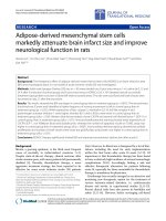
morphology with protruding processes. Immunofluores-
cence staining of β-tubulin, GFAP and GalC demonstrated
positive expressions in a substantial number of cells, sug-
gesting that neurosphere-derived cells were able to differ-
entiate into neuronal, astrocytic and oligodendrocytic
progenies (Figure 1).
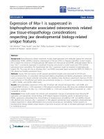
NSCs growth on HA-collagen scaffold
NSCs injected into NT-3-supplemented HA-collagen con-
duits were noted to adhere to the scaffolds and tended to
differentiate, after being cultured for 24 and 48 hours,
respectively (Figure 2). Protruding processes and neurite
outgrowth were evident, compared to that of floating neu-
rospheres.
Recovery of animals
Recovery of the animals includes 2 index: ethology which
is a measurement of animals' behaviour and electromyog-
raphy which is used to measure neuromuscular function.
According to the 2 index, the trend of recovery for the var-
ious groups is different.
1. Ethology
All rabbits were noted to have normal lip muscles, blink
reflex and ear motion prior to the facial nerve transaction.
On 12 weeks post-surgery, rabbits with facial nerve
transection displayed a muscular atrophy of upper lip and
no erection and movement of the ear. Besides, there was
no blink reflex. Injured rabbits implanted with HA-colla-
gen scaffold (n = 7), or NSC and HA-collagen scaffold (n
= 8), or NT-3-supplemented HA-collagen scaffold (n = 6),
presented atrophic muscles of upper lip, torpid blink
reflex and ear palsy. Injured rabbits with implantation of
NSC-embedded NT-3-supplemented HA-collagen com-
posite scaffold (n = 11) demonstrated slight blink reflex
and ear movement but no erection. Muscular atrophy of
upper lip was evident.
2. Electromyography
Electromyography is a measurement of neuromuscular
function. The prolongation of time-latency, increase of
current threshold and decrease of voltage amplitude to
stimuli may be attributed to an impairment of neuromus-
cular function after injury. When injury is recovering,
shrink of time-latency and threshold, increase of ampli-
tude is proposed to be observed. The mean ± SEM time
latency of the study cohort of 39 rabbits before micro-sur-
gery was 1.67 ± 0.30 ms, comparable to 1.68 ± 0.16 ms
shortly after micro-surgery. Additional file 1 shows the
time latency of rabbits with and without nerve fiber defect
and scaffold implant at different time points. Minimal
currents to elicit a visually detectable response in the ani-
mal cohort were depicted in Additional file 2. An increase
of current threshold to stimulate nerves was evident. Rab-
bits which had facial nerve fiber defect, with and without
scaffold implantation, exhibited higher current thresholds
over 12 weeks of monitoring, compared to those derived
from the normal counterparts. Thresholds shot up to the
apexes on week four post-surgery, which were signifi-
cantly higher than those derived from the normal control
animals (p < 0.05), and declined gradually over 12 weeks.
Rabbits which were untreated for facial nerve defect expe-
rienced the highest thresholds over 12 weeks post injury
among their counterparts having implanted with different
scaffolds. Readouts were correlated to the ethological
Journal of Translational Medicine 2008, 6:67 />Page 5 of 11
(page number not for citation purposes)
assessments of animals displayed neuromuscular defects
attributed to the atrophy of the upper lip and impairment
of erection and movement of ipsilateral ears.
Current thresholds derived from rabbits implanted with
HA-collagen scaffold (n = 7), NSC and HA-collagen scaf-
fold (n = 8), and NT-3-supplemented HA-collagen scaf-
fold (n = 6) over 12 weeks were comparable. On week 12,
thresholds were still significantly higher than those before
transection. In the arm of rabbits having implanted with
NSC-embedded NT-3-supplemented HA-collagen com-
posite scaffold for nerve fiber transection, the mean
threshold on week eight was significantly less than that on
week one (6.06 mA vs. 7.08 mA), though statistically
higher than that derived from the normal controls (6.06
mA vs. 3.41 mA). On week 12, the mean threshold was
comparable to that derived from the normal controls
(4.11 mA vs. 3.41 mA). In concordance with the etholog-
ical observation, the animals were noted to have slight
blink reflex and ear movement but no erection. A muscu-
lar atrophy of upper lip was still evident. Data suggested
that acute facial palsy rested on the capacity of segmental
nerve fibers to propagate a stimulus, albeit at a higher
threshold, than that of normal fibers, and the rate and
extent of regeneration. NSCs-embedded NT-3-supple-
mented HA-collagen composite scaffold was effective to
enhance nerve fiber regeneration.
The amplitude of action potential derived from electro-
myography is the reflection of the neuromuscular
response. Additional file 3 shows that voltage amplitudes
derived from rabbits with facial nerve defect over 12
weeks decreased significantly, compared to that of rabbits
before surgery (p < 0.05), attesting persistent facial nerve
fiber defect.
Regeneration of facial nerve
Light and Electron microscope, morphometric analysis
and immunohistochemistry were used to examine regen-
eration of injured nerve.
1. Light microscope
Light microscope observation of toluidine blue stained
tissue sections revealed a significant dysplasia of myeli-
nated nerve fibers in the lesioned tissue of rabbits after 12
weeks of nerve fiber transaction (Figure 3A). There was no
infiltration of macrophages to the site of implant of NSC-
embedded NT-3-supplemented HA-collagen composite
scaffold or NSC and HA-collagen scaffold (data not
shown), suggesting that the xenograft in conduit may be
non- inflammatory, non-antigenic and immunologically
tolerated by the recipient, without any sign of graft rejec-
tion, over 12 weeks of monitoring. Fascicles of various
sizes and disorganized nerve fibers were noted to develop
in rabbits having implant of HA-collagen scaffold, NSC
and HA-collagen scaffold, and NT-3-supplemented HA-
collagen composite scaffold (Figure 3B). Fascicles and
nerve fibers were more remarkable and organized in rab-
bits having NSC-embedded NT-3-supplemented HA-col-
lagen composite scaffold (Figure 3C), which resembled to
that of normal rabbit tissues (data not shown). However,
the degeneration was explicit.
2. Morphometric analysis
The extents of nerve fiber regeneration in the cohort of
rabbits implanted with different scaffold assemblies were
assayed in the term of the absolute number, thickness, cir-
Immunofluorescence staining of β-tubulin, glial fibrillary acidic protein (GFAP) and galactocerebroside (GalC) in neurosphere-derived cells cultured on poly-L-ornithine- and laminin-coated coverslips displaying a microglial morphologyFigure 1
Immunofluorescence staining of β-tubulin, glial fibrillary acidic protein (GFAP) and galactocerebroside (GalC)
in neurosphere-derived cells cultured on poly-L-ornithine- and laminin-coated coverslips displaying a micro-
glial morphology. A: Cells with BrdU-labeled nuclei (green fluorescence) expressing β-tubulin (red fluorescence). B: Cells
with propidium iodide-counterstained nucleus (red fluorescence) expressing GFAP (green fluorescence) and C: GalC (green
fluorescence)-expressing cell counterstained with Hoechst 333442 (blue fluorescence).
Journal of Translational Medicine 2008, 6:67 />Page 6 of 11
(page number not for citation purposes)
cumference and area at the proximal and distal nerve
stumps 12 weeks after surgery (Additional file 4). Rabbits
which had no management of nerve fiber damage were
not enrolled to the assessment as there was little regener-
ation. The mean number of myelinated nerve fibers
derived from rabbits having undergone implantation of
NSC-embedded NT-3-supplemented HA-collagen com-
posite scaffold was comparable to that of normal control
(p < 0.05). The mean areas and circumferences of myeli-
nated nerve fibers derived from rabbits having implanted
NSC and HA-collagen scaffold, and NSC-embedded NT-
3-supplemented HA-collagen scaffold, were similar to
those of normal controls (area and circumference; p <
0.05). Data suggested that NSC in conjunction with HA-
collagen composite scaffold can enhance nerve fiber
regeneration.
Distinct thinning of the myelin sheath was noted in the
two arms of rabbits with HA-collagen scaffold and NT-3-
supplemented HA-collagen, respectively, compared to
that of the normal control (p < 0.05). Conversely, the
mean values of myelin sheath thickness of nerve fibers in
tissue sections of normal rabbits and rabbits implanted
with NSC-embedded NT-3-supplemented HA-collagen
Representative image of scanning electron microscopy of neural stem cell (NSC) at passage three derived from the neural cor-tex of E16 Sprague-Dawley rat embryos implanted on neurotrophon-3-supplemented hyaluronic acid (HA)-collagen composite scaffold and light microscopy of NSC in cultureFigure 2
Representative image of scanning electron microscopy of neural stem cell (NSC) at passage three derived
from the neural cortex of E16 Sprague-Dawley rat embryos implanted on neurotrophon-3-supplemented
hyaluronic acid (HA)-collagen composite scaffold and light microscopy of NSC in culture. A: HA-collagen scaffold
showing a conduit morphology with high porosity and surface area (1,000× magnification). B: The adhesion of a cell with spher-
ical morphology and multiple short villi the scaffold after 24 hour culture (1,000× magnification). C: Cells with processes and
protrusions adhered to the scaffold after culture for 48 hours (3,000× magnification). D: Cells segregated and formed neuro-
spheres in culture without scaffold after 48 hours (400× magnification).
Journal of Translational Medicine 2008, 6:67 />Page 7 of 11
(page number not for citation purposes)
composite scaffold were comparable. The myelin sheath
of rabbits receiving NSC and HA-collagen scaffold was
noted even thicker (Additional file 4). It suggests that NSC
and HA-collagen composite graft is effective in alleviating
the extent of degeneration mediated by facial nerve fiber
defect.
3. Electron microscope
Scanning electron microscopy showed NSC adhered to
the scaffold in 24 hours. Long axons were noted after cul-
turing for three days. Transmission electron microscopy
demonstrated intact myelin sheath, microfilament and
microtubule in nerve fibers of tissue sections of normal
control rabbits. In line with light microscopy, transmis-
sion electron microscopy illustrated the prevalence of
connective tissues and hyperplasia of blood vessels in rab-
bits without management of facial nerve fiber defect. Mye-
linated nerve fibers were sporadically encountered in
tissue sections of rabbits implanted with HA-collagen
scaffold, NSC and HA-collagen scaffold, and NT-3-supple-
mented HA-collagen scaffold. Degeneration was evident.
Observation by the light microscope also helped in the
observation of similar phenomena. A thickening of mye-
lin sheath was noted in the arm of rabbits having grafted
with NSC and HA-collagen scaffold (data not shown). In
rabbits with implant of NSC-embedded NT-3-supple-
mented HA-collagen composite scaffold, the alignment of
myelinated nerve fibers resembled to that of normal con-
trol, however degeneration was explicit.
4. Immunohistochemistry
Immunohistochemical staining demonstrated BrdU
+
cells
in tissues of rabbits implanted with NSC together with
HA-collagen scaffold, and NSC-embedded NT-3-supple-
mented HA-collagen composite scaffold suggesting
implanted cells could survive for at least 12 weeks. (Figure
4A). It was notable that the donor cells migrated and
homed to lesioned junctions of transected tissues. S-100
staining revealed regular waves of nerve fibers in normal
facial muscles of control rabbits with normal plasticity
(Figure 4B), which were in contrast to the predominance
of connective tissues and apparent angiogenesis in a cha-
otic manner in rabbits without management of nerve fiber
truncation (data not shown). A lesser degree of angiogen-
esis and a few irregularly aligned nerve fibers were noted
in three arms of rabbits implanted with HA-collagen scaf-
fold, NT-3-supplemented HA-collagen scaffold, or NSC
and HA-collagen scaffold. Figure 4C illustrated waves of
nerve fibers, though less organized and hypertrophic tis-
sues from rabbits implanted with NSC-embedded NT-3-
supplemented HA-collagen composite scaffold for nerve
fiber damage.
Discussion
Injury to peripheral nerves presents a challenge to the
recovery of nerve function. Despite nerve auto-graft
remaining as a widely practiced micro-surgical technique
for peripheral nerve defect, NSCs therapy and nerve graft-
ing of synthetic conduit made up of biomaterials may be
Representative images of toluidine blue-stained tissue sectionsFigure 3
Representative images of toluidine blue-stained tissue sections. Nuclei and cytoplasm were stained bluish-purple and
light blue, respectively. A: Connective tissue with unremarkable feature of rabbits undergone facial nerve fiber transection for
12 weeks (400× magnification). B: Sporadic clustering of nerve fibers and fascicles of various sizes developed in tissues of rab-
bits with facial nerve fiber transection and implant of NSC and HA-collagen scaffold for 12 weeks (400× magnification). C: An
array of fascicles of relatively uniform size in tissues of rabbit after facial nerve fiber transection and implantation of NSC-
embedded NT-3-supplemented HA-collagen composite scaffold for 12 weeks (400× magnification).
Journal of Translational Medicine 2008, 6:67 />Page 8 of 11
(page number not for citation purposes)
potential modalities for repair. In this study we managed
peripheral nerve injury by grafting NSCs-embedded NT-3-
supplemented HA-collagen composite scaffold to bridge
facial nerve fiber gaps in rabbit models. Donor cells were
noted to home to lesioned areas. Tissue regeneration was
evident with a remarkable development of fascicles and
nerve fibers. Degeneration was reduced as shown by
apparently normal thickness of myelin sheath of nerve
fibers. Ethology could not display any significant neu-
romuscular recovery.
Pertaining to the super biocompatibility, hydrophilic
activity, non-immunogenic property, biodegradability
and inertness in mediating scarring and fibrosis, synthetic
biomaterials have drawn much attention in tissue recon-
struction and regeneration research [3,27]. HA was noted
to play a supporting role for developmentally immature
neural cells in vivo [25]. HA matrix was also shown to
induce neurite outgrowth without glial scar development
in vivo [13]. Cell differentiation and synapse formation
was evident in ex vivo studies of NSC in three-dimensional
collagen gels [20]. The architecture of HA-collagen com-
posite scaffold provides a conduit of high surface area and
porosity for cell adhesion and guide for the nerve fibers
[26]. Not only it is requisite to nerve regeneration, but
also it is vital to accommodate effecter molecules and
cells. Various signals and neural factors were incorporated
into the conduit to minimize infiltration of fibrous tissue
and enhance neurite outgrowth [13].
In the study it was noted that the conjunct nerve was
embedded with connective tissues 12 weeks after implan-
tation. There were neither signs of inflammation, accre-
tion nor destruction. When dissected, no remnants of the
composite scaffold were noted. Readouts suggested that
the HA-collagen scaffold was biocompatible and biode-
gradable. The clearance rate of the scaffold was primarily
in phase with that of regeneration.
The potential of signalling molecules, inducing factors,
cytokines, or effecter cells embedded in synthetic compos-
ite scaffolds for tissue regeneration, especially in the treat-
ment of peripheral nervous system injuries and defects,
has drawn much interest. NT-3 which is a neurotrophic
factor in the nerve growth factor family of neurotrophins
helps support the survival, growth and differentiation of
both existing and new neurons and synapses in vivo and ex
vivo. In the study the supplement of NT-3 to NSCs embed-
ded in HA-collagen composite scaffold not only enhanced
NSCs differentiation and neurite outgrowth, but also pro-
vided growth factor to promote endogenous regeneration
and lessen degeneration.
Peripheral nerve regeneration was evident with the
implantation of conduits pre-seeded with Schwann cells
which secrete neurotropic and neuroprotective factors and
re-myelinate defect nerve [5]. NSCs, which are able to dif-
ferentiate ex vivo into neurons, astrocytes and oli-
godendrocytes and express constitutively neurotropic and
neuroprotective factors, were reported to promote exten-
sive host axonal growth after spinal cord injury [1,8,19].
The fate of implanted NSCs was noted to be dictated by
the in vivo micro-environment [16]. The reactive niche
might induce NSC into progenitors and effecter cells of
the neural lineage that would enhance regeneration and
alleviate degeneration. Besides, low immunogenicity and
antigenicity are the fortes of NSCs. It was reported that all-
ogeneic NSC survived at least four weeks in a non-
immune-privileged site, during which they neither sensi-
tized their hosts nor expressed detectable levels of major
histocompatibility complex class I or II, suggesting that
NSCs lack immunogenicity and resist rejection [11,12]. In
this study, although rat NSCs were used as implanted cells
to rabbits and no immunosuppressant was used, no evi-
dent immunal rejection was observed. This is consistent
with previous reports, even if more solid evidences and
proof are still needed.
Readouts of the ethology, electromyography, light micro-
scopy, morphometric analysis, immunohistochemistry
and transmission electron microscopy suggested that ani-
mals, which had no treatment for peripheral nerve injury,
displayed an extremely limited auto-regeneration. The
conduit provided guides to the regenerating nerve fibers.
Despite the results derived from morphometric analyses
were not totally in line with those of electromyography,
the degree of regeneration from animals with peripheral
nerve defect and implanted with NSC-embedded NT-3-
supplemented HA-collagen composite scaffold appeared
to have the greatest extent of regeneration among all arms
of injured animals. It might be attributable to the differen-
tiation of NSC into effecter glial cells and oligodendro-
cytes participating in regeneration. However, more work
is needed to test the hypothesis. NSC-derived neuro-
trophic and neuroprotective factors also have roles in this
issue. Besides, HA-collagen composite scaffold offered a
favourable platform for cell anchoring and trafficking,
guiding axonal sprouting from nerve stumps, and re-
innervations, not to mention nutrition conveyance.
The impaired transmission of neural impulses resulted
from facial nerve fiber and axonal discontinuity. Minimal
neuromuscular excitability in terms of current threshold
and voltage amplitude was hampered shortly after periph-
eral nerve injury of animals with and without implant of
nerve graft. Despite the current threshold of animals
implanted with NSC-embedded NT-3-supplemented HA-
collagen composite scaffold reached a comparatively nor-
mal level, the neuromuscular function displayed no sig-
nificant improvement.
Journal of Translational Medicine 2008, 6:67 />Page 9 of 11
(page number not for citation purposes)
Representative images of immunohistochemical staining of BrdU and S-100Figure 4
Representative images of immunohistochemical staining of BrdU and S-100. A: Localization of darkly brownish
stained BrdU
+
cells to transected tissues of rabbits having a segment of facial nerve fiber removed and implanted with NSC and
HA-collagen scaffold, or NSC-embedded NT-3-supplemented HA-collagen composite scaffold, for 12 weeks (800× magnifica-
tion). B: Brownish stained S-100+ facial nerve fibers in regular waves in normal tissue section of rabbits. No hyperplasia was
detected (400× magnification). C: Waves of S-100
+
nerve fibers in a less organized manner and hyperplasia of connective tissue
were noted in tissues of rabbits after facial nerve fiber transection and implantation of NSC-embedded NT-3-supplemented
HA-collagen composite scaffold for 12 weeks (400× magnification).
Journal of Translational Medicine 2008, 6:67 />Page 10 of 11
(page number not for citation purposes)
A hypertrophy of myelin sheath of nerve fibers was noted
in animals implanted with NSC and HA-collagen scaffold
for peripheral nerve fiber defect, which also appeared but
was not so evident in injured animals having implant of
NSC embedded NT-3-supplemented HA-collagen scaf-
fold. Reasons were not clear. It is unknown whether NT-3
supplement or NSCs transplantation in arresting the
thickening of myelin sheath in this setting. Conversely,
electron microscope observation of the tissue sections of
facial nerve defect animals, having undergone implant of
NSC-embedded NT-3-supplemented HA-collagen com-
posite scaffold, revealed that a number of nerve fibers
were still un-myelinated. Degeneration and swelling of
myelin lamellae was also evident. Data suggested that
there is still room for improvement of the cell scaffold.
In conclusion, the in vivo study described an alternative to
manage peripheral nerve defect and enhance regeneration
by grafting NSC-embedded NT-3 supplemented HA-colla-
gen composite scaffold to bridge the nerve gap. This high-
lights the importance of optimizing the cell scaffold for
translational medicine.
Competing interests
The authors declare that they have no competing interests.
Authors' contributions
HZ and TWY collected and analyzed data. KST interpreted
data and wrote the manuscript. CRS, JL and HH acquired
data. FZC analyzed and interpret data. YHA designed the
study and approved the manuscript.
Additional material
Acknowledgements
Research support: This study is supported in part by the grants from the
7042014 of the National Science Foundation of Beijing, China, the
50573044 of the National Natural Science Foundation of China and the
2005CB623905 of the National Basic Research Program of China.
References
1. Androutsellis-Theotokis A, Murase S, Boyd JD, Park DM, Hoeppner
DJ, Ravin R, McKay RD: Generating neurons from stem cells.
Methods Mol Biol 2008, 438:31-38.
2. Bain JR, Mackinnon SE, Hudson AR, Wade J, Evans P, Makino A,
Hunter D: The peripheral nerve allograft in the primate
immunosuppressed with Cyclosporin A: I. Histologic and
electrophysiologic assessment. Plast Reconstr Surg 1992,
90:1036-1046.
3. Brannvall K, Bergman K, Wallenquist U, Svahn S, Bowden T, Hilborn
J, Forsberg-Nilsson K: Enhanced neuronal differentiation in a
three-dimensional collagen-hyaluronan matrix. J Neurosci Res
2007, 85:2138-2146.
4. Chu K, Kim M, Jeong SW, Kim SU, Yoon BW: Human neural stem
cells can migrate, differentiate, and integrate after intrave-
nous transplantation in adult rats with transient forebrain
ischemia. Neurosci Lett 2003, 343:129-133.
5. Evans GR, Brandt K, Katz S, Chauvin P, Otto L, Bogle M, Wang B,
Meszlenyi RK, Lu L, Mikos AG, Patrick CW Jr: Bioactive poly(L-
lactic acid) conduits seeded with Schwann cells for periph-
eral nerve regeneration. Biomaterials 2002, 23:841-848.
6. Fansa H, Keilhoff G: Comparison of different biogenic matrices
seeded with cultured Schwann cells for bridging peripheral
nerve defects. Neurol Res 2004, 26:167-173.
7. Fansa H, Keilhoff G, Wolf G, Schneider W: Tissue engineering of
peripheral nerves: A comparison of venous and acellular
muscle grafts with cultured Schwann cells. Plast Reconstr Surg
2001, 107:485-494.
8. Fong SP, Tsang KS, Chan AB, Lu G, Poon WS, Li K, Baum LW, Ng HK:
Trophism of neural progenitor cells to embryonic stem cells:
neural induction and transplantation in a mouse ischemic
stroke model. J Neurosci Res 2007, 85:1851-1862.
9. Francel PC, Francel TJ, Mackinnon SE, Hertl C: Enhancing nerve
regeneration across a silicone tube conduit by using inter-
posed short-segment nerve grafts. J Neurosurg 1997,
87:887-892.
10. Frerichs O, Fansa H, Schicht C, Wolf G, Schneider W, Keilhoff G:
Reconstruction of peripheral nerves using acellular nerve
grafts with implanted cultured Schwann cells. Microsurgery
2002, 22:311-315.
11. Heath CA: Cells for tissue engineering. Trends Biotechnol 2000,
18:17-19.
12. Hori J, Ng TF, Shatos M, Klassen H, Streilein JW, Young MJ: Neural
progenitor cells lack immunogenicity and resist destruction
as allografts. 2003. Ocul Immunol Inflamm 2007, 15:261-273.
13. Hou S, Tian W, Xu Q, Cui F, Zhang J, Lu Q, Zhao C: The enhance-
ment of cell adherence and inducement of neurite out-
growth of dorsal root ganglia co-cultured with hyaluronic
acid hydrogels modified with Nogo-66 receptor antagonist in
vitro. Neuroscience 2006, 137:519-529.
14. Hudson TW, Evans GR, Schmidt CE: Engineering strategies for
peripheral nerve repair. Orthop Clin North Am 2000, 31:485-498.
15. Jeong SW, Chu K, Jung KH, Kim SU, Kim M, Roh JK: Human neural
stem cell transplantation promotes functional recovery in
rats with experimental intracerebral hemorrhage. Stroke
2003, 34:2258-2263.
16. Kelly S, Bliss TM, Shah AK, Sun GH, Ma M, Foo WC, Masel J, Yenari
MA, Weissman IL, Uchida N, Palmer T, Steinberg GK: Transplanted
Additional file 1
The time latency between distal stimulation and recording of electro-
myography of rabbits before and after facial nerve transection with
and without implant of scaffold for repair. The programme required to
open this file is ACDSee
Click here for file
[ />5876-6-67-S1.tiff]
Additional file 2
The current threshold of electromyography of rabbits before and after
facial nerve transection with and without implant of scaffold for
repair. The programme required to open this file is ACDSee
Click here for file
[ />5876-6-67-S2.tiff]
Additional file 3
The voltage amplitude of electromyography of rabbits before and after
facial nerve transection with and without implant of scaffold for
repair. The programme required to open this file is ACDSee
Click here for file
[ />5876-6-67-S3.tiff]
Additional file 4
Morphometric analysis of peripheral nerve regeneration.
Click here for file
[ />5876-6-67-S4.doc]
Publish with Bio Med Central and every
scientist can read your work free of charge
"BioMed Central will be the most significant development for
disseminating the results of biomedical research in our lifetime."
Sir Paul Nurse, Cancer Research UK
Your research papers will be:
available free of charge to the entire biomedical community
peer reviewed and published immediately upon acceptance
cited in PubMed and archived on PubMed Central
yours — you keep the copyright
Submit your manuscript here:
/>BioMedcentral
Journal of Translational Medicine 2008, 6:67 />Page 11 of 11
(page number not for citation purposes)
human fetal neural stem cells survive, migrate, and differen-
tiate in ischemic rat cerebral cortex. Proc Natl Acad Sci USA
2004, 101:11839-11844.
17. Le Belle JE, Caldwell MA, Svendsen CN: Improving the survival of
human CNS precursor-derived neurons after transplanta-
tion. J Neurosci Res 2004, 76:174-183.
18. Lin WL, Zehr C, Lewis J, Hutton M, Yen SH, Dickson DW: Progres-
sive white matter pathology in the spinal cord of transgenic
mice expressing mutant (P301L) human tau. J Neurocytol 2005,
34:397-410.
19. Lu P, Jones LL, Snyder EY, Tuszynski MH: Neural stem cells con-
stitutively secrete neurotrophic factors and promote exten-
sive host axonal growth after spinal cord injury. Exp Neurol
2003, 181:115-129.
20. Ma W, Fitzgerald W, Liu QY, O'Shaughnessy TJ, Maric D, Lin HJ,
Alkon DL, Barker JL: CNS stem and progenitor cell differentia-
tion into functional neuronal circuits in three-dimensional
collagen gels. Exp Neurol 2004, 190:276-288.
21. Mackinnon SE, Doolabh VB, Novak CB, Trulock EP: Clinical out-
come following nerve allograft transplantation. Plast Reconstr
Surg 2001, 107:1419-1429.
22. Midha R, Munro CA, Dalton PD, Tator CH, Shoichet MS: Growth
factor enhancement of peripheral nerve regeneration
through a novel synthetic hydrogel tube. J Neurosurg 2003,
99:555-565.
23. Millesi H: Techniques for nerve grafting. Hand Clin 2000,
16:73-91.
24. Mligiliche N, Endo K, Okamoto K, Fujimoto E, Ide C: Extracellular
matrix of human amnion manufactured into tubes as con-
duits for peripheral nerve regeneration. J Biomed Mater Res
2002, 63:591-600.
25. Rauch U: Extracellular matrix components associated with
remodeling processes in brain. Cell Mol Life Sci 2004,
61:2031-2045.
26. Tang S, Vickers SM, Hsu HP, Spector M: Fabrication and charac-
terization of porous hyaluronic acid-collagen composite scaf-
folds.
J Biomed Mater Res A 2007, 82:323-335.
27. Tian WM, Hou SP, Ma J, Zhang CL, Xu QY, Lee IS, Li HD, Spector M,
Cui FZ: Hyaluronic acid-poly-D-lysine-based three-dimen-
sional hydrogel for traumatic brain injury. Tissue Eng 2005,
11:513-525.