báo cáo hóa học:" A highly invasive human glioblastoma pre-clinical model for testing therapeutics" pot
Bạn đang xem bản rút gọn của tài liệu. Xem và tải ngay bản đầy đủ của tài liệu tại đây (1.04 MB, 13 trang )
Journal of Translational Medicine
BioMed Central
Open Access
Research
A highly invasive human glioblastoma pre-clinical model for testing
therapeutics
Qian Xie*1, Ryan Thompson1, Kim Hardy2, Lisa DeCamp3, Bree Berghuis4,
Robert Sigler4, Beatrice Knudsen5, Sandra Cottingham6, Ping Zhao7,
Karl Dykema8, Brian Cao7, James Resau4, Rick Hay2 and George F Vande
Woude*1
Address: 1Laboratory of Molecular Oncology, Van Andel Research Institute, 333 Bostwick Avenue NE, Grand Rapids, MI 49503, USA, 2Laboratory
of Noninvasive Imaging and Radiation Biology, Van Andel Research Institute, 333 Bostwick Avenue NE, Grand Rapids, MI 49503, USA,
3Transgenic Core Program, Van Andel Research Institute, 333 Bostwick Avenue NE, Grand Rapids, MI 49503, USA, 4Laboratory of Analytical,
Cellular, and Molecular Microscopy, Van Andel Research Institute, 333 Bostwick Avenue NE, Grand Rapids, MI 49503, USA, 5Program in Cancer
Biology, Fred Hutchinson Cancer Research Center, Division of Public Health Sciences, 1100, Fairview Avenue North, Seattle, WA 98109, USA,
6Department of Neuropathology, Spectrum Health Hospitals, 100 Michigan Street NE, Grand Rapids, MI 49503, USA, 7Laboratory of Antibody
Technology, Van Andel Research Institute, 333 Bostwick Avenue NE, Grand Rapids, MI 49503, USA and 8Laboratory of Bioinformatics, Van Andel
Research Institute, 333 Bostwick Avenue NE, Grand Rapids, MI 49503, USA
Email: Qian Xie* - ; Ryan Thompson - ; Kim Hardy - ;
Lisa DeCamp - ; Bree Berghuis - ; Robert Sigler - ;
Beatrice Knudsen - ; Sandra Cottingham - ; Ping Zhao - ;
Karl Dykema - ; Brian Cao - ; James Resau - ; Rick Hay - ;
George F Vande Woude* -
* Corresponding authors
Published: 3 December 2008
Journal of Translational Medicine 2008, 6:77
doi:10.1186/1479-5876-6-77
Received: 31 October 2008
Accepted: 3 December 2008
This article is available from: />© 2008 Xie et al; licensee BioMed Central Ltd.
This is an Open Access article distributed under the terms of the Creative Commons Attribution License ( />which permits unrestricted use, distribution, and reproduction in any medium, provided the original work is properly cited.
Abstract
Animal models greatly facilitate understanding of cancer and importantly, serve pre-clinically for evaluating
potential anti-cancer therapies. We developed an invasive orthotopic human glioblastoma multiforme
(GBM) mouse model that enables real-time tumor ultrasound imaging and pre-clinical evaluation of antineoplastic drugs such as 17-(allylamino)-17-demethoxy geldanamycin (17AAG). Clinically, GBM metastasis
rarely happen, but unexpectedly most human GBM tumor cell lines intrinsically possess metastatic
potential. We used an experimental lung metastasis assay (ELM) to enrich for metastatic cells and three of
four commonly used GBM lines were highly metastatic after repeated ELM selection (M2). These GBMM2 lines grew more aggressively orthotopically and all showed dramatic multifold increases in IL6, IL8,
MCP-1 and GM-CSF expression, cytokines and factors that are associated with GBM and poor prognosis.
DBM2 cells, which were derived from the DBTRG-05MG cell line were used to test the efficacy of 17AAG
for treatment of intracranial tumors. The DMB2 orthotopic xenografts form highly invasive tumors with
areas of central necrosis, vascular hyperplasia and intracranial dissemination. In addition, the orthotopic
tumors caused osteolysis and the skull opening correlated to the tumor size, permitting the use of realtime ultrasound imaging to evaluate antitumor drug activity. We show that 17AAG significantly inhibits
DBM2 tumor growth with significant drug responses in subcutaneous, lung and orthotopic tumor
locations. This model has multiple unique features for investigating the pathobiology of intracranial tumor
growth and for monitoring systemic and intracranial responses to antitumor agents.
Page 1 of 13
(page number not for citation purposes)
Journal of Translational Medicine 2008, 6:77
/>
Background
Methods
Human glioblastoma multiforme (GBM) is one of the
most devastating cancers. Extensive tumor cell invasion
occurs into normal brain parenchyma, making it virtually
impossible to remove the tumor completely by surgery
and inevitably causing recurrent disease [1]. There is
therefore a compelling need for more reliable in vivo preclinical models for studying the disease and for testing
new drugs and therapies. For GBM cell lines in common
use, comparison of gene expression profiles from cell culture, subcutaneous xenografts, or intracranial xenografts
can differ significantly within the same cell line; yet different GBM cell lines from orthotopic models exhibit similar
gene profiling patterns [2]. Recent progress has been made
in optimizing experimental models relevant to GBM. For
example, glial progenitor cells can form invasive orthotopic glioblastoma tumors when driven by plateletderived growth factor (PDGF) [3]. Lee et al. [4] established
a culture system that allows tumor stem cells to grow in
culture with basic fibroblast growth factor (bFGF) and
epidermal growth factor (EGF) without serum, maintaining both genotype and phenotype similar to that of the
primary tumor. Moreover, sorting of CD133-positive
tumor stem cells from glioblastoma tumors yields highly
angiogenic and aggressive orthotopic tumors in mice [5].
All experiments were performed as approved by the Institutional Animal Care and Use Committee (IACUC) and
the Safety Committee of the Van Andel Research Institute.
Significant progress also is being made in developing
mouse models that are genetically engineered to develop
GBM [6,7]. Another approach is to improve the orthotopic human xenograft GBM models. Most commonly
used human GBM cell lines grow slowly as orthotopic
xenografts or generate poorly invasive tumors in the
mouse brain, bearing little resemblance to human GBM.
Interestingly, although extracranial GBM metastases rarely
happen [8-13], most human GBM tumor cell lines are
metastatic from subcutaneous xenografts [14]. We used
experimental lung metastasis (ELM) assays to enrich for
metastatic cells. In this model, three of four commonly
used GBM lines were highly metastatic, grew more aggressively in the brain and, after two cycles (M2), expressed
highly elevated levels of Interleukin-6 (IL6), Interleukin-8
(IL8) and granulocyte macrophage colony-stimulating
factor (GM-CSF), thereby resembling GBM in patients
[15-18]. We further characterized one line, DBM2, which,
when inoculated orthotopically, triggers vascular hyperplasia, and forms areas of central necrosis that are lined by
a crowded aggregate of cancer cells. As DBM2 grows
orthotopically it creates, in proportion to tumor growth,
an opening in the calvarium that allows the use of imaging technologies for non-invasively evaluating and monitoring of therapeutic responses. Here we show that the
HSP90 inhibitor 17-(allylamino)-17-demethoxy geldanamycin (17AAG) [19,20] significantly inhibits GBM
DBM2 orthotopic growth.
Cell culture
DBTRG-05MG, U87, and U118 are human glioma cell
lines originally purchased from American Type Culture
Collection (ATCC, Manassas, VA). DBM2 is a subclone of
DBTRG-05MG derived through lung metastases after
mouse tail vein injection as described below. U251 cells
were provided by Dr. Han-mo Koo of the Van Andel
Research Institute. All cells were grown in Dulbecco's
Modified Eagle's Medium (DMEM) (GibcoTM, Invitrogen
Corporation, Carlsbad, CA) supplemented with 10% fetal
bovine serum (FBS) (Invitrogen Corporation) and penicillin and streptomycin (Invitrogen Corporation).
Recovery of invasive GBM cells from lung metastasis
DBTRG-05MG, U251, U87 and U118 cells (106) in 100 μl
PBS were injected into nude mice via the tail vein. Individual mice were euthanized when moribund; the pulmonary lesions were collected at necropsy and transplanted
subcutaneously into the flank of fresh host mice to propagate the tumors. To generate primary cultures, subcutaneous tumors were harvested at necropsy, washed in PBS,
minced, and treated with 0.25% trypsin (Invitrogen Corporation) for 45 min. Released cells were collected at
1500 rpm and resuspended in complete DMEM containing 10% FBS. This procedure was repeated twice to obtain
GBM-M2 cell lines. U251-M1 cells were harvested after 1
cycle of selection.
Grading criteria of experimental metastasis
To compare the metastatic potential of GBM cell lines, 106
cells in 100 μl PBS were injected intravenously into nude
mice. By time of necropsy, lungs were harvested and a
scoring system was established as follows. If no visible
lesions were observed in lungs or other organs, mice were
scored as (-); if visible and/or hematoxylin and eosin
(H&E)-stainable lung lesions were confined to ≤ 50% of
the tissue section area, animals were scored as (+); if
lesions in the lung exceeded 50% of tissue section area,
animals were scored as (++); and if most of the lung was
involved and a lesion was present in at least one other
organ, animals were scored as (+++).
Expression of cytokines and growth factors
To prepare GBM-conditioned media, 5 × 10⁵ cells were
seeded into 10-cm dishes and grown to 80% confluency.
Cells were washed with PBS twice, and complete medium
was replaced with DMEM lacking serum. After culture for
an additional 24 hrs medium was collected and spun at
13,000 × rpm for 5 min (Sorvall RT7 Plus) and the supernatant fraction was collected and stored at -80C for Multi-
Page 2 of 13
(page number not for citation purposes)
Journal of Translational Medicine 2008, 6:77
Analyte Profile (MAP) testing (Rules-Based Medicine, Austin, TX). To do the data analysis, the concentration levels
of cytokines and growth factors from each cell line was
normalized based on cell numbers. The fold change in
expression of 89 cytokines and proteins are determined by
comparing expression levels of GBM-M2 sub-lines to their
parental DBTRG-05MG, U87 and U251 cell lines. R version 2.6.1 was used to generate the heat-map of the
expression level fold change.
Intracranial injection
Immunocompromised [athymic nude (nu/nu)] mice at
about six weeks of age were used for intracerebral injections. Mice were anesthetized using isoflurane gas
anesthesia (~2%) and placed into the ear bars of a stereotaxic frame. A burr hole was created through the skull 2
mm posterior to the bregma, and 5 × 10⁵ cells in 5 μl PBS
were injected into the brain at 3 mm depth.
Immunohistochemistry staining of GBM orthotopic tumors
Tumor tissues were harvested, fixed with formalin, and
embedded in paraffin. Paraffin blocks were sectioned to
perform H&E and immunohistochemistry (IHC) staining
for microscopic evaluation. IHC was performed using the
Discovery XT Staining Module (Ventana Medical Systems,
Inc., Tucson, Arizona). Briefly, deparaffinized sections
were incubated in Tris/Borate/EDTA, pH 8 at 95°C for 8
minutes and at 100°C for 36 minutes for antigen retrieval.
For Met staining, slides were then incubated with primary
antibodies MET4, a mouse monoclonal antibody (mAb)
against the extracellular domain of human MET [21] at
1:250 dilution (8 μg/ml), anti-uPAR (R&D, Minneapolis,
MN) at 1:200, and anti-CD31 (Neomarkers, Fremont,
CA) at 1:200 for 60 minutes. The slides were then incubated with a universal secondary antibody, which is an
anti-mouse and rabbit cocktail (Ventana Medical Systems,
Inc.) for 30 minutes followed by diaminobenzidine
(DAB) staining (Ventana Medical Systems, Inc.).
Treatment of DBM2 mouse tumor models with 17AAG
17AAG was purchased from LC Laboratory (Woburn,
MA). 17AAG was first dissolved in 100% DMSO and
stored at -80°C and then freshly diluted with vehicle PBST
(PBS with 0.05% Tween 80) just prior to injection [22].
For all tumor models, host mice (6-week old female nude
mice) were given vehicle alone (control), 17AAG in vehicle at a daily dose of 20 mg/kg (single injection daily), or
60 mg/kg body weight (administered as two divided doses
6 hrs apart), all administered by intraperitoneal injection
[22]. For drug testing in the GBM subcutaneous xenograft
model, tumor volume (Vt) was measured with manual
calipers twice a week (Vt = length × width × depth). Results
are expressed as mean ± SE.
With the orthotopic GBM xenograft model, DBM2 cells
were inoculated intracranially and tumor growth was
/>
monitored by serial high-resolution ultrasound as
described in the supplementary figures [Additional Files 1
and 2]. Weekly measured tumor volume was normalized
with the initial tumor size upon group to achieve the fold
change of tumor volume. Result is expressed as mean ± SE.
With lung metastasis model, 28 nude mice were divided
into control (n = 8), 20 mg/kg (n = 10) and 60 mg/kg (n
= 10) groups. Each mouse received a single intravenous
tail vein injection of 106 DBM2 cells in 100 μl PBS. Treatment started the second day after the cells were injected
and continued for 8 weeks, by which time most of the
control mice were moribund. At necropsy, lungs were harvested and scored as described above; body weight and
lung weight of each mouse were also recorded.
Statistical analysis
Statistical analysis of 17AAG-treated DBM2 intracranial
tumor growth was performed with a student's "t" test.
Log-rank test was used to analyze survival time. Chisquare test was used for comparison of 17AAG treatments
against DBM2 pulmonary metastases.
Results
GBM tumor cells have metastatic potential
Primary and metastatic brain tumors are often aggressive
and exceedingly difficult to treat. Evaluating the efficacy of
the novel targeted agents against brain tumors is problematic due to the inadequacy of relevant pre-clinical models.
In contrast to metastasic cancers, GBM is highly invasive
into the brain parenchyma and rarely fully resectable.
Xenograft mouse models for human GBM inadequately
recapitulate the human disease because of slow growth
and invasion at the orthotopic location.
We tested if we could enhance the growth and invasiveness of commonly used GBM lines by selecting metastatic
cell populations from experimental lung metastasis
(ELM). Clark et al. [23] used this approach to enrich for
highly metastatic and invasive melanoma tumor cells.
GBM extra-cranial metastases are rare [8,9,11-13], but surprisingly, most GBM cell lines tested have been shown to
be metastatic from subcutaneous (SQ) tumor xenografts
[14]. Here we show that three out of four GBM tumor
lines are metastatic in ELM assays (Figure 1) and are more
malignant when orthotopically grown (Table 1).
We started by injecting DBTRG-05MG cells into the tail
vein of athymic nu/nu mice. DBTRG-05MG is a human
glioma cell line that is highly invasive in vitro in response
to hepatocyte growth factor (HGF), but grows poorly as
SQ tumor xenografts [24,25]. Starting at 8 weeks after tail
vein injection, we sacrificed mice individually and, when
pulmonary tumor lesions were observed, we collected the
lesions and propagated them in vivo as SQ tumors followed by a second cycle of ELM selection (M2). These
cells, DBM2, were highly invasive and metastatic in ELM
Page 3 of 13
(page number not for citation purposes)
Journal of Translational Medicine 2008, 6:77
/>
Table 1: Metastatic potential of commonly used GBM cell lines.
Cell line
Mouse NO (n)
(+)
(++)
(+++)
U118
5
0
0
0
U251
U251-M1
U251-M2
5
5
8
0
0
0
1
2
1
1
3
7
U87
U87-M1
U87-M2
5
7
10
0
0
0
0
3
3
2
4
7
DBTRG-05MG*
DBM2*
7
7
1
0
5
3
1
4
§To determine if invasive potential of GBM cells can be selected for in
vivo, DBTRG 05MG, U251, U87 and U118 cells were subjected to
experimental metastasis. 106 cells in 100 μl PBS were injected through
the tail vein of nude mice. Mice were sacrificed when they were
moribund, and lungs with tumors were scored and transplanted as
described in Materials and Methods.
*For the comparison between DBTRG-05MG and DBM2, mice were
sacrificed 8 weeks after tumor inoculation.
assays (Figure 1A, B). Tail vein injection of DBM2 cells
produced extensive tumors almost replacing the lungs
(Figure 1B, c–d, Table 1) compared to parental DBTRG05MG cells, which only formed occasional and organ
confined lung tumors (Figure 1B, a–b). DBM2 cells also
formed extensive metastases in skeletal muscles (Figure
1B, e) diaphragm (Figure 1B, f), lymph nodes along the
spine (Figure 1B, g), and in the chest cavity (Figure 1B, h).
DBM2 cancer cells invaded skeletal muscle (Figure 1B, k
left 2 arrows) and caused an osteolytic bone reaction consistent with the skull-erosion phenotype described below.
DBM2 cells also grow more rapidly in vitro compared to
parental DBTRG-05MG [Additional File 3] and especially
in vivo as a xenograft, even compared to the GBM U251
line [Additional File 3][25].
We questioned whether more metastatic tumor cell populations can be selected by ELM from other commonly
used GBM cell lines (U87, U251, U118): We were successful in selecting U87-M2 and U251-M2 cell lines after two
ELM cycles. Both lines not only grew more rapidly, but as
with DBM2, they showed extensive metastasis to lungs
and other organs (Table 1). A comparison of tumor
growth of U87 to U87-M2 either orthotopically or by ELM
assay showed enhanced aggressive biological behavior of
U87-M2 in both assays [Additional File 3]. When tested,
all three GBM-M2 ELM lines showed significant growth
enhancement in ELM, SQ or orthotopic xenograft mouse
models (Table 1). By contrast, U118 GBM cells, which
grow well as a SQ xenograft, did not form lung tumors in
the ELM assay. Interestingly, when inoculated orthotopically, none of the GBM-M2 lines formed extracranial
metastases. Why the metastatic potential of these intercra-
nial tumors is not realized is curious, since these cancers
are highly vascularized [Additional File 1;B,b], elicit
marked angiogenesis (Figure 3C, e–f), and even display
tumor cells in the tumor-associated vasculature (Figure
3C, d).
Elevated expression levels of cytokines and growth factors
in GBM-M2 cells
The expression of a number of factors and interleukins is
increased in patient GBM and is associated with glioma
stage and aggressive tumor behavior [15-18]. Of note are
pro-angiogenic cytokines and interleukins that are
responsible for the vascular proliferation, a hallmark of
GBM. We assayed 24 hr conditioned medium from the
three GBM-M2 cell lines including U251-M1A and U251M1B compared to their parental lines on a platform that
queries expression of 89 proteins (Multi-Analyte Profile;
Rules-Based Medicine, Austin, TX) es
basedmedicine.com. Figure 2 shows a heat map with fold
changes described in the supplementary table [Additional
File 4], revealing four cytokines and growth factors in all
three GBM-M2 lines, GM-CSF, IL-6, BDNF, and IL-8 that
were highly elevated in GBM-M2 cells (DBM2, U87-M2
and U251-M2) compared to their parental cell lines
(DBTRG-05MG, U87 and U251). In addition, GM-CSF,
IL-6 and IL-8 are all reported to be associated with poor
prognosis in patient GBM [16,18]. In addition, monocyte
chemotactic protein-1 (MCP-1), which is elevated in
patients with GBM [26], is also highly elevated in U87 and
U251 sub-lines. It is striking that GBM-M2 ELM selection
of three separate cell lines markedly enhanced the expression of the same interleukins and cytokines that are of
prognostic significance in GBM tumors. These results
encouraged us to analyze the growth and histopathologic
characteristics of this animal model for intracranial tumor
growth.
DBM2 orthotopic tumors are highly invasive in mouse
brain and exhibit features associated with malignant GBM
Metastatic DBM2 cells grow orthotopically in mouse
brain with a diffuse tumor boundary (Figure 3A, a–c) and
finger-like protrusions (Figure 3A, c) indicative of infiltrative growth. Insufficient intracranial growth of parental
DBTRG-05MG cells led to compare DBM2 intracranial
growth with the orthotopic growth of parental U251
xenograft tumors. In contrast to DBM2 tumors, U251
tumors maintained a distinct border with the brain parenchyma with little localized invasion (Figure 3A, d–f).
Analysis of tissue sections from DBM2 tumors for human
c-MET and uPAR expression pinpointed the location of
invasive glioblastoma cells in the brain parenchyma and
at the same time examined an important mechanism for
cellular invasion (Figure 3B). c-MET oncoprotein signaling promotes the activation of urokinase and its receptor
(uPAR) [27] and both are associated with GBM invasion
Page 4 of 13
(page number not for citation purposes)
Journal of Translational Medicine 2008, 6:77
/>
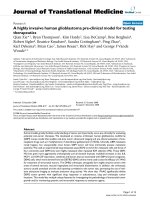
Figure 1
In an experimental metastasis model, DBM2 cells produce tumors in various tissues
In an experimental metastasis model, DBM2 cells produce tumors in various tissues. (A) Clonal selection through
experimental metastasis. The DBTRG-05MG cells were injected into the tail vein of athymic nude mice. Mice were sacrificed
either when they became moribund (~12 weeks) or after 8 weeks. At necropsy, lung lesions were transplanted into nude mice
subcutaneously. From these tumors, cells were harvested and injected into nude mice via tail vein. After the second cycle (M2)
cells were expanded ex-vivo in culture. (B) DBTRG-05MG or DBM2 cells were injected via the tail vein into nude mice. After
eight weeks mice inoculated with DBTRG-05MG cells had only a few pulmonary tumors (a, b). By contrast, lungs from mice
bearing DBM2 cells were almost fully replaced with tumors (c, d), and metastatic foci were found in skeletal muscle (e), diaphragm (f), lymph nodes adjacent to the spinal cord (g) and in the chest cavity (h). H&E staining of formalin fixed sections from
lungs of DBTRG-05MG cells (i) or DBM2 cells (j) eight weeks after tail vein injection. Invasion of DBM2 tumors into skeletal
muscle (left 2 arrows) induces bone resorption (right arrow) (k) and replaces nearly the entire lymph node (arrow) (l, insert at
low magnification).
Page 5 of 13
(page number not for citation purposes)
Journal of Translational Medicine 2008, 6:77
DB
U87
M2
M2
in patient tumors [24,27-29]. Adjacent to the main tumor
xenograft, we observed human c-MET and uPAR staining
of cells invading the normal brain parenchyma (Figure
3B) showing that DBM2 cells are highly invasive.
U251
M2A
M1A
/>
M1B
Certain pathological features are associated with aggressive behavior of many cancer types, including GBM
[15,30]. DBM2 orthotopic tumors show many of these
features. They are markedly pleomorphic and possess
regions of central necrosis lined by a row of crowded
tumor cells (Figure 3Ca, b arrows). Further, the orthotopic
tumors exhibit extensive vascular hyperplasia (Figure
3Ce), vascular invasion (Figure 3Cd) as well as invasion of
vessel walls (Figure 3Cc arrow), thrombus formation (Figure 3Cd). Glomeruloid body-like abnormal vasculature
formation was observed upon staining with CD31 antibody (Figure 3Cf). Together, the invasive and aggressive
growth behavior and cytokine profile of ELM selected
xenografts strongly resemble human disease and validate
this animal model for testing of drugs for inhibition of
intracranial tumor growth.
Fold Change
(log2)
-7 -3.5
0
3.5
7
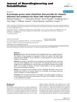
Figure
Elevated2cytokines and growth factors in GBM-M2 cells
Elevated cytokines and growth factors in GBM-M2
cells. Identification of cytokines and growth factors in common in the 24 hr conditioned medium for all three GBM-M2
tumor lines and the fold increases in their expression compared to the parental GBM cells. Heat map shows fold differences based upon the of expression ratios of 89 cytokines
and proteins between parental and GBM-M2 lines determined as described in the materials and methods section.
The fold change in protein expression level is indicated by
color. GM-CSF, IL-6, IL-8 and BDNF were found highly elevated in all three GBM-M2 lines (fold changes are summarized in the supplementary table [Additional File 4]).
Real-time imaging of DBM2 tumor growth and vascularity
As DBM2 orthotopic tumors grow, we observed that the
opening created for tumor cell inoculation increases in
size, allowing both intra and extracranial tumor growth
[Additional File 1]. This opening allows high-resolution
intravital imaging of DBM2 tumor growth [Additional
File 1;B]. Ultrasound imaging revealed poorly distinct
tumor margins, consistent with invasive growth. Further,
ultrasound measurements demonstrated that the increase
of tumor volume was accompanied by a proportional
increase of the skull erosion at the DBM2 cell inoculation
site [Additional File 2]. This was confirmed by CT technology (data not shown). We compared the dimensions of
the skull erosion obtained by ultrasound [Additional File
1;A,c], the distance between the arrows) to measurements
with conventional calipers [Additional File 1;A,d] at the
time of necropsy and observed good correlation between
the two approaches (γ = 0.87, n = 10). Beneath the skull
erosion, tumor volume was determined from the ultrasound images [Additional File 2;C]. Moreover, we found
a high correlation (γ = 0.95, n = 96), [Additional File 2;D]
between tumor volume and the size of the skull opening
measured by ultrasound. Thus, the skull opening provides
a simple way to monitor tumor growth during therapeutic
intervention.
We found that, with Doppler and contrast injection ultrasound, both the amount of blood flow and the direction
of the flow in the orthotopic DBM2 tumor can easily be
visualized. Under the Doppler mode [Additional File 1;B,
a], we see strong energy signals that accumulate in the
skin, indicating the existence of "macro" blood vessels
with high blood flow in these tissues. However, the tumor
Page 6 of 13
(page number not for citation purposes)
Journal of Translational Medicine 2008, 6:77
/>
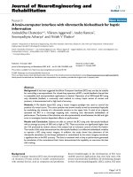
Figure growth and GBM properties of orthotopic DBM2 intracranial tumors
Invasive 3
Invasive growth and GBM properties of orthotopic DBM2 intracranial tumors. (A) Orthotopic DBM2 tumors
exhibit extensive infiltration into the mouse brain parenchyma (a, b). The arrows point to areas of cranial erosion. (c) Higher
magnification of DBM2 tumor demonstrating extensive infiltration into the brain parenchyma. Compared to DBM2, U251
tumors form a sharper cranial margin (d, e) and are less invasive (f). (B) Met (a, b) and uPAR (c, d) expression in invasive DMB2
orthotopic tumors. (C) H&E staining of formalin fixed DBM2 tumors shows central necrosis with the crowding of cancer cells
lining the necrotic area (a, b arrows). Vascular invasion of DBM2 tumors along the perivascular space (arrow) and in vessels in
the surrounding brain (c) with tumor-thrombus formation (d). Higher magnification showing a glomeruloid body-like structure
(d, insert). CD31 staining highlights vascular proliferation (e). Enlargement of (e) showing glomeruloid body-like structure with
multiple layers of endothelial cells is stained by CD31 antibody (f).
Page 7 of 13
(page number not for citation purposes)
Journal of Translational Medicine 2008, 6:77
mass is mostly dark, indicating that the tumor vasculature
does not emit a Doppler signal. To enhance the visualizing of tumor blood vessels, we injected a contrast reagent
through the tail vein before ultrasound measurement. Following injection, we saw a rich vascular network extending from the bone-tumor margins along the intracranial
boundary of the tumor [Additional File 1;B,b]. Strikingly,
almost all the tumor provided a contrast signal, indicating
that the DBM2 orthotopic tumors have micro-blood vessels with a lower flow rate than abundant large, mature
blood vessels. This makes the DBM2 intracranial glioblastoma model particularly useful as a preclinical model to
evaluate novel therapeutic interventions against vascular
flow and formation. Given the resemblance of this animal
model to patient GBM we proceeded with the evaluation
of the 17AAG for inhibition of intracranial tumor growth.
17AAG inhibition of DBM2 tumor growth and metastasis
17AAG is an HSP90 inhibitor that is in clinical phase I trials targeting different types of cancers, but its use has not
been reported against glioblastoma [19,20,31]. With the
SQ model, 17AAG at 60 mg/kg gave significant growth
inhibition after 4 weeks of dosing (Figure 4A, P < 0.05 at
day of 32). When the orthotopic model was used, however, results with the 60 mg/kg-day group growth rate was
significantly lower than that of mice in the non-treated
DBM2 control group (Figure 4B, P < 0.05 at day 21).
Moreover, administration of 17AAG at 60 mg/kg-day significantly prolong the survival of mice bearing DBM2
intracranial tumors in dose-dependent manner (Figure
4C, p < 0.05).
We also tested if 17AAG can inhibit DBM2 ELM metastasis, for the purpose of determining whether the drug
would inhibit this invasion dependent metastasis assay.
Our results show that, at 60 mg/kg-day, 17AAG can significantly block DBM2 metastasis formation in lungs and
other organs (Table 2, P < 0.05). Moreover, the harvested
lungs from the 60 mg/kg-day group demonstrated significantly less tumor burden than those from the 20 mg/kgday and control groups (Table 2, P < 0.05). We conclude
that 17AAG inhibits intracranial DBM2 tumor growth at
the same dose (60 mg/day) as tumor growth and metastasis formation in the SQ and ELM models. This strongly
encourages testing of a novel application for 17AAG in
patients with GBM.
Discussion
The limited number of preclinical models that recapitulate the invasive GBM tumor growth is a major hurdle to
drug development. Subjecting human melanoma cells to
ELM yielded highly metastatic cells with higher proliferative and invasive potential [23,32]. We applied this
method to GBM cell lines for the purpose of improving
their invasiveness in orthotopic models. The ELM assay
has been used to select for metastatic cancer cells in a
/>
number of other cancer types [33-35], but has not been
tested previously with GBM, most likely because of the
notion that extracranial metastases of human GBM are
clinically rare.
Here we show that GBM cell lines can be highly invasive
after ELM selection, but they still are not metastatic when
implanted in the brain. The lack of extracranial metastasis
of the derivative GBM-M2 cell lines strongly suggests that
rapid tumor growth or the unique CNS environment curtails the escape of tumor cells [14]. A previous study confirms the intrinsic metastatic nature of GBM tumor cells:
GBM tumor cells were metastatic in spontaneous metastasis assays and no different than other types of cancer cells
when tested in these assays [14]. Although stem cells isolated from primary tumor tissues [4,36] have not yet been
tested for metastatic potential, the stem-cell like sub-populations from rat C6 glioma cells form neurospheres and
like our GBM-M2 cells, are metastatic to lungs, as well as
to other organs in nude mice upon intraperitoneal (i.p.)
injection [37], again supporting that GBM tumor cells
have intrinsic metastatic potential. Consistent with these
reports we show that three of four commonly used GBM
lines are highly metastatic in ELM assays (Table 1) and
form metastasis in lungs and lymph nodes, similar to the
destinations of some of the rare clinical GBM metastases
in patients [8,9,11-13]. It is quite remarkable that GBM
tumor cell lines, which came from primary tumors that
have never grown as metastases and are selected to grow
in vitro in tissue culture, have the capacity to be highly
metastatic. This indicates that some aspect of GBM malignancy also satisfies the requirements for the metastatic
process, or that the metastatic genotype is acquired early
in tumor progression as has been proposed [38,39]. We
have proposed that once cells acquire an invasive phenotype, they have the ability to acquire a proliferative phenotype again to become a metastatic colony [40].
The changes in cytokine and growth factor expression that
occur after ELM GBM-M2 selection are similar to those
that predict aggressive disease and poor patient outcome,
demonstrating the similarity of cell lines to the scenario in
patients. Interestingly, after ELM selection, all three GBMM2 lines show highly elevated GM-CSF, IL-6, IL-8 and
Brain-derived neurotrophic factor (BDNF) compared with
parental cell lines (Figure 2, [Additional File 4]). Both
GM-CSF and its receptor are absent in normal brain but
expressed at high levels in glioma tissues [17]. In vitro,
GM-CSF stimulates glioma cells to both proliferate and
migrate [17]. IL-6 gene amplification in patients distinguishes GBM from low-level astrocytoma and is associated with poor prognosis [18]. In addition, IL-8
expression is highly associated with gliomagenesis and
tumoral angiogenesis. Taken together, the co-elevation of
these 3 cytokines appears to be an important indicator for
GBM or poor prognosis. BDNF, a member of the neuroPage 8 of 13
(page number not for citation purposes)
Journal of Translational Medicine 2008, 6:77
/>
Figure
17 AAG4inhibition of DBM2 tumor growth
17 AAG inhibition of DBM2 tumor growth. (A) 17AAG at 60 mg/kg-d inhibits DBM2 subcutaneous tumor growth. DBM2
cells were inoculated into the flanks of nude mice at 5 × 10⁵ cells in 100 ul PBS. After 2 weeks, mice with size-matched tumors
(100 – 200 mm3) were assigned into control and treatment (60 mg/kg-d) groups (n = 19) and treatment started. Error bar represents for standard error. (B) 17AAG at 60 mg/kg-d inhibits DBM2 orthotopic tumor growth. DBM2 cells were inoculated
intracranially into nude mice at 5 × 10⁵ cells in 5 ul PBS. The tumor growth was monitored by Ultrasound. After 2 weeks, sizematched tumors were grouped into control and treatment groups (n = 10). Fold change of tumor volume = Weekly measured
tumor size/Initial tumor size upon grouping. (C) The survival time of nude mice bearing orthotopic DBM2 tumor xenografts
treated with 17AAG. DBM2 cells were inoculated intracranially of nude mice at 5 × 10⁵ cells in 5 ul PBS. After 3 weeks, sizematched tumors were grouped into control (n = 6) and 2 treatment groups (20 mg/kg, 60 mg/kg, n = 8). The arrow points to
the day treatment started after orthotopic tumor inoculation. Treatment was administered until individual mice became moribund according to IACUC guild-line and survival time was recorded.
Page 9 of 13
(page number not for citation purposes)
Journal of Translational Medicine 2008, 6:77
/>
trophin family, plays an important role in neuronal development and survival [41]. Although a role for BDNF in
GBM is not elucidated, its downstream signaling through
Ras, ERK as well as PI3K pathways [42], would suggest it
could play a role in GBM disease. Furthermore all of the
GBM lines express high levels of MCP-1, also a marker of
poor prognosis in patient gliomas [26]. All of these markers are consistent with the GBM nature of the GBM-M2
cells.
We chose to further develop DBM2 cells as an orthotopic
model. DBM2 cells, when inoculated orthotopically, not
only show significant invasive growth, but also central
necrosis, extensive vascular hyperplasia, and glomeruloid
body-like vasculature formation. Brat et al. (2004, 2005)
have reported the pathological features associated with
poor diagnosis in GBM patients as well as the possible
mechanisms. Necrosis is a hallmark of glioblastoma
occurring in 60% of GBM patients while intravascular
tumor-thrombus formation is found in over 90% of GBM
cases. In addition, vascular hyperplasia is a characteristic
of GBM and associated with poor prognosis [15,30,43].
As an explanation for their highly invasive nature, we
show that DBM2 tumors not only express both c-Met and
uPAR, the receptor of urokinase signaling pathway, but
also strongly respond to HGF (data not shown) indicating
that the c-Met signaling pathway may play an important
role in the invasion of DBM2 orthotopic tumors into the
brain parenchyma [24,27,40,44]. Brain tumors seldom
invade the skull, but there are reports of GBM with skullerosion phenotypes and metastases to other organs
[45,46]. The exact mechanism of the osteolytic phenotype
of DBM2 is unknown. It is possibly mediated through
activation of bone-resorbing osteoclasts and may be facilitated by elevated IL-6 and IL-8 levels [47,48].
Real-time noninvasive imaging technologies permit longitudinal monitoring of tumor progression. Magnetic resonance imaging (MRI) is commonly used for human
brain tumor imaging and is being refined in preclinical
models [7]. Bioluminescence-based in vivo imaging systems are also used to rapidly measure tumor volume and
evaluate drug efficacy in animal models [49]. Cranial window models have been developed in which part of the
mouse skull is replaced with a cover glass so that the
blood vessels can be observed microscopically [50]. Here,
taking advantage of the osteolytic phenotype, we show
high-resolution ultrasound can be used to monitor realtime, non-invasive imaging of brain tumor growth and
vascularization. In addition, with Doppler and contrast
injection ultrasound, directional blood flow can easily be
visualized in the tumor.
We show that our xenograft model is versatile in that it
can be used with SQ implantation for measuring tumor
growth potential [25], with systemic injection for measuring invasive and metastatic growth potential in EML
assays [51], or with orthotopic administration of tumor
cells for measuring tumor growth in a macro- and microenvironment that recapitulates GBM in patients. Thus this
model is particularly suitable for testing therapeutics. We
chose here to test the drug, 17AAG, because of its diversity
in targeting the destabilization of numerous oncoproteins
[52]. 17AAG, a derivative of geldanamycin, an HSP90
inhibitor that has been in clinical trials in patients with
advanced cancer [19,20]. It has not been considered for
GBM treatment largely, we suspect, because of anticipated
blood brain barrier interference with drug delivery. We
show here that in all three tumor settings, 17AAG at 60
mg/kg, significantly inhibits tumor growth (Table 2, Figure 4). Thus 17AAG prevents SQ xenograft formation, the
formation of metastatic lesions in ELM assays and importantly, at the same dose, inhibits DBM2 orthotopic tumor
growth and prolongs animal survival time. It is certainly
possible that the highly invasive GBM tumors compromise the BBB in our DBM2 orthotopic model leading to
significant 17AAG anti-tumor activity. Studies with orthotopic GBM mouse models have shown that imaging reagents can leak from the intracranial tumors, indicating
that the BBB is compromised [7] and anti-HGF mAbs,
Table 2: 17AAG inhibits the development of DBM2 pulmonary lesions.
Lung grade
Group
1 (n = 8)
2 (n = 10)
3 (n = 10§)
17AAG dose (mg/kg-d)
Vehicle only
20
60
Body weight (g)
Lung weight (g)
+
++
+++
17.79 ± 1.88
19.88 ± 1.68*
20.17 ± 0.89*
0.477 ± 0.19
0.412 ± 0.17
0.276 ± 0.11*
2 (25%)
3 (30%)
8 (80%)
3 (37.5%)
2 (20%)
2 (20%)
3 (37.5%)
5 (50%)
0
*Compared with group 1; Student's t test was used (p < 0.05)
§Compared with group 1; Chi-square was used for statistical analysis P < 0.05.
For drug testing in the lung metastasis model, 28 nude mice (6-week-old females) were divided into three groups: a control group (n = 8), and
17AAG groups treated with either 20 mg/kg (n = 10) or 60 mg/kg (n = 10). Each mouse received a single intravenous tail vein injection of 106 DBM2
cells in 100 μl PBS. Treatment started the second day after the cells were injected and continued for 8 weeks, by which time most of the control
mice were moribund. At necropsy, lungs were harvested and scored; body weight and lung weight of each mouse were also recorded.
Page 10 of 13
(page number not for citation purposes)
Journal of Translational Medicine 2008, 6:77
/>
despite their large molecular size can inhibit orthotopic
tumor growth in the brain [53,54]. Our results indicate
that 17AAG may be used clinically to treat malignant
GBM patients providing there is limited BBB interference
with drug penetration.
Additional file 3
GBM-M2 cells show enhanced malignancy in vitro and in vivo compared to GBM cells. The data provided include the growth curves and survival time of GBM-M2 cells compared with the parental cell lines.
Click here for file
[ />
In conclusion, we report that commonly used GBM cells
have metastatic potential which can easily be selected in
ELM assays. When implanted in the brain, the metastatic
potential of GBM cells can be converted to a highly invasive phenotype. Importantly we show that 17AAG is an
effective inhibitor of orthotopic tumor growth and that
the response to treatment can be measured in real-time by
ultrasound. We anticipate that this orthotopic model with
high-resolution ultrasound technology will serve as a valuable tool in preclinical screening for drugs effective in
targeting GBM.
Additional file 4
Fold increases of cytokines and growth factors in GBM sub-lines. The
data provided represent the fold changes of cytokines and growth factors
amongst all three GBM-M2 lines.
Click here for file
[ />
Additional file 5
Supplementary Materials & Methods. The data provided represent the
materials and methods used for Additional Files 1, 2, 3, 4 (this file is not
cited in the paper; it is the Materials and Methods used for the supplementary figures).
Click here for file
[ />
Competing interests
The authors declare that they have no competing interests.
Authors' contributions
QX designed study, isolated and characterized cell lines,
performed ultrasound imaging, performed data analysis
and interpretation and prepared manuscript. RT performed animal experimentation. KH served as sonographer. LD performed ultrasound imaging and assisted with
animal studies. BB performed immunohistochemistry,
staining procedures and evaluation. RS reviewed pathological slides and provided interpretation. BK served as
pathologist and assisted with preparation of manuscript.
SC served as pathologist. PZ prepared Met4 antibody. KD
performed statistical analysis. BC prepared Met4 antibody. JR performed histology and immunohistochemistry. RH performed ultrasound imaging. GVW developed
the concept and designed study, interpreted data, prepared manuscript, and supervised study.
Additional material
Additional file 1
DBM2 orthotropic tumor growth promotes cranial osteolysis. The data
provided demonstrate the rationale of using cranial osteolysis phenotype to
perform ultrasound imaging.
Click here for file
[ />
Additional file 2
DBM2 orthotopic tumor growth promotes cranial osteolysis-continued. Ultrasound imaging reveals that the cranial osteolysis generated by
DBM2 orthotopic tumor growth results in an opening that is proportional
to tumor size.
Click here for file
[ />
Additional file 6
Supplementary Figure Legends. This file contains the figure legends for
supplementary Figures 1 and 2 (this file is not cited in the paper; it contains the supplementary figure legends).
Click here for file
[ />
Acknowledgements
We are grateful to Drs. David Wenkert and Yuehai Shen for 17AAG characterization and to Drs. Jacob Zhang and Kyle Furge for statistical analysis.
We thank Michelle Bassett for assistance in the preparation of the manuscript. We thank Dr. Richard Lister (Molecular Therapeutics, Inc.) and Dr.
Justi Rao (University of Illinois at Chicago) for their help with the intracranial model. This work was generously supported, in part, by the Jay and
Betty Van Andel Foundation.
References
1.
2.
3.
4.
5.
Berens ME, Giese A: "...those left behind." Biology and oncology of invasive glioma cells. Neoplasia 1999, 1:208-219.
Camphausen K, Purow B, Sproull M, Scott T, Ozawa T, Deen DF,
Tofilon PJ: Influence of in vivo growth on human glioma cell
line gene expression: convergent profiles under orthotopic
conditions. Proc Natl Acad Sci USA 2005, 102:8287-8292.
Assanah M, Lochhead R, Ogden A, Bruce J, Goldman J, Canoll P: Glial
progenitors in adult white matter are driven to form malignant gliomas by platelet-derived growth factor-expressing
retroviruses. J Neurosci 2006, 26:6781-6790.
Lee J, Kotliarova S, Kotliarov Y, Li A, Su Q, Donin NM, Pastorino S,
Purow BW, Christopher N, Zhang W, et al.: Tumor stem cells
derived from glioblastomas cultured in bFGF and EGF more
closely mirror the phenotype and genotype of primary
tumors than do serum-cultured cell lines. Cancer Cell 2006,
9:391-403.
Bao S, Wu Q, McLendon RE, Hao Y, Shi Q, Hjelmeland AB, Dewhirst
MW, Bigner DD, Rich JN: Glioma stem cells promote radiore-
Page 11 of 13
(page number not for citation purposes)
Journal of Translational Medicine 2008, 6:77
6.
7.
8.
9.
10.
11.
12.
13.
14.
15.
16.
17.
18.
19.
20.
21.
22.
23.
sistance by preferential activation of the DNA damage
response. Nature 2006, 444:756-760.
Dai C, Celestino JC, Okada Y, Louis DN, Fuller GN, Holland EC:
PDGF autocrine stimulation dedifferentiates cultured astrocytes and induces oligodendrogliomas and oligoastrocytomas from neural progenitors and astrocytes in vivo. Genes
Dev 2001, 15:1913-1925.
McConville P, Hambardzumyan D, Moody JB, Leopold WR, Kreger
AR, Woolliscroft MJ, Rehemtulla A, Ross BD, Holland EC: Magnetic
resonance imaging determination of tumor grade and early
response to temozolomide in a genetically engineered
mouse model of glioma. Clin Cancer Res 2007, 13:2897-2904.
Beauchesne P, Soler C, Mosnier JF: Diffuse vertebral body metastasis from a glioblastoma multiforme: a technetium-99m
Sestamibi single-photon emission computerized tomography study. J Neurosurg 2000, 93:887-890.
Chelly I, Mekni A, Ferchichi L, Houissa S, Kchir N, Haouet S, Khaldi
M, Zitouna M: [Bone metastasis from a glioblastoma: An unusual course!]. Neurochirurgie 2006, 52:367-370.
Chivukula M, Dincer HE, Biller JA, Krouwer HG, Simon G, Shidham
V: FNAB cytology of extra-cranial metastasis of glioblastoma
multiforme may resemble a lung primary: a diagnostic pitfall. Cytojournal 2005, 2:9.
Saad AG, Sachs J, Turner CD, Proctor M, Marcus KJ, Wang L, Lidov
H, Ullrich NJ: Extracranial metastases of glioblastoma in a
child: case report and review of the literature. J Pediatr Hematol Oncol 2007, 29:190-194.
Ueda S, Mineta T, Suzuyama K, Furuta M, Shiraishi T, Tabuchi K: Biologic characterization of a secondary glioblastoma with
extracranial progression and systemic metastasis. Neuro
Oncol 2003, 5:14-18.
Utsuki S, Tanaka S, Oka H, Iwamoto K, Sagiuchi T, Fujii K: Glioblastoma multiforme metastasis to the axis. Case report. J Neurosurg 2005, 102:540-542.
Huang P, Allam A, Taghian A, Freeman J, Duffy M, Suit HD: Growth
and metastatic behavior of five human glioblastomas compared with nine other histological types of human tumor
xenografts in SCID mice. J Neurosurg 1995, 83:308-315.
Brat DJ, Bellail AC, Van Meir EG: The role of interleukin-8 and its
receptors in gliomagenesis and tumoral angiogenesis. Neuro
Oncol 2005, 7:122-133.
Goswami S, Gupta A, Sharma SK: Interleukin-6-mediated autocrine growth promotion in human glioblastoma multiforme
cell line U87MG. J Neurochem 1998, 71:1837-1845.
Mueller MM, Herold-Mende CC, Riede D, Lange M, Steiner HH,
Fusenig NE: Autocrine growth regulation by granulocyte colony-stimulating factor and granulocyte macrophage colonystimulating factor in human gliomas with tumor progression. Am J Pathol 1999, 155:1557-1567.
Tchirkov A, Khalil T, Chautard E, Mokhtari K, Veronese L, Irthum B,
Vago P, Kemeny JL, Verrelle P: Interleukin-6 gene amplification
and shortened survival in glioblastoma patients. Br J Cancer
2007, 96:474-476.
Bagatell R, Gore L, Egorin MJ, Ho R, Heller G, Boucher N, Zuhowski
EG, Whitlock JA, Hunger SP, Narendran A, et al.: Phase I pharmacokinetic and pharmacodynamic study of 17-N-allylamino17-demethoxygeldanamycin in pediatric patients with recurrent or refractory solid tumors: a pediatric oncology experimental therapeutics investigators consortium study. Clin
Cancer Res 2007, 13:1783-1788.
Ramanathan RK, Egorin MJ, Eiseman JL, Ramalingam S, Friedland D,
Agarwala SS, Ivy SP, Potter DM, Chatta G, Zuhowski EG, et al.: Phase
I and pharmacodynamic study of 17-(allylamino)-17-demethoxygeldanamycin in adult patients with refractory advanced
cancers. Clin Cancer Res 2007, 13:1769-1774.
Knudsen BS, Zhao P, Resau J, Cottingham S, Gherardi E, Xu E, Resau
J, Berghuis B, Daugherty J, Grabinski T, et al.: A Novel Multipurpose Monoclonal Antibody for Evaluating Human c-Met
Expression in Preclinical and Clinical Settings. Appl Immunohistochem Mol Morphol 2008.
Burger AM, Fiebig HH, Stinson SF, Sausville EA: 17-(Allylamino)17-demethoxygeldanamycin activity in human melanoma
models. Anticancer Drugs 2004, 15:377-387.
Clark EA, Golub TR, Lander ES, Hynes RO: Genomic analysis of
metastasis reveals an essential role for RhoC. Nature 2000,
406:532-535.
/>
24.
25.
26.
27.
28.
29.
30.
31.
32.
33.
34.
35.
36.
37.
38.
39.
40.
41.
42.
43.
44.
45.
Koochekpour S, Jeffers M, Rulong S, Taylor G, Klineberg E, Hudson
EA, Resau JH, Woude GF Vande: Met and hepatocyte growth factor/scatter factor expression in human gliomas. Cancer Res
1997, 57:5391-5398.
Gao CF, Xie Q, Su YL, Koeman J, Khoo SK, Gustafson M, Knudsen
BS, Hay R, Shinomiya N, Woude GF Vande: Proliferation and invasion: plasticity in tumor cells. Proc Natl Acad Sci USA 2005,
102:10528-10533.
Liang Y, Bollen AW, Gupta N: CC chemokine receptor-2A is frequently overexpressed in glioblastoma. J Neurooncol 2008,
86:153-163.
Jeffers M, Rong S, Woude GF Vande: Enhanced tumorigenicity
and invasion-metastasis by hepatocyte growth factor/scatter
factor-met signalling in human cells concomitant with induction of the urokinase proteolysis network. Mol Cell Biol 1996,
16:1115-1125.
Arrieta O, Garcia E, Guevara P, Garcia-Navarrete R, Ondarza R,
Rembao D, Sotelo J: Hepatocyte growth factor is associated
with poor prognosis of malignant gliomas and is a predictor
for recurrence of meningioma. Cancer 2002, 94:3210-3218.
Moriyama T, Kataoka H, Koono M, Wakisaka S: Expression of
hepatocyte growth factor/scatter factor and its receptor cMet in brain tumors: evidence for a role in progression of
astrocytic tumors (Review). Int J Mol Med 1999, 3:531-536.
Brat DJ, Van Meir EG: Vaso-occlusive and prothrombotic mechanisms associated with tumor hypoxia, necrosis, and accelerated growth in glioblastoma. Lab Invest 2004, 84:397-405.
Grem JL, Morrison G, Guo XD, Agnew E, Takimoto CH, Thomas R,
Szabo E, Grochow L, Grollman F, Hamilton JM, et al.: Phase I and
pharmacologic study of 17-(allylamino)-17-demethoxygeldanamycin in adult patients with solid tumors. J Clin Oncol
2005, 23:1885-1893.
Cruz-Munoz W, Man S, Xu P, Kerbel RS: Development of a preclinical model of spontaneous human melanoma central
nervous system metastasis. Cancer Res 2008, 68:4500-4505.
Luu HH, Kang Q, Park JK, Si W, Luo Q, Jiang W, Yin H, Montag AG,
Simon MA, Peabody TD, et al.: An orthotopic model of human
osteosarcoma growth and spontaneous pulmonary metastasis. Clin Exp Metastasis 2005, 22:319-329.
Chao TC, Greager JA: Experimental pulmonary sarcoma
metastases in athymic nude mice.
J Surg Oncol 1997,
65:123-126.
Corti C, Pratesi G, DeCesare M, Pellegrini R, Giardini R, Supino R,
Zunino F: Spontaneous lung metastases in a human lung
tumor xenograft: a new experimental model. J Cancer Res Clin
Oncol 1996, 122:154-160.
Bao S, Wu Q, Sathornsumetee S, Hao Y, Li Z, Hjelmeland AB, Shi Q,
McLendon RE, Bigner DD, Rich JN: Stem cell-like glioma cells
promote tumor angiogenesis through vascular endothelial
growth factor. Cancer Res 2006, 66:7843-7848.
Kondo T, Setoguchi T, Taga T: Persistence of a small subpopulation of cancer stem-like cells in the C6 glioma cell line. Proc
Natl Acad Sci USA 2004, 101:781-786.
Bernards R, Weinberg RA: A progression puzzle. Nature 2002,
418:823.
Wouw AJ van de, Janssen-Heijnen ML, Coebergh JW, Hillen HF: Epidemiology of unknown primary tumours; incidence and population-based survival of 1285 patients in Southeast
Netherlands, 1984–1992. Eur J Cancer 2002, 38:409-413.
Knudsen BS, Woude G Vande: Showering c-MET-dependent
cancers with drugs. Curr Opin Genet Dev 2008, 18:87-96.
Davis MI: Ethanol-BDNF interactions: still more questions
than answers. Pharmacol Ther 2008, 118:36-57.
Reichardt LF: Neurotrophin-regulated signalling pathways.
Philos Trans R Soc Lond B Biol Sci 2006, 361:1545-1564.
Brat DJ, Castellano-Sanchez AA, Hunter SB, Pecot M, Cohen C, Hammond EH, Devi SN, Kaur B, Van Meir EG: Pseudopalisades in
glioblastoma are hypoxic, express extracellular matrix proteases, and are formed by an actively migrating cell population. Cancer Res 2004, 64:920-927.
Gao C, Furge K, Koeman J, Dykema K, Su Y, Cutler ML, Werts A,
Haak P, Woude GF Vande: Chromosome instability, chromosome transcriptome, and clonal evolution of tumor cell populations. Proc Natl Acad Sci USA 2007, 104:8995-9000.
Gheyi V, Hui FK, Doppenberg EM, Todd W, Broaddus WC: Glioblastoma multiforme causing calvarial destruction: an unu-
Page 12 of 13
(page number not for citation purposes)
Journal of Translational Medicine 2008, 6:77
46.
47.
48.
49.
50.
51.
52.
53.
54.
/>
sual manifestation revisited. AJNR Am J Neuroradiol 2004,
25:1533-1537.
Vik A, Kvikstad A, Unsgard G, Jorgensen JV, Torp SH: A 54-year-old
man with a large subcutaneous skull erosion and focal epileptic seizures. Tidsskr Nor Laegeforen 2006, 126:2386-2387.
Koreny T, Tunyogi-Csapo M, Gal I, Vermes C, Jacobs JJ, Glant TT:
The role of fibroblasts and fibroblast-derived factors in
periprosthetic osteolysis. Arthritis Rheum 2006, 54:3221-3232.
Tanaka R, Yasunaga Y, Hisatome T, Yamasaki T, Iwamori H, Ochi M:
Serum interleukin 8 levels correlate with synovial fluid levels
in patients with aseptic loosening of hip prosthesis. J Arthroplasty 2005, 20:1049-1054.
Fomchenko EI, Holland EC: Mouse models of brain tumors and
their applications in preclinical trials. Clin Cancer Res 2006,
12:5288-5297.
Kawamura S, Schurer L, Goetz A, Kempski O, Schmucker B, Baethmann A: An improved closed cranial window technique for
investigation of blood-brain barrier function and cerebral
vasomotor control in the rat. Int J Microcirc Clin Exp 1990,
9:369-383.
Fidler IJ: Tumor heterogeneity and the biology of cancer invasion and metastasis. Cancer Res 1978, 38:2651-2660.
Richter K, Buchner J: Hsp90: chaperoning signal transduction. J
Cell Physiol 2001, 188:281-290.
Kim KJ, Wang L, Su YC, Gillespie GY, Salhotra A, Lal B, Laterra J: Systemic anti-hepatocyte growth factor monoclonal antibody
therapy induces the regression of intracranial glioma
xenografts. Clin Cancer Res 2006, 12:1292-1298.
Martens T, Schmidt NO, Eckerich C, Fillbrandt R, Merchant M,
Schwall R, Westphal M, Lamszus K: A novel one-armed anti-cMet antibody inhibits glioblastoma growth in vivo. Clin Cancer
Res 2006, 12:6144-6152.
Publish with Bio Med Central and every
scientist can read your work free of charge
"BioMed Central will be the most significant development for
disseminating the results of biomedical researc h in our lifetime."
Sir Paul Nurse, Cancer Research UK
Your research papers will be:
available free of charge to the entire biomedical community
peer reviewed and published immediately upon acceptance
cited in PubMed and archived on PubMed Central
yours — you keep the copyright
BioMedcentral
Submit your manuscript here:
/>
Page 13 of 13
(page number not for citation purposes)