báo cáo hóa học:" Comparative study on the immunogenicity between an HLA-A24-restricted cytotoxic T-cell epitope derived from survivin and that from its splice variant survivin-2B in oral cancer patients" pot
Bạn đang xem bản rút gọn của tài liệu. Xem và tải ngay bản đầy đủ của tài liệu tại đây (616.17 KB, 11 trang )
BioMed Central
Page 1 of 11
(page number not for citation purposes)
Journal of Translational Medicine
Open Access
Research
Comparative study on the immunogenicity between an
HLA-A24-restricted cytotoxic T-cell epitope derived from survivin
and that from its splice variant survivin-2B in oral cancer patients
Jun-ichi Kobayashi
1,2
, Toshihiko Torigoe*
1
, Yoshihiko Hirohashi
1
,
Satomi Idenoue
3
, Akihiro Miyazaki
2
, Akira Yamaguchi
2
,
Hiroyoshi Hiratsuka
2
and Noriyuki Sato
1
Address:
1
Department of Pathology, Sapporo Medical University School of Medicine, Sapporo, Japan,
2
Department of Oral Surgery, Sapporo
Medical University School of Medicine, Sapporo, Japan and
3
Department of Surgery, Sapporo Medical University School of Medicine, Sapporo,
Japan
Email: Jun-ichi Kobayashi - ; Toshihiko Torigoe* - ; Yoshihiko Hirohashi - ;
Satomi Idenoue - ; Akihiro Miyazaki - ; Akira Yamaguchi - ;
Hiroyoshi Hiratsuka - ; Noriyuki Sato -
* Corresponding author
Abstract
Background: We previously reported an HLA-A24-restricted cytotoxic T-cell epitope, Survivin-
2B80-88, derived from a splice variant of survivin, survivin-2B. In this report, we show a novel HLA-
A24-restricted T-cell epitope, Survivin-C58, derived from a wild type survivin, and compared their
immunogenicity in oral cancer patients.
Methods: By stimulating peripheral blood lymphocytes of HLA-A24-positive cancer patients with
Survivin-C58 peptide in vitro, the peptide-specific CTLs were induced. In order to compare the
immunogenic potential between C58 peptide and 2B80-88 peptide, peripheral blood T-cells from
thirteen HLA-A24-positive oral cancer patients were stimulated with either or both of these two
peptides.
Results: Survivin-2B80-88 peptide-specific CTLs were induced from four patients, and C58
peptide-specific CTLs were induced from three out of eight patients with over stage II progression.
The CTLs exerted cytotoxicity against HLA-A24-positive tumor cells. In contrast, CTL induction
failed from a healthy volunteer and all four patients with cancer stage I.
Conclusion: It was indicated that a splicing variant-derived peptide and wild type survivin-derived
peptide might have a comparable potency of CTL induction, and survivin targeting immunotherapy
using survivin-2B80-88 and C58 peptide cocktail should be suitable for HLA-A24+ oral cancer
patients.
Published: 6 January 2009
Journal of Translational Medicine 2009, 7:1 doi:10.1186/1479-5876-7-1
Received: 30 July 2008
Accepted: 6 January 2009
This article is available from: />© 2009 Kobayashi et al; licensee BioMed Central Ltd.
This is an Open Access article distributed under the terms of the Creative Commons Attribution License ( />),
which permits unrestricted use, distribution, and reproduction in any medium, provided the original work is properly cited.
Journal of Translational Medicine 2009, 7:1 />Page 2 of 11
(page number not for citation purposes)
Background
Survivin, an inhibitor of apoptosis protein, is highly
expressed in the vast majority of cancers [1,2]. Survivin
has been shown to increase tumor resistance to apoptotic
stimuli, such as radiation and chemotherapy [3,4]. In
agreement with these findings, a number of reports dem-
onstrate that survivin expression in cancer cells has a prog-
nostic value and is associated with increased tumor
recurrence and shorter patient survival [5-10], although
the opposite correlation is reported in certain cancers
[11]. So far, four different splicing variants of human sur-
vivin have been described, including survivin-2α, sur-
vivin-2B, survivin-ΔEx3, and survivin-3B [12-15]. While
survivin-2α and survivin-3B are truncated forms, survivin-
2B results from alternating splicing at the interface
between exon 2 and exon 3, leading to insertion of an
additional exon, termed exon 2B, in BIR domain. Since
BIR domain is a functional domain that is important for
the anti-apoptotic activity of survivin, survivin-2B is pre-
dicted to be non-anti-apoptotic [16,17].
Survivin was originally detected only in normal thymus,
testis and placenta; however, low levels of wild type sur-
vivin was detected in other normal tissues, such as acti-
vated T-cells, vascular endothelial cells, and
hematopoietic cells by more sensitive methods [3,18,19].
Wild type survivin is known to have an essential role in
the mitosis [3,18,20]. It forms a complex with the chro-
mosomal passenger proteins during mitosis and regulates
mitotic progression. In contrast, the protein levels as well
as the mRNA levels of survivin-2B and other survivin var-
iants are far less than that of wild type survivin, and they
are dispensable in such a mitotic checkpoint [17,21].
Since survivin expression is very low in normal differenti-
ated adult tissues as compared with that in cancer tissues,
survivin is considered to be an ideal molecular target for
cancer immunotherapy. With this mind, we attempted to
identify a HLA-A24-restricted cytotoxic T-lymphocyte
(CTL) epitopes of survivin that were suitable for cancer
vaccine, since HLA-A24 was the most frequent allele in
Japanese. In our previous report, three peptides derived
from survivin and its splicing variant survivin-2B were
examined for HLA-A24-binding affinity and immuno-
genicity [22]. It was shown that Survivin-2B80-88 peptide
(amino acid sequence AYACNTSTL), which was derived
from a splicing variant survivin-2B-specific exon2B, was
capable of inducing CTLs that had killing activity to HLA-
A24
+
cancer cells. Following this report, we provided fur-
ther evidence that Survivin-2B80-88 was highly immuno-
genic in various cancer patients, including those with
gastric cancer, breast cancer, and colorectal cancer [23].
Based on these results ex vivo, we have conducted phase I
clinical trials assessing the adverse event and efficacy of
Survivin-2B80-88 peptide vaccination in patients with
advanced colorectal cancer, breast cancer, lung cancer,
bladder cancer, and oral cancer [24-26].
Though we failed to identify an HLA-A24-restricted CTL
epitope derived from wild type survivin in the initial
study, a number of epitopes have been identified from
wild type survivin that are restricted to other HLA class I
alleles, such as A1, A2, A11, and B35 [27-29], some of
which have been applied for clinical trials [30,31]. More
recently, Andersen, et al. demonstrated that wild type sur-
vivin-derived Sur20-28 peptide (amino acid sequence
STFKNWPFL) was capable of inducing the peptide-spe-
cific CD8-positive T-cells from PBMCs of HLA-A24
+
can-
cer patients, although HLA-A24-restricted killing activity
of the peptide-specific T-cells against survivin-positive
cancer cells has not been assessed [32]. In this study, we
present a novel CTL epitope Survivin-C58 peptide derived
from wild type survivin. The peptide-specific CTLs
induced from peripheral blood mononuclear cells
(PBMCs) of oral cancer patient exerted HLA-A24-
restricted cytotoxicity against the tumor cells. Then, we
stimulated PBMCs of oral cancer patients with either or
both Survivin-C58 and Survivin-2B80-88 peptides, and
the consequent CTLs were examined for the peptide-spe-
cificity and cytotoxicity against HLA-A24+ tumor cells. We
demonstrate here for the first time a comparative study on
the potency of inducing CTLs in vitro between wild type
survivin-derived peptide and survivin-2B-derived peptide,
which indicates the comparable potency of CTL induction
in oral cancer patients.
Materials and methods
Patients and samples
Surgically-resected cancer specimens and PBMCs used in
this study were obtained from HLA-A*2402
+
patients with
breast cancer or oral cancer who were hospitalized at Sap-
poro Medical University Hospital after obtaining their
informed consent. The patients' clinicopathological pro-
files were listed on the table.
Cell lines and culture media
Human breast cancer cell lines, HMC-1 and HMC-2,
human oral squamous cell carcinoma (OSCC) cell lines,
OSC19, OSC20, OSC30, OSC40, OSC70, and POT1 were
established in our laboratory. OSCC cell lines HO-1-NH,
KOSC-3, HSC-2, HSC- 3, and HSC-4 were purchased from
the Human Science Research Resources Bank (HSRRB,
Osaka, Japan). OSCC cell line SAS was obtained from the
Institute of Development, Aging and Cancer Tohoku Uni-
versity (Tohoku, Japan). Human embryonic kidney cell
line 293T, breast cancer cell line MCF7, lymphoma cell
line Daudi and leukemia cell line K562 were purchased
from American Type Culture Collection (Manassas, VA).
C1R-A24 and C1R-A31, lymphoblastoid cell line C1R
transfectants with HLA-A*2402 and HLA-A*31012 cDNA
Journal of Translational Medicine 2009, 7:1 />Page 3 of 11
(page number not for citation purposes)
respectively, were kind gifts from Dr. M. Takiguchi
(Kumamoto University School of Medicine, Kumamoto,
Japan). T2-A24, a stable transfectant of T2 cells with HLA-
A*2402 cDNA was a kind gift from Dr. K. Kuzushima
(Aichi Cancer Research Institute, Nagoya, Japan).
293T cells and breast cancer lines were cultured in Dul-
becco's modified Eagle's medium (DMEM, Sigma-
Aldrich, St. Louis, MO) with 2 mM L-glutamine, 10%
heat-inactivated fetal bovine serum, 100 U/ml penicillin
G, and 100 mg/ml streptomycin at 37°C in humidified
5% CO
2
atmosphere. All the OSCC cell lines were cul-
tured in RPMI 1640 (Sigma-Aldrich, St. Louis, MO)
medium supplemented with 2 mM L-glutamine, 10%
fetal bovine serum, 100 U/ml penicillin G, and 100 μg/ml
streptomycin at 37°C in a humidified 5% CO2 atmos-
phere. OSC20-A24, a stable transfectant of OSC20 with
HLA-A*2402 cDNA was cultured in a medium supple-
mented with 800 ng/ml of puromycin (Sigma-Aldrich, St.
Louis, MO). Hygromycin B (0.5 mg/ml, WAKO chemi-
cals, Osaka, Japan) or G418 (800 μg/ml, GIBCO/Invitro-
gen Corp., Carlsbad, CA) was continuously added to the
culture medium for C1R transfectants and T2 transfectant,
respectively.
RT-PCR Analysis
A set of total RNA from normal human adult tissues was
purchased from Clontech (human total RNA master
panel). Total RNA was isolated from cultured cells by
using ISOGEN reagent (Nippon Gene, Tokyo, Japan). The
cDNA mixture was synthesized from 1 mg of total RNA by
reverse transcription using Superscript II and oligo(dT)
primer (Life Technologies, Inc., Gaithersburg, MD)
according to the manufacturer's protocol. PCR amplifica-
tion was performed in 50 ml of PCR mixture containing 1
mL of the cDNA mixture, KOD Plus DNA polymerase
(Toyobo, Osaka, Japan), and 50 pmol of primers. The
PCR mixture was initially incubated at 94°C for 2 min,
followed by 30 cycles of denaturation at 94°C for 15 s,
annealing at 57°C for 30 s, and extension at 68°C for 1
min. Primer pairs used for RT-PCR analysis were 5'-
TCAAGGACCACCGCATCTCTAC-3' and 5'-GCACTTTCT-
TCGCAGTTTCCTC-3' as a forward and a reverse primer,
respectively. Expected sizes of PCR products for wild type
survivin, survivin-2B, and survivin-DEx3 were 355 bp,
424 bp, and 236 bp, respectively. As an internal control
glyceraldehyde-3-phosphate dehydrogenase (G3PDH)
was detected by using a forward primer 5'-ACCACAGTC-
CATGCCATCAC-3' and a reverse primer 5'-TCCACCAC-
CCTGTTGCTGTA-3' with an expected PCR product of 452
bp. The PCR products were visualized with ethidium bro-
mide staining under UV light after electrophoresis on
1.0% agarose gel. Nucleotide sequence of the PCR prod-
ucts was confirmed by direct sequencing using ABI
Genetic analyzer PRIM 310 and an AmpliCycle sequenc-
ing kit (Perkin-Elmer, Foster City, CA).
Western blotting
Cultured cells were washed in ice-cold PBS, lysed by incu-
bation on ice in a lysis buffer [50 mmol/L Tris-HCl (pH
8.0), 150 mmol/L NaCl, 1% NP40, protease inhibitor
cocktail; Complete, Roche Diagnostics, Inc., Basel, Swit-
zerland], and clarified by centrifugation at 15,000 rpm for
20 minutes at 4°C. The whole-cell lysates were boiled for
5 minutes in the presence of SDS sample buffer, resolved
by 10% SDS-PAGE, and electrophoretically transferred to
polyvinylidene difluoride (PVDF) membranes (Immo-
bilon-P, Millipore, Billerica, MA). The membranes were
then incubated with blocking buffer (5% nonfat dry milk
in PBS) for 1 hour at room temperature and then incu-
bated for 40 minutes with mouse anti-human survivin
monoclonal antibody (Santa Cruz Biotechnology) or
mouse anti-β-actin monoclonal antibody AC-15 (Sigma-
Aldrich). After washing three times with wash buffer
(0.1% Tween-20 in PBS), the membrane was reacted with
peroxidase-labeled goat anti-mouse IgG antibody (KPL,
Gaithersburg, MD) for two hours. Finally, the signal was
visualized by using an enhanced chemiluminescence
(ECL) detection system (Amersham Life Science, Arling-
ton Heights, IL) according to the manufacturer's protocol.
Peptides and Cytokines
Wild type survivin-derived peptides carrying HLA-A24
binding motif Survivin-C58 (amino acid sequence
FFCFKELEGW), a splicing variant survivin-2B-derived
peptide Survivin-2B80-88 (AYACNTSTL) [22], EBV LMP2-
derived HLA-A24 binding peptide (TYGPVFMSL) [33],
HIV env-derived HLA-A24 binding peptide (RYL-
RDQQLLGI) [34], CMV pp65-derived HLA-A24 binding
peptide (QVDPVAALF), mouse VSV-derived peptide VSV8
(RGYVYQGL), and synovial sarcoma chromosomal trans-
location product SYT-SSX-derived SS393 peptide and K9I
peptide (GYDQIMPKK and GYDQIMPKI respectively)
[35,36] were purchased from SIGMA Genosys (Ishikari,
Japan). They were resolved in DMSO at the concentration
of 5 mg/ml and stored at -80°C. Human recombinant
interleukin (IL)-2 was a kind gift from Takeda Pharmaceu-
tical Co. (Osaka, Japan). Human recombinant GM-CSF
was a kind gift from Kirin (Tokyo, Japan) and Novartis
Pharmaceutical (Basel, Switzerland). Human recom-
binant IL-4 and IL-7 were purchased from Invitrogen (San
Diego, CA).
Peptide Binding Assay
Peptide binding affinity to HLA-A24 was assessed by HLA-
A24 stabilization assay as described previously [22].
Briefly, after incubation of T2-A24 cells in culture medium
at 26°C for 18 h, cells (2 × 10
5
) were washed with PBS and
suspended with 1 ml of Opti-MEM (Life Technologies,
Journal of Translational Medicine 2009, 7:1 />Page 4 of 11
(page number not for citation purposes)
Inc.) containing 3 μg/ml of β 2-microglobulin with or
without 100 μg of peptide, followed by incubation at
26°C for 3 h and then at 37°C for 3 h. After washing with
PBS, the cells were incubated with anti-HLA-A24 mono-
clonal antibody (c7709A2.6, kindly provided by Dr. P. G.
Coulie, Ludwig Institute for Cancer Research, Brussels
Branch) at 4°C for 30 min, followed by incubation with
FITC-conjugated rabbit anti-mouse IgG at 4°C for 30 min.
The cells were then suspended with 1 ml of PBS contain-
ing 1% formaldehyde and analyzed by FACScan (Becton
Dickinson, Mountain View, CA). Binding affinity was
evaluated by comparing mean fluorescence intensity of
HLA-A24 expression in the presence of peptide pulsation
to mean fluorescence intensity in the absence of the pep-
tide.
Peptide specific CTL induction with immature dendritic
cells and phytohemagglutinin blasts
CTLs were induced from PBMCs by using autologous den-
dritic cells (DCs) and phytohemagglutinin (PHA) blasts
as antigen presenting cells (APC). Briefly, PBMCs were
isolated from one healthy volunteer and 12 cancer
patients (one breast cancer and eleven oral cancer) by
standard density gradient centrifugation on Lymphoprep
(Nycomed, Oslo, Norway) and cultured in AIM-V
medium (Life Technologies) at 37°C for 2 h to separate
adherent cells and non-adherent cells. Autologous imma-
ture DCs were generated from adherent cells in the plastic
flask by culturing in AIM-V medium supplemented with
HEPES (10 mmol/L), 2-mercaptoethanol (50 μmol/L),
GM-CSF (1000 units/mL) and IL-4 (1000 units/mL) for 7
days. CD8
+
cells were isolated from non-adherent cells in
the plastic flask by the MACS separation system (Miltenyi
Boitech, Bergish Blabach, Germany) using anti-CD8 mon-
oclonal antibody coupled with magnetic microbeads
according to manufacturer's instruction. PHA blasts were
derived from CD8
-
cells by culturing in AIM-V medium
containing IL-2 (100 units/mL) and PHA (1 μg/mL, Wako
Chemicals, Osaka, Japan) for 3 days, followed by washing
and cultivation in the presence of IL-2 (100 units/ml) for
4 days. DCs and PHA blasts were cultured in AIM-V
medium supplemented with 50 μmol/L of peptide at
room temperature for 2 h, washed with AIM-V, and then
irradiated (100 Gy) before use.
CTL induction procedure was initiated by stimulating 2 ×
10
6
CD8
+
cells with peptide-pulsed autologous DCs at a
20:1 effecter/APC ratio in AIM-V supplemented with IL-7
(10 ng/mL) for 7 days at 37°C. The following stimulation
was performed with peptide-pulsed PHA blasts at a 5:1
effector/APC ratio. On the next day of the second stimula-
tion, IL-2 was added to the culture at a concentration of 50
units/mL. The same CTL stimulation cycle with PHA
blasts was then performed twice more over the period of
two weeks. One week after the 4th stimulation, cytotoxic
activity of the CTLs was measured by
51
Cr release assay.
Cytotoxicity assay
The cytotoxic activities of CTLs were measured by
51
Cr
release assay as described previously [22]. Briefly, target
cells were labeled with 100 μCi of
51
Cr for 1 hr at 37°C,
washed thrice, and resuspended in AIM-V medium. Then,
3 × 10
3 51
Cr-labeled target cells were incubated with
effecter cells at various effector/target (E/T) ratios at 37°C
for 6 h in V-bottom 96-well microtiter plates. Then super-
natants were collected and the radioactivity was measured
by a gamma-counter. The percentage of specific lysis was
calculated as following: % specific lysis = (test sample
release - spontaneous release) × 100/(maximum release -
spontaneous release). For preparation of peptide-pulsed
target cells, T2-A24 cells or C1R-A24 cells were incubated
with 50 μg/mL of peptide at room temperature for 2 h
before the assay. For preparation of tumor target cells, tar-
get cells were treated with 100 units/ml of IFN-γ for 48–72
h before the assay.
Results
Survivin expression in oral cancer cells
We previously showed that the survivin mRNA level was
elevated in various cancer cell lines, including gastric can-
cer cells, colon cancer cells, breast cancer cells, lung cancer
cells, bladder cancer cells, renal cancer cells, and
melanoma cells [22]. In the present study, we focused on
the survivin expression in oral cancer cell lines. In concur-
rence with previous reports [5], survivin was highly
expressed in oral cancer tissues as well as oral cancer cell
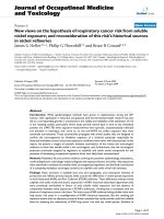
lines. In the RT-PCR analysis, three bands were detected,
corresponding to survivin-2B, wild type survivin, and sur-
vivin-ΔEx3 respectively (Fig. 1A), which were confirmed
by DNA sequence analysis. By the same RT-PCR method,
wild type survivin expression was detected only in the pla-
centa, thymus, and testis among normal adult tissues;
however, survivin-2B and survivin-ΔEx3 were barely
detected (Fig. 1B). By using more sensitive RT-PCR analy-
sis, expression of these splicing variants was shown only
in the thymus [22].
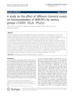
We then analyzed the survivin expression in the protein
level. In all the oral cancer cell lines examined in the
present study, wild type survivin was detected, but not in
normal oral mucosal tissue by Western blotting (Fig. 2). A
small amount of survivin-2B protein was also detected in
some cell lines. These data indicate that the expression of
survivin-2B was more restricted to cancer tissues, though
its level was far less as compared to that of wild type sur-
vivin.
Journal of Translational Medicine 2009, 7:1 />Page 5 of 11
(page number not for citation purposes)
HLA-A24-binding analysis of survivin-derived peptides
To evaluate if wild type survivin might become a target of
immunotherapy as well as a splicing variant survivin-2B,
we re-screened the total amino acid sequence of wild type
survivin protein for peptides containing HLA-A24-bind-
ing motif. In our previous report, two peptides,
survivin85-93 and survivin92-101, derived from exon 3-
encoded region were examined; however, they did not
have a significant binding affinity to HLA-A24 [22]. In the
present study, we identified another peptide, designated
as survivin-C58 (amino acid sequence FFCFKELEGW),
which was derived from exon 2-encoded region. Survivin-
C58 and survivin-2B80-88 were assessed for the binding
ability to HLA-A24 molecule by HLA stabilization assay
using transporters associated with antigen processing
(TAP) deficient and HLA-A*2402-transfected cell line, T2-
A24 cells, as described previously [35,36]. Two positive
control peptides, HLA-A24-restricted CMV-pp65 epitope
and HIV-env epitope, and a negative control peptide VSV8
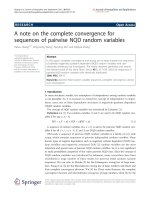
were used in the assay. HLA-A24 level on the cell surface
of T2-A24 cells is up-regulated in the presence of HLA-
A24-binding peptides. Up-regulation of mean fluores-
cence intensity (MFI) of cell surface HLA-A24 was
detected by flow cytometer (Fig. 3). Both CMV-pp65-
derived peptide and HIV-env-derived peptide increased
MFI of HLA-A24 clearly, while VSV8-derived peptide
failed, indicating adequate qualification of this assay.
Both survivin-2B80-88 and survivin-C58 peptides were
capable of up-regulating the HLA-A24 levels, though sur-
vivin-C58 showed less binding capacity than survivin-
2B80-88.
Expression of survivin mRNA as assessed by RT-PCR in normal tissues, and oral cancer cell lines and primary oral cancer tis-suesFigure 1
Expression of survivin mRNA as assessed by RT-PCR in normal tissues, and oral cancer cell lines and primary
oral cancer tissues. (A) Expression of survivin mRNA in oral cancer cell lines and primary oral cancer tissues from two
patients. G3PDH expression was detected as an internal control. (B) Expression of survivin mRNA in normal adult tissues.
293T cells transfected with myc-tagged survivin cDNA (293T-survivin) was used as a positive control for survivin expression.
G3PDH expression was detected as an internal control.
Journal of Translational Medicine 2009, 7:1 />Page 6 of 11
(page number not for citation purposes)
CTL induction from PBMCs of HLA-A*2402
+
cancer
patients
In order to know if HLA-A24-restricted peptide-specific
CTLs are induced from PBMCs of cancer patients, PBMCs
were collected from HLA-A*2402-positive cancer patients
(one breast cancer patient and one oral cancer patient),
and stimulated in vitro with survivin-C58 peptide in the
presence of autologous monocyte-derived DC or autolo-
gous PHA blasts. After 4 times stimulation, cytotoxic activ-
ity against peptide-pulsed target cells was examined by
51
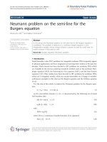
Cr release assay. As shown in Fig. 4A, CTLs induced from
PBMCs of a breast cancer patient were capable of killing
survivin-C58-pulsed T2-A24 target cells, but they failed in
killing SYT-SSX-derived peptide-pulsed T2-A24 cells or
survivin-C58-pulsed HLA-A24-negative target cells. The
same CTLs showed a significant cytotoxicity to HLA-
A*2402-positive breast cancer cells, HMC2 and HMC1,
but not to HLA-A*2402-negative breast cancer cells
MCF7. The similar result was shown in Fig. 4B, when CTLs
were induced from an oral cancer patient (Case #13 in
Table 1). Survivin-C58 peptide-specific CTLs showed
cytotoxicity against HLA-A*2402-transfected oral cancer
cell line OSC20. Therefore, it was indicated that wild type
survivin-derived survivin-C58 peptide could be presented
on tumor cells in the context of HLA-A24 and recognized
by CTLs.
CTL induction efficiency with survivin-2B80-88 or survivin-
C58 from PBMCs of HLA-A24+ oral cancer patients
Previously we showed that survivin-2B80-88-specific
CTLs were induced efficiently from PBMCs of HLA-A24+
patients with survivin-positive breast cancer, colorectal
cancer, and gastric cancer [23]. In the present study, we
examined if survivin-2B80-88-specific CTLs and survivin-
C58-specific CTLs could be induced from PBMCs of HLA-
A24+ oral cancer patients. PBMCs were collected from
thirteen patients with survivin-positive oral cancer and
one healthy volunteer with HLA-A*2402 genotype (Table
1), and stimulated with either or both of these two pep-
tides in vitro in the presence of autologous DC or PHA
blasts as APCs. After 4 times stimulation over a period of
four weeks, CTLs were examined for their peptide-specific
killing activity by 51-Cr release assay using peptide-pulsed
T2-A24 target cells. Survivin-2B-specific CTLs were
induced from four patients out of twelve patients exam-
ined, and survivin-C58-specific CTLs were induced from
three patients out of twelve patients examined. Though
the number of patients in this study was too few to discuss
the exact correlation, it is possible that the CTL induction
efficiency might be related to the disease progression stage
of the patients, since CTLs could not be induced from any
of four patients with stage I (cases #2, #9, #11, and #12),
nor from a healthy volunteer.
PBMCs from eleven patients were stimulated with sur-
vivin-2B80-88 and survivin-C58 peptides in separate
Western blotting analysis of survivin protein in oral cancer cell linesFigure 2
Western blotting analysis of survivin protein in oral cancer cell lines. Lysates from oral cancer cell lines or normal
oral mucosal tissue were resolved by 10% SDS-PAGE and transferred to PVDF membranes. The membranes were then incu-
bated with mouse anti-human survivin monoclonal antibody (upper panel) or mouse anti-β-actin monoclonal antibody AC-15
(lower panel).
Journal of Translational Medicine 2009, 7:1 />Page 7 of 11
(page number not for citation purposes)
HLA-A24-binding assay of peptidesFigure 3
HLA-A24-binding assay of peptides. Binding affinity of peptide to HLA-A24 molecule was evaluated by mean fluorescent
intensity (MFI) shift of cell surface HLA-A24 level on T2-A24 cells that were pulsed with each peptide. CMV pp65-derived
HLA-A24-binding peptide (QVDPVAALF) and HIV env-derived HLA-A24-binding peptide (RYLRDQQLLGI) were used as pos-
itive controls. VSV-derived peptide VSV8 (RGYVYQGL) was used as a negative control. Histograms of MFI shift were displayed
for each peptide. MFI shift was calculated as; MFI shift = (MFI of T2-A24 cells pulsed with the peptide) - (MFI of T2-A24 cells
without peptide pulsation).
Table 1: CTL induction from PBMCs of oral cancer patients
CTL induction Survivin
Case no. age sex stage Origin, histology Prior treatment 2B80-88 specific CTL Survivin-C58-specific CTL expression
#1 63 M Stage II buccal mucosa, SCC, well Chem, Surg - + +
#2 69 M Stage I tongue, SCC, basaloid Surg - - +
#3 60 F Stage II mandibular, SCC, m Chem, Surg - - +
#4 60 M Stage III oropharynx, SCC, mod Chem, Surg + - +
#5 50 F Stage II tongue, SCC, well Chem, Surg + + +
#6 83 F Stage IVA maxillary gingiva, SCC, well Chem, Surg - - +
#7 64 M Stage II tongue, SCC, well Chem, Surg + n.d. +
#8 50 F Stage II submaxillary gland, Adenoid
cystic
Chem, Surg - - +
#9 65 F Stage I tongue, SCC, well Surg - - +
#10 66 F Stage II tongue, SCC, mod Chem, Surg + - +
#11 73 F Stage I tongue, SCC, well Surg - - +
#12 50 F Stage I tongue, SCC, well Surg - - +
#13 67 M Stage IVA oral floor, SCC, poorly Chem, Surg n.d. + +
H#1 35 M - healthy volunteer - - -
Journal of Translational Medicine 2009, 7:1 />Page 8 of 11
(page number not for citation purposes)
wells. CTLs with specificity to either of the two peptides
were induced from three cases (case #1 specific to sur-
vivin-C58, and cases #4 and #10 specific to survivin-2B80-
88), and both survivin-2B80-88-specific CTLs and sur-
vivin-C58-specific CTLs were successfully induced from
one case (case #5) (Fig. 5). These data indicate that sur-
vivin-2B80-88 and survivin-C58 peptides have a compa-
rable potency of CTL induction in oral cancer patients.
Discussion
Survivin is overexpressed in a variety of cancer tissues, and
at least four different splicing variants have been identi-
fied so far. Wild type survivin is known to have an impor-
tant role in the mitotic checkpoint in normal cells and an
anti-apoptotic function in cancer cells [3,18]. In contrast,
the splicing variants are dispensable in the mitotic check-
point [21], and anti-apoptotic function is lost in some
splicing variants such as survivin-2B, in which BIR
domain is disrupted by the insertion of exon 2B [17]. Sur-
vivin-2B and other splicing variant proteins are unstable
Induction of survivin-C58 peptide-specific CTLs and their cytotoxicity against survivin-positive cancer cell linesFigure 4
Induction of survivin-C58 peptide-specific CTLs and their cytotoxicity against survivin-positive cancer cell
lines. CTLs were induced from PBMCs of an HLA-A*2402
+
breast cancer patient by stimulating with survivin-C58 peptide-
pulsed APCs. After four times stimulation, CTLs were subjected to standard
51
Cr release assay at the indicated effector/target
(E/T) ratio. In the left panel, T2-A24 cells and C1R-A31 cells were pulsed with or without survivin-C58 peptide (C58) or SYT-
SSX-derived SS393 peptide (SYT), serving as target cells. In the right panel, survivin-positive breast cancer cell lines with HLA-
A*2402 (HMC1 and HMC2) or without HLA-A*2402 (MCF7 and K562) were used as target cells. (A) CTLs were induced
from PBMCs of an HLA-A*2402
+
oral cancer patient (case #13 in Table 1) by stimulating with survivin-C58 peptide-pulsed
APCs. After four times stimulation, CTLs were subjected to standard
51
Cr release assay at the indicated effector/target (E/T)
ratio. In the left panel, T2-A24 cells were pulsed with or without survivin-C58 peptide (C58), serving as target cells. In the right
panel, survivin-positive HLA-A*2402-negative oral cancer cells (OSC20) and OSC20 transfectants with HLA-A*2402 cDNA
(OSC20-A24) were used as target cells.
Journal of Translational Medicine 2009, 7:1 />Page 9 of 11
(page number not for citation purposes)
Peptide-specific CTL induction using survivin-2B80-88 peptide and survivin-C58 peptide from PBMCs of HLA-A*2402
+
oral cancer patientsFigure 5
Peptide-specific CTL induction using survivin-2B80-88 peptide and survivin-C58 peptide from PBMCs of HLA-
A*2402
+
oral cancer patients. PBMCs of HLA-A*2402
+
oral cancer patients were stimulated in vitro with survivin-2B80-88
peptide-pulsed APCs (APC+2B80-88) and survivin-C58 peptide-pulsed APCs (APC+C58) separately, followed by assessment
of the peptide-specific cytotoxic activity by
51
Cr release assay at the indicated effector/target (E/T) ratio. T2-A24 cells were
pulsed with HIV-env peptide (HIV+), SYT-SSX-derived peptide (K9I+), survivin-2B80-88 peptide (2B+), or survivin-C58 pep-
tide (C+), serving as target cells. P(-) indicates T2-A24 target cells without peptide pulsation. K562 target cells were used for
monitoring natural killer activity and lymphokine-activated non-specific cytotoxicity.
Journal of Translational Medicine 2009, 7:1 />Page 10 of 11
(page number not for citation purposes)
in cells, thereby degraded rapidly. Therefore, survivin
splicing variants do not appear to be suitable for the target
molecules in targeting cancer therapy. However, survivin-
2B is an attractive target antigen for cancer immuno-
therapy, since it contains a unique amino acid sequence
and is barely expressed in normal adult tissue including
thymus, where T-cell tolerance is induced. We have iden-
tified HLA-A24-restricted CTL epitope survivin-2B80-88
derived from survivin-2B previously and reported that it
had a high potency of CTL induction in various cancer
patients including breast cancer, colorectal cancer, and
gastric cancer patients [23]. On the basis of these findings
in vitro, clinical trials of survivin-2B80-88 peptide immu-
notherapy have been conducted for advanced cancers
such as colorectal cancer, breast cancer, lung cancer, and
oral cancer [24,26], in which tumor regression (partial
response) was observed in certain cases. Other groups
have identified the other HLA-restricted CTL epitopes
from wild type survivin and applied for clinical trials
[30,31]. More recently, a novel HLA-A24-restricted CTL
epitope Sur20-28 was identified from wild type survivin
by the screening of a peptide library of overlapping non-
amers spanning the full length of survivin protein [32].
Though the peptide was shown to induce peptide-specific
perforin-positive CD8+ T-cells from PBMCs of cancer
patients, it remains to be determined whether the peptide-
specific T-cells have a capability of killing cancer cells in
an HLA-A24-restricted manner. However, it may be true
that wild type survivin is also immunogenic to cancer host
as well as its splicing variant survivin-2B. Therefore, we re-
screened to find a novel CTL epitope derived from wild
type survivin in the present study. Survivin-C58 peptide-
specific CTLs were successfully induced from PBMCs of
advanced oral cancer patients and exerted HLA-A24-
restricted cytotoxicity against oral cancer cells. The CTL
induction efficiency of survivin-C58 peptide was almost
comparable to that of survivin-2B80-88 peptide, and it
was noted that CTL could not be induced from PBMCs of
oral cancer patients with stage I. These findings contrast
with our previous report that survivin-specific CTLs were
induced successfully from PBMCs of breast cancer
patients and colorectal cancer patients with stage I [23]. It
is speculated that immunogenicity of tumor-expressed
survivin may be lower in the early oral cancer than that in
other cancers. It is possible that the peptide-specific CTL
efficiency might be related to the expression levels of sur-
vivin or survivin-2B proteins in the tumor tissues. As
shown in Table 1, survivin expression was detected in all
the cases by immunostaining. Though there were some
differences in the staining intensity among the cases, we
couldn't find any correlation between the staining inten-
sity and the CTL induction efficiency.
Why does survivin have so immunogenic feature despite
the abundant expression in thymus? The exact answer
remains unknown. Interestingly, we observed that sur-
vivin-positive cells in thymus are mainly cortical thymo-
cytes, but not medullary epithelial cells or dendritic cells
that mediate negative selection and T-cell tolerance. It
may explain at least in part the incomplete peripheral tol-
erance and immunogenic feature of survivin.
Conclusion
In conclusion, we provided evidence that wild type sur-
vivin is an attractive target for the immunotherapy against
oral cancer as well as survivin-2B, and survivin targeting
immunotherapy using survivin-2B80-88 and C58 peptide
cocktail should be suitable for HLA-A24+ cancer patients.
Abbreviations
CTL: cytotoxic T-lymphocyte; PBMC: peripheral blood
mononuclear cells; OSCC: oral squamous cell carcinoma;
DC: dendritic cell; PHA: phytohemagglutinin; APC: anti-
gen presenting cell.
Competing interests
The authors declare that they have no competing interests.
Authors' contributions
JK carried out the CTL induction, killing assays and
drafted the manuscript. TT and YH participated in the
design of the study and performed the evaluation of the
data. TT helped to draft the manuscript. SI contributed to
the HLA-A24-binding assay and CTL induction from
PBMCs. AM, AY and HH contributed to collecting
patients' samples with the informed consent. HH and NS
contributed to the design and coordination of this study
as well as reviewing the manuscript. All authors have read
and approved the final manuscript.
Acknowledgements
We thank Dr. P. G. Coulie for providing anti-HLA-A24 mAb C7709A2.6.
We thank Dr. M. Takiguchi for providing C1R-A*2402 and C1R-A*31012
cells and Dr. K. Kuzushima for providing T2-A24 cells. We are also grateful
to Dr. Hisami Ikeda of Hokkaido Red Cross Blood Center for generous
help with our study. This study was supported in part by a grant-aid from
Ministry of Education, Culture, Sports, Science and Technology of Japan and
a grant-aid for Clinical Cancer Research from the Ministry of Health, Labor
and Welfare of Japan.
References
1. Ambrosini G, Adida C, Altieri DC: A novel anti-apoptosis gene,
survivin, expressed in cancer and lymphoma. Nat Med 1997,
3:917-921.
2. Fukuda S, Pelus LM: Survivin, a cancer target with an emerging
role in normal adult tissues. Mol Cancer Ther 2006, 5:1087-1098.
3. Altieri DC: Survivin, versatile modulation of cell division and
apoptosis in cancer. Oncogene 2003, 22:8581-8589.
4. Asanuma H, Torigoe T, Kamiguchi K, Hirohashi Y, Ohmura T, Hirata
K, Sato M, Sato N: Survivin expression is regulated by coex-
pression of human epidermal growth factor receptor 2 and
epidermal growth factor receptor via phosphatidylinositol 3-
kinase/AKT signaling pathway in breast cancer cells. Cancer
Res 2005, 65:11018-11025.
Publish with Bio Med Central and every
scientist can read your work free of charge
"BioMed Central will be the most significant development for
disseminating the results of biomedical research in our lifetime."
Sir Paul Nurse, Cancer Research UK
Your research papers will be:
available free of charge to the entire biomedical community
peer reviewed and published immediately upon acceptance
cited in PubMed and archived on PubMed Central
yours — you keep the copyright
Submit your manuscript here:
/>BioMedcentral
Journal of Translational Medicine 2009, 7:1 />Page 11 of 11
(page number not for citation purposes)
5. Lo Muzio L, Pannone G, Staibano S, Mignogna MD, Rubini C, Mariggio
MA, Procaccini M, Ferrari F, De Rosa G, Altieri DC: Survivin
expression in oral squamous cell carcinoma. Br J Cancer 2003,
89:2244-2248.
6. Kawasaki H, Altieri DC, Lu CD, Toyoda M, Tenjo T, Tanigawa N:
Inhibition of apoptosis by survivin predicts shorter survival
rates in colorectal cancer. Cancer Res 1998, 58:5071-5074.
7. Kato J, Kuwabara Y, Mitani M, Shinoda N, Sato A, Toyama T, Mitsui
A, Nishiwaki T, Moriyama S, Kudo J, Fujii Y: Expression of survivin
in esophageal cancer: correlation with the prognosis and
response to chemotherapy. Int J Cancer 2001, 95:92-95.
8. Lo Muzio L, Farina A, Rubini C, Pezzetti F, Stabellini G, Laino G,
Santarelli A, Pannone G, Bufo P, de Lillo A, Carinci F: Survivin as
prognostic factor in squamous cell carcinoma of the oral cav-
ity. Cancer Lett 2005, 225:27-33.
9. Rosato A, Pivetta M, Parenti A, Iaderosa GA, Zoso A, Milan G, Man-
druzzato S, Del Bianco P, Ruol A, Zaninotto G, Zanovello P: Survivin
in esophageal cancer: An accurate prognostic marker for
squamous cell carcinoma but not adenocarcinoma. Int J Can-
cer 2006, 119:1717-1722.
10. Tanaka K, Iwamoto S, Gon G, Nohara T, Iwamoto M, Tanigawa N:
Expression of survivin and its relationship to loss of apoptosis
in breast carcinomas. Clin Cancer Res 2000, 6:127-134.
11. Freier K, Pungs S, Sticht C, Flechtenmacher C, Lichter P, Joos S,
Hofele C: High survivin expression is associated with favora-
ble outcome in advanced primary oral squamous cell carci-
noma after radiation therapy. Int J Cancer 2007, 120:942-946.
12. Caldas H, Honsey LE, Altura RA: Survivin 2alpha: a novel Sur-
vivin splice variant expressed in human malignancies. Mol
Cancer 2005, 4:11.
13. Li F: Role of survivin and its splice variants in tumorigenesis.
Br J Cancer 2005, 92:212-216.
14. Ryan B, O'Donovan N, Browne B, O'Shea C, Crown J, Hill AD,
McDermott E, O'Higgins N, Duffy MJ: Expression of survivin and
its splice variants survivin-2B and survivin-DeltaEx3 in breast
cancer.
Br J Cancer 2005, 92:120-124.
15. Zheng W, Ma X, Wei D, Wang T, Ma Y, Yang S: Molecular cloning
and bioinformatics analysis of a novel spliced variant of sur-
vivin from human breast cancer cells. DNA Seq 2005,
16:321-328.
16. Caldas H, Jiang Y, Holloway MP, Fangusaro J, Mahotka C, Conway EM,
Altura RA: Survivin splice variants regulate the balance
between proliferation and cell death. Oncogene 2005,
24:1994-2007.
17. Mahotka C, Liebmann J, Wenzel M, Suschek CV, Schmitt M, Gabbert
HE, Gerharz CD: Differential subcellular localization of func-
tionally divergent survivin splice variants. Cell Death Differ
2002, 9:1334-1342.
18. Li F, Ambrosini G, Chu EY, Plescia J, Tognin S, Marchisio PC, Altieri
DC: Control of apoptosis and mitotic spindle checkpoint by
survivin. Nature 1998, 396:580-584.
19. Song J, So T, Cheng M, Tang X, Croft M: Sustained survivin
expression from OX40 costimulatory signals drives T cell
clonal expansion. Immunity 2005, 22:621-631.
20. Lens SM, Wolthuis RM, Klompmaker R, Kauw J, Agami R, Brum-
melkamp T, Kops G, Medema RH: Survivin is required for a sus-
tained spindle checkpoint arrest in response to lack of
tension. Embo J 2003, 22:2934-2947.
21. Noton EA, Colnaghi R, Tate S, Starck C, Carvalho A, Ko Ferrigno P,
Wheatley SP: Molecular analysis of survivin isoforms: evidence
that alternatively spliced variants do not play a role in mito-
sis. J Biol Chem 2006, 281:1286-1295.
22. Hirohashi Y, Torigoe T, Maeda A, Nabeta Y, Kamiguchi K, Sato T,
Yoda J, Ikeda H, Hirata K, Yamanaka N, Sato N: An HLA-A24-
restricted Cytotoxic T Lymphocyte Epitope of a Tumor-
associated Protein, Survivin. Clin Cancer Res 2002, 8:1731-1739.
23. Idenoue S, Hirohashi Y, Torigoe T, Sato Y, Tamura Y, Hariu H,
Yamamoto M, Kurotaki T, Tsuruma T, Asanuma H, Kanaseki T, Ikeda
H, Kashiwagi K, Okazaki M, Sasaki K, Sato T, Ohmura T, Hata F,
Yamaguchi K, Hirata K, Sato N: A potent immunogenic general
cancer vaccine that targets survivin, an inhibitor of apoptosis
proteins. Clin Cancer Res 2005, 11:
1474-1482.
24. Tsuruma T, Hata F, Torigoe T, Furuhata T, Idenoue S, Kurotaki T,
Yamamoto M, Yagihashi A, Ohmura T, Yamaguchi K, Katsuramaki T,
Yasoshima T, Sasaki K, Mizushima Y, Minamida H, Kimura H, Akiyama
M, Hirohashi Y, Asanuma H, Tamura Y, Shimozawa K, Sato N, Hirata
K: Phase I clinical study of anti-apoptosis protein, survivin-
derived peptide vaccine therapy for patients with advanced
or recurrent colorectal cancer. J Transl Med 2004, 2:19.
25. Tsuruma T, Hata F, Furuhata T, Ohmura T, Katsuramaki T, Yamaguchi
K, Kimura Y, Torigoe T, Sato N, Hirata K: Peptide-based vaccina-
tion for colorectal cancer. Expert Opin Biol Ther 2005, 5:799-807.
26. Tsuruma T, Iwayama Y, Ohmura T, Katsuramaki T, Hata F, Furuhata
T, Yamaguchi K, Kimura Y, Torigoe T, Toyota N, Yagihashi A, Hiro-
hashi Y, Asanuma H, Shimozawa K, Okazaki M, Mizushima Y, Nomura
N, Sato N, Hirata K: Clinical and immunological evaluation of
anti-apoptosis protein, survivin-derived peptide vaccine in
phase I clinical study for patients with advanced or recurrent
breast cancer. J Transl Med 2008, 6:24.
27. Andersen MH, Pedersen LO, Becker JC, Straten PT: Identification
of a cytotoxic T lymphocyte response to the apoptosis inhib-
itor protein survivin in cancer patients. Cancer Res 2001,
61(3):869-872.
28. Bachinsky MM, Guillen DE, Patel SR, Singleton J, Chen C, Soltis DA,
Tussey LG: Mapping and binding analysis of peptides derived
from the tumor-associated antigen survivin for eight HLA
alleles. Cancer Immun 2005, 5:6.
29. Hadrup SR, Gehl J, Sorensen RB, Geertsen PF, Straten PT, Andersen
MH: Persistence of survivin specific T cells for seven years in
a melanoma patient during complete remission. Cancer Biol
Ther 2006, 5:480-482.
30. Otto K, Andersen MH, Eggert A, Keikavoussi P, Pedersen LO, Rath
JC, Bock M, Brocker EB, Straten PT, Kampgen E, Becker JC: Lack of
toxicity of therapy-induced T cell responses against the uni-
versal tumour antigen survivin. Vaccine 2005, 23:884-889.
31. Wobser M, Keikavoussi P, Kunzmann V, Weininger M, Andersen MH,
Becker JC: Complete remission of liver metastasis of pancre-
atic cancer under vaccination with a HLA-A2 restricted pep-
tide derived from the universal tumor antigen survivin.
Cancer Immunol Immunother 2006, 55:
1294-1298.
32. Andersen MH, Soerensen RB, Becker JC, Thor Straten P: HLA-A24
and survivin: possibilities in therapeutic vaccination against
cancer. J Transl Med 2006, 4:38.
33. Lee SP, Tierney RJ, Thomas WA, Brooks JM, Rickinson AB: Con-
served CTL epitopes within EBV latent membrane protein
2: a potential target for CTL-based tumor therapy. J Immunol
1997, 158:3325-3334.
34. Ikeda-Moore Y, Tomiyama H, Miwa K, Oka S, Iwamoto A, Kaneko Y,
Takiguchi M: Identification and characterization of multiple
HLA-A24-restricted HIV-I CTL epitopes: strong epitopes
are derived from V regions of HIV-I. J Immunol 1997,
159:6242-6252.
35. Sato Y, Nabeta Y, Tsukahara T, Hirohashi Y, Syunsui R, Maeda A,
Sahara H, Ikeda H, Torigoe T, Ichimiya S, Wada T, Yamashita T,
Hiraga H, Kawai A, Ishii T, Araki N, Myoui A, Matsumoto S, Umeda
T, Ishii S, Kawaguchi S, Sato N: Detection and induction of CTLs
specific for SYT-SSX-derived peptides in HLA-A24(+)
patients with synovial sarcoma. J Immunol 2002, 169:1611-1618.
36. Ida K, Kawaguchi S, Sato Y, Tsukahara T, Nabeta Y, Sahara H, Ikeda
H, Torigoe T, Ichimiya S, Kamiguchi K, Wada T, Nagoya S, Hiraga H,
Kawai A, Ishii T, Araki N, Myoui A, Matsumoto S, Ozaki T, Yoshikawa
H, Yamashita T, Sato N: Crisscross CTL induction by SYT-SSX
junction peptide and its HLA-A*2402 anchor substitute. J
Immunol 2004, 173:1436-1443.