báo cáo hóa học:" Semi-allogeneic vaccines and tumor-induced immune tolerance" pdf
Bạn đang xem bản rút gọn của tài liệu. Xem và tải ngay bản đầy đủ của tài liệu tại đây (189.51 KB, 6 trang )
BioMed Central
Page 1 of 6
(page number not for citation purposes)
Journal of Translational Medicine
Open Access
Research
Semi-allogeneic vaccines and tumor-induced immune tolerance
Jin Yu
1
, Mark S Kindy
1,3,4
and Sebastiano Gattoni-Celli*
2,3,4
Address:
1
Department of Neurosciences, Medical University of South Carolina, Charleston, SC 29425, USA,
2
Department of Radiation Oncology,
Medical University of South Carolina, Charleston, SC 29425, USA,
3
Ralph H. Johnson VA Medical Center, Charleston, SC 29401, USA and
4
SemiAlloGen Inc., 3384 Shagbark Circle, Mt. Pleasant, SC 29466, USA
Email: Jin Yu - ; Mark S Kindy - ; Sebastiano Gattoni-Celli* -
* Corresponding author
Abstract
Experimental results from studies with inbred mice and their syngeneic tumors indicated that the
inoculation of semi-allogeneic cell hybrids (derived from the fusion between syngeneic tumor cells
and an allogeneic cell line) protects the animal host from a subsequent lethal challenge with
unmodified syngeneic tumor cells. Semi-allogeneic somatic cell hybrids were generated by the
fusion of EL-4 T lymphoma cells (H-2
b
) and BALB/c-derived renal adenocarcinoma RAG cells (H-
2
d
). Cell hybrids were injected intra-peritoneally (i.p.) in C57BL/6 mice (H-2
b
) before challenging
the mice with a tumorigenic dose of EL-4 cells. Semi-allogeneic tumor cell hybrids could not form
a tumor in the animal host because they expressed allogeneic determinants (H-2
d
) and were
rejected as a transplant. However, they conferred protection against a tumorigenic challenge of EL-
4 cells compared to control mice that were mock-vaccinated with i.p injected phosphate-buffered
saline (PBS) and in which EL-4 lymphomas grew rapidly to a large size in the peritoneal cavity.
Screening of spleen-derived RNA by means of focused microarray technology showed up-
regulation of genes involved in the Th-1-type immune response and in the activation of dendritic
antigen-presenting cells (APC). The results of our studies confirm the role of APC in mediating the
immune protection induced by semi-allogeneic vaccines by activating a Th-1 response; these studies
also reveal that semi-allogeneic vaccines are able to interfere with or even block the tumor-
mediated induction of immune tolerance, a key mechanism underlying the suppression of anti-
tumor immunity in the immune competent host.
Background
Almost a century has passed since Paul Erlich first pro-
posed that the immune system has the potential to eradi-
cate cancer even though tumor cells arise from normal
cells. Fifty years later the immune surveillance theory put
forth that lymphocytes have the capacity to survey and
destroy newly arising tumor cells that continuously
appear in the body [1]. The conviction that the immune
system can be mobilized as well as manipulated to eradi-
cate tumor cells has invigorated the field of tumor immu-
nology, one of the most active fields in immunology. The
parallel discoveries of histocompatibility antigens in
humans and mice are a good example of how studies in
animal models and humans may go hand in hand [2]. In
fact, animal studies continue as a basis for important
advances because they have allowed the evaluation of
multiple parameters in tumor immunology that are not
possible in clinical studies [3].
Published: 8 January 2009
Journal of Translational Medicine 2009, 7:3 doi:10.1186/1479-5876-7-3
Received: 17 October 2008
Accepted: 8 January 2009
This article is available from: />© 2009 Yu et al; licensee BioMed Central Ltd.
This is an Open Access article distributed under the terms of the Creative Commons Attribution License ( />),
which permits unrestricted use, distribution, and reproduction in any medium, provided the original work is properly cited.
Journal of Translational Medicine 2009, 7:3 />Page 2 of 6
(page number not for citation purposes)
Despite a reasonable understanding of anti-tumor effector
mechanisms, clinical studies investigating spontaneous
anti-tumor immune responses have yet to lead to repro-
ducible or consistent tumor regression. Thus, the question
of why tumors continue to grow and metastasize in
immunological competent cancer patients remains unan-
swered. Several observations have demonstrated that
tumors evade and actively suppress the immune system.
Tumor evasion of the immune system, termed immune
escape, may occur through several mechanisms, including
(i) tolerance or anergy induction; (ii) the genetic instabil-
ity of tumors; (iii) modulation of tumor antigens; and (iv)
decreased major histocompatibility complex class I
(MHC-I) expression [4]. In addition to evasion of the
immune system, tumors actively suppress the immune
system directly through production of immune suppres-
sive cytokines and indirectly through the induction of
immune inhibitory cells [5]. This secretion of soluble fac-
tors is thought to contribute to the Th2-skewed immune
responses observed in cancer patients and to induce the
development of CD4
+
CD25
+
T regulatory cells [6]. The
ability of these cells to suppress cytotoxic T lymphocyte
(CTL) effector function has been demonstrated in cancer
patients [7,8]. This may explain how tumors can indirectly
suppress anti-tumor immunity; therefore, immuno-
therapy modalities aimed at concurrently stimulating
anti-tumor immune reactivity, while diminishing tumor-
induced immune suppression will be the key to clinical
success.
One of the approaches used to increase the immunogenic-
ity of a tumor is called heterogenization, which can be
achieved by fusing tumor cells with various allogeneic
cells [9,10]. The purpose of heterogenization is to force
the host immune response to recognize tumor-associated
antigens in the context of allogeneic MHC-I or II mole-
cules or in proximity of strong non-self antigens. The all-
ogeneic/non-self antigen would provide a strong
costimulatory signal to enhance anti-tumor immune
responses [11]. This approach stemmed from studies with
inbred mice and their syngeneic tumors; these studies
indicated that the inoculation of semi-allogeneic cell
hybrids (derived from the fusion between syngeneic
tumor cells and an allogeneic cell line) can protect the ani-
mal host from a subsequent lethal challenge with
unmodified syngeneic tumor cells [12-14]. We recently
reported [15] that semi-allogeneic somatic cell hybrids,
generated by the fusion of EL-4 T lymphoma cells (H-2
b
)
and BALB/c-derived renal adenocarcinoma RAG cells (H-
2
d
), conferred protection against a tumorigenic challenge
of EL-4 cells compared to control mice that were mock-
vaccinated with phosphate-buffered saline (PBS). Screen-
ing of spleen-derived RNA by means of focused microar-
ray technology revealed up-regulation of genes involved
in the Th-1-type immune response and in the activation of
dendritic antigen-presenting cells (APC). We now report
experimental evidence suggesting that, in addition to acti-
vating APC and a Th-1-type immune response, semi-allo-
geneic vaccines also inhibit tumor-induced immune
tolerance and anergy.
Methods
Cells and semi-allogeneic hybrids
RAG cells are a non-reverting, 8-azaguanine-resistant
clone of the Renal-2a cell line, originally derived from a
kidney adenocarcinoma of a BALB/c mouse (H-2
d
haplo-
type). RAG cells are deficient in the X-linked hypoxan-
thine-guanine phosphoribosyl transferase gene (HGPRT
-
); therefore, they are killed in culture media containing a
supplement of hypoxanthine, aminopterin, and thymi-
dine (HAT). These cells grow as a monolayer. EL-4 cells
were established from a T-cell lymphoma induced in a
C57BL mouse (H-2
b
haplotype) by the chemical carcino-
gen 9,10-dimethyl-1,2-benzanthracene. These cells grow
in suspension. RAG and EL-4 cell lines were purchased
from the American Type Culture Collection (ATCC). Both
cell lines were propagated in Dulbecco's modified Eagle's
medium (DMEM) supplemented with 10% fetal bovine
serum (FBS), glutamax and antibiotics (Gibco/Invitro-
gen).
RAG cell monolayers were trypsinized, mixed with EL-4
cells, and fused in 50% polyethylene glycol (PEG)-1450
(cell-culture grade from the ATCC and diluted in serum-
free DMEM); after fusion, cells were plated in selective
medium (DMEM + 10% FBS and HAT supplement).
Under these culture conditions only RAG × EL-4 semi-all-
ogeneic somatic cell hybrids will survive, since RAG cells
are killed and EL-4 cells are lost because they grow in sus-
pension and do not attach to the plastic substrate like
somatic cell hybrids do. Resulting cell hybrids were prop-
agated in selective medium and used in vaccination ani-
mal studies.
Animals
Pathogen-free C57BL/6 male mice were obtained through
the Jackson Laboratories (Bar Harbor, ME). All mice were
housed and bred in the VA animal facility located on the
seventh floor of the Strom Thurmond Biomedical
Research Bldg. After vaccination or mock-vaccination and
challenge, mice were monitored very closely for growth of
i.p. tumors and sacrificed when their abdomen became
clearly extended, generally within three to four weeks.
Necropsy was performed on each animal to document the
presence of EL-4-derived i.p. tumors. All animal studies
were carried out according to the PHS Policy on Humane
Care and Use of Laboratory Animals, 2002 and approved
by the Ralph H. Johnson VA Medical Center IACUC.
Journal of Translational Medicine 2009, 7:3 />Page 3 of 6
(page number not for citation purposes)
PCR arrays
Total RNAs were isolated from spleens of mock-vacci-
nated that developed tumors, and from vaccinated mice
that did not develop tumors. These two RNA pools were
analyzed for T-cell and B-cell activation (SA Biosciences,
cat. # PAMM-053), and for T-cell anergy and immune tol-
erance (SA Biosciences, cat. # PAMM-074). These analyses
combine the multi-gene profiling capabilities of a micro-
array with the performance of real-time PCR; therefore,
the results of the PCR studies are both qualitative and
quantitative. The relative or ratio of gene expression, also
known as the fold-change or fold regulation, was calcu-
lated for each gene using the '2
-ΔΔCt
method' [16]. To more
easily determine the genes that were up-regulated or
down-regulated by at least 1.5 fold, a scatter plot compar-
ison was used. Scatter plots compare the normalized, rel-
ative expression of each gene2
-ΔCt
and allow a 'fold-change
boundary' to be drawn within the plot. The 'fold-change
boundary' segregates the genes up or down regulated
based upon the predetermined fold-change value. Scatter
plot comparisons were performed by the microarray man-
ufacturer and included only genes that showed either up-
regulation or down-regulation by 1.5 fold or more. There
are no statistical manipulations within a scatter plot. It
simply allows you to visualize the data in a comprehen-
sive fashion.
Statistical analysis
Data from the animal experiments were analyzed by one-
way analysis of variance for analyses of statistical signifi-
cance, with p < 0.05 indicating statistical significance,
using GraphPad Prism software program (GraphPad Soft-
ware Inc., La Jolla, CA).
Results and Discussion
In vivo animal studies
We set to establish the minimum tumorigenic dose of EL-
4 cells injected intraperitoneally (i.p.) in C57BL/6 mice
(10 mice per group) and tested decreasing numbers of EL-
4 cells (1 × 10
4
, 5 × 10
3
, 2 × 10
3
, 1 × 10
3
, 5 × 10
2
, and 2 ×
10
2
per mouse, respectively) in PBS (0.2 mL per mouse).
We found that, in these experimental conditions, 1 × 10
3
EL-4 cells were very close to the minimum tumorigenic
dose for C57BL/6 mice, most of which developed abdom-
inal tumors within three to four weeks. Even at 2 × 10
2
cells per mouse we observed tumor formation, a clear evi-
dence of the highly malignant phenotype of these cells.
Subsequently, we set to investigate whether irradiated
RAG × EL-4 semi-allogeneic somatic cell hybrids could
protect C57BL/6 mice from a lethal challenge with 1 × 10
3
EL-4 cells. Ten-week-old C57BL/6 male mice were
injected intraperitoneally (i.p.) with 1 × 10
6
RAG × EL-4
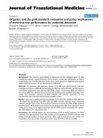
Survival of vaccinated vs. mock-vaccinated miceFigure 1
Survival of vaccinated vs. mock-vaccinated mice. Ten C57BL/6 male mice were vaccinated i.p. with 1 × 10
6
RAG × EL-4
semi-allogeneic somatic cell hybrids [irradiated with 30 Gy (3,000 rad) in a
137
Cs irradiator]. As a control, ten age-matched
mice were mock-vaccinated i.p. with 0.5 mL PBS. Four weeks after vaccination or mock-vaccination each mouse was chal-
lenged by i.p. injection with 1 × 10
3
EL-4 and mice were monitored daily for ten more weeks. Mice with enlarging abdominal
tumors were euthanized and the presence of tumor was confirmed at necropsy (P < 0.0001 between the two survival curves).
0 20 40 60 80 100
0
50
100
Mock-Vaccinated
Vaccinated
Days
Percent sur vival
Journal of Translational Medicine 2009, 7:3 />Page 4 of 6
(page number not for citation purposes)
Table 1: Differential expression of genes involved in T-cell anergy and immune tolerance.
GENE FUNCTION Fold UP-Regulation
(DOWN-Regulation)
BTLA Induced during activation of T cells;
Expressed on Th1 cells;
Interacts with B7 homolog B7H4.
3.0; 2.7
CD40 Co-stimulatory molecule expressed by B cells, dendritic cells, and follicular dendritic cells. 2.6; 2.7
CD40L Expressed by activated T cells;
Binds to CD40 on APC.
1.6, 2.1
CD70 Expressed by activated T and B cells;
Induces proliferation of co-stimulated T cells;
Enhances the generation of CTLs.
3.0, 1.7
FASLG Interacts with FAS and triggers apoptosis. 2.1, 2.2
GZMB Granzyme B is crucial for apoptosis of target
Cells by CTLs.
3.2, 2.4
HDAC9 Histone deacetylase 9, transcriptional repressor. 3.4, 2.7
ICOS Inducible T-cell co-stimulator. 2.2, 1.7
IFNG Th1- and dendritic cell-specific cytokine. 5.5
LTA Lymphotoxin α or tumor necrosis factor β. 2.1, 1.7
PRF1 Perforin, key CTL effector molecule. 2.4, 2.6
TBX21 Th1-specific transcription factor that controls the
expression of IFN-γ.
2.6
TNFRSF4 Receptor involved in CD4+ T cell response. 2.0, 2.1
TNFSF10 TNF-like cytokine;
Induces apoptosis of tumor cells.
2.1, 1.6
TNFSF8 TNF-like cytokine;
Induces apoptosis of some lymphoma cells.
2.1, 1.7
CCR4 Receptor for CC chemokines. (2.9, 2.0)
GATA3 Transcription factor that favors expression of
Th2-type cytokines.
(2.4, 1.5)
IL5 Cytokine for growth and differentiation of B cells
and eosinophils.
(1.3, 4.1)
IL6 Inhibits T cell activation;
Inhibits the CD40L system;
Induces a Th2-type cytokine response.
(5.9, 6.7)
LAT Required for TCR-mediated signaling;
Possibly associated with overstimulation and
apoptosis of T cells.
(2.8, 2.1)
PDCD1 Induction and maintenance of T-cell tolerance. (2.7, 2.0)
Journal of Translational Medicine 2009, 7:3 />Page 5 of 6
(page number not for citation purposes)
semi-allogeneic somatic cell hybrids [irradiated with 30
Gy (3,000 rad) in a
137
Cs irradiator] in 0.5 mL PBS. As a
control, age-matched mice were mock-vaccinated i.p. with
0.5 mL PBS. Four weeks after vaccination or mock-vacci-
nation each mouse was challenged by i.p. injection with 1
× 10
3
EL-4 cells in 0.2 mL PBS. Figure 1 shows that less
than four weeks after challenge, nine of ten mock-vacci-
nated mice had to be euthanized because of large abdom-
inal tumors (verified at necropsy); in contrast, only one of
ten mice vaccinated with irradiated RAG × EL-4 semi-allo-
geneic somatic cell hybrids had to be euthanized almost
five weeks after challenge, because of a large abdominal
tumor. No further changes were observed at ten weeks
after challenge, considered a safe time-frame for measur-
ing established anti-tumor protection. We have per-
formed several experiments of vaccination followed by
challenge, obtaining comparable results (complete pro-
tection from tumor at more than ten weeks after chal-
lenge).
Studies with PCR arrays
Total RNAs were isolated from spleens of mock-vacci-
nated that developed tumors, and from vaccinated mice
that did not develop tumors. We purified RNA from pro-
tected mice at ten weeks after challenge, under the
assumption that those mice had true immune protection
against the EL-4-derived tumor. Obviously, we had to
purify RNA from the spleen of tumor-bearing mice much
earlier (before they would die). These two RNA pools were
analyzed for T-cell and B-cell activation (SA Biosciences,
cat. # PAMM-053), and for T-cell anergy and immune tol-
erance (SA Biosciences, cat. # PAMM-074). The results of
these experiments confirmed to a large extent what we
reported previously [15], including the enhanced expres-
sion of CD80 and CD86. However, the transcriptomic
profile of genes associated with T-cell anergy and immune
tolerance yielded the most informative results. The value
of these microarray studies also stems from the fact that it
combines the multi-gene profiling capabilities of a micro-
array with the performance of real-time PCR; therefore,
the results of the microarray studies are both qualitative
and quantitative. Table 1 shows the summary of these
analyses for genes that were either over-expressed or
down-regulated at the transcription level.
These studies were undertaken to further our understand-
ing of the mechanisms underlying the specific anti-tumor
response induced by semi-allogeneic vaccines. The results
of our animal studies and PCR array experiments confirm
that semi-allogeneic vaccines trigger the activation of den-
dritic APC and CTL to specifically recognize and kill their
target tumor cells. These studies also reveal that semi-all-
ogeneic vaccines are able to interfere with or even block
the tumor-mediated establishment of immune tolerance,
a key mechanism underlying the suppression of anti-
tumor immunity in the immunocompetent host. The
results reported in this short communication represent an
additional building block for future studies aimed at
assessing, by fluorescence-activated cell sorting (FACS),
the phenotypic profile of splenocytes of vaccinated and
mock-vaccinated mice at various time points before and
after vaccination and/or challenge. Furthermore, we plan
to undertake functional analysis of splenocyte subsets to
corroborate, by intracellular staining, the results of the
microarray studies and document the differential expres-
sion of select proteins and cytokines.
Competing interests
MSK and SCG have interests in SemiAlloGen.
Authors' contributions
JY was responsible for conducting the animal experi-
ments, MSK designed the animal experiments and per-
formed the statistical analysis, and SGC was responsible
for overall experimental design and wrote the manuscript.
All authors read and approved the final manuscript.
Acknowledgements
The authors wish to acknowledge support by the National Science Founda-
tion EPSCoR grants (MSK, EPS-0132573 and EPS-0447660), Veterans
Administration Merit Review (MSK) and support from SCLaunch to Semi-
AlloGen.
References
1. Burnet FM: The concept of immunological surveillance. Prog
Exp Tumor Res 1970, 13:1-27.
2. Klein J: Natural history of the major histocompatibility com-
plex. Wiley-Interscience, New York; 1986.
3. Snell GD, Dausset J, Natheson S: Histocompatibility. Academic
Press, New York; 1976.
4. Lathers DMR, Gattoni-Celli S: Tumor Immunology. In Medical
Immunology Volume Chapter 26. 6th edition. Edited by: Virella G.
Informa Healthcare, NY; 2007:369-377.
RNF128 Involved in induction of anergic phenotype (5.4, 4.5)
TNFRSF8 Positive regulator of apoptosis;
Limits proliferation of CD+ effector T cells.
(4.7, 2.8)
Expression was measured by real-time RT-PCR. Total RNA was purified from splenocytes of vaccinated immune mice and compared to RNA from
control, non-immune mice.
Table 1: Differential expression of genes involved in T-cell anergy and immune tolerance. (Continued)
Publish with BioMed Central and every
scientist can read your work free of charge
"BioMed Central will be the most significant development for
disseminating the results of biomedical research in our lifetime."
Sir Paul Nurse, Cancer Research UK
Your research papers will be:
available free of charge to the entire biomedical community
peer reviewed and published immediately upon acceptance
cited in PubMed and archived on PubMed Central
yours — you keep the copyright
Submit your manuscript here:
/>BioMedcentral
Journal of Translational Medicine 2009, 7:3 />Page 6 of 6
(page number not for citation purposes)
5. Smyth MJ, Cretney E, Kershaw MH, Hayakawa Y: Cytokines in can-
cer immunity and immunotherapy. Immunol Rev 2004,
202:275-293.
6. Sakaguchi S: Naturally arising CD4+ regulatory t cells for
immunologic self-tolerance and negative control of immune
responses. Annu Rev Immunol 2004, 22:531-562.
7. Woo EY, Chu CS, Goletz TJ, Schlienger K, Yeh H, Coukos G, Rubin
SC, Kaiser LR, June CH: Regulatory CD4(+)CD25(+) T cells in
tumors from patients with early-stage non-small cell lung
cancer and late-stage ovarian cancer. Cancer Res 2001,
61:4766-72.
8. Wang HY, Lee DA, Peng G, Guo Z, Li Y, Kiniwa Y, Shevach EM, Wang
RF: Tumor-specific human CD4+ regulatory T cells and their
ligands: implications for immunotherapy. Immunity 2004,
20:107-118.
9. Newton DA, Romano C, Gattoni-Celli S: Semiallogeneic cell
hybrids as therapeutic vaccines for cancer. J Immunother 2000,
23:246-254.
10. Newton DA, Acierno PM, Metts MC, Baron PL, Brescia FJ, Gattoni-
Celli S: Semiallogeneic cancer vaccines formulated with gran-
ulocyte-macrophage colony-stimulating factor for patients
with metastatic gastrointestinal adenocarcinomas: a pilot
phase I study. J Immunother 2001, 24:19-26.
11. Grene E, Newton DA, Brown EA, Berzofsky JA, Gattoni-Celli S,
Shearer GM: Semi-allogeneic cell hybrids stimulate HIV-1
envelope-specific cytotoxic T lymphocytes. Aids 2000,
14:1497-1506.
12. Jami J, Ritz E: Expression of tumor-specific antigens in mouse
somatic cell hybrids. Cancer Res 1973, 33:2524-2528.
13. Parkman R: Tumor hybrid cells: an immunotherapeutic agent.
J Natl Cancer Inst 1974, 52:1541-1545.
14. Kim BS: Tumor-specific immunity induced by somatic
hybrids. II. Elicitation of enhanced immunity against the par-
ent plasmacytoma. J Immunol 1979, 123:739-744.
15. Yu J, Kindy MS, Gattoni-Celli S: Semi-Allogeneic Vaccine for T-
Cell Lymphoma. J Translational Medicine 2007, 5:
39-46.
16. Livak KJ, Schmittgen TD: Analysis of relative gene expression
data using real-time quantitative PCR and the 2
ΔΔCT
method.
Methods 2001, 25:402-408.