báo cáo hóa học:" Non-expanded adipose stromal vascular fraction cell therapy for multiple sclerosis" pptx
Bạn đang xem bản rút gọn của tài liệu. Xem và tải ngay bản đầy đủ của tài liệu tại đây (690.39 KB, 9 trang )
BioMed Central
Page 1 of 9
(page number not for citation purposes)
Journal of Translational Medicine
Open Access
Review
Non-expanded adipose stromal vascular fraction cell therapy for
multiple sclerosis
Neil H Riordan
1
, Thomas E Ichim*
1
, Wei-Ping Min
2
, Hao Wang
2
,
Fabio Solano
3
, Fabian Lara
3
, Miguel Alfaro
4
, Jorge Paz Rodriguez
5
,
Robert J Harman
6
, Amit N Patel
7
, Michael P Murphy
8
, Roland R Lee
9,10
and
Boris Minev
11,12
Address:
1
Medistem Inc, San Diego, CA, USA,
2
Department of Surgery, University of Western Ontario, London, Ontario, Canada,
3
Cell Medicine
Institutes, San Jose, Costa Rica,
4
Hospital CIMA, San Jose, Costa Rica,
5
Cell Medicine Institutes, Panama City, Panama,
6
Vet-Stem, Inc. Poway, CA,
USA,
7
Dept of Cardiothoracic Surgery, University of Utah, Salt Lake City, Utah, USA,
8
Division of Medicine, Indiana University School of Medicine,
Indiana, USA,
9
Department of Radiology, University of Canlfornia San Diego, San Diego, CA, USA,
10
Veterans Administration, San Diego, CA,
USA,
11
Moores Cancer Center, University of California, San Diego, CA, USA and
12
Department of Medicine, Division of Neurosurgery, University
of California San Diego, San Diego, CA, USA
Email: Neil H Riordan - ; Thomas E Ichim* - ; Wei-Ping Min - ;
Hao Wang - ; Fabio Solano - ; Fabian Lara - ;
Miguel Alfaro - ; Jorge Paz Rodriguez - ; Robert J Harman - ;
Amit N Patel - ; Michael P Murphy - ; Roland R Lee - ;
Boris Minev -
* Corresponding author
Abstract
The stromal vascular fraction (SVF) of adipose tissue is known to contain mesenchymal stem cells
(MSC), T regulatory cells, endothelial precursor cells, preadipocytes, as well as anti-inflammatory
M2 macrophages. Safety of autologous adipose tissue implantation is supported by extensive use of
this procedure in cosmetic surgery, as well as by ongoing studies using in vitro expanded adipose
derived MSC. Equine and canine studies demonstrating anti-inflammatory and regenerative effects
of non-expanded SVF cells have yielded promising results. Although non-expanded SVF cells have
been used successfully in accelerating healing of Crohn's fistulas, to our knowledge clinical use of
these cells for systemic immune modulation has not been reported. In this communication we
discuss the rationale for use of autologous SVF in treatment of multiple sclerosis and describe our
experiences with three patients. Based on this rationale and initial experiences, we propose
controlled trials of autologous SVF in various inflammatory conditions.
1. Introduction
Adipose tissue has attracted interest as a possible alterna-
tive stem cell source to bone marrow. Enticing character-
istics of adipose derived cells include: a) ease of
extraction, b) higher content of mesenchymal stem cells
(MSC) as compared to bone marrow, and c) ex vivo
expandability of MSC is approximately equivalent, if not
superior to bone marrow [1]. With one exception [2], clin-
ical trials on adipose derived cells, to date, have been lim-
ited to ex vivo expanded cells, which share properties with
bone marrow derived MSC [3-8]. MSC expanded from
adipose tissue are equivalent, if not superior to bone mar-
Published: 24 April 2009
Journal of Translational Medicine 2009, 7:29 doi:10.1186/1479-5876-7-29
Received: 16 March 2009
Accepted: 24 April 2009
This article is available from: />© 2009 Riordan et al; licensee BioMed Central Ltd.
This is an Open Access article distributed under the terms of the Creative Commons Attribution License ( />),
which permits unrestricted use, distribution, and reproduction in any medium, provided the original work is properly cited.
Journal of Translational Medicine 2009, 7:29 />Page 2 of 9
(page number not for citation purposes)
row in terms of differentiation ability [9,10], angiogenesis
stimulating potential [11], and immune modulatory
effects [12]. Given the requirements and potential con-
taminations associated with ex vivo cellular expansion, a
simpler procedure would be the use of primary adipose
tissue derived cells for therapy. Indeed it is reported that
over 3000 horses with various cartilage and bone injuries
have been treated with autologous lipoaspirate fractions
without cellular expansion [13]. In double blind studies
of canine osteoarthritis statistically significant improve-
ments in lameness, range of motion, and overall quality
of life have been described [14,15].
If such approaches could be translated clinically, an easy-
to-use autologous stem cell therapy could be imple-
mented that is applicable to a multitude of indications.
Indeed, this is the desire of commercial entities that are
developing bench top closed systems for autologous adi-
pose cell therapy, such as Cytori's Celution™ system [16]
and Tissue Genesis' TGI 1000™ platform [17], which are
presently entering clinical trials. Unfortunately, since the
majority of scientific studies have focused on in vitro
expanded adipose derived cells, relatively little is known
about the potential clinical effects of the whole lipoaspi-
rate that contains numerous cell populations besides
MSC. From a safety perspective the process of autologous
fat grafting has been commonly used in cosmetic surgery
[18,19], so at least theoretically, autologous cell therapy,
with the numerous cellular populations besides MSC that
are found in adipose tissue, should be relatively innocu-
ous. However, from an efficacy or disease-impact perspec-
tive, it is important to consider the various cellular
components of adipose tissue and to develop a theoretical
framework for evaluating activities that these components
may mediate when administered systemically. For exam-
ple, while attention is focused on the MSC component of
adipose tissue, the high concentrations of monocytes/
macrophages, and potential impact these may have on a
clinical indication is often ignored.
In this paper we will discuss the potential use of the adi-
pose derived cells for the treatment of inflammatory con-
ditions in general, with specific emphasis on multiple
sclerosis. Due to the chronic nature of the disease, the fact
that in some situations remission naturally occurs, as well
as lack of therapeutic impact on long term progression of
current treatments, we examine the possibility of using
autologous adipose derived cells in this condition. We
will discuss the cellular components of adipose tissue, the
biology of these components, how they may be involved
in suppression of inflammatory/immunological aspects
of MS, and conclude by providing case reports of three
patients treatment with autologous adipose derived cells.
2. Components of Adipose Tissue
Mesenchymal Stem Cells
The mononuclear fraction of adipose tissue, referred to as
the stromal vascular fraction (SVF) was originally
described as a mitotically active source of adipocyte pre-
cursors by Hollenberg et al. in 1968 [20]. These cells mor-
phologically resembled fibroblasts and were
demonstrated to differentiate into pre-adipocytes and
functional adipose tissue in vitro [21]. Although it was
suggested that non-adipose differentiation of SVF may
occur under specific conditions [22], the notion of "adi-
pose-derived stem cells" was not widely recognized until
a seminal paper in 2001, where Zuk et al demonstrated
the SVF contains large numbers of mesenchymal stem
cells (MSC)-like cells that could be induced to differenti-
ate into adipogenic, chondrogenic, myogenic, and osteo-
genic lineages [23]. Subsequent to the initial description,
the same group reported after in vitro expansion the SVF
derived cells had surface marker expression similar to
bone marrow derived MSC, comprising of positive for
CD29, CD44, CD71, CD90, CD105/SH2, and SH3 and
lacking CD31, CD34, and CD45 expression [24]. Boquest
et al characterized fresh CD45 negative, CD34 positive,
CD105 positive SVF cells based on CD31 expression. They
demonstrated that the CD31 negative cells exhibited mes-
enchymal properties and could be expanded in vitro,
whereas the CD31 positive cells possessed endothelial-
like properties with poor in vitro expansion capacity [25].
Mesenchymal cells with pluripotent potential have also
been isolated from the liposuction aspirate fluid, which is
the fluid portion of liposuction aspirates [26].
Endothelial Progenitor Cells
In addition to MSC content, it was identified that SVF con-
tains endothelial precursor cells (EPC). A common notion
is that vasculature tissue continually replenishes damaged
endothelial cells de novo from circulating bone marrow
derived EPC [27], and that administration of exogenous
EPC in animals having damaged vasculature can inhibit
progression of atherosclerosis or restenosis [28,29].
Miranville et al demonstrated that human SVF cells iso-
lated from subcutaneous or visceral adipose tissue contain
a population of cells positive for CD34, CD133 and the
drug efflux pump ABCG2 [30]. These cells had endothe-
lial colony forming ability in vitro, and in vivo could
induce angiogenesis in a hindlimb ischemia model. Inter-
estingly, the concentrations of cells with the phenotype
associated with in vivo angiogenic ability, CD31 negative
and CD34 positive, was positively associated with body
mass index. This suggests the possibility that endothelial
precursor cell entrapment in adipose tissue of obese
patients may be related to the reduced angiogenic func-
tion seen in obesity [31]. Several other groups have
reported CD34 positive cells in the SVF capable of stimu-
lating angiogenesis directly or through release of growth
Journal of Translational Medicine 2009, 7:29 />Page 3 of 9
(page number not for citation purposes)
factors such as IGF-1, HGF-1 and VEGF [32-35]. The exist-
ence of a CD34 positive subset in the SVF may indicate
possibility of cells with not only endothelial but also
hematopoietic potential. Indeed at least one report exists
of a bipotent hematopoietic and angiopoietic phenotype
isolated from the SVF [36]. Thus from these data it appears
that SVF contains at least 2 major populations of stem
cells, an MSC compartment and an EPC compartment
that may have some hematopoietic activity. When these
cells are quantified, one author describes that from pri-
mary isolated SVF, approximately 2% of the cells have the
hematopoietic-associated CD34+ CD45+ phenotype, and
6.7% having a mesenchymal CD105+ CD146+ pheno-
type [37]. Many studies using SVF perform in vitro expan-
sion of the cells, this causes selection for certain cell
populations such as MSC and decreases the number of
CD34 cells [38]. Thus in vitro expanded SVF derived cells
can not be compared with primary isolated SVF cells.
Immune Regulatory Monocytes/Macrophages
In addition to its stem/progenitor cell content, the SVF is
known to contain monocytes/macrophages. Although
pluripotency of monocytic populations has previously
been described [39,40], we will focus our discussion to
immunological properties. Initial experiments suggested
that macrophage content of adipose tissue was associated
with the chronic low grade inflammation found in obese
patients. This was suggested by co-culture experiments in
which adipocytes were capable of inducing TNF-alpha
secretion from macrophage cell lines in vitro [41]. Clinical
studies demonstrated that adipocytes also directly release
a constitutive amount of TNF-alpha and leptin, which are
capable of inducing macrophage secretion of inflamma-
tory mediators [42]. It appears from several studies in
mice and humans that when monocytes/macrophages are
isolated from adipose tissue, they in fact possess anti-
inflammatory functions characterized by high expression
of IL-10 and IL-1 receptor antagonist [43-45]. These adi-
pose derived macrophages have an "M2" phenotype,
which physiologically is seen in conditions of immune
suppression such as in tumors [46], post-sepsis compen-
satory anti-inflammatory syndrome [47,48], or pregnancy
associated decidual macrophages [49]. It is estimated that
the monocytic/macrophage compartment of the SVF is
approximately 10% based on CD14 expression [37].
Interestingly, administrations of ex vivo generated M2
macrophages have been demonstrated to inhibit kidney
injury in an adriamycin-induced model [50]. In the con-
text of MS, alternatively activated, M2-like microglial cells
are believed to inhibit progression in the EAE model [51].
Thus the anti-inflammatory activities of M2 cells are a
potential mechanism of therapeutic effect of SVF cells
when isolated from primary sources and not expanded.
T Regulatory Cells
It has been reported by us and others, that activation of T
cells in the absence of costimulatory signals leads to gen-
eration of immune suppressive CD4+ CD25+ T regulatory
(Treg) cells [52,53]. Thus local activation of immunity in
adipose tissue would theoretically be associated with
reduced costimulatory molecule expression by the M2
macrophages, which theoretically may predispose to Treg
generation. Conversely, it is known that Tregs are
involved in maintaining macrophages in the M2 pheno-
type [54]. Supporting the possibility of Treg in adipose tis-
sue also comes from the high concentration of local MSC
which are known to secrete TGF-beta [55] and IL-10 [56],
both involved in Treg generation [57]. Indeed numerous
studies have demonstrated the ability of MSC to induce
Treg cells [56,58-60]. To test the possibility that Treg exist
in the SVF, we performed a series of experiments isolating
CD4, CD25 positive cells from the SVF of BALB/c mice
and compared frequency between other tissues, (lymph
node and spleen). We observed a 3 fold increase in the
CD4+, CD25+ compartment as compared to control tis-
sues. Functionally, these cells were capable of suppressing
ConA stimulated syngeneic CD4+ CD25+ negative cells
(manuscript in preparation).
3. Treatment of Autoimmunity with Adipose
Cells
In general, MSC, whether derived from the bone marrow,
adipose, or other sources, have been demonstrated to
exert dual functions that are relevant to autoimmunity
[61-65]. These conditions are usually exemplified by acti-
vation of innate immune components, breakdown of self
tolerance of the adaptive immune response, and subse-
quent destruction of tissues. Although these are generali-
zations, an initial insult either by foreign microorganisms,
or other means, causes tissue damage and activation of
innate immunity, which under proper genetic back-
ground leads to re-activation/escape from anergy of "self"-
recognizing T cell clones, thus causing more tissue dam-
age, activation of immunity, and lose of function. MSC
inhibit innate immune activation by blocking dendritic
cell maturation [66,67], by suppressing macrophage acti-
vation [68], and by producing agents such as IL-1 receptor
antagonist [69] and IL-10 [70] that directly block inflam-
matory signaling. Perhaps the strongest example of MSC
inhibiting the innate immune response is the recent pub-
lication of Nemeth et al, which demonstrated that admin-
istration of MSC can block onset of sepsis in the aggressive
cecal ligation and puncture model [68]. Through inhibit-
ing DC activation, MSC suppress subsequent adaptive
immunity by generating T regulatory (Treg) cells [59], as
well as blocking cytotoxic activities of CD8 cells. In some
situations, increased immunoregulatory activity is
reported with expanded MSC compartment of SVF as
reported by Mcintosh et al. [71].
Journal of Translational Medicine 2009, 7:29 />Page 4 of 9
(page number not for citation purposes)
In addition to inhibiting pathological innate and adaptive
immunity, MSC have the ability to selectively home to
areas of tissue damage, and mediate direct or indirect
repair function. As an example, CXCR-4 expression of
MSC allows homing toward injured/hypoxic tissue after
intravenous administration. Indeed this has allowed for
numerous studies demonstrating positive effects of intra-
venously administered MSC causing regeneration in
many tissues such as CNS injury [72,73], transplant rejec-
tion [59], toxin-induced diabetes [74], nephropathy [75],
and enteropathy [76]. The regenerative effects of MSC
have been postulated to be mediated by differentiation
into damaged tissue, although this is somewhat contro-
versial, as well as through secretion of growth factors/
antiapoptotic factors which induce tissue regeneration
[77,78].
The ability of MSC to inhibit immune response, while
offering the possibility of inducing/accelerating healing of
tissue that has already been damaged, makes this popula-
tion attractive for treatment of autoimmune disorders.
While numerous studies clinical studies are using
expanded MSC derived from the bone marrow [79-81],
here we chose an indication of autologous adipose SVF
based on the immunological profile, the length of disease
progress allowing several interventions, and the fact that
the disease naturally has periods of remission during
which the rationale would be to amplify a process that
already is underway.
4. Multiple Sclerosis
Multiple sclerosis (MS) is an autoimmune condition in
which the immune system attacks the central nervous sys-
tem (CNS), leading to demyelination. It may cause
numerous physical and mental symptoms, and often
progresses to physical and cognitive disability. Disease
onset usually occurs in young adults, and is more com-
mon in women [82]. MS affects the areas of the brain and
spinal cord known as the white matter. Specifically, MS
destroys oligodendrocytes, which are the cells responsible
for creating and maintaining the myelin sheath, which
helps the neurons carry electrical signals. MS results in a
thinning or complete loss of myelin and, less frequently,
transection of axons [83].
Current therapies for MS include steroids, immune sup-
pressants (cyclosporine, azathioprine, methotrexate),
immune modulators (interferons, glatiramer acetate), and
immune modulating antibodies (natalizumab). At
present none of the MS treatment available on the market
selectively inhibit the immune attack against the nervous
system, nor do they stimulate regeneration of previously
damaged tissue.
Treg cells modulate MS
Induction of remission in MS has been associated with
stimulation of T regulatory cells. For example, patients
responding to the clinically used immune modulatory
drug glatiramer acetate have been reported to have
increased levels of CD4+, CD25+, FoxP3+ Treg cells in
peripheral blood and cerebral spinal fluid [84]. Interferon
beta, another clinically used drug for MS induces a renor-
malization of Treg activity after initiation of therapy
through stimulation of de novo regulatory cell generation
[85]. In the animal model of MS, experimental allergic
encephalomyelitis (EAE), disease progression is exacer-
bated by Treg depletion [86], and natural protection
against disease in certain models of EAE is associated with
antigen-specific Treg [87]. Thus there is some reason to
believe that stimulation of the Treg compartment may be
therapeutically beneficial in MS.
Endogenous neural stem cells affect MS recovery
In addition to immune damage, MS patients are known to
have a certain degree of recovery based on endogenous
repair processes. Pregnancy associated MS remission has
been demonstrated to be associated with increased white
matter plasticity and oligodendrocyte repair activity [88].
Functional MRI (fMRI) studies have suggested that vari-
ous behavioral modifications may augment repair proc-
esses at least in a subset of MS patients [89]. Endogenous
stem cells in the sub-ventricular zone of brains of mice
and humans with MS have been demonstrated to possess
ability to differentiate into oligodendrocytes and to some
extent assist in remyelination [89]. For example, an 8-fold
increase in de novo differentiating sub-ventricular zone
derived cells was observed in autopsy samples of MS
patients in active as compared to non-active lesions [90].
Stem Cell Therapy for MS
The therapeutic effects of MSC in MS have been demon-
strated in several animal studies. In one of the first studies
of immune modulation, Zappia et al. demonstrated
administration of MSC subsequent to immunization with
encephalomyelitis-inducing bovine myelin prevented
onset of the mouse MS-like disease EAE. The investigators
attributed the therapeutic effects to stimulation of Treg
cells, deviation of cytokine profile, and apoptosis of acti-
vated T cells [73]. It is interesting to note that the MSC
were injected intravenously. Several other studies have
shown inhibition of EAE using various MSC injection pro-
tocols [91,92].
To our knowledge there is only one publication describ-
ing clinical exploration of MSC in MS. An Iranian group
reported using intrathecal injections of autologous culture
expanded MSC in treatment unresponsive MS patients
demonstrated improvement in one patient (EDSS score
from 5 to 2.5), no change in 4 patients, and progressive
Journal of Translational Medicine 2009, 7:29 />Page 5 of 9
(page number not for citation purposes)
disease in 5 patients based on EDSS score. Functional sys-
tem assessment revealed six patients had improvement in
their sensory, pyramidal, and cerebellar functions. One
showed no difference in clinical assessment and three
deteriorated [93].
5. Case Reports
Given the rationale that autologous SVF cells have a rea-
sonable safety profile, and contain both immune modu-
latory and regenerative cell populations, a physician-
initiated compassionate-use treatment was explored in 3
patients. Here we describe their treatments and histories.
#CR-231
In 2005, a 50-year-old man was diagnosed with Relaps-
ing-remitting MS, presenting with tonic spasms, stiffness,
gait imbalance, excessive hearing loss, loss of coordina-
tion, numbness in both feet, sexual dysfunction, severe
pain all over his body, fatigue and depression. In 2005,
the patient experienced refractory spells of tonic flexion
spasms, occurring for several minutes at a time and multi-
ple times throughout the day. He was treated with muscle
relaxants, I.V. steroids and Tegretol, and his condition had
improved. However, in 2006 he experienced severe
uncontrollable tonic extensions of all four extremities
lasting about two minutes and associated with significant
pain. Cranial MRI done at that time revealed at least 30
periventricular white matter lesions. Patient also reported
excellent response to Solu-Medrol infusions. Therefore,
the combination of response to steroids, characteristic
MRI abnormalities and positive oligoclonal banding
strongly suggested a diagnosis of Relapsing Remitting MS.
Infusions of Tysabri (Natalizumab, Biogen Idec) every
four weeks were prescribed in November 2006, with excel-
lent results and no significant side effects. However, in
March 2007 patient reported spasticity approximately
three weeks after the infusions, leading to alteration of his
Tysabri infusion regimen to Q3 weeks. By June 2007 the
patient had began complaining of significant memory
loss and by September 2007 he has had recurrence of his
tonic spasms with multiple attacks daily. He was treated
with Solu-Medrol, Baclofen, Provigil, Tegretol, Trileptal,
Tysabri, Vitamins, Omega-3 and Zanaflex with some
improvement of his neurologic symptoms. However, he
complained of severe abdominal pain, decreased appetite
and melanotic stools, consistent with stress ulcer second-
ary to steroid treatment. By November 2007 the patient
was still somewhat responsive to Tysabri and I.V. Solu-
Medrol, but continued to experience multiple severe tonic
spasms at a rate of 30 – 40 spasms per month.
In May 2008, the patient was treated with two I.V. infu-
sions of 28 million SVF cells and multiple intrathecal and
intravenous infusions of allogeneic CD34+ and MSC cells.
MSC were third party unmatched and CD34 were
matched by mixed lymphocyte reaction. Infusions were
performed within a 9-day period and were very well toler-
ated without any adverse or side effects. No other treat-
ments were necessary during the patient's stay. After the
second stem cell infusion the patient reported a signifi-
cant decrease of his generalized pain. However, he contin-
ued to experience severe neck and shoulder pain and was
re-evaluated by his neurologist. Two months after the
stem cell therapy, the volume of his hearing aids had to be
lowered once per week over 4 weeks. Three months after
the stem cell infusions the patient reported a significant
improvement of his cognition and almost complete
reduction of the spasticity in his extremities. He men-
tioned that he has had 623 tonic seizures in the past and
confirmed that he has not experienced any more seizures
since the completion of the stem cell therapy. A neurolog-
ical evaluation performed three months after the stem cell
infusions revealed an intact cranial nerve (II-XII) function
and no nystagmus, normal motor function without any
atrophy or fasciculations, and intact sensory and cerebel-
lar functions and mental status. New MRI images,
obtained 6 months after the stem cell treatment showed
lesions, very similar to the lesions observed before the
stem cell treatment (Figure 1). The patient also reported
significantly improved memory, sexual function, and
energy level. Currently, the patient is taking only multivi-
tamin, minerals and Omega 3.
#233
Second patient: A 32-year-old man was diagnosed in 2001
with relapsing-remitting MS, presenting with fatigue and
depression, uneven walk pattern, cognitive dysfunction,
and a progressive decline in his memory without any spe-
cific neurological symptoms. In 2002 he was started on
weekly intramuscular Avonex (IFN-b1a, Biogen Idec) and
has had no further exacerbations and no evidence of pro-
gressive deterioration. Patient's fatigue was treated well
with Provigil, and his mood improved significantly due to
treatment with Wellbutrin SR. In 2007, the patient com-
plained of some mood changes, with more agitation, irri-
tability, mood destabilization, and cognitive slowing. As
depression was suspected in playing a central role in
patient's condition, Razadyne was added to the antide-
pressant regimen.
In 2008, the patient was treated with two I.V. infusions of
25 million autologous adipose-derived SVF cells and mul-
tiple intrathecal and intravenous infusions of allogeneic
CD34+ and MSC cells. MSC were third party unmatched
and CD34 were matched by mixed lymphocyte reaction.
All infusions were performed within a 10-day period and
were very well tolerated without any significant side
effects. The treatment plan also included physical therapy
sessions.
Journal of Translational Medicine 2009, 7:29 />Page 6 of 9
(page number not for citation purposes)
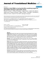
MRI Images obtained before (Panels A and B), and six months after (Panel C) the stem cell treatment of patient 1Figure 1
MRI Images obtained before (Panels A and B), and six months after (Panel C) the stem cell treatment of
patient 1. Panels A and B: Consecutive axial FLuid-Attenuated Inversion Recovery (FLAIR) images through the lateral ven-
tricles show multiple small foci of bright signal in the periventricular and subcortical white matter, consistent with plaques of
multiple sclerosis. Panel C: Axial FLAIR image shows no significant change in the multiple periventricular and subcortical
white-matter plaques. (For the comparison, note that this slice is positioned between those in A and B, and at slightly different
scanning-angle, so it includes lesions of both those slices, as well as others slightly out-of their plane.).
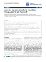
MRI Images obtained before (Panels A and B), and seven months after (Panel C) the stem cell treatment of patient 2Figure 2
MRI Images obtained before (Panels A and B), and seven months after (Panel C) the stem cell treatment of
patient 2. Panels A and B: Consecutive axial FLuid-Attenuated Inversion Recovery (FLAIR) images through the lateral ven-
tricles show multiple small patches of bright signal in the periventricular and subcortical white matter, consistent with plaques
of multiple sclerosis. Panel C: Axial FLAIR image shows no significant change in the multiple periventricular and subcortical
white-matter plaques. (For the comparison, note that this slice is positioned similar to slice A but at slightly different scanning-
angle, so it includes lesions of both slices A and B.).
Journal of Translational Medicine 2009, 7:29 />Page 7 of 9
(page number not for citation purposes)
Three months after the stem cell infusions the patient
reported a significant improvement of his balance and
coordination as well as an improved energy level and
mood. New MRI images, obtained 7 months after the
stem cell treatment showed lesions, very similar to the
lesions observed before the stem cell treatment (Figure 2).
Currently, he is not taking any antidepressants and is
reporting a significantly improved overall condition. His
current treatment regiment includes a weekly injection of
Avonex, vitamins, minerals and Omega 3.
#255
The patient was diagnosed with relapsing-remitting MS in
1993, presenting symptoms were noticeable tingling and
burning sensation in the right leg, followed by paraplegia
lasting almost three weeks. Neurological investigations at
the time uncovered MRI findings suggestive for a demyeli-
nating syndrome. In June of 2008, the patient was treated
with two I.V. infusions of 75 million autologous adipose-
derived SVF cells and multiple intrathecal and intrave-
nous infusions of allogeneic CD34+ and MSC cells. MSC
were third party unmatched and CD34 were matched by
mixed lymphocyte reaction. All infusions were performed
within a 10-day period and were very well tolerated with-
out any significant side effects. His gait, balance and coor-
dination improved dramatically oven a period of several
weeks. His condition continued to improve over the next
few months and he is currently reporting a still continuing
improvement and ability to jog, run and bike for extended
periods of time daily.
Conclusion
The patients treated were part of a compassionate-use
evaluation of stem cell therapeutic protocols in a physi-
cian-initiated manner. Previous experiences in MS
patients using allogeneic CD34+ cord blood cells together
with MSC did not routinely result in substantial improve-
ments observed in the three cases described above. While
obviously no conclusions in terms of therapeutic efficacy
can be drawn from the above reports, we believe that fur-
ther clinical evaluation of autologous SVF cells is war-
ranted in autoimmune conditions.
Competing interests
Thomas E Ichim and Neil H Riordan are management and
shareholders of Medistem Inc, a company that has filed
intellectual property on the use of adipose stromal vascu-
lar fraction cells for immune modulation.
Authors' contributions
All authors read and approved the final manuscript. NHR,
TEI, WPM, HW, FS, FL, MA, JPR, RJH, ANP, MPM, RRL and
BM conceived experiments, interpreted data, and wrote
the manuscript.
Acknowledgements
We thank Victoria Dardov, Rosalia De Necochea Campion, Florica Batu,
and Boris Markosian for stimulating discussions.
References
1. Kern S, Eichler H, Stoeve J, Kluter H, Bieback K: Comparative anal-
ysis of mesenchymal stem cells from bone marrow, umbilical
cord blood, or adipose tissue. Stem Cells 2006, 24:1294-1301.
2. Garcia-Olmo D, Herreros D, Pascual M, Pascual I, De-La-Quintana P,
Trebol J, Garcia-Arranz M: Treatment of enterocutaneous fis-
tula in Crohn's Disease with adipose-derived stem cells: a
comparison of protocols with and without cell expansion. Int
J Colorectal Dis 2009, 24:27-30.
3. Garcia-Olmo D, Garcia-Arranz M, Herreros D, Pascual I, Peiro C,
Rodriguez-Montes JA: A phase I clinical trial of the treatment of
Crohn's fistula by adipose mesenchymal stem cell transplan-
tation. Dis Colon Rectum 2005, 48:1416-1423.
4. Stillaert FB, Di Bartolo C, Hunt JA, Rhodes NP, Tognana E, Monstrey
S, Blondeel PN: Human clinical experience with adipose pre-
cursor cells seeded on hyaluronic acid-based spongy scaf-
folds. Biomaterials 2008, 29:3953-3959.
5. Garcia-Olmo D, Garcia-Arranz M, Herreros D: Expanded adipose-
derived stem cells for the treatment of complex perianal fis-
tula including Crohn's disease. Expert Opin Biol Ther 2008,
8:1417-1423.
6. Fang B, Song YP, Liao LM, Han Q, Zhao RC: Treatment of severe
therapy-resistant acute graft-versus-host disease with
human adipose tissue-derived mesenchymal stem cells. Bone
Marrow Transplant 2006, 38:389-390.
7. Fang B, Song Y, Zhao RC, Han Q, Lin Q: Using human adipose tis-
sue-derived mesenchymal stem cells as salvage therapy for
hepatic graft-versus-host disease resembling acute hepatitis.
Transplant Proc 2007, 39:1710-1713.
8. Fang B, Song Y, Liao L, Zhang Y, Zhao RC: Favorable response to
human adipose tissue-derived mesenchymal stem cells in
steroid-refractory acute graft-versus-host disease. Transplant
Proc 2007, 39:3358-3362.
9. Hayashi O, Katsube Y, Hirose M, Ohgushi H, Ito H: Comparison of
osteogenic ability of rat mesenchymal stem cells from bone
marrow, periosteum, and adipose tissue. Calcif Tissue Int 2008,
82:238-247.
10. Noel D, Caton D, Roche S, Bony C, Lehmann S, Casteilla L, Jorgensen
C, Cousin B:
Cell specific differences between human adipose-
derived and mesenchymal-stromal cells despite similar dif-
ferentiation potentials. Exp Cell Res 2008, 314:1575-1584.
11. Kim Y, Kim H, Cho H, Bae Y, Suh K, Jung J: Direct comparison of
human mesenchymal stem cells derived from adipose tis-
sues and bone marrow in mediating neovascularization in
response to vascular ischemia. Cell Physiol Biochem 2007,
20:867-876.
12. Keyser KA, Beagles KE, Kiem HP: Comparison of mesenchymal
stem cells from different tissues to suppress T-cell activa-
tion. Cell Transplant 2007, 16:555-562.
13. Vet-Stem [
]
14. Black LL, Gaynor J, Gahring D, Adams C, Aron D, Harman S, Gin-
gerich DA, Harman R: Effect of adipose-derived mesenchymal
stem and regenerative cells on lameness in dogs with chronic
osteoarthritis of the coxofemoral joints: a randomized, dou-
ble-blinded, multicenter, controlled trial. Vet Ther 2007,
8:272-284.
15. Black LL, Gaynor J, Adams C, Dhupa S, Sams AE, Taylor R, Harman S,
Gingerich DA, Harman R: Effect of intraarticular injection of
autologous adipose-derived mesenchymal stem and regen-
erative cells on clinical signs of chronic osteoarthritis of the
elbow joint in dogs. Vet Ther 2008, 9:192-200.
16. Lin K, Matsubara Y, Masuda Y, Togashi K, Ohno T, Tamura T, Toy-
oshima Y, Sugimachi K, Toyoda M, Marc H, Douglas A: Characteri-
zation of adipose tissue-derived cells isolated with the
Celution system. Cytotherapy 2008, 10:417-426.
17. Tissue genesis cell isolation system [suegene
sis.com/TGI%201000%20Product%20Brochure.pdf]
18. Hang-Fu L, Marmolya G, Feiglin DH: Liposuction fat-fillant
implant for breast augmentation and reconstruction. Aes-
thetic Plast Surg 1995, 19:427-437.
Journal of Translational Medicine 2009, 7:29 />Page 8 of 9
(page number not for citation purposes)
19. Klein AW: Skin filling. Collagen and otherinjectables of the
skin. Dermatol Clin 2001, 19:491-508. ix
20. Hollenberg CH, Vost A: Regulation of DNA synthesis in fat cells
and stromal elements from rat adipose tissue. J Clin Invest
1969, 47:2485-2498.
21. Gaben-Cogneville AM, Aron Y, Idriss G, Jahchan T, Pello JY, Swierc-
zewski E: Differentiation under the control of insulin of rat
preadipocytes in primary culture. Isolation of homogeneous
cellular fractions by gradient centrifugation. Biochim Biophys
Acta 1983, 762:437-444.
22. Glick JM, Adelman SJ: Established cell lines from rat adipose tis-
sue that secrete lipoprotein lipase. In Vitro 1983, 19:421-428.
23. Zuk PA, Zhu M, Mizuno H, Huang J, Futrell JW, Katz AJ, Benhaim P,
Lorenz HP, Hedrick MH: Multilineage cells from human adipose
tissue: implications for cell-based therapies. Tissue Eng 2001,
7:211-228.
24. Zuk PA, Zhu M, Ashjian P, De Ugarte DA, Huang JI, Mizuno H,
Alfonso ZC, Fraser JK, Benhaim P, Hedrick MH: Human adipose
tissue is a source of multipotent stem cells. Mol Biol Cell 2002,
13:4279-4295.
25. Boquest AC, Shahdadfar A, Fronsdal K, Sigurjonsson O, Tunheim SH,
Collas P, Brinchmann JE: Isolation and transcription profiling of
purified uncultured human stromal stem cells: alteration of
gene expression after in vitro cell culture. Mol Biol Cell 2005,
16:1131-1141.
26. Yoshimura K, Shigeura T, Matsumoto D, Sato T, Takaki Y, Aiba-
Kojima E, Sato K, Inoue K, Nagase T, Koshima I, Gonda K: Charac-
terization of freshly isolated and cultured cells derived from
the fatty and fluid portions of liposuction aspirates. J Cell Phys-
iol 2006, 208:64-76.
27. Asahara T, Murohara T, Sullivan A, Silver M, Zee R van der, Li T, Wit-
zenbichler B, Schatteman G, Isner JM: Isolation of putative pro-
genitor endothelial cells for angiogenesis. Science 1997,
275:964-967.
28. Rauscher FM, Goldschmidt-Clermont PJ, Davis BH, Wang T, Gregg
D, Ramaswami P, Pippen AM, Annex BH, Dong C, Taylor DA: Aging,
progenitor cell exhaustion, and atherosclerosis.
Circulation
2003, 108:457-463.
29. Sata M, Fukuda D, Tanaka K, Kaneda Y, Yashiro H, Shirakawa I: The
role of circulating precursors in vascular repair and lesion
formation. J Cell Mol Med 2005, 9:557-568.
30. Miranville A, Heeschen C, Sengenes C, Curat CA, Busse R, Bouloumie
A: Improvement of postnatal neovascularization by human
adipose tissue-derived stem cells. Circulation 2004, 110:349-355.
31. Urbich C, Dimmeler S: Risk factors for coronary artery disease,
circulating endothelial progenitor cells, and the role of
HMG-CoA reductase inhibitors. Kidney Int 2005, 67:1672-1676.
32. Planat-Benard V, Silvestre JS, Cousin B, Andre M, Nibbelink M, Tam-
arat R, Clergue M, Manneville C, Saillan-Barreau C, Duriez M, Tedgui
A, Levy B, Penicaud L, Casteilla L: Plasticity of human adipose lin-
eage cells toward endothelial cells: physiological and thera-
peutic perspectives. Circulation 2004, 109:656-663.
33. Rehman J, Traktuev D, Li J, Merfeld-Clauss S, Temm-Grove CJ, Bov-
enkerk JE, Pell CL, Johnstone BH, Considine RV, March KL: Secre-
tion of angiogenic and antiapoptotic factors by human
adipose stromal cells. Circulation 2004, 109:1292-1298.
34. Cai L, Johnstone BH, Cook TG, Liang Z, Traktuev D, Cornetta K,
Ingram DA, Rosen ED, March KL: Suppression of hepatocyte
growth factor production impairs the ability of adipose-
derived stem cells to promote ischemic tissue revasculariza-
tion. Stem Cells 2007, 25:3234-3243.
35. Sumi M, Sata M, Toya N, Yanaga K, Ohki T, Nagai R: Transplanta-
tion of adipose stromal cells, but not mature adipocytes,
augments ischemia-induced angiogenesis. Life Sci 2007,
80:559-565.
36. Minana MD, Carbonell-Uberos F, Mirabet V, Marin S, Encabo A:
IFATS collection: Identification of hemangioblasts in the
adult human adipose tissue. Stem Cells 2008, 26:2696-2704.
37. Astori G, Vignati F, Bardelli S, Tubio M, Gola M, Albertini V, Bambi F,
Scali G, Castelli D, Rasini V, Soldati G, Moccetti T: "In vitro" and
multicolor phenotypic characterization of cell subpopula-
tions identified in fresh human adipose tissue stromal vascu-
lar fraction and in the derived mesenchymal stem cells.
J
Transl Med 2007, 5:55.
38. Varma MJ, Breuls RG, Schouten TE, Jurgens WJ, Bontkes HJ, Schu-
urhuis GJ, van Ham SM, van Milligen FJ: Phenotypical and func-
tional characterization of freshly isolated adipose tissue-
derived stem cells. Stem Cells Dev 2007, 16:91-104.
39. Ruhnke M, Ungefroren H, Nussler A, Martin F, Brulport M, Schor-
mann W, Hengstler JG, Klapper W, Ulrichs K, Hutchinson JA, Soria
B, Parwaresch RM, Heeckt P, Kremer B, Fandrich F: Differentiation
of in vitro-modified human peripheral blood monocytes into
hepatocyte-like and pancreatic islet-like cells. Gastroenterology
2005, 128:1774-1786.
40. Ruhnke M, Nussler AK, Ungefroren H, Hengstler JG, Kremer B,
Hoeckh W, Gottwald T, Heeckt P, Fandrich F: Human monocyte-
derived neohepatocytes: a promising alternative to primary
human hepatocytes for autologous cell therapy. Transplanta-
tion 2005, 79:1097-1103.
41. Suganami T, Nishida J, Ogawa Y: A paracrine loop between adi-
pocytes and macrophages aggravates inflammatory
changes: role of free fatty acids and tumor necrosis factor
alpha. Arterioscler Thromb Vasc Biol 2005, 25:2062-2068.
42. Bastard JP, Maachi M, Lagathu C, Kim MJ, Caron M, Vidal H, Capeau
J, Feve B: Recent advances in the relationship between obes-
ity, inflammation, and insulin resistance. Eur Cytokine Netw
2006, 17:4-12.
43. Zeyda M, Stulnig TM: Adipose tissue macrophages. Immunol Lett
2007, 112:61-67.
44. Odegaard JI, Ricardo-Gonzalez RR, Goforth MH, Morel CR, Subrama-
nian V, Mukundan L, Eagle AR, Vats D, Brombacher F, Ferrante AW,
Chawla A: Macrophage-specific PPARgamma controls alter-
native activation and improves insulin resistance. Nature
2007, 447:1116-1120.
45. Zeyda M, Farmer D, Todoric J, Aszmann O, Speiser M, Gyori G, Zlab-
inger GJ, Stulnig TM: Human adipose tissue macrophages are of
an anti-inflammatory phenotype but capable of excessive
pro-inflammatory mediator production. Int J Obes (Lond) 2007,
31:1420-1428.
46. Mantovani A, Sozzani S, Locati M, Allavena P, Sica A:
Macrophage
polarization: tumor-associated macrophages as a paradigm
for polarized M2 mononuclear phagocytes. Trends Immunol
2002, 23:549-555.
47. Mehta A, Brewington R, Chatterji M, Zoubine M, Kinasewitz GT, Peer
GT, Chang AC, Taylor Jr FB, Shnyra A: Infection-induced modu-
lation of m1 and m2 phenotypes in circulating monocytes:
role in immune monitoring and early prognosis of sepsis.
Shock 2004, 22:423-430.
48. Song GY, Chung CS, Jarrar D, Chaudry IH, Ayala A: Evolution of an
immune suppressive macrophage phenotype as a product of
P38 MAPK activation in polymicrobial sepsis. Shock 2001,
15:42-48.
49. Gustafsson C, Mjosberg J, Matussek A, Geffers R, Matthiesen L, Berg
G, Sharma S, Buer J, Ernerudh J: Gene expression profiling of
human decidual macrophages: evidence for immunosup-
pressive phenotype. PLoS ONE 2008, 3:e2078.
50. Wang Y, Wang YP, Zheng G, Lee VW, Ouyang L, Chang DH, Mahajan
D, Coombs J, Wang YM, Alexander SI, Harris DC: Ex vivo pro-
grammed macrophages ameliorate experimental chronic
inflammatory renal disease. Kidney Int 2007, 72:290-299.
51. Ponomarev ED, Maresz K, Tan Y, Dittel BN: CNS-derived inter-
leukin-4 is essential for the regulation of autoimmune
inflammation and induces a state of alternative activation in
microglial cells. J Neurosci 2007, 27:10714-10721.
52. Zhang X, Li M, Lian D, Zheng X, Zhang ZX, Ichim TE, Xia X, Huang
X, Vladau C, Suzuki M, Garcia B, Jevnikar AM, Min WP: Generation
of therapeutic dendritic cells and regulatory T cells for pre-
venting allogeneic cardiac graft rejection. Clin Immunol 2008,
127:313-321.
53. Ichim TE, Zhong R, Min WP: Prevention of allograft rejection by
in vitro generated tolerogenic dendritic cells. Transpl Immunol
2003, 11:295-306.
54. Tiemessen MM, Jagger AL, Evans HG, van Herwijnen MJ, John S,
Taams LS: CD4+CD25+Foxp3+ regulatory T cells induce
alternative activation of human monocytes/macrophages.
Proc Natl Acad Sci USA 2007, 104:19446-19451.
55. Ryan JM, Barry F, Murphy JM, Mahon BP: Interferon-gamma does
not break, but promotes the immunosuppressive capacity of
adult human mesenchymal stem cells. Clin Exp Immunol 2007,
149:353-363.
Journal of Translational Medicine 2009, 7:29 />Page 9 of 9
(page number not for citation purposes)
56. Ye Z, Wang Y, Xie HY, Zheng SS: Immunosuppressive effects of
rat mesenchymal stem cells: involvement of CD4+CD25+
regulatory T cells. Hepatobiliary Pancreat Dis Int 2008, 7:608-614.
57. Askenasy N, Kaminitz A, Yarkoni S: Mechanisms of T regulatory
cell function. Autoimmun Rev 2008, 7:370-375.
58. Gonzalez-Rey E, Gonzalez MA, Varela N, O'Valle F, Hernandez-
Cortes P, Rico L, Buscher D, Delgado M: Human adipose-derived
mesenchymal stem cells reduce inflammatory and T-cell
responses and induce regulatory T cells in vitro in rheuma-
toid arthritis. Ann Rheum Dis 2009 in press.
59. Casiraghi F, Azzollini N, Cassis P, Imberti B, Morigi M, Cugini D, Cav-
inato RA, Todeschini M, Solini S, Sonzogni A, Perico N, Remuzzi G,
Noris M: Pretransplant infusion of mesenchymal stem cells
prolongs the survival of a semiallogeneic heart transplant
through the generation of regulatory T cells. J Immunol 2008,
181:3933-3946.
60. Di Ianni M, Del Papa B, De Ioanni M, Moretti L, Bonifacio E, Cecchini
D, Sportoletti P, Falzetti F, Tabilio A: Mesenchymal cells recruit
and regulate T regulatory cells. Exp Hematol 2008, 36:309-318.
61. Zannettino AC, Paton S, Arthur A, Khor F, Itescu S, Gimble JM,
Gronthos S: Multipotential human adipose-derived stromal
stem cells exhibit a perivascular phenotype in vitro and in
vivo. J Cell Physiol 2008, 214:413-421.
62. Hoogduijn MJ, Crop MJ, Peeters AM, Van Osch GJ, Balk AH, Ijzer-
mans JN, Weimar W, Baan CC: Human heart, spleen, and peri-
renal fat-derived mesenchymal stem cells have
immunomodulatory capacities. Stem Cells Dev 2007,
16:597-604.
63. Chao KC, Chao KF, Fu YS, Liu SH: Islet-like clusters derived from
mesenchymal stem cells in Wharton's Jelly of the human
umbilical cord for transplantation to control type 1 diabetes.
PLoS ONE 2008, 3:e1451.
64. Jo YY, Lee HJ, Kook SY, Choung HW, Park JY, Chung JH, Choung YH,
Kim ES, Yang HC, Choung PH: Isolation and characterization of
postnatal stem cells from human dental tissues. Tissue Eng
2007, 13:767-773.
65. He Q, Wan C, Li G: Concise review: multipotent mesenchymal
stromal cells in blood. Stem Cells 2007, 25:
69-77.
66. Djouad F, Charbonnier LM, Bouffi C, Louis-Plence P, Bony C, Appa-
railly F, Cantos C, Jorgensen C, Noel D: Mesenchymal stem cells
inhibit the differentiation of dendritic cells through an inter-
leukin-6-dependent mechanism. Stem Cells 2007, 25:2025-2032.
67. English K, Barry FP, Mahon BP: Murine mesenchymal stem cells
suppress dendritic cell migration, maturation and antigen
presentation. Immunol Lett 2008, 115:50-58.
68. Nemeth K, Leelahavanichkul A, Yuen PS, Mayer B, Parmelee A, Doi
K, Robey PG, Leelahavanichkul K, Koller BH, Brown JM, Hu X, Jelinek
I, Star RA, Mezey E: Bone marrow stromal cells attenuate sep-
sis via prostaglandin E(2)-dependent reprogramming of host
macrophages to increase their interleukin-10 production.
Nat Med 2009, 15:42-49.
69. LA Ortiz, Dutreil M, Fattman C, Pandey AC, Torres G, Go K, Phinney
DG: Interleukin 1 receptor antagonist mediates the antiin-
flammatory and antifibrotic effect of mesenchymal stem
cells during lung injury. Proc Natl Acad Sci USA 2007,
104:11002-11007.
70. Nasef A, Chapel A, Mazurier C, Bouchet S, Lopez M, Mathieu N,
Sensebe L, Zhang Y, Gorin NC, Thierry D, Fouillard L: Identifica-
tion of IL-10 and TGF-beta transcripts involved in the inhibi-
tion of T-lymphocyte proliferation during cell contact with
human mesenchymal stem cells. Gene Expr 2007, 13:217-226.
71. McIntosh K, Zvonic S, Garrett S, Mitchell JB, Floyd ZE, Hammill L,
Kloster A, Di Halvorsen Y, Ting JP, Storms RW, Goh B, Kilroy G, Wu
X, Gimble JM: The immunogenicity of human adipose-derived
cells: temporal changes in vitro. Stem Cells 2006, 24:1246-1253.
72. Karussis D, Kassis I: The potential use of stem cells in multiple
sclerosis: an overview of the preclinical experience. Clin Neu-
rol Neurosurg 2008, 110:889-896.
73. Zappia E, Casazza S, Pedemonte E, Benvenuto F, Bonanni I, Gerdoni
E, Giunti D, Ceravolo A, Cazzanti F, Frassoni F, Mancardi G, Uccelli
A: Mesenchymal stem cells ameliorate experimental
autoimmune encephalomyelitis inducing T-cell anergy. Blood
2005, 106:1755-1761.
74. Boumaza I, Srinivasan S, Witt WT, Feghali-Bostwick C, Dai Y, Garcia-
Ocana A, Feili-Hariri M: Autologous bone marrow-derived rat
mesenchymal stem cells promote PDX-1 and insulin expres-
sion in the islets, alter T cell cytokine pattern and preserve
regulatory T cells in the periphery and induce sustained nor-
moglycemia. J Autoimmun
2009, 32:33-42.
75. Zhou K, Zhang H, Jin O, Feng X, Yao G, Hou Y, Sun L: Transplan-
tation of human bone marrow mesenchymal stem cell amel-
iorates the autoimmune pathogenesis in MRL/lpr mice. Cell
Mol Immunol 2008, 5:417-424.
76. Parekkadan B, Tilles AW, Yarmush ML: Bone marrow-derived
mesenchymal stem cells ameliorate autoimmune enteropa-
thy independently of regulatory T cells. Stem Cells 2008,
26:1913-1919.
77. Arthur A, Zannettino A, Gronthos S: The therapeutic applica-
tions of multipotential mesenchymal/stromal stem cells in
skeletal tissue repair. J Cell Physiol 2009, 218:237-245.
78. Mishra PK: Bone marrow-derived mesenchymal stem cells for
treatment of heart failure: is it all paracrine actions and
immunomodulation? J Cardiovasc Med (Hagerstown) 2008,
9:122-128.
79. Centeno CJ, Busse D, Kisiday J, Keohan C, Freeman M, Karli D:
Increased knee cartilage volume in degenerative joint dis-
ease using percutaneously implanted, autologous mesenchy-
mal stem cells. Pain Physician 2008, 11:343-353.
80. Katritsis D: Cellular replacement therapy for arrhythmia
treatment: early clinical experience. J Interv Card Electrophysiol
2008, 22:99-105.
81. Slavin S, Kurkalli BG, Karussis D: The potential use of adult stem
cells for the treatment of multiple sclerosis and other neuro-
degenerative disorders. Clin Neurol Neurosurg 2008, 110:943-946.
82. Rosati G: The prevalence of multiple sclerosis in the world: an
update. Neurol Sci 2001, 22:117-139.
83. Pittock SJ, Lucchinetti CF: The pathology of MS: new insights
and potential clinical applications. Neurologist 2007, 13:45-56.
84. Saresella M, Marventano I, Longhi R, Lissoni F, Trabattoni D, Men-
dozzi L, Caputo D, Clerici M: CD4+CD25+FoxP3+PD1- regula-
tory T cells in acute and stable relapsing-remitting multiple
sclerosis and their modulation by therapy. FASEB J 2008,
22:3500-3508.
85. Korporal M, Haas J, Balint B, Fritzsching B, Schwarz A, Moeller S, Fritz
B, Suri-Payer E, Wildemann B: Interferon beta-induced restora-
tion of regulatory T-cell function in multiple sclerosis is
prompted by an increase in newly generated naive regula-
tory T cells. Arch Neurol 2008, 65:1434-1439.
86. Akirav EM, Bergman CM, Hill M, Ruddle NH: Depletion of
CD4(+)CD25(+) T cells exacerbates experimental autoim-
mune encephalomyelitis induced by mouse, but not rat, anti-
gens. J Neurosci Res 2009 in press.
87. Reddy J, Illes Z, Zhang X, Encinas J, Pyrdol J, Nicholson L, Sobel RA,
Wucherpfennig KW, Kuchroo VK: Myelin proteolipid protein-
specific CD4+CD25+ regulatory cells mediate genetic resist-
ance to experimental autoimmune encephalomyelitis. Proc
Natl Acad Sci USA 2004, 101:15434-15439.
88. Gregg C, Shikar V, Larsen P, Mak G, Chojnacki A, Yong VW, Weiss
S: White matter plasticity and enhanced remyelination in
the maternal CNS. J Neurosci 2007, 27:1812-1823.
89. Penner IK, Kappos L, Rausch M, Opwis K, Radu EW: Therapy-
induced plasticity of cognitive functions in MS patients:
insights from fMRI. J Physiol Paris 2006, 99:455-462.
90. Nait-Oumesmar B, Picard-Riera N, Kerninon C, Decker L, Seilhean
D, Hoglinger GU, Hirsch EC, Reynolds R, Baron-Van Evercooren A:
Activation of the subventricular zone in multiple sclerosis:
evidence for early glial progenitors. Proc Natl Acad Sci USA 2007,
104:4694-4699.
91. Kassis I, Grigoriadis N, Gowda-Kurkalli B, Mizrachi-Kol R, Ben-Hur T,
Slavin S, Abramsky O, Karussis D: Neuroprotection and immu-
nomodulation with mesenchymal stem cells in chronic
experimental autoimmune encephalomyelitis. Arch Neurol
2008, 65:753-761.
92. Bai L, Lennon DP, Eaton V, Maier K, Caplan AI, Miller SD, Miller RH:
Human bone marrow-derived mesenchymal stem cells
induce Th2-polarized immune response and promote
endogenous repair in animal models of multiple sclerosis.
Glia 2009 in press.
93. Mohyeddin Bonab M, Yazdanbakhsh S, Lotfi J, Alimoghaddom K,
Talebian F, Hooshmand F, Ghavamzadeh A, Nikbin B: Does mesen-
chymal stem cell therapy help multiple sclerosis patients?
Report of a pilot study. Iran J Immunol 2007, 4:50-57.