báo cáo hóa học:" CTLA4 blockade increases Th17 cells in patients with metastatic melanoma" pdf
Bạn đang xem bản rút gọn của tài liệu. Xem và tải ngay bản đầy đủ của tài liệu tại đây (486.1 KB, 13 trang )
BioMed Central
Page 1 of 13
(page number not for citation purposes)
Journal of Translational Medicine
Open Access
Research
CTLA4 blockade increases Th17 cells in patients with metastatic
melanoma
Erika von Euw
1
, Thinle Chodon
2
, Narsis Attar
2
, Jason Jalil
1
, Richard C Koya
1
,
Begonya Comin-Anduix
1
and Antoni Ribas*
1,2,3
Address:
1
Department of Surgery, Division of Surgical Oncology, University of California, Los Angeles (UCLA), Los Angeles, California, USA,
2
Department of Medicine, Division of Hematology/Oncology, UCLA, Los Angeles, California, USA and
3
Jonsson Comprehensive Cancer Center,
UCLA, Los Angeles, California, USA
Email: Erika von Euw - ; Thinle Chodon - ; Narsis Attar - ;
Jason Jalil - ; Richard C Koya - ; Begonya Comin-Anduix - ;
Antoni Ribas* -
* Corresponding author
Abstract
Background: Th17 cells are CD4+ cells that produce interleukin 17 (IL-17) and are potent
inducers of tissue inflammation and autoimmunity. We studied the levels of this T cell subset in
peripheral blood of patients treated with the anti-CTLA4 antibody tremelimumab since its major
dose limiting toxicities are inflammatory and autoimmune in nature.
Methods: Peripheral blood mononuclear cells (PBMC) were collected before and after receiving
tremelimumab within two clinical trials, one with tremelimumab alone (21 patients) and another
together with autologous dendritic cells (DC) pulsed with the melanoma epitope MART-1
26–35
(6
patients). Cytokines were quantified directly in plasma from patients and after in vitro stimulation
of PBMC. We also quantified IL-17 cytokine-producing cells by intracellular cytokine staining (ICS).
Results: There were no significant changes in 13 assayed cytokines, including IL-17, when analyzing
plasma samples obtained from patients before and after administration of tremelimumab. However,
when PBMC were activated in vitro, IL-17 cytokine in cell culture supernatant and Th17 cells,
detected as IL-17-producing CD4 cells by ICS, significantly increased in post-dosing samples. There
were no differences in the levels of Th17 cells between patients with or without an objective tumor
response, but samples from patients with inflammatory and autoimmune toxicities during the first
cycle of therapy had a significant increase in Th17 cells.
Conclusion: The anti-CTLA4 blocking antibody tremelimumab increases Th17 cells in peripheral
blood of patients with metastatic melanoma. The relation between increases in Th17 cells and
severe autoimmune toxicity after CTLA4 blockade may provide insights into the pathogenesis of
anti-CTLA4-induced toxicities.
Trial Registration: Clinical trial registration numbers: NCT0090896 and NCT00471887
Published: 20 May 2009
Journal of Translational Medicine 2009, 7:35 doi:10.1186/1479-5876-7-35
Received: 18 February 2009
Accepted: 20 May 2009
This article is available from: />© 2009 von Euw et al; licensee BioMed Central Ltd.
This is an Open Access article distributed under the terms of the Creative Commons Attribution License ( />),
which permits unrestricted use, distribution, and reproduction in any medium, provided the original work is properly cited.
Journal of Translational Medicine 2009, 7:35 />Page 2 of 13
(page number not for citation purposes)
Introduction
Monoclonal antibodies blocking the cytotoxic T lym-
phocyte associated antigen 4 (CTLA4), a key negative reg-
ulator of the immune system, induce regression of tumors
in mice and humans, and are being pursued as treatment
for cancer [1-4]. CTLA4 blocking antibodies break toler-
ance to self tissues, as clearly demonstrated by the autoim-
mune phenomena in CTLA4 knock out mice [5,6], which
results in autoimmune toxicities in patients. Understand-
ing the immunological mechanisms guiding antitumor
responses and anti-self toxicities may allow improving the
use of this class of agents in the clinic.
The emerging clinical data suggests that a minority of
patients with metastatic melanoma (in the range of 10%)
achieve durable objective tumor responses when treated
with CTLA4 blocking monoclonal antibodies, with most
being relapse-free up to 7 years later. However, a signifi-
cant proportion of patients (in the range of 20–30%)
develop clinically-relevant toxicities, most often autoim-
mune or inflammatory in nature [2-4]. There is a preva-
lent thought that toxicity and response are correlated after
therapy with anti-CTLA4 blocking monoclonal antibod-
ies. This conclusion is based mainly on statistical correla-
tions in 2 × 2 tables grouping patients with toxicities and/
or objective responses. However, even though patients
with a response are more likely to have toxicities in these
series, most patients with toxicity do not have a tumor
response and there are occasional patients with an objec-
tive tumor response who never developed clinically-rele-
vant toxicities [2,7], thereby suggesting to us that the
relationship between toxicity and response is not linear. If
we assume that both phenomena (toxicity and response)
are mediated by activation of lymphocytes, then we need
to question their antigen specificity, since it is unlikely
that the same T cells that mediate toxicity in the gut, for
example, will be responsible for antitumor activity against
melanoma. It is more likely that the same threshold of
CTLA4 blockade may lead to activation of lymphocytes
reactive to self-tissues and cancer. Therefore, we studied a
differentiated subset of cells termed Th17, which have
emerged as key mediators of autoimmunity and inflam-
mation for their potential implication in toxicity and
responses after anti-CTLA4 therapy.
The description of Th17 cells has substantially advanced
our understanding of T cell-mediated inflammation and
immunity [8]. These cells are characterized as preferential
producers of IL-17A (also known as IL-17), IL-17F, IL-21,
IL-22, and IL-26 in humans. The production of IL-17 is
used to identify Th17 cells and differentiate them from
IFN-γ-producing Th1 cells, or IL-4-producing Th2 cells.
The transcription factor retinoic-acid-related orphan
receptor-γτ (ROR-γτ) and IL-1β and IL-23 are important
for the generation of human Th17 cells in vitro and in vivo
[8,9]. Th17 cells are potent inducers of tissue inflamma-
tion, and dysregulated expression of IL-17 appears to ini-
tiate organ-specific autoimmunity; this has been best
characterized in mouse models of colitis [10], experimen-
tal autoimmune encephalomyelitis (EAE) [11,12], rheu-
matoid arthritis [13] and autoimmune myocarditis [14].
In these models, mice treated with anti-IL-17 antibodies
have lower incidence of disease, slower progression of dis-
ease and reduced scores of disease severity. Treatment
with anti-IL-17 antibodies nine days after inducing EAE
significantly delayed the onset of paralysis. When the
treatment was started at the peak of paralysis, disease pro-
gression was attenuated [15]. Cytokines like IL-17A and
IL-17F, as well as IL-22 (a member of the IL-10 family) are
produced by Th17 and evoke inflammation largely by
stimulating fibroblasts, endothelial cells, epithelial cells
and macrophages to produce chemokines, cytokines and
matrix metalloproteinases (MMP), with the subsequent
recruitment of polymorphonuclear leukocytes to sites of
inflammation [16]. In addition, Th17 cells have been
associated with effective tumor immunity in a model of
adoptive transfer of TCR transgenic CD4+ T cells specific
for the shared self-tumor antigen tyrosinase-related pro-
tein 1 (TRP1) [17]. These cells were used for the treatment
of the poorly immunogenic B16 murine melanoma, and
the therapeutic efficacy of Th1, Th17, and Th0 CD4+ T cell
subsets was studied. The investigators demonstrated that
the tumor-eradicating population was the Th17 cells [17].
Tremelimumab is a fully human IgG2 monoclonal antibody
with high binding affinity for human CTLA-4 [18]. This anti-
body is in late stages of clinical development in patients with
metastatic melanoma [3,4,19]. It has a long plasma half life
of 22 days, which is identical to the half life of endogenous
IgG2s. When administered at doses of 10 to 15 mg/kg,
plasma levels of tremelimumab beyond 30 μg/ml are achiev-
able for 1 to 3 months [19]. This sustained antibody concen-
tration in plasma correlates with the in vitro concentrations
required to have a biological effect of CTLA4 blockade [18],
suggesting that sustained therapeutic levels of this antibody
can be achieved with the doses administered to patients. The
remarkably durable antitumor activity of tremelimumab in a
small subset of patients is mediated by T cell-induced tumor
regressions [20], but its use is limited by autoimmune and
inflammatory toxicities [3,4]. Therefore, understanding the
mechanisms that lead to toxicity and antitumor response are
of great importance to the development of CTLA4 blocking
antibodies. Here we report the increase in Th17 cells in
patients with metastatic melanoma after treatment with
tremelimumab with or without DC vaccines, and its prefer-
ential increase in patients that develop clinically-relevant
inflammatory and autoimmune toxicities.
Patients and methods
Description of Clinical Trials
Peripheral blood samples were obtained from leukapher-
esis procedures from 27 patients with metastatic melanoma
Journal of Translational Medicine 2009, 7:35 />Page 3 of 13
(page number not for citation purposes)
that had been treated at UCLA in two investigator-initiated
research protocols that included the anti-CTLA4 blocking
antibody tremelimumab (Pfizer, New London, CT). In
both clinical trials, patients underwent pre- and post-dos-
ing apheresis collecting PBMC and plasma, and the UCLA
IRB approved informed consent forms described their
banking for immune monitoring assays. Six patients were
treated in a phase I clinical trial of three biweekly intrader-
mal (i.d.) administrations (study days 1, 14 and 28) of a
fixed dose of 1 × 10
7
autologous DC pulsed with the MART-
1
26–35
immunodominant peptide epitope (MART-1
26–35
/
DC) manufactured as previously described [21], concomi-
tantly with a dose escalation of tremelimumab at 10 (3
patients) and 15 mg/kg (3 other patients) every 3 months
(UCLA IRB# 03-12-023, IND# 11579, Trial Registration
number NCT0090896). The samples from these patients
were coded with the study denomination of NRA and a
patient-specific number. The remaining 21 patients were
enrolled in a phase II clinical trial of single agent tremeli-
mumab (UCLA IRB# 06-06-093, IND# 100453, Trial Reg-
istration number NCT00471887) administered at 15 mg/
kg every 3 months. The samples from these patients were
coded with the study denomination of GA and a patient-
specific number. Objective clinical responses were recorded
following a slightly modified Response Evaluation Criteria
in Solid Tumors (RECIST) [22]. The modification was to
consider measurable disease lesions in the skin and subcu-
taneous lesions detectable by physical exam, but not by
imaging exams, if they were adequately recorded at base-
line using a camera with a measuring tape or ruler. Toxici-
ties were recorded during the first 3 months of therapy (one
cycle of tremelimumab-based therapy), since the post-dos-
ing leukapheresis was performed only during the first cycle
of therapy, most frequently between 30 and 60 days from
the first dose of tremelimumab. The post-dosing leuka-
pheresis were performed a median of 41 days after the dose
of tremelimumab (range 28 to 81, with 6 cases out of the
30–60 day range). In all cases, concentrations of tremeli-
mumab in peripheral blood should have been above 10
μg/ml at the time of cell harvesting by leukapheresis, which
is the minimum concentration of tremelimumab that stim-
ulated a biological effect consistent with CTLA4 blockade
in preclinical studies [18]. Adverse events attributed to
tremelimumab by the study investigators were graded
according to the NCI common toxicity criteria version 2.0
[23]. Dose limiting toxicities (DLTs) were prospectively
defined in both studies as any treatment-related toxicity
equal or greater than grade 3, or the clinical evidence of
grade 2 or higher autoimmune reaction in critical organs
(heart, lung, kidney, bowel, bone marrow, musculoskele-
tal, central nervous system and the eye).
Sample Procurement and Processing
PBMC were collected from patients receiving tremelimu-
mab-containing experimental immunotherapy by a leu-
kapheresis procedure. Leukaphereses were planned as part
of the pre-dosing procedures, and one to two months after
receiving the first dose. Leukapheresis products were used
to isolate PBMC by Ficoll-Hypaque (Amersham Pharma-
cia, Piscataway, New Jersey, USA) gradient centrifugation.
PBMC were cryopreserved in liquid nitrogen in Roswell
Park Memorial Institute medium (RPMI, Gibco-BRL,
Gaithersburg, Maryland, USA) supplemented with 20%
(all percentages represent v/v) heat-inactivated human AB
serum (Omega Scientific, Tarzana, California, USA) and
10% dimethylsulfoxide (Sigma, St. Louis, Missouri, USA).
One hundred milliliters of plasma were collected during
the same apheresis procedures and were frozen at -20°C
in 1 to 10 ml single use aliquots. Plasma samples were
thawed and used immediately to measure cytokines.
Cytokine Detection in Plasma
Plasma samples from patients enrolled in the GA study
were assessed for 12 cytokines using a cytokine suspen-
sion array detection system. The cytokines quantified were
IL-1β, IL-2, IL-4, IL-5, IL-6, IL-10, IL-12 (p70), IL-13,
tumor necrosis factor alpha (TNF-α), IFN-γ, granulocyte
colony-stimulating factor (G-CSF), monocyte chemoat-
tractant protein 1 (MCP-1/MCAF) and Chemokine (C-C
motif) ligand 5, CCL-5 (RANTES). The assay was done
according to the manufacturer's instructions in 96-well
plates (Millipore, Billerica, Massachusetts, USA). Samples
were analyzed using the Bio-Plex suspension array system
(Bio-Rad Laboratories, Hercules, California, USA) and the
Bio-Plex manager software with 5PL curve fitting. In addi-
tion, IL-17, a cytokine not represented in the multiplex
cytokine detection kit described above, was quantified in
plasma using a commercially available ELISA according to
the manufacturer's instructions (eBioscience, San Diego,
California, USA). Cytokine concentrations were analyzed
in neat (undiluted) samples. The ranges of detection were
from 6.9 to 5000 pg/ml for IL-4, IL-5, IL-6, IL-10, IL-13,
TNF-α, from 12.3 to 9000 pg/ml for INF-γ and MCP-1,
from 4.1 to 3000 pg/ml for RANTES and from 3.9 to 500
pg/ml for IL-17.
Cytokine Detection in Culture Supernatants
Cryopreserved PBMC aliquots collected before and after
administration of tremelimumab within the GA and NRA
studies were thawed and immediately diluted with RPMI
complete media consisting of 10% human AB serum and
1% penicillin, streptomycin, and amphotericin (Omega
Scientific). Cells were washed and subjected to enzymatic
treatment with DNAse (0.002%, Sigma) for 1 hour at
37°C. Cells were washed again, and an aliquot of each
sample was sorted using CD4+ magnetic cell sorting beads
following the manufacturer's instructions (Miltenyi Biotec
Inc., Auburn, California, USA). 2 × 10
6
pre- and post-dos-
ing PBMC, and the same number of magnetic colum-
sorted CD4+ cells, were incubated for 4 days with 50 ng/
Journal of Translational Medicine 2009, 7:35 />Page 4 of 13
(page number not for citation purposes)
ml of anti-CD3 (OKT3, Ortho-Biotech, Bridgewater, New
Jersey, USA) and 1 μg/ml of anti-CD28 (BD Biosciences,
San Diego, California, USA) in 6-well plates. Cells were
spun down, and the supernatants were collected for IL-17
by ELISA assay. All samples were measured in duplicates.
Intracellular Flow Cytometry for IL-17
To enumerate Th17 cells by ICS, PBMC or sorted CD4+
cells were activated as described above for 4 days in anti-
CD3 and anti-CD28, and then re-stimulated for 5 hours
with 5 μg/μl PMA and 5 μg/μl ionomycin in the presence
of 1 μl/ml of a protein transport inhibitor containing
brefeldin A (GolgiPlug, BD Biosciences) in FACS tubes.
Cells were then surface stained with phycoerythrin (PE)
anti-human CD4 and peridinin-chlorophyll-protein com-
plex (PerCP) anti-CD3 (BD Biosciences) at room temper-
ature for 15 minutes, permeabilized and then stained
intracellularly with APC anti-IL-17 according to the man-
ufacturer's instructions (eBioscience). Isotype antibody
controls were used to enable correct compensation and to
confirm antibody specificity. Flow cytometry analysis was
conducted using FACSCalibur (BD Biosciences), and the
data was analyzed using FlowJo software (Tree Star, Inc.,
San Carlos, California, USA).
Statistical analysis
Statistically significant differences in the concentration or
percentage of IL-17 cytokine and Th17 cells between pre-
and post-treatment samples were analyzed using a two-
sided Student's paired t test using the Prism package
(GraphPad Software, Inc., San Diego, California, USA).
For all statistical analysis, the p value was set at p < 0.05.
There was no correction for multiple comparisons, and all
statistical analysis should be considered exploratory. All
error bars shown in this paper are standard errors of the
means (SEM).
Results
Patient Characteristics, Response and Toxicity
Table 1 provides a description of the study patients, their
baseline characteristics, the treatment received and the
outcome after therapy. Two thirds of the patients had M1c
metastatic melanoma (visceral metastasis and/or high
LDH), and most of the remaining patients had either in
transit (stage IIIc) or soft tissue and nodal metastasis
(M1a). There were 6 patients with objective tumor
responses among the 27 study patients, resulting in sus-
tained and durable tumor regressions in 5 of them, all
with either stage IIIc or M1a metastatic melanoma. Two of
these responses were among the 6 patients enrolled in the
NRA study that included both tremelimumab (one at 10
mg/kg and the other at 15 mg/kg, in both cases adminis-
tered every 3 months) and the MART-1
26–35
peptide
pulsed DC vaccine. The other 3 patients with an objective
response were among the 21 patients enrolled in the GA
study administering single agent tremelimumab at 15 mg/
kg every 3 months. For this study we graded toxicities dur-
ing the first 3 months of therapy, which is considered one
cycle. Among these patients there were 3 with toxicities
that met the definition of DLTs as included in the clinical
trial protocols, all in the GA study. These included two
cases of grade 3 diarrhea or colitis and one patient with
symptomatic panhypopituitarism (grade 2 hypophysitis).
None of these patients received corticosteroids before the
post-dosing apheresis.
No Change in IL-17 in Plasma of Patients Receiving
Tremelimumab
We analyzed the amount of IL-17 at baseline compared to
post-tremelimumab aliquots of cryopreserved plasma
obtained by apheresis. The concentration was very low in
all samples (median of 4 pg/ml), and there were no evi-
dent differences between pre- and post-dosing samples
(Figure 1A). We then analyzed an extended panel of
cytokines in the same plasma samples using a multicy-
tokine array to determine if a preferential cytokine profile
was evident after CTLA4 blockade in patients. Levels of
IL1-β, IL-2 and IL-12 were under the limit of detection for
all samples. Levels of IL-4, IL-5, IL-6, IL-10, IL-13, TNF-α,
INF-γ, MCP-1 and RANTES were detectable above the
assay background, with no differences between pre- and
post-dosing samples in most patients resulting in non-sig-
nificant differences using a paired t test (Figure 1B). How-
ever, the results of one of the patients, GA18, are worth
noting as an outlier in this group of patients. This patient
entered the study with in transit skin metastasis that pro-
gressed after adjuvant interferon alpha 2b and GM-CSF,
this last treatment stopped approximately two months
before initiating tremelimumab. This patient went onto
have a complete response that is ongoing over 1 year from
study initiation. Table 2 provides complete results of the
cytokine analysis in this patient, which demonstrates
post-dosing increases in IL-4, IL-6, IL-10, IL-13, TNF-α,
MCP-1 and RANTES (but not IL-5, IL-17 and INF-γ). These
changes were not noted in any of the other 5 patients with
an objective tumor response in this series, nor in patients
with clinically-significant toxicities. In conclusion, there
were no significant changes in circulating levels of
cytokines after the administration of tremelimumab in
most patients included in this series, and in particular
there were no significant changes in circulating levels of
IL-17 in the plasma of any patient.
IL-17 Production Increases in Ex Vivo Activated PBMC
We examined the difference in the amount of IL-17
cytokine secreted by ex vivo activated cells obtained from
pre- and post-dosing leukapheresis. The spontaneous
cytokine production of non-stimulated PBMC was under
the limit of detection for IL-17, as was for the rest of the
cytokines measured by array (data not shown). Therefore,
Journal of Translational Medicine 2009, 7:35 />Page 5 of 13
(page number not for citation purposes)
Table 1: Patient characteristics
Patient ID Sex Age Stage Location of Metastasis Treme-limumab
(mg/kg q3mo)
MART-1/DC Toxicities During the First Cycle Tumor Response
NRA11 M 57 M1c LN, Muscle 10 Y - PD
NRA12 M 55 M1c Lung, Liver 10 Y - PD
NRA13 F 34 M1c SC, LN, Muscle, Breast 10 Y - PD
NRA14 M 57 IIIc SC 15 Y - CR
NRA15 M 48 M1a LN 15 Y - PR
NRA16 F 61 M1a S.C. 15 Y - PD
GA 5 M 65 M1c Skin, LN, Adrenal 15 N - PR, then PD
GA 7 M 62 IIIc Skin 15 N G2 Pruritus PD
GA 8 F 48 M1c SC 15 N G2 Diarrhea PD
GA 9 M 52 M1c LN, Bone 15 N - PD
GA 11 M 47 M1c LN 15 N - PD
GA 12 M 76 M1c Skin 15 N G3 Colitis PD
GA 13 M 37 M1a LN 15 N G2 Hypophysitis PD
GA 14 M 38 M1c SC, Muscle 15 N - PD
GA 15 M 58 M1c Brain, Bowel, Liver 15 N - PD
GA 18 F 49 M1a Skin 15 N - CR
GA 19 M 55 M1c LN, Brain 15 N G2 Diarrhea PD
GA 21 M 71 M1c Skin, SC, LN, Liver, Spleen 15 N - PD
GA 23 M 27 M1b Lung 15 N - PD
GA 24 M 81 M1c SC, Lung 15 N - PD
GA 25 M 71 M1c LN 15 N - PD
GA 26 M 68 M1b LN, Lung 15 N G3 Diarrhea PD
GA 27 M 52 M1c SC 15 N G2 Pruritus PD
GA 28 M 48 M1c LN, Lung 15 N - PD
GA 29 F 79 IIIc Skin, SC 15 N G2 Diarrhea CR
GA 32 M 36 M1c Muscle 15 N - PD
GA 33 F 49 IIIc Skin 15 N - CR
MART-1/DC: MART-1
26–35
peptide pulsed dendritic cells; G: grade; LN: lymph node; SC: subcutaneous; M: male; F: female; Y: yes; N: no; PD:
progressive disease; SD: stable disease; PR: partial response; CR: complete response.
Journal of Translational Medicine 2009, 7:35 />Page 6 of 13
(page number not for citation purposes)
pre- and post-treatment whole PBMC and CD4-sorted
cells were non-specifically stimulated with anti-CD3/anti-
CD28 for 4 days and then analyzed for the amount of IL-
17 in the culture supernatants by ELISA. IL-17 levels were
significantly increased in the post-treatment samples as
compared to the pre-treatment samples, with a similar
profile in both supernatants from whole PBMC (Figure
2A) and magnetic column-sorted CD4 cells (Figure 2B).
The culture supernatants from activated whole PBMC
were also analyzed for an extended panel of cytokines by
muticytokine array (Figure 2C). There were no differences
in the concentrations of IL-1β, IL-2, IL-4, IL-5, IL-10, IL-
13, TNF-α, and RANTES between pre- and post-dosing
cultures. However, there was a significant decrease in IL-
12(p70) in activated PBMC obtained after the administra-
tion of tremelimumab as compared to the secretion of this
cytokine in activated baseline samples. Taken together,
these data suggests a preferential increase in IL-17 produc-
tion post-dosing.
Th17 Cells Increase after CTLA4 Blockade
The number of IL-17-producing cells was analyzed by ICS
after ex vivo stimulation of whole PBMC and isolated CD4
cells with anti-CD3/anti-CD28 for 4 days. To capture
intracellular IL-17, these cultures were additionally stimu-
lated for 5 hours with mitogens while cytokine secretion
was inhibited with a protein transport inhibitor (see
Materials and Methods). The lymphocyte population was
gated first by morphology, followed by detection of T cells
by anti-CD3 staining, and then Th17 quantitation as dou-
ble positive CD4 cells with intracellular IL-17. Represent-
ative flow cytometric plots from one patient (NRA12)
using CD4-sorted and stimulated cells (Figure 3A) dem-
onstrate the increase in the population of Th17 cells when
comparing pre- and post-dosing samples (Figure 3B).
Double staining with anti-IL-17 and anti-CD4 antibodies
in the samples from GA and NRA study patients revealed
a statistically significant increase in the number of Th17
cells after tremelimumab treatment both in whole PBMC
and in isolated CD4 cells (Figure 3C). Similar results were
obtained when calculating the change in Th17 cells as an
absolute number as opposed to a proportion (pre-dosing
mean of 73,711 with 95% confidence interval of 46,912–
100,510, compared with post-dosing mean of 101,066
with 95% confidence interval of 70,644–131,488, p =
0.026). We also analyzed the background values of IL-17
positive cells among unstimulated CD4+ cells. As
expected, these values are low, with mean of 0.46 pre-dos-
ing (95% confidence interval 0.22–0.7) and 0.62 post-
dosing (95% confidence interval 0.49–0.75), with a trend
(p = 0.15) in favor of increase in the post-dosing samples.
Taken together with the cytokine profile in the culture
supernatants, we conclude that there is a reproducible
increase in IL-17-producing cells among activated blood
cells after the administration of tremelimumab, suggest-
ing an increase in Th17 cells with CTLA4 blockade in
patients with metastatic melanoma.
Preferential Increase in Th17 Cells in Patients with
Autoimmune Toxicity after CTLA4 Blockade
Since Th17 cells have been associated with inflammation,
autoimmunity and antitumor responses, we explored the
changes in pre- and post-dosing levels of IL-17-producing
cells among patients with toxicity or response to tremeli-
mumab-based therapy. There were no differences between
samples from patients with or without an objective tumor
response, either analyzed by IL-17 secretion in culture
supernatants or by ICS for CD4 cells producing IL-17
(data not shown). Similarly, there were no differences
between samples from the GA study administering treme-
limumab alone and the NRA study where patients
received both tremelimumab and an autologous DC vac-
cine. We then analyzed samples from patients with clini-
cally significant toxicities during the first cycle of
tremelimumab-based therapy (within 3 months from first
dosing), meeting the prospectively-defined criteria for
DLTs in these two studies described in the filing of the
Investigator New Drug (IND) applications with the US
Food and Drug Administration. This analysis demon-
strated that the increase in Th17 cells is driven mostly by
patients with toxicities. In PBMC from patients with toxic-
ities the IL-17 increases were 2.3 and 2.2 fold when com-
paring pre- and post-dosing samples by ELISA and ICS,
respectively, while in PBMC from patients without toxic-
ity the respective increments were 1.5 and 1.1 fold. IL-17
increment in sorted CD4+ cells was 3.4 and 1.7 fold in
patients with toxicity measured by ELISA and ICS, respec-
tively, and 1.8 and 1.2 fold in PBMC from patients with-
out toxicity. Even though the number of patients with
toxicities is small in this series, the increase in IL-17-pro-
ducing cells in patients with significant toxicities was
highly reproducible, since it was evident and statistically
significant when comparing IL-17 cytokine production in
culture supernatants of activated whole PBMC and CD4-
sorted cells (Figure 4A and 4B), as well as in the number
of IL-17-producing cells determined by ICS, in both
whole PBMC and CD4-sorted cells (Figure 4C and 4D).
Discussion
Dose-escalation studies with CTLA4 blocking monoclonal
antibodies provide clear evidence that increasing the anti-
body dose and exposure results in increasing toxicities
consistent with breaking tolerance to self tissues, and at
higher dosing levels, some patients benefit with durable
tumor regressions [4,19,24]. Understanding the mecha-
nism of both phenomena is of critical importance for this
class of agents. It seems highly unlikely that the lym-
phocytes that mediate melanoma antitumor responses are
the same as the ones that mediate toxicities like colitis,
hypophysitis or thyroiditis, since there is little evidence of
Journal of Translational Medicine 2009, 7:35 />Page 7 of 13
(page number not for citation purposes)
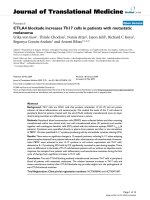
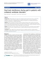
Cytokine quantitation in patient's plasmaFigure 1
Cytokine quantitation in patient's plasma. A) ELISA analysis of IL-17 in cryopreserved plasma samples taken from
patients before and after tremelimumab dosing. B) Multicytokine array quantifying IL-4, IL-5, IL-6, IL-10, IL-13, TNFα,, INF-γ,
MCP-1 and RANTES in cryopreserved plasma before and after dosing with tremelimumab.
0
5
10
15
20
25
30
35
1
2
3
4
5
6
0
50
100
150
200
0
10
20
30
40
50
60
70
0
25
50
75
100
125
0
100
200
300
3
4
5
6
7
8
9
0
100
200
300
400
500
600
700
800
900
0
50
100
150
200
0
2
4
6
8
10
12
IL-17
A
B
IL-4 IL-5 IL-6
IL-10 IL-13 TNF-
IFN-Ȗ MCP-1 RANTES
pre post
pre post pre post pre post
pre post pre post pre post
pre post pre post pre post
pg/ml
pg/mlpg/mlpg/ml
pg/ml
pg/ml
pg/ml
pg/ml
pg/ml
pg/ml
Journal of Translational Medicine 2009, 7:35 />Page 8 of 13
(page number not for citation purposes)
shared antigen profiles recognized by effector T cells
among these tissues. Therefore, many studies have
focused on studying immune cell subsets that are impli-
cated in maintenance of peripheral tolerance. In particu-
lar, a lot of effort has been focused on detecting if Treg are
decreased or functionally impaired in patients receiving
CTLA4 blocking monoclonal antibodies. The interest is
based on several lines of evidence, including the overlap-
ping phenotype of autoimmune conditions in CTLA4 and
FoxP3 deficient mice, and evidence that Treg-specific defi-
ciency in CTLA4 expression impairs the suppressive func-
tion of Tregs [25]. The relatively high basal level of CTLA4
on Treg compared to activated T effector cells (which is the
prime target for these blocking antibodies), and the clini-
cal evidence of the modulation of peripheral tolerance
with CTLA4 blocking antibodies, provided grounds for
studying the implication of Treg in patient-derived sam-
ples. Most data reported to date demonstrate that the
number of circulating cells with a Treg phenotype (CD4,
CD25, FoxP3 positive) does not decrease after the admin-
istration of CTLA4 antibodies. In fact, there is a clear trend
towards an increase in these cells [26-29], a finding that is
not that surprising taking into account that these antibod-
ies are blocking but not depleting antibodies for CTLA4
positive cells. Also, the number of cells staining positive
for FoxP3 by immunohistochemistry increases in tumor
biopsies of regressing lesions after CTLA4 blockade [20].
Data on functional modulation of Treg is not that clear,
with mixed results on the detection of Treg-mediated sup-
pression of effector T cells [26,28,29].
An alternative possibility studied by us is that Th17 cells,
an immune cell subset implicated in mediating autoim-
munity and in chronic inflammatory conditions, may be
modulated by CTLA4 blocking antibodies. There is a
reciprocal negative correlation between Treg and Th17
mediated by IL-2 [30], suggesting that their effects may be
mutually exclusive as opposed to redundant. There is evi-
dence that CTLA4 is expressed on murine Th17 cells at lev-
els that are higher than Th1 cells [31], while CTLA4 has
also been demonstrated on human Th17 cells [32]. Since
both tremelimumab and ipilimumab, the two CTLA4
blocking antibodies in clinical development, inhibit
CTLA4 negative signaling without inducing antibody-
dependent cellular cytotoxicity (ADCC) [18,33], it is cer-
tainly possible that these antibodies would release nega-
tive signaling in Th17 resulting in increased number or
function. In this study we analyzed IL-17 cytokine and
cytokine-producing cells in peripheral blood of patients
treated with tremelimumab with the goal of exploring if
Th17 may be involved in the clinical events in patients
receiving CTLA4 blocking monoclonal antibodies. Our
data provides preliminary evidence that this may be the
case. The modulation of Th17 levels is not large in magni-
tude, but is was highly reproducible among different assay
conditions. Although we could not detect differences in
IL-17 cytokine levels after dosing in plasma samples
obtained directly from peripheral blood, the cells that had
ability to produce IL-17 upon non-specific ex vivo stimula-
tion increased in post-dosing blood cell samples from
patients. This could be detected by quantifying soluble
cytokine in culture supernatants and by determining the
number of cells with intracellular IL-17 by flow cytometry.
In addition, the results were comparable when we ana-
lyzed cultures from whole PBMC (including many
immune and non-immune cell subsets other than CD4 T
helper cells) and with sorted populations containing CD4
cells alone.
Th17 may be implicated in toxicities as well as responses
after administration of anti-CTLA4 antibodies. Besides the
well recognized implication of Th17 in murine and
human inflammatory and autoimmune conditions [8], it
is becoming clearer that they may also have a role in medi-
ating antitumor immunity [17]. Therefore, we explored if
the increases in Th17 cells were more prominent in the
subsets of patients with toxicity or tumor responses.
Although we found no correlation between IL-17 produc-
tion and responses to therapy, our exploratory analysis
suggests that the post-dosing increase in the levels of IL-17
in culture supernatants and by intracellular flow cytome-
try were higher in the small number of patients with tox-
icity. For this analysis, we restricted to clinically-
significant toxicities that followed the prospective defini-
tion of DLTs in the clinical trial protocols, and which hap-
pened during the first cycle of therapy, the closest time to
the obtaining of post-dosing samples in these patients.
When samples from these patients were analyzed sepa-
rately from samples from patients with lower levels of tox-
Table 2: Cytokine levels in plasma of patient GA18
Pre-dosing Post-dosing
IL-4 3.31 32.78
IL-5 3.11 5.56
IL-6 0 181.45
IL-10 0 67.26
IL-13 0 122.46
IL-17 4.34 4
TNF-α 0 294.85
INF-γ 4.32 5.77
MCP-1 0 811.45
RANTES 102.67 141.16
Journal of Translational Medicine 2009, 7:35 />Page 9 of 13
(page number not for citation purposes)
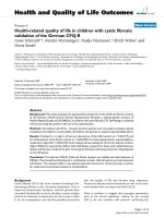
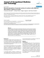
IL-17 quantification by ELISAFigure 2
IL-17 quantification by ELISA. A and B) Pre- and post-dosing IL-17 cytokine determined in culture supernatants of whole
PBMC (A) or CD4+-sorted cells (B) after stimulation for 4 days with anti-CD3 and anti-CD28. The supernatant was collected
for IL-17 quantitation using an ELISA assay (p values by pairwise t-test). C) Multicytokine array in the same ex vivo stimulated
samples quantifying IL-1β, IL-2, IL-4, IL-5, IL-10, IL-12(p70), IL-13, TNFα, and RANTES.
IL-17 PBMC IL-17 CD4
A B
C
IL-1 IL-2 IL-4
IL-5 IL-10 IL-12
IL-13 TNF-
RANTES
pre post pre post
pre post pre post pre post
pre post pre post pre post
pre post pre post pre post
pg/ml x 10
6
cells
pg/ml
pg/ml
pg/ml
pg/ml
pg/ml
** p= 0.0028
* p= 0.038
pg/ml x 10
6
cells
0
500
1000
1500
2000
2500
3000
3500
0
500
1000
1500
2000
2500
0
100
200
300
0
200
400
600
800
1000
1200
0
1000
2000
0
10
20
0
10000
20000
0
20000
40000
60000
80000
0
10000
20000
pg/ml
pg/ml
pg/ml
pg/ml
Journal of Translational Medicine 2009, 7:35 />Page 10 of 13
(page number not for citation purposes)
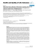
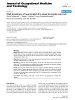
Increase in Th17 cells after tremelimumab-based therapy by intracellular cytokine stainingFigure 3
Increase in Th17 cells after tremelimumab-based therapy by intracellular cytokine staining. A) Gating strategy for
IL-17 intracellular staining. Starting from either whole PBMC or CD-4 sorted cells (as depicted here), the lymphocyte popula-
tion was gated on by FSC-H and SSC-H dot plot. Live cells were gated in the same graphic. A second gate was performed in
CD3 and SSC-H dot plot. We analyzed for IL-17-producing cells among CD4+ T cells after gating. B) Example of IL-17 intracel-
lular staining. After 4-day activation of CD4-sorted cells with anti-CD3 and anti-CD28, cells were additionally stimulated with
PMA and ionomycin while inhibiting protein transport, and the number of Th17 cells was determined by flow cytometry.
Depicted are the plots of gated Th17 cells from patient NRA12. The left column is the baseline pre-dosing sample, and the
right column the post-dosing sample. C) Th17 quantification by flow cytometry. Pre- and post-dosing whole PBMC (left graph)
or CD4+ cells (right graph) analyzed by flow cytometry for Th17 cells as described above (p values by pairwise t-test).
B
60% 95%
11%
60% 95%
11%
C
CD4
CD4 PE
IL-17 APC
7%
14.5%
A
**
p= 0.0096
%
CD3+ CD4+ IL17+
*
p= 0.037
PBMC CD4
%
CD3+ CD4+ IL17+
pre post
Journal of Translational Medicine 2009, 7:35 />Page 11 of 13
(page number not for citation purposes)
icity or no toxicities, differences between pre- and post-
dosing samples were only evident in samples from
patients with DLTs. The significance of increases in Th17
disappeared from the group of patients with non-DLT tox-
icities. Of note, patients with the highest levels of Th17
cells were not the ones who developed toxicities, suggest-
ing to us that it is a doubling of the number of Th17 after
tremelimumab may be linked to toxicities as opposed to
the absolute number at any given time point. Our explor-
atory analysis is obviously limited by the small number of
patients in this series, and will need to be confirmed in
larger groups. However, the findings are reproducible in
all of the different experimental conditions used to ana-
lyze IL-17-producing cells, which provides confidence in
these results. From this work we conclude that Th17 may
be implicated in the clinical effects of CTLA4 blocking
monoclonal antibodies, and further study of their role in
treatment-induced toxicities may help in elucidating how
toxicities and responses may be differentially modulated
with this mode of therapy.
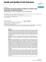
IL-17 Intracellular Staining and IL-17 ELISA According to the Development of Inflammatory or Autoimmune ToxicityFigure 4
IL-17 Intracellular Staining and IL-17 ELISA According to the Development of Inflammatory or Autoimmune
Toxicity. A and B) IL-17 secretion detected by ELISA as described in Figure 1, and Th17 by intracellular staining (ICS) as
described in Figure 2, comparing the assay results in whole PBMC cultures from patients with Grade 3 or higher toxicity and
the rest of the patients (p values by pairwise t-test). C and D) The same analysis with CD4-sorted cultures.
C D
A B
%
CD3+ CD4+ IL-17+
%
CD3+ CD4+ IL-17+
pg/ml x 10
6
cells
pg/ml x 10
6
cells
pre post pre post
pre post pre post
pre post pre post
pre post pre post
PBMC
CD4
*p= 0.044
*p= 0.046
*p= 0.019
*p= 0.046
Non Toxicity DLTs Non Toxicity DLTs
Non Toxicity DLTsNon Toxicity DLTs
Journal of Translational Medicine 2009, 7:35 />Page 12 of 13
(page number not for citation purposes)
Competing interests
AR has received research funding and honoraria from
Pfizer. The other authors have no competing interests on
this work.
Authors' contributions
EVE and AR conceived and designed the study. EVE, TC
and NA carried out the experiments. JJ and BC-A provided
the human samples for analysis. RCK and BC-A contrib-
uted to the assay conduct and data interpretation. EVE and
AR wrote the manuscript. All authors read and approved
the final manuscript.
Acknowledgements
EvE was supported by grants from the Consejo Nacional de Investigaciones
Científicas y Técnicas (CONICET), and the Fundación Sales, Buenos Aires,
Argentina. AR was supported by the Harry J. Lloyd Charitable Trust, P50
CA086306, U54 CA119347 and RN2-00902-1 New Faculty Award 2 from
the California Institute for Regenerative Medicine (CIRM). Flow cytometry
assays were performed in the UCLA Jonsson Comprehensive Cancer
Center (JCCC) and Center for AIDS Research Flow Cytometry Core Facil-
ity that is supported by National Institutes of Health awards CA-16042 and
AI-28697 and by the JCCC, the UCLA AIDS Institute, and the David Geffen
School of Medicine at UCLA. Patients were treated at the UCLA General
Clinical Research Center (G-CRC), which is supported by USPHS Grant
M01-RR-0865.
References
1. Leach DR, Krummel MF, Allison JP: Enhancement of antitumor
immunity by CTLA-4 blockade. Science 1996, 271:1734-1736.
2. Downey SG, Klapper JA, Smith FO, Yang JC, Sherry RM, Royal RE,
Kammula US, Hughes MS, Allen TE, Levy CL, Yellin M, Nichol G,
White DE, Steinberg SM, Rosenberg SA: Prognostic factors
related to clinical response in patients with metastatic
melanoma treated by CTL-associated antigen-4 blockade.
Clin Cancer Res 2007, 13:6681-6688.
3. Ribas A, Hauschild A, Kefford R, Punt CA, Haanen JB, Marmol M,
Garbe C, Gomez-Navarro J, Pavlov D, Marshall M: Phase III, Open-
Label, Randomized, Comparative Study of Tremelimumab
(CP-675,206) and Chemotherapy (Temozolomide or Dacar-
bazine) in Patients with Advanced Melanoma. J Clin Oncol
2008, 26(Suppl):9011.
4. Camacho LH, Antonia S, Sosman J, Kirkwood JM, Gajewski TF, Red-
man B, Pavlov D, Bulanhagui C, Bozon VA, Gomez-Navarro J, Ribas
A: Phase I/II trial of Tremelimumab in patients with meta-
static melanoma. J Clin Oncol 2009, 27:1075-1081.
5. Waterhouse P, Penninger JM, Timms E, Wakeham A, Shahinian A, Lee
KP, Thompson CB, Griesser H, Mak TW: Lymphoproliferative
disorders with early lethality in mice deficient in Ctla-4. Sci-
ence 1995, 270:985-988.
6. Tivol EA, Borriello F, Schweitzer AN, Lynch WP, Bluestone JA,
Sharpe AH: Loss of CTLA-4 leads to massive lymphoprolifer-
ation and fatal multiorgan tissue destruction, revealing a
critical negative regulatory role of CTLA-4. Immunity 1995,
3:541-547.
7. Attia P, Phan GQ, Maker AV, Robinson MR, Quezado MM, Yang JC,
Sherry RM, Topalian SL, Kammula US, Royal RE, Restifo NP, Haworth
LR, Levy C, Mavroukakis SA, Nichol G, Yellin MJ, Rosenberg SA:
Autoimmunity correlates with tumor regression in patients
with metastatic melanoma treated with anti-cytotoxic T-
lymphocyte antigen-4. J Clin Oncol 2005, 23:6043-6053.
8. Bettelli E, Oukka M, Kuchroo VK: T(H)-17 cells in the circle of
immunity and autoimmunity. Nat Immunol 2007, 8:345-350.
9. Bettelli E, Korn T, Oukka M, Kuchroo VK: Induction and effector
functions of T(H)17 cells. Nature 2008, 453:1051-1057.
10. Zhang Z, Zheng M, Bindas J, Schwarzenberger P, Kolls JK:
Critical
role of IL-17 receptor signaling in acute TNBS-induced coli-
tis. Inflamm Bowel Dis 2006, 12:382-388.
11. Hofstetter HH, Ibrahim SM, Koczan D, Kruse N, Weishaupt A, Toyka
KV, Gold R: Therapeutic efficacy of IL-17 neutralization in
murine experimental autoimmune encephalomyelitis. Cell
Immunol 2005, 237:123-130.
12. Komiyama Y, Nakae S, Matsuki T, Nambu A, Ishigame H, Kakuta S,
Sudo K, Iwakura Y: IL-17 plays an important role in the devel-
opment of experimental autoimmune encephalomyelitis. J
Immunol 2006, 177:566-573.
13. Nakae S, Nambu A, Sudo K, Iwakura Y: Suppression of immune
induction of collagen-induced arthritis in IL-17-deficient
mice. J Immunol 2003, 171:6173-6177.
14. Sonderegger I, Rohn TA, Kurrer MO, Iezzi G, Zou Y, Kastelein RA,
Bachmann MF, Kopf M: Neutralization of IL-17 by active vacci-
nation inhibits IL-23-dependent autoimmune myocarditis.
Eur J Immunol 2006, 36:2849-2856.
15. Rohn TA, Jennings GT, Hernandez M, Grest P, Beck M, Zou Y, Kopf
M, Bachmann MF: Vaccination against IL-17 suppresses
autoimmune arthritis and encephalomyelitis. Eur J Immunol
2006, 36:2857-2867.
16. Jin D, Zhang L, Zheng J, Zhao Y: The inflammatory Th 17 subset
in immunity against self and non-self antigens. Autoimmunity
2008, 41:154-162.
17. Muranski P, Boni A, Antony PA, Cassard L, Irvine KR, Kaiser A, Paulos
CM, Palmer DC, Touloukian CE, Ptak K, Gattinoni L, Wrzesinski C,
Hinrichs CS, Kerstann KW, Feigenbaum L, Chan CC, Restifo NP:
Tumor-specific Th17-polarized cells eradicate large estab-
lished melanoma. Blood 2008, 112:362-373.
18. Ribas A, Hanson DC, Noe DA, Millham R, Guyot DJ, Bernstein SH,
Canniff PC, Sharma A, Gomez-Navarro J: Tremelimumab (CP-
675,206), a cytotoxic T lymphocyte associated antigen 4
blocking monoclonal antibody in clinical development for
patients with cancer. Oncologist 2007, 12:873-883.
19. Ribas A, Camacho LH, Lopez-Berestein G, Pavlov D, Bulanhagui CA,
Millham R, Comin-Anduix B, Reuben JM, Seja E, Parker CA, Sharma
A, Glaspy JA, Gomez-Navarro J: Antitumor activity in melanoma
and anti-self responses in a phase I trial with the anti-cyto-
toxic T lymphocyte-associated antigen 4 monoclonal anti-
body CP-675,206. J Clin Oncol 2005, 23:8968-8977.
20. Ribas A, Comin-Anduix B, Economou JS, Donahue TR, de la Rocha P,
Morris LF, Jalil J, Dissette VB, Shintaku IP, Glaspy JA, Gomez-Navarro
J, Cochran AJ: Intratumoral Immune Cell Infiltrates, FoxP3,
and Indoleamine 2,3-Dioxygenase in Patients with
Melanoma Undergoing CTLA4 Blockade. Clin Cancer Res 2009,
15:390-399.
21. Ribas A, Glaspy JA, Lee Y, Dissette VB, Seja E, Vu HT, Tchekmedyian
NS, Oseguera D, Comin-Anduix B, Wargo JA, Amarnani SN, McBride
WH, Economou JS, Butterfield LH: Role of dendritic cell pheno-
type, determinant spreading, and negative costimulatory
blockade in dendritic cell-based melanoma immunotherapy.
J Immunother 2004, 27:354-367.
22. Therasse P, Arbuck SG, Eisenhauer EA, Wanders J, Kaplan RS, Rubin-
stein L, Verweij J, Van Glabbeke M, van Oosterom AT, Christian MC,
Gwyther SG: New guidelines to evaluate the response to
treatment in solid tumors [see comments]. J Natl Cancer Inst
2000, 92:205-216.
23. Criteria NCT: The Revised Common Toxicity Criteria: Ver-
sion 2.0. CTEP Website 1999 [
].
24. Weber JS, O'Day S, Urba W, Powderly J, Nichol G, Yellin M, Snively
J, Hersh E: Phase I/II study of ipilimumab for patients with
metastatic melanoma. J Clin Oncol 2008, 26:5950-5956.
25. Wing K, Onishi Y, Prieto-Martin P, Yamaguchi T, Miyara M, Fehervari
Z, Nomura T, Sakaguchi S: CTLA-4 control over Foxp3+ regula-
tory T cell function. Science 2008, 322:271-275.
26. Maker AV, Attia P, Rosenberg SA: Analysis of the cellular mech-
anism of antitumor responses and autoimmunity in patients
treated with CTLA-4 blockade. J Immunol 2005, 175:7746-7754.
27. Comin-Anduix B, Lee Y, Jalil J, Algazi A, de la Rocha P, Camacho LH,
Bozon VA, Bulanhagui CA, Seja E, Villanueva A, Straatsma BR, Gual-
berto A, Economou JS, Glaspy JA, Gomez-Navarro J, Ribas A:
Detailed analysis of immunologic effects of the cytotoxic T
lymphocyte-associated antigen 4-blocking monoclonal anti-
body tremelimumab in peripheral blood of patients with
melanoma. J Transl Med 2008, 6:22.
28. Menard C, Ghiringhelli F, Roux S, Chaput N, Mateus C, Grohmann U,
Caillat-Zucman S, Zitvogel L, Robert C: Ctla-4 blockade confers
lymphocyte resistance to regulatory T-cells in advanced
Publish with BioMed Central and every
scientist can read your work free of charge
"BioMed Central will be the most significant development for
disseminating the results of biomedical research in our lifetime."
Sir Paul Nurse, Cancer Research UK
Your research papers will be:
available free of charge to the entire biomedical community
peer reviewed and published immediately upon acceptance
cited in PubMed and archived on PubMed Central
yours — you keep the copyright
Submit your manuscript here:
/>BioMedcentral
Journal of Translational Medicine 2009, 7:35 />Page 13 of 13
(page number not for citation purposes)
melanoma: surrogate marker of efficacy of tremelimumab?
Clin Cancer Res 2008, 14:5242-5249.
29. Kavanagh B, O'Brien S, Lee D, Hou Y, Weinberg V, Rini B, Allison JP,
Small EJ, Fong L: CTLA4 blockade expands FoxP3+ regulatory
and activated effector CD4+ T cells in a dose-dependent
fashion. Blood 2008, 112:1175-1183.
30. Kryczek I, Wei S, Zou L, Altuwaijri S, Szeliga W, Kolls J, Chang A, Zou
W: Cutting edge: Th17 and regulatory T cell dynamics and
the regulation by IL-2 in the tumor microenvironment. J
Immunol 2007, 178:6730-6733.
31. Nakae S, Iwakura Y, Suto H, Galli SJ: Phenotypic differences
between Th1 and Th17 cells and negative regulation of Th1
cell differentiation by IL-17. J Leukoc Biol 2007, 81:1258-1268.
32. Zhou M, Yang B, Ma R, Wu C: Memory Th-17 cells specific for
C. albicans are persistent in human peripheral blood. Immu-
nol Lett 2008, 118:72-81.
33. Keler T, Halk E, Vitale L, O'Neill T, Blanset D, Lee S, Srinivasan M,
Graziano RF, Davis T, Lonberg N, Korman A: Activity and safety
of CTLA-4 blockade combined with vaccines in cynomolgus
macaques. J Immunol 2003, 171:6251-6259.