báo cáo hóa học:" Isolation and culture of fibroblasts from endoscopic duodenal biopsies of celiac patients" pdf
Bạn đang xem bản rút gọn của tài liệu. Xem và tải ngay bản đầy đủ của tài liệu tại đây (636.65 KB, 8 trang )
BioMed Central
Page 1 of 8
(page number not for citation purposes)
Journal of Translational Medicine
Open Access
Research
Isolation and culture of fibroblasts from endoscopic duodenal
biopsies of celiac patients
Leda Roncoroni
1,2
, Luca Elli*
1
, Luisa Doneda
3
, Luca Piodi
4
,
Michele M Ciulla
5
, Roberta Paliotti
5
and Maria Teresa Bardella
1,2
Address:
1
Center for Prevention and Diagnosis of Celiac Disease, Fondazione IRCCS Ospedale Maggiore Policlinico, Mangiagalli e Regina Elena,
Milan, Italy,
2
Department of Medical Sciences, University of Milan, Italy,
3
Department of Biology and Genetic for the Health Sciences, University
of Milan, Italy,
4
Gastroenterology II, Fondazione IRCCS Ospedale Maggiore Policlinico, Mangiagalli e Regina Elena, Milan, Italy and
5
Cardiothoracic Department, Institute of Cardiovascular Medicine, Center of Clinical Physiology and Hypertension, Laboratory of Clinical
Informatics and Cardiovascular Imaging, University of Milan, Italy
Email: Leda Roncoroni - ; Luca Elli* - ; Luisa Doneda - ;
Luca Piodi - ; Michele M Ciulla - ; Roberta Paliotti - ;
Maria Teresa Bardella -
* Corresponding author
Abstract
Background: Fibroblasts are actually considered pivotal in inflammation and tissue remodelling
process and for these reasons they are involved in the pathogenesis of autoimmune disorders such
as celiac disease. Investigations to define the role of fibroblasts in celiac diseases are obstructed by
the absence of specific models. Our objective is to isolate and culture primary fibroblasts from
endoscopic duodenal biopsies of celiac and non-celiac subjects, to analyze their growth patterns
and the morphometric characteristics.
Methods: 60 duodenal bioptic specimens from 20 celiac patients and 114 from 38 non-celiac
subjects were mechanically chopped and enzymatically digested in order to obtain primary cell
cultures. Growth patterns, karyotype (Q-banding analysis), expression of typing proteins (fibroblast
surface protein and cytokeratin 20) and morphometric parameters (diameters and their ratio,
perimeter, area and perimeter/area ratio at computerised image analysis) were investigated on
cultured cells.
Results: Primary cells were successfully cultured in 78% of the collected duodenal biopsies.
Cultured cells, expressing the fibroblast surface protein, were negative for cytokeratine 20 and
maintained a normal kariotype. Cells grew slowly without differences between the celiac and the
non celiac group. Morphometric analysis of celiac fibroblasts revealed significantly increased
dimensions, with a preserved diameters ratio, and a reduced perimeter/area ratio.
Conclusion: For the first time this study demonstrates the feasibility of culturing primary
fibroblast cell from endoscopic duodenal biopsies in celiac and non-celiac subjects, opening a new
window of opportunity in studies intended to establish the role of fibroblasts as a possible partaker
in the pathogenesis of the celiac mucosal damage.
Published: 4 June 2009
Journal of Translational Medicine 2009, 7:40 doi:10.1186/1479-5876-7-40
Received: 11 February 2009
Accepted: 4 June 2009
This article is available from: />© 2009 Roncoroni et al; licensee BioMed Central Ltd.
This is an Open Access article distributed under the terms of the Creative Commons Attribution License ( />),
which permits unrestricted use, distribution, and reproduction in any medium, provided the original work is properly cited.
Journal of Translational Medicine 2009, 7:40 />Page 2 of 8
(page number not for citation purposes)
Introduction
Celiac disease (CD), the most common chronic enteropa-
thy in Western countries, affects genetically predisposed
subjects carrying HLA-DQ2 or DQ8 after the ingestion of
prolamins (gliadins) present in wheat, rye and barley;
Although the CD pathogenesis is largely unknown, it is
considered an autoimmune disease due to the abnormal
activation of immune system and the presence of autoan-
tibodies [1,2]. Different cell types (enterocytes, lym-
phocytes B and T, macrophages, dendritic and
mesenchymal cells) participate in the development of the
CD small bowel mucosal damage, characterised by lym-
phocytic infiltration and villous architectural rearrange-
ment [3,4], and in particular fibroblasts (FBs) seem to
have a central role due to their involvement in inflamma-
tory mechanisms and tissue remodelling. The traditional
idea of FBs has been evolved from merely extracellular
matrix (ECM) producers to transducers of complex envi-
ronmental stimuli, supporting their central role in the
pathogenesis of different human pathologies such as
fibrotic diseases, infections, tumors and autoimmune dis-
orders [5-7]. The biological functions exerted by FBs are
linked to the secretion of enzymes (metalloproteases-
MMPs, tissue inhibitor of metalloprotease-TIMP, trans-
glutaminase type 2-TG2) [8-12], cytokines and chemok-
ines (transforming growth factor β-TGFβ, tumor necrosis
factor α-TNFα, interferon γ-IFNγ, interleukins-ILs, mono-
cyte and granulocyte chemotactic proteins, RANTES) [13-
17], prostaglandines [18], proteins of the extracellular
matrix (ECM) [19]. Moreover, they take part in the inter-
cellular network through the presence on their cell mem-
brane and in the intracellular space of different types of
receptors (receptors for E series of prostaglandins, insulin-
like growth factor 1 receptor, 5-HT receptor-associated
proteins, nuclear fibroblast growth factor receptor-1 and
cytokine receptors) [5,20-22]. Researches about the
involvement of FBs in CD are actually obstructed by the
absence of a specific models; we therefore aimed this
study to isolate and culture primary FBs from endoscopic
duodenal biopsies of CD and non-CD subjects, to analyze
the growth patterns of the cultures and to compare the
basic morphometric characteristics of FBs.
Methods
Patients
From September 2006 to January 2008, 58 consecutive
subjects undergoing EGDS and agreeing to the study, were
enrolled. Twenty CD (9 males and 11 females, median age
41, range 25–55), 11 (5 males and 6 females, median age
40, range 25–43) following a gluten containing diet and 9
(4 males and 5 female, median age 48, range 30–55) fol-
lowing a gluten free diet (GFD) (median years on a GFD
7, range 1–20), and 38 non-CD (18 males and 20 females,
median age 45, range 24–56) patients. CD diagnosis was
based on the presence of the serological markers anti-tis-
sue-transglutaminase (ELISA or radioimmunoassay tests)
and/or anti-endomysium (immunofluorescence tech-
nique) IgA antibodies and a Marsh-Oberhuber III duode-
nal histology [23,24]. Marsh-Oberhuber grading was used
to evaluate duodenal histology [24]. Adherence to the
GFD was based on negativization of serological CD mark-
ers. Non-CD group was composed by dyspeptic subjects
without endoscopic or histological lesions, not referring
other autoimmune or intestinal diseases.
From each patient 3 duodenal biopsies were taken for a
total of 60 CD and 114 non-CD specimens.
The study was approved by the ethical committee of the
"Fondazione IRCCS Ospedale Maggiore Policlinico,
Mangiagalli e Regina Elena – Milano".
Duodenal specimens and cell cultures
During EGDS (Olympus endoscopes, Japan), duodenal
tissue specimens were taken by the use of standard endo-
scopic forceps (Boston Scientific, USA); they were rapidly
dipped into sterile tubes (Becton and Dickinson, Italy)
containing 3 mL of medium composed by DMEM
(GIBCO, Italy) supplemented with 4% penicillin 100 U/
mL-streptomycin 100 μg/mL (GIBCO, Italy) during the
transport from the endoscopy room to the cell culture lab-
oratory (approximately 15 minutes).
At the laboratory, biopsy samples were gently washed
three-times with 4 mL of PBS without Ca
2
and Mg
2
(GIBCO, Italy), moved into a tissue culture dish (60 × 15
mm) (Corning, Italy) and finely chopped with a disposa-
ble surgery knife for approximately 10 minutes; samples
were incubated in Ham's F12 medium (GIBCO, Italy),
containing liberase blendzyme 2 (1.4 W/mL) (Roche,
Italy) at 37°C for three hours in CO
2
. Digestion termi-
nated by centrifugation (1000 × g for 5 minutes) and the
obtained tissutal pieces and floating cells were seeded
onto the cell culture Petri dishes (35 × 10 mm) (Nunc,
Italy) in 2 mL of medium composed by Ham's F-12
(GIBCO, Italy), foetal bovine serum 10% (GIBCO, Italy)
supplemented with 4% penicillin 100 U/mL-streptomy-
cin 100 μg/mL (GIBCO, Italy), covered with cover glasses
and incubated at 37°C in 5% CO
2
. The medium was
replaced every 6 days.
After first passage cells were passed in T25 flasks (Corning,
Italy) and pooled for each patient; passages were enzymat-
ically performed by a 1:2 split. Cells at passage 3 were
used for the studies.
Supplemental 10 bioptic specimens from CD and non-
CD group were rapidly dipped into 2 mL cryovials (Corn-
ing, Italy), nitrogen frozen and successively weighted
(Gibertini E42S, Italy).
Journal of Translational Medicine 2009, 7:40 />Page 3 of 8
(page number not for citation purposes)
Mycoplasma contamination was routinely checked and
excluded by mean of Hoechst method [25].
Cell cultures were observed by phase contrast microscopy
to verify growth, and viability was routinely checked by a
trypan blue-dye exclusion assay (Sigma, Italy). Cultures
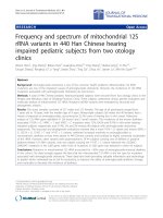
showing a viability > 95% were used. Materials used are
shown in figure 1.
Immunocytochemistry
FBs were typed by using a conventional marker (FB sur-
face protein-FSP, monoclonal anti-human FSP, Clone
1B10; Sigma, Italy) and epithelial types were carefully
excluded performing cytokeratine analysis (anti-human
Cytokeratin 20; Sigma, Italy); primary antibodies were
used at the manufacturer recommended dilutions. Cells
were seeded onto 24 well plates at a concentration of
20.000 cells/plate; after 48 hours they were washed twice
in PBS and fixed with 3.7% formaldehyde in PBS for 15
minutes at room temperature (RT). Fixed cells were per-
meabilised with 0.1% triton X-100 (Sigma, Italy) in PBS
for 15 minutes at RT. Non specific binding of secondary
antibody was blocked by incubation with normal foetal
serum for 30 minutes at RT. After immunostaining cells
were rinsed with PBS and incubated with fluorochrome
conjugated secondary antibody for 45 minutes at RT,
according to donor species of the primary antibodies. PBS
was used as the negative control in place of the primary
antibody. Counterstaining was performed using DAPI;
the glass coverslip was mounted on glass slides with pro-
long gold antifade reagent (Invitrogen, Italy). Images were
obtained by fluorescence microscope (Leica, Italy).
Q-Banding
Cells in log phase were cultured with 50 μL of colchicine
for 4 hours and mitotic cells were gently blown with a
pipette. Cells were centrifuged at 235 × g for 10 minutes
and the supernatant fluid removed. KCl at 37°C (0.075
M) was added to the cells and the mixture incubated at
37°C for 30 minutes. Cells were then fixed with 3:1 meth-
anol/acetic acid and cell suspensions dropped onto slides,
air dried and stained with quinacrine stain for 20 minutes.
Slides were observed with oil immersion at fluorescence
microscopy (Leica, Italy) [26].
Image capture and morphometric analysis
Culture growth and FBs morphometric analysis were per-
formed on low-power fields (10× magnification) with a
microscope (Nikon, Italy) coupled with a digital CCD
camera. Images were stored on a personal computer
(Power Mac G4, 1.25 GHz, 512 MB RAM, Apple, Cuper-
tino, CA) in TIFF format. Stored images were analyzed in
the Laboratory of Clinical Informatics and Cardiovascular
Imaging, University of Milan by a single experienced
reader blinded to image sequence and assignment. Analy-
sis algorithms were developed as a set of macros executed
with NIH Image, an integrated image-processing software
distributed on a freeware basis by the National Institutes
of Health (Bethesda, USA). Before the analysis, an auto-
mated threshold process was performed on the images to
minimize the influence of light variation in the micro-
scope field and in the operator subjective settings. This
process cuts off any object below the minimum signal
intensity. FBs were recognized on the basis of their sizes
and intensity signal by using a cell count algorithm that
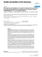
Left panel: disposable materials used in primary fibroblast cultures; A endoscopic forceps, B and C tubes, D laboratory forceps, E T25 flask, F and G Petri dishes, H cover glasses, I surgical knifeFigure 1
Left panel: disposable materials used in primary fibroblast cultures; A endoscopic forceps, B and C tubes, D
laboratory forceps, E T25 flask, F and G Petri dishes, H cover glasses, I surgical knife. Right Panel: duodenal endo-
scopic biopsy procedure.
Journal of Translational Medicine 2009, 7:40 />Page 4 of 8
(page number not for citation purposes)
draws a region of interest (ROI) around each discrete
object whithin the image. The minimum sizes in pixels of
the objects to be included in the count was previously
defined by accurately measuring 12 representative FBs.
Objects below the minimum size were not included in the
count, cells closely adjacent to each other (touching
edges) were excluded. The culture growth was determined
on days 12, 20, and 30 by counting the number of recog-
nized FBs over the area (microscopic field). The morpho-
metric evaluation included the major orthogonal
diameters and their ratio, as index of circularity, the
perimeter, the area, and their ratio, as index of complexity.
Statistical analysis
Data were expressed as mean ± standard deviation (SD) or
median and range. A comparison of the morphometric
data obtained from culture of CD and non-CD subjects
was done using one way ANOVA. All statistical analysis
was performed using statistical computer software (SPSS
13, SPSS, USA). A p value of 0.05 was considered signifi-
cant.
Results
Sixty CD and 114 non-CD duodenal bioptic specimens
were successfully obtained from EGDSs and from each
biopsy a similar amount of tissue weight was processed
from CD and non-CD (54.9 mg ± 6.4 vs 56.7 mg ± 4.8;
respectively; p = ns). All the CD patients on a gluten con-
taining diet had villous atrophy (type 3 lesion); among
the CD patients on GFD, all serologically negative, histol-
ogy showed type 0, 1, 2 and 3a in 2 cases each and type 3b
in 1 case. Non CD patients were all classified as type 0.
After 8–12 days of culture, FB-like cells growing radially
from the chopped and enzymatically digested bioptic
pieces were observed; their lateral spreading increased
dimensionally during the culture and the first passage was
performed at day 30 (range 25–35) with confluent cells
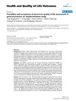
(Figure 2 upper panels). Next passages were performed
monthly (range 25–40 days). Primary cultures survived
for at least six passages and usually died after 180 days
(range 170–200) of culture. The duplication time was
about 8 days both for CD and non-CD cells (Figure 2
lower panels), with no statistical differences.
Out of the 174 duodenal specimens, 135 (78%; 45 CD, 90
non-CD) completed the entire cycle of culture. The major
reasons of unsuccessful were bacterial contamination
(18%) and insufficient bioptic material (4%), equally dis-
tributed between the 2 groups (data not shown). Sex, age,
Cellular growth from a chopped and enzymatically digested fragment of endoscopic duodenal biopsy at different times after seeding as visualised at microscopy (10 × magnification, upper panels) and after computer image analysis skeletonizing objects compatible with cells (fibroblasts) evidencing growth pattern radially spreading from the tissue sampleFigure 2
Cellular growth from a chopped and enzymatically digested fragment of endoscopic duodenal biopsy at differ-
ent times after seeding as visualised at microscopy (10 × magnification, upper panels) and after computer
image analysis skeletonizing objects compatible with cells (fibroblasts) evidencing growth pattern radially
spreading from the tissue sample.
Journal of Translational Medicine 2009, 7:40 />Page 5 of 8
(page number not for citation purposes)
clinical and dietary status in the CD group (patients fol-
lowing a gluten-containing or a gluten-free diet) did not
influence the successful rate or growth indexes of cell cul-
tures.
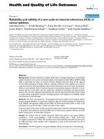
Immunocytochemistry was positive for FSP and negative
for cytokeratin 20 in all the cultured and examined cells
(Figure 3).
Q-banding analysis of FBs from CD and non-CD subjects
demonstrated a normal and stable karyotype (data not
shown).
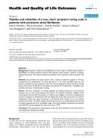
Morphometrical analysis performed on CD and non-CD
FBs images obtained at the same day of culture (Figure 4)
showed some significant differences; in particular, CD FBs
were greater, with a longer diameter and perimeter and
the area was wider even if the circularity index was similar;
on the other side the complexity index was decreased, sug-
gesting a change in the cellular membrane-cytoplasm
ratio (Table 1). In particular dietary status and the Marsh-
Oberhuber histological grading of CD patients did not
influenced morphometric parameters.
Discussion
FBs are known to be involved in inflammation and tissue
remodelling, and they play a pivotal role in CD [27].
Unfortunately, till now no experiences have been reported
on culturing primary cells obtained from endoscopic duo-
denal biopsies, the most reliable source of primary intes-
tinal cells. In this study, for the first time, we describe a
suitable technique to obtain long-standing primary
human FB cultures from endoscopic biopsies.
In the absence of standardised systems, we based FBs
extraction method on those used for cultures from surgi-
cal pieces, muscle and skin tissue samples [28]. Differ-
ently from these specimens, intestinal endoscopic
biopsies contain a small amount of a soft tissue and have
an important bacterial contamination caused by the com-
mon intestinal flora, the manual management of the
endoscope and endoscopic forceps, and their passage
through the endoscopic channel together with the
patients' gastric juice and saliva. For these reasons we used
a higher dose of antibiotics and the proteolytic cocktail of
enzymes liberase, rather than the traditional collagenase
cocktails that are known to contain endotoxin and exert
cytotoxic effects on primary cultures with an increase of
lipidic intracellular droplets [29]. Liberase is a blend of
highly purified enzymes used to improve the isolation
and cultures derived from small tissutal specimens, not
suitable for mechanical isolation [30-32].
In our study 96% of the endoscopic samples resulted ade-
quate to obtain cells without differences between the
specimens obtained from CD patients and those from
control subjects. All the cultured cells were FBs with nor-
mal karyotype, as demonstrated by the FSP positivity,
cytokeratine 20 negativity, and the Q banding analysis.
The successful rate of cell cultures was 78%, higher than
those obtained from transbronchial lung endoscopic
biopsies (successful rate 54%) [33], the only available to
make a comparison, since there are no data on mesenchy-
mal cell extraction from endoscopic duodenal biopsies.
This success rate was not affected by other possible covari-
ates such as the clinical and demographic characteristics
of patients suggesting that the stabilization of cell culture
is almost technique-dependent. We judged our rate of suc-
cess acceptable, taking into consideration the technical
difficulties and the bacterial load, the most important
cause of withdrawal (18%).
Fibroblast surface protein immunocytochemistry of primary cells from duodenal endoscopic biopsies from celiac (upper panel) and non celiac (lower panel) patients; DAPI counter-stained cellular nucleiFigure 3
Fibroblast surface protein immunocytochemistry of
primary cells from duodenal endoscopic biopsies
from celiac (upper panel) and non celiac (lower
panel) patients; DAPI counterstained cellular nuclei.
Journal of Translational Medicine 2009, 7:40 />Page 6 of 8
(page number not for citation purposes)
In CD, FBs are known to take part in the development of
the intestinal damage (villous atrophy) regulating the
deposition, degradation and remodeling of the ECM
through the secretion of collagen, MMPs, TIMPs, and
TG2, usually altered in CD intestinal mucosa [34-37].
Moreover, FBs cooperate in the establishment of the CD
immunomediated reaction and enterocyte differentiation
through the secretion of TGFβ and as a target of the celiac
autoantibodies, which finally influence their cell cycle
inducing an S phase shifting and the TGFβ secretion
[38,39]. However, these observations are derived from
studies on immortalised cell lines (NIH 3T3 and IMR90
FB), human umbilical chord-derived FBs and cultured
duodenal biopsies. Although these techniques provide
important high-technology resources, they have some
constrains: immortalised cell lines are important to study
cytotoxic effects in a simplified protein/xenobiotic-cell
microenvironment, but they are not disease-specific [40-
42]; cultured duodenal biopsies are a human and disease-
specific technique, but they survive in laboratory setting
for a maximum of 72 hours, conditioning the study of
chronic long-term mechanisms [43]. Furthermore, there
are no suitable animal models for investigating CD: the
Irish setter dog gluten-induced enteropathy and the rhe-
sus macaques non-infectious diarrhoea are the most CD-
specific, but they are expensive, not accessible to a high
number of researchers, non-human and involve ethical
aspects [44,45].
Thus, the cultures of primary CD FBs represent an impor-
tant research aid, easy to obtain because all CD patients
undergo to EGDS.
Contrast microscopy images of celiac and non celiac (control) fibroblasts at the third passage (upper panel) and skeletonized computer image analysis used for morphometric measurementsFigure 4
Contrast microscopy images of celiac and non celiac (control) fibroblasts at the third passage (upper panel)
and skeletonized computer image analysis used for morphometric measurements.
Table 1: Morphometrical characteristics of cultured fibroblasts
Parameter Non-CD CD p
Feret Diameter (μm) 40.15 ± 5.15 46.79 ± 8.09 0.025
Perimeter (μm) 91.93 ± 15.43 113.30 ± 19.11 0.0064
Area (μm
2
) 88.79 ± 22.52 122.86 ± 19.11 0.0018
Circularity index 0.13 ± 0.02 0.12 ± 0.02 ns
Complexity index (μm
-1
) 1.06 ± 0.16 0.93 ± 0.11 0.033
Feret Diameter: longest axis; Circularity index = Feret Diameter/
Short axis length; Complexity index = perimeter/area.
A p < 0.05 was considered significant; ns = not significant
Journal of Translational Medicine 2009, 7:40 />Page 7 of 8
(page number not for citation purposes)
By using a morphometric approach, based on five param-
eters, we found significant differences between cultured
control and celiac FBs; in particular, celiac FBs were sub-
stantially longer and wider, with a preserved circularity
but a reduced complexity index (perimeter/area ratio) if
compared with control FBs. These characteristics are spe-
cific of celiac FBs independently by the dietary status of
the patients and the Marsh-Hoberhuber histologic grad-
ing, suggesting a "permanent" alteration. Since it is well
known that shape and size of cells are the result of the spa-
tial arrangement of the microtubule cytoskeleton and are
closely related to cell function, we cannot exclude that
these differences reflect, at least in part, a different func-
tional state and/or a phenotype. It is noteworthy that the
reduced perimeter/area ratio suggests for cultured celiac
FBs a lower shape complexity, a parameter that normally
is under tight control to ensure a normal cell architecture
and tissue pattern [46]. At this regard it should be noticed
that standard in vitro cell culture models do not represent
the in vivo structure, nonetheless the differences observed
between control and celiac groups were obtained in the
same culture conditions.
Conclusion
Primary cell cultures from duodenal endoscopic biopsies
provide human disease-specific material and are easily
suitable in all patients; in fact previous studies using pri-
mary cultures from the GI tract were performed only by
sampling surgical pieces, thus excluding non-surgical
patients, that are known to represent the majority of
affected ones. The method of cell culture here described
could help in the establishment of novel experiments to
study the role of FBs in the pathogenesis of the mucosal
damage and to test new therapies alternative to the gluten-
free diet. In this context, endoscopy can revalue its role
from a simple diagnostic and therapeutic method to a
determinant technique in basic translational research.
Competing interests
The authors declare that they have no competing interests.
Authors' contributions
Conception and Design: LR, LE.
Data Analysis: MC, RP.
Drafting the article: LD, LP.
Critical Revision and Final Approval: MTB.
References
1. Elli L, Bardella MT: Motility disorders in patients with celiac dis-
ease. Scand J Gastroenterol 2005, 40:743-749.
2. Koning F, Schuppan D, Cerf-Bensussan N, Sollid LM: Pathomecha-
nisms in celiac disease. Best Pract Res Clin Gastroenterol 2005,
19:373-387.
3. Green PH, Cellier C: Celiac disease. N Engl J Med 2007,
357:1731-1743.
4. Bardella MT, Velio P, Cesana BM, Prampolini L, Casella G, Di Bella C,
Lanzini A, Gambarotti M, Bassotti G, Villanacci V: Coeliac disease:
a histological follow-up study. Histopathology 2007, 50:465-471.
5. Smith TJ: Insights into the role of fibroblasts in human autoim-
mune diseases. Clin Exp Immunol 2005, 141:388-397.
6. Okamoto H, Hoshi D, Kiire A, Yamanaka H, Kamatani N: Molecular
targets of rheumatoid arthritis. Inflamm Allergy Drug Targets
2008, 7:53-66.
7. Khoo TK, Bahn RS: Pathogenesis of Graves' ophthalmopathy:
the role of autoantibodies. Thyroid 2007, 17:1013-1018.
8. Huang Y, Lu MQ, Li H, Xu C, Yi SH, Chen GH: Occurrence of
cGMP/nitric oxide-sensitive store-operated calcium entry in
fibroblasts and its effect on matrix metalloproteinase secre-
tion. World J Gastroenterol 2006, 12:5483-5489.
9. Andoh A, Fujino S, Okuno T, Fujiyama Y, Bamba T: Intestinal sub-
epithelial myofibroblasts in inflammatory bowel diseases. J
Gastroenterol 2002, 37(Suppl 14):33-37.
10. Andoh A, Shimada M, Bamba S, Okuno T, Araki Y, Fujiyama Y, Bamba
T: Extracellular signal-regulated kinases 1 and 2 participate
in interleukin-17 plus tumor necrosis factor-alpha-induced
stabilization of interleukin-6 mRNA in human pancreatic
myofibroblasts. Biochim Biophys Acta 2002, 1591:69-74.
11. Neaud V, Rosenbaum J: A red wine polyphenolic extract
reduces the activation phenotype of cultured human liver
myofibroblasts. World J Gastroenterol 2008,
14:2194-2199.
12. Mishra S, Melino G, Murphy LJ: Transglutaminase 2 kinase activ-
ity facilitates protein kinase A-induced phosphorylation of
retinoblastoma protein. J Biol Chem 2007, 282:18108-18115.
13. Rossini A, Zacheo A, Mocini D, Totta P, Facchiano A, Castoldi R, Sor-
dini P, Pompilio G, Abeni D, Capogrossi MC, Germani A: HMGB1-
stimulated human primary cardiac fibroblasts exert a para-
crine action on human and murine cardiac stem cells. J Mol
Cell Cardiol 2008, 44:683-693.
14. Koumas L, King AE, Critchley HO, Kelly RW, Phipps RP: Fibroblast
heterogeneity: existence of functionally distinct Thy 1(+) and
Thy 1(-) human female reproductive tract fibroblasts. Am J
Pathol 2001, 159:925-935.
15. Sciaky D, Brazer W, Center DM, Cruikshank WW, Smith TJ: Cul-
tured human fibroblasts express constitutive IL-16 mRNA:
cytokine induction of active IL-16 protein synthesis through
a caspase-3-dependent mechanism. J Immunol 2000,
164:3806-3814.
16. Pierer M, Rethage J, Seibl R, Lauener R, Brentano F, Wagner U,
Hantzschel H, Michel BA, Gay RE, Gay S, Kyburz D: Chemokine
secretion of rheumatoid arthritis synovial fibroblasts stimu-
lated by Toll-like receptor 2 ligands. J Immunol 2004,
172:1256-1265.
17. Karnoub AE, Dash AB, Vo AP, Sullivan A, Brooks MW, Bell GW, Rich-
ardson AL, Polyak K, Tubo R, Weinberg RA: Mesenchymal stem
cells within tumour stroma promote breast cancer metasta-
sis. Nature 2007, 449:557-563.
18. Stichtenoth DO, Thoren S, Bian H, Peters-Golden M, Jakobsson PJ,
Crofford LJ: Microsomal prostaglandin E synthase is regulated
by proinflammatory cytokines and glucocorticoids in pri-
mary rheumatoid synovial cells. J Immunol 2001, 167:469-474.
19. Friedman SL: Mechanisms of hepatic fibrogenesis. Gastroenterol-
ogy 2008, 134:1655-1669.
20. Odaka T, Kobayashi K, Takahashi K, Nakamura H, Matsuoka T:
Effect of prostaglandin E(2) on urokinase-type plasminogen
activator production by human lung fibroblasts. Scand J Clin
Lab Invest. 2009, 69(2):225-233.
21. Pritchard J, Han R, Horst N, Cruikshank WW, Smith TJ:
Immu-
noglobulin activation of T cell chemoattractant expression
in fibroblasts from patients with Graves' disease is mediated
through the insulin-like growth factor I receptor pathway. J
Immunol 2003, 170:6348-6354.
22. Stachowiak MK, Maher PA, Stachowiak EK: Integrative nuclear
signaling in cell development – a role for FGF receptor-1.
DNA Cell Biol 2007, 26:811-826.
23. Schuppan D, Dennis MD, Kelly CP: Celiac disease: epidemiology,
pathogenesis, diagnosis, and nutritional management. Nutr
Clin Care 2005, 8:54-69.
Publish with Bio Med Central and every
scientist can read your work free of charge
"BioMed Central will be the most significant development for
disseminating the results of biomedical research in our lifetime."
Sir Paul Nurse, Cancer Research UK
Your research papers will be:
available free of charge to the entire biomedical community
peer reviewed and published immediately upon acceptance
cited in PubMed and archived on PubMed Central
yours — you keep the copyright
Submit your manuscript here:
/>BioMedcentral
Journal of Translational Medicine 2009, 7:40 />Page 8 of 8
(page number not for citation purposes)
24. Oberhuber G, Granditsch G, Vogelsang H: The histopathology of
coeliac disease: time for a standardized report scheme for
pathologists. Eur J Gastroenterol Hepatol 1999, 11:1185-1194.
25. Alves MP, Carrasco CP, Balmelli C, Ruggli N, McCullough KC, Sum-
merfield A: Mycoplasma contamination and viral immu-
nomodulatory activity: dendritic cells open Pandora's box.
Immunol Lett 2007, 110:101-109.
26. Roncoroni L, Elli L, Dolfini E, Erba E, Dogliotti E, Terrani C, Doneda
L, Grimoldi MG, Bardella MT: Resveratrol inhibits cell growth in
a human cholangiocarcinoma cell line. Liver Int 2008,
28:1426-1436.
27. Schuppan D: Current concepts of celiac disease pathogenesis.
Gastroenterology 2000, 119:234-242.
28. Miller RC, Enno M, Yamane M, Nishiki M: Recovery from X-ray
induced damage in primary cultures of human skin fibroblast
cells. J Radiat Res (Tokyo) 1985, 26:339-345.
29. Linetsky E, Bottino R, Lehmann R, Alejandro R, Inverardi L, Ricordi C:
Improved human islet isolation using a new enzyme blend,
liberase. Diabetes 1997, 46:1120-1123.
30. Georges P, Muirhead RP, Williams L, Holman S, Tabiin MT, Dean SK,
Tuch BE: Comparison of size, viability, and function of fetal pig
islet-like cell clusters after digestion using collagenase or lib-
erase. Cell Transplant 2002, 11:539-545.
31. Cavanagh TJ, Lakey JR, Dwulet F, Wright MJ, Wile K, Albertson T,
Fetterhoff T: Improved pig islet yield and post-culture recov-
ery using Liberase PI purified enzyme blend. Transplant Proc
1998, 30:367.
32. Lakey JR, Cavanagh TJ, Zieger MA, Wright M: Evaluation of a puri-
fied enzyme blend for the recovery and function of canine
pancreatic islets. Cell Transplant 1998, 7:365-372.
33. Tamm M, Roth M, Malouf M, Chhajed P, Johnson P, Black J, Glanville
A: Primary fibroblast cell cultures from transbronchial biop-
sies of lung transplant recipients. Transplantation 2001,
71:337-339.
34. Ciccocioppo R, Di Sabatino A, Bauer M, Della Riccia DN, Bizzini F,
Biagi F, Cifone MG, Corazza GR, Schuppan D: Matrix metallopro-
teinase pattern in celiac duodenal mucosa.
Lab Invest 2005,
85:397-407.
35. Daum S, Bauer U, Foss HD, Schuppan D, Stein H, Riecken EO, Ullrich
R: Increased expression of mRNA for matrix metalloprotei-
nases-1 and -3 and tissue inhibitor of metalloproteinases-1 in
intestinal biopsy specimens from patients with coeliac dis-
ease. Gut 1999, 44:17-25.
36. Daum S, Bauer U, Foss HD, Wahnschaffe U, Schuppan D, Stein H,
Riecken EO, Ullrich R: Expression of matrix metalloprotease-1
and collagen I mRNA in biopsies from patients with celiac
disease. Ann N Y Acad Sci 1998, 859:254-257.
37. Maiuri L, Ciacci C, Ricciardelli I, Vacca L, Raia V, Rispo A, Griffin M,
Issekutz T, Quaratino S, Londei M: Unexpected role of surface
transglutaminase type II in celiac disease. Gastroenterology
2005, 129:1400-1413.
38. Barone MV, Caputo I, Ribecco MT, Maglio M, Marzari R, Sblattero D,
Troncone R, Auricchio S, Esposito C: Humoral immune response
to tissue transglutaminase is related to epithelial cell prolif-
eration in celiac disease. Gastroenterology 2007, 132:1245-1253.
39. Halttunen T, Maki M: Serum immunoglobulin A from patients
with celiac disease inhibits human T84 intestinal crypt epi-
thelial cell differentiation. Gastroenterology 1999, 116:566-572.
40. Dolfini E, Elli L, Dasdia T, Bufardeci B, Colleoni MP, Costa B, Floriani
I, Falini ML, Guerrieri N, Forlani F, Bardella MT: In vitro cytotoxic
effect of bread wheat gliadin on the LoVo human adenocar-
cinoma cell line. Toxicol In Vitro 2002, 16:331-337.
41. Dolfini E, Elli L, Ferrero S, Braidotti P, Roncoroni L, Dasdia T, Falini
ML, Forlani F, Bardella MT: Bread wheat gliadin cytotoxicity: a
new three-dimensional cell model. Scand J Clin Lab Invest 2003,
63:135-141.
42. Elli L, Dolfini E, Bardella MT: Gliadin cytotoxicity and in vitro cell
cultures. Toxicol Lett 2003, 146:1-8.
43. Stenman SM, Lindfors K, Korponay-Szabo IR, Lohi O, Saavalainen P,
Partanen J, Haimila K, Wieser H, Maki M, Kaukinen K: Secretion of
celiac disease autoantibodies after in vitro gliadin challenge
is dependent on small-bowel mucosal transglutaminase 2-
specific IgA deposits. BMC Immunol 2008,
9:6.
44. Polvi A, Garden OA, Houlston RS, Maki M, Batt RM, Partanen J:
Genetic susceptibility to gluten sensitive enteropathy in Irish
setter dogs is not linked to the major histocompatibility
complex. Tissue Antigens 1998, 52:543-549.
45. Bethune MT, Borda JT, Ribka E, Liu MX, Phillippi-Falkenstein K, Jan-
dacek RJ, Doxiadis GG, Gray GM, Khosla C, Sestak K: A non-
human primate model for gluten sensitivity. PLoS ONE 2008,
3:e1614.
46. Rafelski SM, Marshall WF: Building the cell: design principles of
cellular architecture. Nat Rev Mol Cell Biol 2008, 9:593-602.