báo cáo hóa học:" High dose concentration administration of ascorbic acid inhibits tumor growth in BALB/C mice implanted with sarcoma 180 cancer cells via the restriction of angiogenesis" potx
Bạn đang xem bản rút gọn của tài liệu. Xem và tải ngay bản đầy đủ của tài liệu tại đây (1.37 MB, 9 trang )
BioMed Central
Page 1 of 9
(page number not for citation purposes)
Journal of Translational Medicine
Open Access
Research
High dose concentration administration of ascorbic acid inhibits
tumor growth in BALB/C mice implanted with sarcoma 180 cancer
cells via the restriction of angiogenesis
Chang-Hwan Yeom
1
, Gunsup Lee
2
, Jin-Hee Park
2
, Jaelim Yu
2
, Seyeon Park
3
,
Sang-Yeop Yi
4
, Hye Ree Lee
5
, Young Seon Hong
6
, Joosung Yang
2
and
Sukchan Lee*
2
Address:
1
Department of Palliative Medicine, Seoul St Mary's Hospital, The Catholic University of Korea, Seoul, 137-701, Korea,
2
Department of
Genetic Engineering, Sungkyunkwan University, Suwon, 440-746, Korea,
3
Department of Applied Chemistry, Dongduk Women's University,
Seoul, 136-714, Korea,
4
Department of Pathology, Kwandong University, College of Medicine, Goyang, 412-270, Korea,
5
Department of Family
Medicine, Gangnam Severance Hospital, Yonsei University College of Medicine, Seoul, 135-720, Korea and
6
Department of Medical Oncology,
Seoul St. Mary's Hospital, The Catholic University of Korea, Seoul, 137-701, Korea
Email: Chang-Hwan Yeom - ; Gunsup Lee - ; Jin-Hee Park - ;
Jaelim Yu - ; Seyeon Park - ; Sang-Yeop Yi - ;
Hye Ree Lee - ; Young Seon Hong - ; Joosung Yang - ;
Sukchan Lee* -
* Corresponding author
Abstract
To test the carcinostatic effects of ascorbic acid, we challenged the mice of seven experimental
groups with 1.7 × 10
-4
mol high dose concentration ascorbic acid after intraperitoneal
administrating them with sarcoma S-180 cells. The survival rate was increased by 20% in the group
that received high dose concentration ascorbic acid, compared to the control. The highest survival
rate was observed in the group in which 1.7 × 10
-4
mol ascorbic acid had been continuously injected
before and after the induction of cancer cells, rather than just after the induction of cancer cells.
The expression of three angiogenesis-related genes was inhibited by 0.3 times in bFGF, 7 times in
VEGF and 4 times in MMP2 of the groups with higher survival rates. Biopsy Results, gene expression
studies, and wound healing analysis in vivo and in vitro suggested that the carcinostatic effect induced
by high dose concentration ascorbic acid occurred through inhibition of angiogenesis.
Background
Despite advances in medical science, both the number of
cancer patients and the death rate due to cancer is increas-
ing. Although new approaches and new carcinostatic
agents have been developed, their effects on cancer
patients are not sufficient [1]. Since Klenner and col-
leagues applied vitamin C (ascorbic acid) to cure cancer
patients in 1949, cell experiments, model animal experi-
ments and clinical trials have been carried out [2,3]. Linus
Pauling and Ewan Cameron reported that the administra-
tion of high dose concentrations of ascorbic acid (1.7 ×
10
-4
mol) to cancer patients in the terminal stage
improved the quality of life and extended their lives [4].
Although there are experimental results supporting the
Published: 11 August 2009
Journal of Translational Medicine 2009, 7:70 doi:10.1186/1479-5876-7-70
Received: 19 May 2009
Accepted: 11 August 2009
This article is available from: />© 2009 Yeom et al; licensee BioMed Central Ltd.
This is an Open Access article distributed under the terms of the Creative Commons Attribution License ( />),
which permits unrestricted use, distribution, and reproduction in any medium, provided the original work is properly cited.
Journal of Translational Medicine 2009, 7:70 />Page 2 of 9
(page number not for citation purposes)
carcinostatic effects of ascorbic acid and its use as a thera-
peutic agent to prevent the growth of cancer cells, there is
still controversy over the effects of ascorbic acid. Accord-
ing to the work done by Levin's group [5,6], ascorbic acid
has definite effect as an antitumor agent when adminis-
trated at a high dose concentration. They reported that
high dose concentrations of ascorbic acid, provided intra-
venously, work as a pro-oxidant therapeutic agent in can-
cer by generating ascorbate radicals and hydrogen
peroxide in extracellular fluid in vivo. In addition, clinical
case reports (from kidney cancer and bladder tumors)
strongly indicate that high dose concentration ascorbic
acid therapy in cancer treatment should be reassessed.
These studies were confirmed by histopathologic review
and examined in accordance with National Cancer Insti-
tute (NCI) Best Case Series guidelines [7].
Ascorbic acid mediated direct cytotoxicity effects on can-
cer cells by hydrogen peroxide have been numerously
reviewed [8,9] but in some cases the concentration of
ascorbic acid radicals and hydrogen peroxide have not
been sufficiently induced tumor cell death [6]. Therefore
other action mechanism of ascorbic acid as an anticancer
drug has been investigated. The one possibility of ascorbic
acid mediated angiostatic effects has been recently
reported [10,11]. Mikirova and colleagues showed that
high dose concentration of ascorbic acid inhibited cell
migration ability and gap filling capacity of endothelial
progenitor cells (EPCs). Peyman and colleagues showed
that ascorbic acid inhibited corneal neovascularization in
a rat model. The rat mode was not for angiogenesis study
caused by cancer cells but they showed the neovasculari-
zation was clearly affected by the concentration of ascor-
bic acid.
In our recently published works, intraperitoneal adminis-
tration of a high dose concentration of ascorbic acid quan-
titatively up-regulated Raf kinase inhibitory protein
(RKIP) and annexin A5 expression in a group of BALB/C
mice implanted with S-180 sarcoma cancer cells. The
increase in RKIP protein level suggested that these pro-
teins are involved in the ascorbic acid-mediated suppres-
sion of tumor formation [12].
Based on our previous experiments [12], here we further
investigated the non-cytotoxic antitumor activities of
ascorbic acid by inhibiting angiogenesis ability in vitro
and in vivo. We supported this finding by quantitative real
time RT-PCR as well as wound healing assay to examine
the expression of three angiogenesis-related genes and the
inhibition of angiogenesis in treatment and control
groups. This study supports that high dose concentration
ascorbic acid treatment inhibits the angiogenesis of cancer
cells by one of the antitumor mechanisms triggered by
ascorbic acids.
Methods
Animals and tumor cell lines
Murine sarcoma S180 cells provided by Korea Cell Line
Bank were maintained in RPMI-1640 medium supple-
mented with 10% fetal bovine serum (Hyclone, Aurora,
Canada), 100 U/ml Penicillin-Streptomycin (Hyclone),
and Non-Essential Amino Acids (Sigma), at 37°C in a 5%
CO
2
atmosphere. Female BALB/c mouse (Charles River,
Seongnam, Korea) weighing 1822 g were kept under
standard laboratory conditions (tap water, constant room
temperature 22°C). Principles of laboratory animal care
(NIH publication 85-23, revised 1986) were followed and
all experiment was carried out under AAALAC Interna-
tional (Association for Assessment and Accreditation of
Laboratory Animal Care International) approval.
Treatments with cancer cells and ascorbic acid
Sarcoma 180 cells were cultivated in a CO
2
incubator for
five days, adding 9 ml RPMI 1640 medium, in 8 plates of
100 mm in diameter, and then 5 × 10
5
cells in 200 ml PBS
were injected into the abdominal cavities of experimental
mice using a 21 G injector. The high dose ascorbic acid
dose of 1.7 × 10
-4
mol (30 mg) corresponds to 100 g for a
human of 70 kg. The low dose of ascorbic acid was 3.1 ×
10
-5
mol (5.5 mg). After each group was treated with
ascorbic acid and cancer cells, they were observed and
measured over time, and then livers and kidneys were har-
vested and stored at -70°C for further analysis. BALB/C
mice were divided into 7 groups (A G) with 10 mice per
group (Figure 1). Group A was a control group that was
treated with phosphate buffer saline (PBS), Group B was
treated with low-level ascorbic acid at two-day intervals,
and Group C was treated with high dose concentration
ascorbic acid at two-day intervals. Group D group was
administered Sarcoma 180 cells for cancer induction.
Groups E-G received both cancer cells and ascorbic acid.
Group E was treated twice with PBS at two-day intervals,
injected with S-180 cells, and then treated with high dose
concentration ascorbic acid at two-day intervals for Group
F was injected with low dose ascorbic acid before injecting
cancer cells, and was then treated with high dose concen-
tration ascorbic acid after cancer challenging for 24 days.
Group G group was injected with high dose concentration
ascorbic acid for four days before injecting cancer cells,
and was then treated with high dose concentration ascor-
bic acid for 24 days after cancer challenging (Figure 1).
RNA preparation and quantitative real-time RT-PCR
RNA was isolated from livers and kidneys of each group.
After evenly grinding the samples from each group, 100
mg of each sample were put in 1.5 ml tubes and 1 ml of
Corezol (Corebio System, Seoul, Korea) was added. After
adding 200 μl of chloroform to the tubes, we centrifuged
them at 12,000 g at 4°C for 15 minutes. The supernatants,
which contained the RNA, were placed in new 1.5 ml
Journal of Translational Medicine 2009, 7:70 />Page 3 of 9
(page number not for citation purposes)
tubes and then precipitated with 700 μl isopropanol. After
centrifuging at 12,000 g at 4°C for 15 minutes, we recov-
ered the RNA pellet in 20 μl DEPCed DDH
2
O [13]. The
RNA concentrations were measured by spectrophotome-
ter and electrophoresis. To identify gene expression in the
harvested livers, cDNA was synthesized from 5 μg of total
RNA using oligo (dT) primers and Moloney murine leuke-
mia virus (MMLV) reverse transcriptase (SuperBio Co.
Daejon, Korea). Six ng mRNA was used for reverse tran-
scription. Primers used for quantitative PCR were
designed using Primer3 />index.php and synthesized by Genotech (Daejon, Korea).
Angiogenesis genes detected were bFGF (forward primer:
CGG CTG CTG GCT TCT AAG TG; reverse primer: CCC
GTT TTG GAT CCG AGT TT), VEGF (forward primer: ACA
CGG GAG ACA ATG GGA TG; reverse primer: TCT TGA
CTC AGG GCC AGG AA) and MMP2 (forward primer:
ATG GGG CTG GAA CAC TCT CA; reverse primer: GGG
GCC AGT ACC GTC AG); the housekeeping gene was
GAPDH (forward primer: TTG CAG TGG CAA AGT GGA
GA; reverse primer: GGC TTC CCG TTG ATG ACA AG).
PCR amplification was done in a 20 μl total volume con-
taining 4 or 6 μl of 2 × diluted cDNA (duplicate), 0.25 μM
each primer, 1 μl 20000 × diluted SYBR Green I (Molecu-
lar Probes, Eugene, OR) and 2.5 units Taq DNA polymer-
ase (SuperBio Co. Daejon, Korea) in a reaction buffer
composed of 10 mM Tris/HCl (pH 9), 50 mM KCl, 2 mM
MgCl
2
, 0.5 mM each deoxyribose trinucleotide, and 0.1%
Triton X-100, in a Rotor-Gene 3000 (Corbett Research,
Sydney, Australia). PCR cycling parameters were 40 cycles
of 10 s at 94°C, 15 s at 60°C, and 20 s at 72°C. The prod-
ucts of real-time quantitative PCR were separated by 1%
agarose gel electrophoresis to make sure. Two negative
controls, missing either RNA template or reverse tran-
scriptase, were included in each experiment. Each data
point represents the average of three experiments and the
error bars indicate the standard deviation of individual
experiments unless mentioned otherwise.
Hematoxylin-eosin stain
Specimens were fixed in 10% buffered formalin, serially
sectioned, and embedded in paraffin. The prepared paraf-
fin blocks were cut at 3 μm thickness and then stained
with hematoxylin-eosin [14].
Immunohistochemical stain
Representative 3 μm-thick tissue sections for immunohis-
tochemical analysis were mounted on silane coated slides.
The sections were deparaffinized in xylene and dehy-
drated with distilled water through a graded series of eth-
anol solutions. The slides were pretreated in a microwave
oven (20 min) with citrate acid solution for antigen
retrieval. After rinsing with APK Wash Solution (Ventana
Medical Systems, Tucson, AZ, USA), immunochemistry
was performed in a Ventana NexES IHC automated
immunostainer (Ventana Medical Systems, Tucson, AZ,
USA). The primary antibodies used in this study included
MMP-2 and VEGF (ABcam, Cambridge, UK), and bFGF
(BD Transduction Laboratories™, San Jose, CA, USA). The
prediluted (1:50) primary antibodies were applied for 32
min at 37°C. The sections were then treated for color
development with diaminobenzidine (4 min), and coun-
terstaining was done with hematoxylin (4 min) using the
iVIEW™ DAB Detection Kit (Ventana Medical Systems).
Cell migration and cell culture wound assay
We used a wound healing assay [15] to identify the degree
of migration of cancer cells and normal cells caused by the
treatment with ascorbic acid. Wounds were created in
confluent H-ras NIH3T3 cells (Biochemistry laboratory,
Department of Genetic Engineering, Sungkyunkwan Uni-
versity) using a pipette tip. The cells were then rinsed with
medium to remove any free-floating cells and debris.
Serum-free medium was then added, and culture plates
were incubated at 37°C. Wound healing was observed at
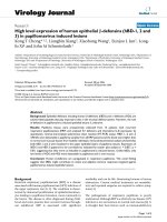
Schematic diagram for S-180 and ascorbic acid challenge pro-tocolFigure 1
Schematic diagram for S-180 and ascorbic acid chal-
lenge protocol. Group A: PBS treatment every two days.
Group B: Low ascorbic acid treatment every two days.
Group C: high dose concentration ascorbic acid treatment
every two days. Group D: PBS treatment twice for 4 days
and then 5 × 10
5
S-180 cells were injected intraperitoneally
followed by PBS treatment every two days. Group E: PBS
treatment and S-180 cells same as Group D, and then high
dose concentration ascorbic acid every two days. Group F:
low dose ascorbic acid twice for 4 days, S-180 cells same as
Group D, and then high dose concentration ascorbic acid
given twice. Group G: high dose concentration ascorbic acid
twice for 4 days, S-180 cells same as Group D, and then high
dose concentration ascorbic acid given twice. Liver samples
of all groups were harvested at 16 days after the first treat-
ment.
Journal of Translational Medicine 2009, 7:70 />Page 4 of 9
(page number not for citation purposes)
0, 12, 24, and 36 hours within the scrape line, and repre-
sentative scrape lines for each cell line were photo-
graphed. Duplicate wells of each condition were
examined for each experiment, and each experiment was
repeated 3 times.
Statistical analysis
We compared angiogenesis gene expression (bFGF, VEGF,
MMP-2), survival rate, and ascites genesis rate between
experiment groups. All analyses were carried out using the
statistic software Sigmaplot (Systat software Inc. Chicago,
USA). Data are presented as mean ± SE.
Results
1. Intraperitoneal cancer progression in each group
Sizes of ascites and intraperitoneal tumors were measured
at 16 days after ascorbate or PBS treatment (Figure 2).
Mice developed ascites containing tumor cells between 6
and 12 days after cancer injection. Group D (no ascorbate
treatment) developed intraperitoneal tumors rapidly.
Groups E, F and G developed tumors both more slowly
and later than Group D. The amounts of ascites were
quantified by recording weights of each mouse. The
weights of Groups A-C were maintained at about 20 g but
Groups D-G increased beginning 6 days after cancer injec-
tion (Figure 3A).
Significant tumor induction was observed in Group D
compared to the other groups. White masses were formed,
indicated by red arrows, in each organ (Figure 2); these
were tumors formed by cancer cells. In addition, more
ascites were generated in Group D than in the other
groups into which cancer cell had been injected.
2. Increased viability and decreased acsites production by
ascorbic acid treatments
Of the 10 mice in each group, 5 were dissected to measure
angiogenesis gene expression and observe abdominal cav-
ities, and 5 mice were observed up to 28 days after inject-
ing ascorbic acid to measure survival rate. Celiectomy was
performed around 14 days after the injection of cancer
cells, and mice were weighed at that time. The greatest
Effects of high dose concentration of ascorbic acid on mouse model experimentsFigure 2
Effects of high dose concentration of ascorbic acid on mouse model experiments. Ascite formation and cancer
induction were shown in cancer cell injected experimental groups (D to G) with different degrees of ascite formation and can-
cer induction. Dissection picture of group D shows the most severe ascite formation and polyps, indicated by red arrows.
Journal of Translational Medicine 2009, 7:70 />Page 5 of 9
(page number not for citation purposes)
average weight, 27.8 g, was found in Group D at the 14
th
day after injecting cancer cells, an increase of 1.39 times
from the start of the experiment. Groups G, E, and F
groups followed in weight order (Figure 3A). The average
body weight at the 18
th
day was 25.2 g of Group E, 24.8 g
of Group F and 26 g of Group G respectively. These data
showed that the treatments of ascorbic acid by challeng-
ing with low dose before cancer infection and then treated
with high dose of ascorbic acid was more effective (Group
E). At the 18
th
day, the body weight of Group F was 0.89
times of Group D. Survival rate was measured up to 28
days from the beginning of the experiment. Group D
showed a survival rate of 0% after 25 days, and Group E
showed a survival rate of 0% after 28 days from the begin-
ning of the treatment. In contrast, F and G groups, which
had been treated with ascorbic acid prior to injecting can-
cer cells, showed a survival rate of 20% at the 28
th
day
(Figure 3B).
3. Inhibition of the Expression of Angiogenesis-related
Genes by Ascorbic acid
Angiogenesis is an important mechanism in cancer gene-
sis and the growth process. We measured gene expression
of genes involved in angiogenesis by staining and real-
time PCR. Cancer genesis in each group was identified by
H&E staining as shown in Figure 4A. We observed blue
staining of giant nuclei followed by cancer cell genesis,
and identified cancer genesis in tissue from Group E (Fig-
ure 4B, e2). No staining was found in the other groups;
they did not differ from the negative control groups to
Ascorbic acid effects in changes of body weight (A) and via-bility (B) in each experimental group after cancer cell injec-tionFigure 3
Ascorbic acid effects in changes of body weight (A)
and viability (B) in each experimental group after
cancer cell injection. (A) The body weights were meas-
ured from 10 mice of each group up to 18 days after injecting
ascorbic acid. (B) Result shows for the changes of survival
rates of 5 mice per each group up to 28 days after injecting
ascorbic acid.
Tumors in high does ascorbic acid treated groups exhibit poorly formedFigure 4
Tumors in high does ascorbic acid treated groups
exhibit poorly formed. Histochemical (A, × 100 and B, ×
200) and immunohistochemical data (C to F) of liver tissues
represent the clear tumor staining in group e. A-B: Cancer
induction was identified by H & E staining in liver tissues
treated with ascorbic acid. Small letters in each figure (a to g)
represent the name of each group. C-F: Expression of angio-
genesis related proteins (bFGF, VEGF and MMP2) were
examined by immunohistochemistry. The name of each
tested groups were shown in (C to A group), (D to D
group), (E to E group) and (F to F group) in Figures. Angio-
genesis related proteins of group D showed dark brown
stains rather than other tested groups.
Journal of Translational Medicine 2009, 7:70 />Page 6 of 9
(page number not for citation purposes)
which cancer cells had not been injected. Thus there was a
remarkable reduction of cancer genesis in the groups
which received prior treatment with ascorbic acid. We also
measured gene expression involved in angiogenesis by
immunohistochemistry (Figure 4B). Additional histo-
chemical staining was made for A, D, E, and F groups. As
a result of staining with antibody of other 3 angiogenesis
related protein, applied to this test, in each tissue, no his-
tochemical staining was made in other groups except D
group (Figure 4B). We also analyzed expression of genes
involved in angiogenesis by Quantitative real-time RT-
PCR (Figure 5). In Group D, expression of bFGF was
increased by about 18 times over the groups that did not
receive injected cancer cells. This increase was 2.5 times,
1.8 times, and about 1.3 times greater than the increase
seen in Groups E, F, and G, respectively, groups which had
been treated with ascorbic acid after injecting cancer cells.
In Group D, expression of VEGF was increased by 4.57; for
MMP2, the increase was about 5 times. The expression of
angiogenesis related genes was thus remarkably reduced
in the groups with ascorbic acid treatment compared to
the group with cancer cell treatment only. These results
suggest that ascorbic acid treatment in high concentration
inhibits angiogenesis by inhibiting the expression of ang-
iogenesis related genes.
4. Inhibition of Cancer by Ascorbic Acid in H-ras NIH-3T3
cells
We used a wound healing assay to compare the inhibition
of the expression of angiogenesis related genes and pro-
tein synthesis by ascorbic acid with the change of cell
migration efficiency (Figure 6). We observed wound
recovery at 0, 12, 24, 36 hrs after treating with 2.5 mM or
10 mM ascorbic acid. The H-ras NIH3T3 cells did not
recover after wounding and high treatment concentration
of ascorbic acid, while artificially formed wounding was
recovered in NIH3T3 cell at 12, 24, 36 hrs by cell migra-
tion even in ascorbic acid in 2.5 mM and ascorbic acid in
10 mM (Figure 6). Therefore, migration was inhibited
according to ascorbic acid concentration in cancer cell and
the treatment time.
Conclusion
Ascorbic acid is known to be a nontoxic substance. Demol
(1934) injected 5 g/kg into guinea pigs, but no specific
adverse reaction was found. The above amount corre-
sponds to 350 g for a human of 70 kg. In our research, no
specific adverse reaction was observed in control groups
(A, B, and C). Several adverse effects have been hypothe-
sized to occur from administration of high dose concen-
tration ascorbic acid; however, these are only known from
in vitro experiments or single case reports in most cases.
These adverse reactions include genomic mutation, birth
defects, cancer, arteriosclerosis, Calculus of kidney,
rebound scurvy, oxidative stress, hyperabsorption of
limatura ferri, deficiency in ascorbic acid B
12
, and erosion
of enamel [16]. However, there is no scientific evidence
that high dose concentration ascorbic acid is toxic, harm-
ful, or unfavorable.
Quantitative real time RT-PCR (qRT-PCR) analysis of the three angiogenesis related genesFigure 5
Quantitative real time RT-PCR (qRT-PCR) analysis
of the three angiogenesis related genes. Expression
patterns of three angiogenesis related genes (bFGF, VEGF
and MMP2) were high in group D and it is correlated with
the immunohistochemistry analysis. Ascorbic acid treated
groups showed suppressed expression of these genes. Each
qRT-PCR is a representative example of data from 3 repli-
cate experiments.
Journal of Translational Medicine 2009, 7:70 />Page 7 of 9
(page number not for citation purposes)
Mayland and coworkers (2005) reported that 30% of pro-
gressive cancer patients were deficient in blood ascorbic
acid [17]. Deficiency in ascorbic acid is related to albu-
min, platelet, and C-reactive protein (CRP), and it has a
negative impact on the prognosis of patients. According to
Schorah and colleagues (1996), ascorbic acid concentra-
tion in critically ill patients is less than 25% of normal
people. In our experiment, injecting ascorbic acid into
mice injected with cancer cells led to an increased survival
rate over mice injected with cancer cells only, both when
ascorbic acid was provided preventively and therapeuti-
cally (Figure 3). The group into which ascorbic acid had
been injected prior to S-180 cancer cell treatment showed
a two times higher survival rate than the group injected
with ascorbic acid after S-180 cancer cell treatment (Figure
3).
Angiogenesis related genes are directly involved in the
growth and metastasis of tumors. It has previously been
shown that expression changes in the angiogenesis related
genes bFGF, VEGF, and MMP-2 are closely related to
tumor growth and metastasis [18-20]. Therefore we tested
that ascorbic acid reduced the expression of three genes
(bFGF, VEGF, and MMP-2) when used preventively and/
or therapeutically in this experiment. The expression of
angiogenesis related genes was lower in the group given
ascorbic acid prior to S-180 cancer cell treatment than the
group which received ascorbic acid after induction of can-
cer cells (Figure 4 and 5). bFGF is related to the growth
and shift of endotheliocyte and proteolysis [21-23].; in
particular, it makes cancer cells grow by activating FGFR-
4 (FGFs including FGF receptor-4) [24]. VEGF induces
endothelial growth and increases permeability of cells, so
it is frequently observed when tumors form new vessels
through which nutrition can be supplied [25-28]. VEGF is
expressed more strongly in metastatic cancer, and is less
well known in primary cancer than the other genes. The
prognosis of metastatic cancer when the primary cancer is
not known is worse than for other cancers; thus Karavasi-
lis and colleagues (2005) suggested VEGF as a target for
therapy [29]. MMP-2 is known to be involved in the
destruction of basement membranes, the most important
process of angiogenesis. Therefore if MMP-2 is high, can-
cer cells can easily invade surrounding tissue, since base-
ment membranes and extracellular matrices are destroyed
[30]. Therefore this suggests that ascorbic acid can prevent
cancer genesis and metastasis by inhibiting induction and
angiogenesis. It appears that ascorbic acid inhibited the
activation of cancer cells, invasion into surrounding tis-
sue, or metastasis in the group into which S-180 cancer
cells were injected.
Roomi and colleagues (2006) reported similar results
from in vitro and in vivo experiments [31-33]. They
observed changes in angiogenesis related gene expression
as an anticancer effect of ascorbic acid, lysine, proline,
arginine, and green tea extract on various cancer cells, and
suggested that such substances, including ascorbic acid,
were affordable as a cancer remedy. By changing the con-
centration of ascorbic acid and time it was administered,
their experiments uncovered a positive effect on the
growth and metastasis of cancer cells in the group to
which ascorbic acid had been injected before injecting
cancer cells into the abdominal cavity. Past research on
the anticancer effects of ascorbic acid had only focused on
inhibition of the expression of angiogenesis related genes.
Data on administration time and concentration for apply-
ing ascorbic acid appears to be fundamental to anticancer
treatment in the future. Also Mikirova et al (2008)
showed similar observations about anti-angiogenesis
effects by high dose concentration ascorbic acid treatment
on endothelial progenitor cells in vitro and they suggested
that nitric oxide (NO) generation can be one of the mech-
anism by which ascorbic acid mediated angiostatic effects.
Our results also supported the finding shown by Mikirova
and Roomi groups and we have demonstrated in vivo and
in vitro that high dose concentration of ascorbic acid sup-
pressed the gene expression of angiogenesis-related genes
and thereby can inhibit angiogenesis.
According to Ashino and colleagues (2003), cytopermea-
bility is increased by endothelial growth factor and
decreased by antioxidant, and ascorbic acid affects angio-
genesis through antioxidation reactions and collagen syn-
thesis. Ashino and colleagues also reported that this
characteristic of ascorbic acid contributes to resistibility to
cancer [34,35]. Ascorbic acid, a strong antioxidant,
reduces unstable oxygen, nitrogen, and sulfa active oxy-
Wound healing assay on NIH3T3 and ras-NIH3T3 cells depending on the concentration of ascorbic acids and the treated timesFigure 6
Wound healing assay on NIH3T3 and ras-NIH3T3
cells depending on the concentration of ascorbic
acids and the treated times. The cell migration of ras-
NIH 3T3 cells was inhibited by the treatments of ascorbic
acid (2.5 mM and 10 mM), 24 hours after treatments.
Journal of Translational Medicine 2009, 7:70 />Page 8 of 9
(page number not for citation purposes)
gen, and may react as a primary protective mechanism
against hydrosoluble active oxygen [36-39]. It would pre-
vent fat-soluble active oxygen by reducing vitamin E. In
addition, ascorbic acid prevents the formation of carcino-
genic nitrosamines by reducing nitrates through the NAD
(nicotinamide adenine dinucleotide)-dependent system
[36,38,40]. HIF-1α (hypoxia-inducible factor-1 alpha) is
involved in tumor growth as a vector to which cells adapt
in hypoxia, and is also involved in cancer metastasis by
increasing the expression of other angiogenesis related
genes (VEGF) [41]. Ascorbic acid inhibits HIF-1α, through
increasing resistibility to cancer [42]. According to our
data, high dose concentration of ascorbic acid inhibited
the angiogenesis but we could not conclude the action
mechanism of ascorbic acid against angiogenesis. This
non-cytotoxic action of ascorbic acid would be intensively
investigated in further experiments.
An ideal antitumor agent would prevent the growth of
cancer cells, extend survival period, and improve the qual-
ity of life. Although to date administration of ascorbic
acid for people has only been supported by a few clinical
research results, recently there has been an increase in the
number of research reports on the clinical cases of cured
cancer patient [7,43]. The study reported here, based on
an animal model, S-180 induced mice, showed both pre-
ventive and therapeutic effects of ascorbic acid. Ascorbic
acid treatment resulted in reduced expression of angio-
genesis related genes involved in the growth and metasta-
sis of cancer as well as increased survival rate. Based on
these experimental results, more clinical experiments
should be tried, as well as additional research on other
cancers.
Competing interests
The authors declare that they have no competing interests.
Authors' contributions
CY and KL performed the mouse experiments and gene
expression analysis. HL, YH and SY carried out the immu-
nohistochemistry, JP, JY, SP and JY collected and analyzed
the wounding healing experiments. CY, KL and SL con-
ceived and designed the experiments and analyzed the
data. The manuscript was written by CY, KL and SL. All
authors read and approved the final manuscript.
Acknowledgements
This work was supported by the Korea Research Foundation Grant funded
by the Korean Government (MOEHRD, Basic Research Promotion Fund,
KRF-2005-003-E00238).
References
1. Jemal A, Thun MJ, Ries LA, Howe HL, Weir HK, Center MM, Ward
E, Wu XC, Eheman C, Anderson R, et al.: Annual report to the
nation on the status of cancer, 19752005featuring trends in
lung cancer, tobacco use, and tobacco control. J Natl Cancer
Inst 2008, 100:1672-1694.
2. Gonzalez MJ, Miranda-Massari JR, Mora EM, Guzman A, Riordan NH,
Riordan HD, Casciari JJ, Jackson JA, Roman-Franco A: Orthomo-
lecular oncology review: ascorbic acid and cancer 25 years
later. Integr Cancer Ther 2005, 4:32-44.
3. Klenner FR: The treatment of poliomyelitis and other virus
diseases with vitamin C. South Med Surg 1949, 111:209-214.
4. Cameron E, Pauling L, Leibovitz B: Ascorbic acid and cancer: a
review. Cancer Res 1979, 39:663-681.
5. Chen Q, Espey MG, Krishna MC, Mitchell JB, Corpe CP, Buettner GR,
Shacter E, Levine M: Pharmacologic ascorbic acid concentra-
tions selectively kill cancer cells: action as a pro-drug to
deliver hydrogen peroxide to tissues. Proc Natl Acad Sci USA
2005, 102:13604-13609.
6. Chen Q, Espey MG, Sun AY, Lee JH, Krishna MC, Shacter E, Choyke
PL, Pooput C, Kirk KL, Buettner GR, Levine M: Ascorbate in phar-
macologic concentrations selectively generates ascorbate
radical and hydrogen peroxide in extracellular fluid in vivo.
Proc Natl Acad Sci USA 2007, 104:8749-8754.
7. Padayatty SJ, Riordan HD, Hewitt SM, Katz A, Hoffer LJ, Levine M:
Intravenously administered vitamin C as cancer therapy:
three cases. CMAJ 2006, 174:937-942.
8. Frei B, Lawson S: Vitamin C and cancer revisited. Proc Natl Acad
Sci USA 2008, 105:11037-11038.
9. de la Lastra CA, Villegas I: Resveratrol as an antioxidant and pro-
oxidant agent: mechanisms and clinical implications. Biochem
Soc Trans 2007, 35:1156-1160.
10. Mikirova NA, Ichim TE, Riordan NH: Anti-angiogenic effect of
high doses of ascorbic acid. J Transl Med 2008, 6:
50.
11. Peyman GA, Kivilcim M, Morales AM, DellaCroce JT, Conway MD:
Inhibition of corneal angiogenesis by ascorbic acid in the rat
model. Graefes Arch Clin Exp Ophthalmol 2007, 245:1461-1467.
12. Park S, Ahn ES, Lee S, Jung M, Park JH, Yi SY, Yeom CH: Proteomic
analysis reveals upregulation of RKIP in S-180 implanted
BALB/C mouse after treatment with ascorbic acid. J Cell Bio-
chem 2009, 106:1136-1145.
13. Chomczynski P: aMK Short technical report. Modification of
the TRIZOL reagent procedure for isolation of RNA from
Polysaccharide-and proteoglycan-rich sources. Biotechniques
1995, 19:942-945.
14. Lynch MJRS, Mellor LD, Spare PD, Inwood JH: Medical Laboratory
Technology and Clinical Pathology 2nd edition. Philadelphia: W. B. Saun-
ders Co; 1969.
15. McLane MA, Zhang X, Tian J, Zelinskas C, Srivastava A, Hensley B,
Paquette-Straub C: Scratching below the surface: wound heal-
ing and alanine mutagenesis provide unique insights into
interactions between eristostatin, platelets and melanoma
cells. Pathophysiol Haemost Thromb 2005, 34:164-168.
16. Barness LA: Safety considerations with high ascorbic acid dos-
age. Ann N Y Acad Sci 1975, 258:523-528.
17. Mayland CR, Bennett MI, Allan K: Vitamin C deficiency in cancer
patients. Palliat Med 2005, 19:17-20.
18. Folkman J: Tumor angiogenesis: therapeutic implications. N
Engl J Med 1971, 285:1182-1186.
19. Folkman J: Tumor angiogenesis. Adv Cancer Res 1985, 43:175-203.
20. Folkman J: Fundamental concepts of the angiogenic process.
Curr Mol Med 2003, 3:643-651.
21. Di Blasio AM, Carniti C, Vigano P, Vignali M: Basic fibroblast
growth factor and ovarian cancer. J Steroid Biochem Mol Biol
1995, 53:
375-379.
22. Crickard K, Gross JL, Crickard U, Yoonessi M, Lele S, Herblin WF,
Eidsvoog K: Basic fibroblast growth factor and receptor
expression in human ovarian cancer. Gynecol Oncol 1994,
55:277-284.
23. Montesano R, Vassalli JD, Baird A, Guillemin R, Orci L: Basic fibrob-
last growth factor induces angiogenesis in vitro. Proc Natl Acad
Sci USA 1986, 83:7297-7301.
24. Johnston CL, Cox HC, Gomm JJ, Coombes RC: bFGF and aFGF
induce membrane ruffling in breast cancer cells but not in
normal breast epithelial cells: FGFR-4 involvement. Biochem
J 1995, 306(Pt 2):609-616.
25. Connolly DT, Heuvelman DM, Nelson R, Olander JV, Eppley BL, Delf-
ino JJ, Siegel NR, Leimgruber RM, Feder J: Tumor vascular perme-
ability factor stimulates endothelial cell growth and
angiogenesis. J Clin Invest 1989, 84:1470-1478.
Publish with BioMed Central and every
scientist can read your work free of charge
"BioMed Central will be the most significant development for
disseminating the results of biomedical research in our lifetime."
Sir Paul Nurse, Cancer Research UK
Your research papers will be:
available free of charge to the entire biomedical community
peer reviewed and published immediately upon acceptance
cited in PubMed and archived on PubMed Central
yours — you keep the copyright
Submit your manuscript here:
/>BioMedcentral
Journal of Translational Medicine 2009, 7:70 />Page 9 of 9
(page number not for citation purposes)
26. Plate KH, Breier G, Weich HA, Risau W: Vascular endothelial
growth factor is a potential tumour angiogenesis factor in
human gliomas in vivo. Nature 1992, 359:845-848.
27. Ramakrishnan S, Olson TA, Bautch VL, Mohanraj D: Vascular
endothelial growth factor-toxin conjugate specifically inhib-
its KDR/flk-1-positive endothelial cell proliferation in vitro
and angiogenesis in vivo. Cancer Res 1996, 56:1324-1330.
28. Yamamoto S, Konishi I, Mandai M, Kuroda H, Komatsu T, Nanbu K,
Sakahara H, Mori T: Expression of vascular endothelial growth
factor (VEGF) in epithelial ovarian neoplasms: correlation
with clinicopathology and patient survival, and analysis of
serum VEGF levels. Br J Cancer 1997, 76:1221-1227.
29. Karavasilis V, Malamou-Mitsi V, Briasoulis E, Tsanou E, Kitsou E, Kalo-
fonos H, Fountzilas G, Fotsis T, Pavlidis N: Angiogenesis in cancer
of unknown primary: clinicopathological study of CD34,
VEGF and TSP-1. BMC Cancer 2005, 5:25.
30. Staack A, Badendieck S, Schnorr D, Loening SA, Jung K: Combined
determination of plasma MMP2, MMP9, and TIMP1
improves the non-invasive detection of transitional cell car-
cinoma of the bladder. BMC Urol 2006, 6:19.
31. Roomi MW, Ivanov V, Kalinovsky T, Niedzwiecki A, Rath M: In vivo
and in vitro antitumor effect of ascorbic acid, lysine, proline,
arginine, and green tea extract on human fibrosarcoma cells
HT-1080. Med Oncol 2006, 23:105-111.
32. Roomi MW, Ivanov V, Netke S, Kalinovsky T, Niedzwiecki A, Rath M:
In vivo and in vitro antitumor effect of ascorbic acid, lysine,
proline and green tea extract on human melanoma cell line
A2058. In Vivo 2006, 20:25-32.
33. Roomi MW, Roomi N, Ivanov V, Kalinovsky T, Niedzwiecki A, Rath
M: Inhibitory effect of a mixture containing ascorbic acid,
lysine, proline and green tea extract on critical parameters
in angiogenesis. Oncol Rep 2005, 14:807-815.
34. Ashino H, Shimamura M, Nakajima H, Dombou M, Kawanaka S,
Oikawa T, Iwaguchi T, Kawashima S: Novel function of ascorbic
acid as an angiostatic factor. Angiogenesis 2003,
6:259-269.
35. Fain O, Mathieu E, Thomas M: Scurvy in patients with cancer.
BMJ 1998, 316:1661-1662.
36. Chan AC: Partners in defense, vitamin E and vitamin C. Can J
Physiol Pharmacol 1993, 71:725-731.
37. Frei B, England L, Ames BN: Ascorbate is an outstanding antioxi-
dant in human blood plasma. Proc Natl Acad Sci USA 1989,
86:6377-6381.
38. Levine M, Dhariwal KR, Washko PW, Butler JD, Welch RW, Wang
YH, Bergsten P: Ascorbic acid and in situ kinetics: a new
approach to vitamin requirements. Am J Clin Nutr 1991,
54:1157S-1162S.
39. Niki E: Action of ascorbic acid as a scavenger of active and sta-
ble oxygen radicals. Am J Clin Nutr 1991, 54:1119S-1124S.
40. Mirvish SS: Experimental evidence for inhibition of N-nitroso
compound formation as a factor in the negative correlation
between vitamin C consumption and the incidence of certain
cancers. Cancer Res 1994, 54:1948s-1951s.
41. Eckardt KU, Bernhardt W, Willam C, Wiesener M: Hypoxia-induc-
ible transcription factors and their role in renal disease.
Semin Nephrol 2007, 27:363-372.
42. Jones DT, Trowbridge IS, Harris AL: Effects of transferrin recep-
tor blockade on cancer cell proliferation and hypoxia-induc-
ible factor function and their differential regulation by
ascorbate. Cancer Res 2006, 66:2749-2756.
43. Drisko JA, Chapman J, Hunter VJ: The use of antioxidants with
first-line chemotherapy in two cases of ovarian cancer. J Am
Coll Nutr 2003, 22:118-123.