báo cáo hóa học:" Clinical values of multiple Epstein-Barr virus (EBV) serological biomarkers detected by xMAP technology" potx
Bạn đang xem bản rút gọn của tài liệu. Xem và tải ngay bản đầy đủ của tài liệu tại đây (675.27 KB, 8 trang )
BioMed Central
Open Access
Page 1 of 8
(page number not for citation purposes)
Journal of Translational Medicine
Research
Clinical values of multiple Epstein-Barr virus (EBV)
serological biomarkers detected by xMAP technology
Ai-Di Gu
†1,2
, Li-Xia Lu
†3
, Yan-Bo Xie
1,2
, Li-Zhen Chen
1,2
, Qi-Sheng Feng
1,2
,
Tiebang Kang
1,2
, Wei-Hua Jia
1,2
and Yi-Xin Zeng*
1,2
Address:
1
State Key Laboratory of Oncology in Southern China, Guangzhou, PR China,
2
Department of Experimental Research, Sun Yat-sen
University Cancer Center, Guangzhou, PR China and
3
Department of Radiotherapy, Sun Yat-sen University Cancer Center, Guangzhou, PR China
Email: Ai-Di Gu - ; Li-Xia Lu - ; Yan-Bo Xie - ; Li-
Zhen Chen - ; Qi-Sheng Feng - ; Tiebang Kang - ; Wei-
Hua Jia - ; Yi-Xin Zeng* -
* Corresponding author †Equal contributors
Abstract
Background: Serological examination of Epstein-Barr virus (EBV) antibodies has been performed
for screening nasopharyngeal carcinoma (NPC) and other EBV-associated diseases.
Methods: By using xMAP technology, we examined immunoglobulin (Ig) A antibodies against
Epstein-Barr virus (EBV) VCA-gp125, p18 and IgA/IgG against EA-D, EBNA1 and gp78 in
populations with distinct diseases, or with different genetic or geographic background. Sera from
Cantonese NPC patients (n = 547) and healthy controls (n = 542), 90 members of high-risk NPC
families and 52 non-endemic healthy individuals were tested. Thirty-five of NPC patients were
recruited to observe the kinetics of EBV antibody levels during and after treatment. Patients with
other EBV-associated diseases were collected, including 16 with infectious mononucleosis, 28 with
nasal NK/T cell lymphoma and 14 with Hodgkin's disease.
Results: Both the sensitivity and specificity of each marker for NPC diagnosis ranged 61–84%, but
if combined, they could reach to 84.5% and 92.4%, respectively. Almost half of NPC patients
displayed decreased EBV immunoactivities shortly after therapy and tumor recurrence was
accompanied with high EBV antibody reactivates. Neither the unaffected members from high-risk
NPC families nor non-endemic healthy population showed statistically different EBV antibody levels
compared with endemic controls. Moreover, elevated levels of specific antibodies were observed
in other EBV-associated diseases, but all were lower than those in NPC.
Conclusion: Combined EBV serological biomarkers could improve the diagnostic values for NPC.
Diverse EBV serological spectrums presented in populations with different EBV-associated
diseases, but NPC patients have the highest EBV activity.
Background
Epstein-Barr virus (EBV) is a ubiquitous γ-herpesvirus
which infects more than 90% of the worldwide popula-
tion [1]. In developing countries, primary EBV infection
usually occurs in the childhood and is asymptomatic [2].
But in western countries, primary infection with EBV can
Published: 23 August 2009
Journal of Translational Medicine 2009, 7:73 doi:10.1186/1479-5876-7-73
Received: 23 June 2009
Accepted: 23 August 2009
This article is available from: />© 2009 Gu et al; licensee BioMed Central Ltd.
This is an Open Access article distributed under the terms of the Creative Commons Attribution License ( />),
which permits unrestricted use, distribution, and reproduction in any medium, provided the original work is properly cited.
Journal of Translational Medicine 2009, 7:73 />Page 2 of 8
(page number not for citation purposes)
be delayed until adolescence with occurrence of infectious
mononucleosis (IM) [3]. EBV could establish a life-long
persistent infection without serious consequences in most
of populations, but a number of documents showed that
EBV infection was involved in many diseases, including
Hodgkin's disease (HD) [4], gastric cancer and lympho-
proliferative diseases [5,6]. Interestingly, EBV is also asso-
ciated with some specific cancers with endemic patterns
[7], such as nasopharyngeal carcinoma (NPC) in south
China and Southeast Asia [8], Burkitt's lymphoma (BL) in
equatorial Africa and Papua New Guinea [9], nasal NK/T-
cell lymphoma in Asia and Latin American [10].
Generally, people infected by EBV will develop specific
antibodies against this virus, even with primary infection
including IM, which is characterized by the first presence
of immunoglobulin (Ig) M antibodies against viral capsid
antigen (VCA) and followed by IgG against VCA, early
antigen (EA) and EBV nuclear antigen 1 (EBNA1) [11].
On the other hand, aberrant antibody levels against EBV
have been evidenced in the EBV-associated carcinomas
due to the specific EBV gene-expression patterns [8]. For
instance, anti-VCA and anti-EA antibody levels are
increased in BL and HD patients prior to and/or at the
time of diagnosis [12]. NPC patients usually have high IgA
and/or IgG reactivities to various EBV antigens, including
VCA, EA, EBNA1, transcription activator Zta and Rta, etc
[13-16]. Notably, the elevated EBV antibody responses
could precede the clinical onset of NPC by 1–5 years
[17,18], indicating that the examination of EBV antibod-
ies is valuable for the diagnosis NPC. In addition, progno-
sis of NPC could be reflected by the fluctuation of EBV
antibody levels after NPC therapy [19]. Thus, EBV serolog-
ical examination may be crucial for the diagnosis and
prognosis of NPC.
Molecular diversity of EBV serological profiles in NPC
patients has been visualized by immunoblot method and
thereby simultaneous examination of several EBV biomar-
kers could improve the efficiency of NPC diagnosis [20].
At present, Luminex multi-analyte profiling (xMAP) tech-
nology has been developed, in which more than one hun-
dred distinct reactions could be carried out
simultaneously [21]. Based on this technology, we have
recently reported that IgA- and IgG-gp78 are novel
biomarkers for NPC diagnosis by screening EBV serologi-
cal parameters [22]. In this study, we performed EBV sero-
logical examination with 8 EBV biomarkers in a large scale
of Cantonese NPC patients and healthy controls in order
to value their clinical values. In addition, various EBV
serological profiles were also revealed among different
populations, such as the high-risk NPC families, the non-
endemic healthy controls and patients with other EBV-
associated diseases.
Methods and Materials
Study populations
A total of 547 NPC patients and 542 healthy controls
from Cantonese population were included in this study.
These NPC patients were newly diagnosed and pathologi-
cally confirmed. The stage of disease progression was clas-
sified according to the 1996 Union International Cancer
Control classification. The NPC case group included 17 at
cancer stage I, 90 at stage II, 286 at stage III and 154 at
stage IV. The healthy volunteers were collected as controls
(Table 1). Additional 35 NPC patients were recruited to
study their EBV antibody levels before, during and after
treatment. The patients were followed-up for 3–12
months. Moreover, 92 individuals were derived from 6
high-risk NPC families, with at least two NPC cases in
each family. 52 sera from the low-risk healthy controls
were collected in Shanxi Province, a non-endemic NPC
area in north China.
Sera from patients with other diseases were obtained from
the serum repository at Sun Yat-Sen University Cancer
Center. Children with IM (n = 16), suffering from fever,
pharyngitis and lymphodenopathy, were diagnosed by
the presence of anti-VCA IgM. Patients with HD (n = 14),
nasal NK/T cell lymphoma (n = 28), and other non-Hodg-
kin's lymphoma (NHL) (n = 49) were confirmed by his-
topathology. Patients with non-NPC solid tumors were
Table 1: Characteristics of Cantonese healthy controls and NPC patients
Healthy (n) NPC (n) NPC cancer stage (n)
Age rang (years) male female male female stage I stage II stage III stage IV
< 30 29 38 13 9 2 5 10 5
30–39 73 52 89 37 0 23 73 30
40–49 78 61 127 50 10 31 85 51
50–59 86 46 109 38 4 24 79 40
^ 60 58 21 53 22 1 7 39 28
total 324 218 391 156 17 90 286 154
NOTE. Data are sample volumes in this study.
Journal of Translational Medicine 2009, 7:73 />Page 3 of 8
(page number not for citation purposes)
collected, including head and neck tumours (n = 94), lung
cancer (n = 49), stomach cancer (n = 19) and intestinal
cancer (n = 27). The Institutional Review Board of our
hospital approved this study and written informed con-
sents were obtained from these participants.
xMAP technology
Synthetic peptide
Immunodominant epitopes on VCA-p18, EBNA1 and
gp78 were defined as described before [23]. Briefly, the
protein sequences were examined by DNAStar software
according to the reported EBV proteomes [24]. The
sequences with high possibility of hydrophilicity, surface-
orientation and flexibility were chosen. About 20 residues
of each peptide were selected and synthesized by adding
six carbon and one biotin at the N terminus (Hanyu,
China), and then further purified by high-performance
liquid chromatography to achieve > 90% purity. The pep-
tide sequences used in this study were shown as follows:
p18 (BFRF3), GGGQPHDTAPRGARKKQ; EBNA1
(BKRF1), GSGPRHRDGVRRPQKRPS; gp78 (BILF2), TST-
SHRPHRRPVSKRPTHK.
xMAP analysis
Coupling of recombinant EBVVCA-gp125, EA-D (Biode-
sign) to the carboxylated beads (Luminex) and bioti-
nylated peptides to LumAvidin microspheres (Luminex)
was performed according to the manufacturer's instruc-
tion. Details and interpretation of the procedure have
been described before [22,25].
The conjugated beads were diluted with storage buffer
according to 1000 beads/50 μl per reaction well and then
added to the 96-well filtration system (Millipore). Sera
diluted to 1:21 in storage buffer (20 μl/well) were added
and incubated with the beads for 30 min and protected
from light at room temperature. After washing thrice, 150
μl of R-phycoerythrin (R-PE) conjugated goat anti-human
IgA or IgG (Jackson ImmunoResearch, 1:200 in PBS) was
added to each reaction well and incubated for 30 min. The
detection analysis was performed by Luminex multi-ana-
lytic system 100 (Bio-Rad). All tests were carried out in
duplicate.
Statistical Analysis
The results were analyzed using the statistics software
SPSS (v. 16.0). The unpaired t test was used to compare
the mean values from NPC and healthy groups. Receiver
operating characteristic (ROC) curve analysis was done to
determine the cutoff values. Logistic regression was used
to create a diagnostic model of NPC. One-way analysis of
variance (ANOVA) was used to compare mean fluores-
cence intensity (FI) of all EBV biomarkers among NPC
patients with different ages and cancer stages or other
patients with different diseases.
Results
Diagnostic values of multiple EBV biomarkers for NPC
By using in-house xMAP assays, we analyzed 8 EBV anti-
bodies, including 5 IgA antibodies against VCA-gp125,
p18, EA-D, EBNA1 or gp78 and 3 IgG antibodies to EA-D,
EBNA1 or gp78, in a large scale of Cantonese healthy sub-
jects and NPC patients. The mean FI values for each anti-
body were significantly higher in NPC patients than that
in healthy controls (P < 0.05) (see Additional file 1).
Therefore, ROC analysis was consequently utilized to
check the diagnostic values of these serological biomark-
ers for NPC. As shown in Table 2, the areas under ROC
curve (AUCs) of IgA-EBNA1, IgA-EA-D and IgG-EA-D
were 0.81 (95% CI, 0.79–0.84), 0.87 (95% CI, 0.85–0.89)
and 0.90 (95% CI, 0.88–0.92), respectively, whereas
those of other biomarkers ranged from 0.68 to 0.77. In
addition, based on the ROC analysis, the cutoff FI values
were also determined. Interestingly, 52 of 542 (9.6%)
healthy controls have lower FI values than the cutoffs for
all eight EBV parameters, consistent with the fact that
more than 90% people worldwide are infected by EBV.
For all eight biomarkers, only 0.4% of NPC patients had
false negative and 0.4% of healthy controls had false pos-
itive. Moreover, 92.6% of NPC patients had higher levels
of at least four markers than the cutoff values, indicating
that the eight parameters may be complementary for NPC
Table 2: ROC analysis of EBV serological parameters for NPC diagnosis
EBV biomarker AUC (95% CI) Cutoff (FI) Sensitivity (95% CI) Specificity (95% CI)
IgA – EA-D 0.87 (0.85–0.89) 500 80% (77–83%) 77% (73–80%)
IgA – gp125 0.78 (0.75–0.80) 700 69% (65–73%) 73% (69–77%)
IgA – EBNA1 0.81 (0.79–0.84) 300 70% (66–74%) 76% (72–80%)
IgA – gp78 0.76 (0.73–0.79) 300 66% (62–70%) 72% (68–76%)
IgA – p18 0.72 (0.69–0.75) 500 60% (56–64%) 74% (70–78%)
IgG – EA-D 0.90 (0.88–0.92) 1000 81% (77–84%) 84% (81–87%)
IgG – EBNA1 0.68 (0.65–0.71) 1400 67% (63–71%) 61% (57–65%)
IgG – gp78 0.74 (0.71–0.76) 1600 62% (58–66%) 71% (67–74%)
NOTE. ROC analysis is made by using the data from the Cantonese panel, including sera from healthy subjects (n = 542) and NPC patients (n =
547) detected by xMAP technology. AUC, area under ROC curve; FI, fluorescence intensity; CI, confidence interval; EA-D, early antigen-diffused;
EBNA1, Epstein-Barr nuclear antigen 1.
Journal of Translational Medicine 2009, 7:73 />Page 4 of 8
(page number not for citation purposes)
diagnosis. Therefore, we performed logistic regression
analysis to establish a diagnostic model for NPC using the
8 EBV parameters. In this model, the sensitivity and spe-
cificity were increased to 84.5% and 92.4%, respectively,
much higher than single EBV biomarkers, further support-
ing our conclusion drawn recently [22].
EBV antibody levels in Cantonese subgroups with different
characteristics
To assess the relationship between EBV antibody concen-
trations and cancer stages, ANOVA analysis was per-
formed. Both IgA and IgG levels against EA-D increase
gradually from lower NPC stages to higher NPC stages,
and there are statistically different (P < 0.05) between any
two NPC stages. For the stage II and stage IV NPC, there
are also statistically different (P < 0.05) for IgA-VCA, IgA-
gp78 and IgG-gp78 (data not shown). However, no statis-
tic differences were observed between stage I and stage II
or IV NPC. Collectively, our results suggest that later NPC
stages have the tendency to induce more EBV antibodies.
Most of the EBV biomarker levels were independent of
their ages for NPC patients. Unexpectedly, anti-EBV anti-
body levels increased in elder healthy populations. For
example, sixties had higher levels than any of the other
groups for IgA-p18 and IgG-gp78 and twenties had lower
levels than any of the other groups for IgA-EA and IgA-
gp78, both with statistic differences. In addition, there is
no significant correlation between gender and any of the
EBV biomarkers (data not shown).
Kinetics of EBV antibody levels during NPC treatment
To examine the fluctuations of EBV antibody reactivities
in NPC patients, we recruited 35 patients to perform a
serial analysis of these EBV parameters during NPC treat-
ment and follow-up. In the most patients, the kinetics of
the eight anti-EBV antibodies was consistent.
In 15 of the 35 patients the levels of anti-EBV antibodies
descended after the therapy while 13 showed small
changes during the follow-up. However, the EBV antibody
levels in 5 patients rose up after therapy and 2 patients
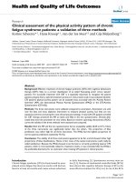
firstly fell down and then rose up. For patient R014, the
xMAP FI of IgG-EA-D was 6303 before treatment and then
rose to 7567 after the initial chemotherapy, but it dropped
to 2385 three months after therapy (Fig. 1). The initial rise
of some EBV antibodies in patients R072, R077, R139
after clinical therapy all reached to ~40 – 70%. Interest-
ingly, the reactivities of IgG-EBNA1 in patient R103 had a
drastic decrease after the starting treatment, with xMAP FIs
ranging from 7014 to 2970, whereas it ascend to 6279 at
the end of treatment (Fig. 1). The disparity of EBV serolog-
ical kinetics in different NPC individuals during treatment
might reflect the different radiosensitivity and immuno-
logical reactivation.
Moreover, patient R057 showed continuous elevation of
EBV immunoactivities one year after treatment. When
NPC recurrence was detected, the antibody levels were
much higher than those of pretreatment. But patient R100
showed a more complicated kinetics of EBV antibody
reactivities. During the therapy, all of the EBV biomarkers
fell down largely or slightly. However, the levels of IgA-
and IgG-EA-D in patient R100 rose up at one month after
finishing clinical treatment, whereas IgG-EBNA1 and IgG-
gp78 had an elevation at three months. But IgA-p18 kept
rising at four months and the time metastasis was detected
(Fig. 1).
EBV serological examination in the high-risk NPC families
In order to evaluate the distribution of EBV antibody lev-
els in NPC high-risk families, we collected 92 sera from
members of 6 families with at least two NPC patients for
each family, including 15 NPC patients, 60 Grade I rela-
tives and 17 Grade II relatives, based on their relationship
to the NPC cases in the family: Grade I (parents, children,
siblings) and Grade II (spouses).
Compared with the general NPC cases, the NPC individu-
als in the high-risk families showed lower EBV antibody
levels except for IgA-EA (see Additional file 1). This may
be due to the fact that a majority of the familiar cases in
our study were after NPC therapy and the EBV seroreactiv-
ity declined. On the other hand, the unaffected individu-
als from high-risk families and community controls
showed no statistical differences in the antibody levels
against any EBV markers. Intriguingly, a couple with both
NPC cases, who are from two separate high-risk families,
didn't show elevated EBV antibody levels and their chil-
dren are healthy.
EBV serological detection in non-endemic healthy
population and patients with other solid cancers
To compare the EBV antibody levels in populations from
distinct geographic origins, we collected 52 sera of healthy
blood donors from Shanxi Province, which is located in
the north China and represents a non-endemic NPC area.
The mean FI value in non-endemic healthy subjects were
lower than those in Cantonese population for each EBV
biomarker tested, although there was no statistical differ-
ence (see Additional file 1). Furthermore, we also exam-
ined sera from patients with other solid cancers. There was
no difference for antibody levels of each EBV marker
between any group of the patients and Cantonese con-
trols.
EBV serological profiles in patients with other EBV-
associated diseases
We further analyzed these 8 EBV antibodies in sera from
patients with different EBV-associated diseases. The mean
FI values of theses markers are also presented in Addi-
Journal of Translational Medicine 2009, 7:73 />Page 5 of 8
(page number not for citation purposes)
tional file 1. Interestingly, all of these disease groups had
much lower EBV antibody levels than NPC group. When
compared with Cantonese healthy controls, IM patients
had significantly higher IgA-gp125 level (P = 0.01) but rel-
atively lower IgG levels. The IgA-p18 level in HD patients
was higher than that in healthy group, but lower than that
in NPC patients. However, neither was statistically differ-
ent (P > 0.05). This may be due to a small number of HD
patients. Compared with the healthy, patients with NK/T
cell tumors had a significantly higher levels of IgG-EA (P
= 0.03), and higher levels of IgA-EA and IgA-gp125 (P >
0.05), and a lower level of IgG-gp78 (P > 0.05); patients
with NHL except for NK/T cell tumors had higher levels of
IgA-EA and IgG-EBNA1 (P > 0.05). The results may indi-
cate that EBV has different activities in various EBV-associ-
ated diseases.
Discussion
EBV serology testing is usually performed by indirect
immuno-fluorescence assay, ELISA or immunoblot
[20,23,26], but these methods could only address one of
two aspects: evaluation of EBV antibody parameters for
the diagnosis of NPC or analysis of molecular diversity of
EBV serological spectrums in different populations. In
contrast, xMAP assay could achieve both simultaneously.
At present, by using xMAP technology, we examined IgA
and IgG levels against a wide spectrum of EBV antigens in
populations with distinct diseases, or with different
genetic or geographic background.
We are presenting a diagnostic model for NPC using logis-
tic regression by combining 8 EBV biomarkers. This
model could reach the sensitivity and specificity of 84.5%
and 92.4%, respectively, to discriminate between NPC
patients and healthy controls. Furthermore, this model
could be further used to predict the risk rate of NPC occur-
rence in a large-scale screening. In addition, our study also
confirmed that single EBV biomarker was not efficient
enough for NPC diagnosis [20,23,27], and that there is a
Fluctuations of EBV antibody levels in four representative NPC patients during and after radiotherapy and/or chemotherapyFigure 1
Fluctuations of EBV antibody levels in four representative NPC patients during and after radiotherapy and/or
chemotherapy. The Y axis represents the mean xMAP fluorescence intensity (FI) for each EBV parameter. X axis, time from
the start of blood sampling, with the day of diagnosis, the day of treatment end and the follow-up period. R014, R103, R57 and
R100 represent different NPC patients.
Journal of Translational Medicine 2009, 7:73 />Page 6 of 8
(page number not for citation purposes)
diversity of EBV-antigen-recognition spectrum within
individuals [20].
Although EBV serological examination has been widely
employed for assisting in NPC diagnosis, the temporal
kinetics of antibody levels in a short period during and
after treatment had been rarely studied. It was reported
that patients with confirmed clinical recurrence 1 year
after completion of radiotherapy had significantly increas-
ing IgG-EA and mainly IgA-EA titers [28]. By using xMAP
technology, we found the EBV antibody levels were also
correlated with early clinical events of NPC patients after
treatment, similar to the studies of plasma EBV DNA [29].
At the time of tumor recurrence, increased EBV antibody
levels were observed. In some patients, an initial rise of
EBV antibody reactivities was detected during NPC treat-
ment, comparable to the initial rise of plasma EBV DNA
after therapy [30]. So it strongly supports the close link of
EBV antibody levels and NPC tumor load.
Familial history is one of the contributors to the risk of
NPC [31-33]. EBV serology testing in Taiwan indicated
that unaffected members of high-risk families had
increased seropositivity rate of anti-VCA IgA and anti-
EBNA1 IgA compared to general healthy population, but
this trend was not observed among Greenlandic Inuit
[34,35]. Our present study using the eight EBV markers
showed that the percentage of positive subjects was iden-
tical in the healthy populations from either high-risk NPC
family or community. The inconsistency might be due to
the distinct age distributions among these studies, since
elder healthy populations usually have higher anti-EBV
antibody levels, which is another interesting finding in
our study. Furthermore, our results showed that no statis-
tical difference is observed between unaffected individu-
als of high-risk families and general controls for all EBV
antibody levels tested, neither is between first-degree rela-
tives and spouses of NPC cases. These are in agreement
with previous studies [34,35]. But a long-term follow-up
study on EBV antibody-elevated population from Taiwan
suggested a significantly higher risk for developing into
NPC than controls [17]. Therefore, EBV infection might
not be the key initiator for NPC, but play an important
role in the high-risk subjects. Other factors such as genetic
susceptibility and environmental factors may be essential
for the incidence and development of NPC as indicated
previously [36-40].
EBV-associated diseases could be characterized by differ-
ent EBV serological features. For example, the acute EBV
infection resulted in IM could be reflected by the appear-
ance of anti-VCA antibodies [41], which support our
results showing that IgA-VCA gp125 levels significantly
increased in IM patients as previously reported [42]. On
the other hand, chronic EBV infection is linked to several
lymphoma diseases with aberrant EBV antibody levels.
HD patients usually have elevated IgG antibodies against
VCA, EA-D and EBNA1 [43]. Interestingly, we found that
compared with healthy controls, IgA-p18 was higher in
HD patients and IgG-EA-D was higher in the patients with
nasal NK/T cell lymphoma. Remarkably, NPC patients
have higher levels of both IgA and IgG classes in a large
spectrum of EBV antigens including VCA gp125 and p18,
EA-D, EBNA1 and gp78 compared with the healthy popu-
lations or populations with other EBV-associated diseases,
indicative of a vigorous viral activity in NPC.
Although viral expression in most EBV-associated tumor
cells is mainly latent, transcription of a variety of lytic
genes was detected in infiltrating lymphoid cells in NPC
by in situ studies [44,45]. Accordingly, it has been sug-
gested that diverse EBV antigens in the lymphoid stroma
of NPC could stimulate EBV antibody reactivity and con-
tributed to the specific serologic feature of this disease
[46]. But the mechanism of uncontrolled EBV activity in
cancer patients remains unclear. Depressed immune con-
trol of the virus might enable EBV more activated, since
increasing EBV antibody levels were usually found in
advanced cancer stage and aging healthy people, which
have lower immunity [47]. In agreement with this specu-
lation, reactivation of latent EBV infection was considered
an important pathogenic mechanism of EBV-related dis-
eases in immunocompromised patients such as those
with PTLD or HIV [48,49]. However, patients with other
solid tumors didn't show higher EBV activities than
healthy controls in this study, suggesting EBV propagation
may undergo in parallel with strong microenvironment
disposition. Further investigations are awaited to charac-
terize the biological activities and functions of EBV in
NPC and lymphoma.
Conclusion
Our results revealed that diverse EBV antibody spectrums
presented in distinct populations with different EBV-asso-
ciated diseases. Moreover, NPC individuals have various
EBV serological profiles and combined EBV biomarkers
could improve the analytic accuracy for diagnosis.
Competing interests
The authors declare that they have no competing interests.
Authors' contributions
YXZ and YBX were responsible for the design of this study.
ADG carried out the experiments and drafted the manu-
script. LXL participated in the data analysis. LZC, QSF and
WHJ helped in serum samples colletion. TK helped in
amending the manuscript. All authors read and approved
the final manuscript.
Journal of Translational Medicine 2009, 7:73 />Page 7 of 8
(page number not for citation purposes)
Additional material
Acknowledgements
This study was supported by 973 projects of the Ministry of Science and
Technology of China (2004CB518604), the Scientific and Technologic
Project of Guangzhou City (2007Z-E4021) and China Postdoctoral Science
Foundation (20070410862). We thank Juan Peng and Miao-Yan Li for the
assistance in serologic tests. We thank all the patients and healthy popula-
tions participated in this study.
References
1. Rickinson AB, Kieff E: Epstein-Barr virus 4th edition. Philadelphia (Penn-
sylvania): Lippincott/Williams & Wilkins; 2001.
2. Crawford DH: Biology and disease associations of Epstein-
Barr virus. Philos Trans R Soc Lond B Biol Sci 2001, 356:461-473.
3. Henle G, Henle W, Diehl V: Relation of Burkitt's tumor-associ-
ated herpes-ytpe virus to infectious mononucleosis. Proc Natl
Acad Sci USA 1968, 59:94-101.
4. Ambinder RF: Epstein-barr virus and hodgkin lymphoma.
Hematology Am Soc Hematol Educ Program 2007, 2007:204-209.
5. List AF, Greco FA, Vogler LB: Lymphoproliferative diseases in
immunocompromised hosts: the role of Epstein-Barr virus.
J Clin Oncol 1987, 5:1673-1689.
6. Imai S, Koizumi S, Sugiura M, Tokunaga M, Uemura Y, Yamamoto N,
Tanaka S, Sato E, Osato T: Gastric carcinoma: monoclonal epi-
thelial malignant cells expressing Epstein-Barr virus latent
infection protein. Proc Natl Acad Sci USA 1994, 91:9131-9135.
7. de The G: Sero epidemiology of EBV and associated malignancies
Wymondham: Caister Academic Press; 2005.
8. Tao Q, Young LS, Woodman CB, Murray PG: Epstein-Barr virus
(EBV) and its associated human cancers – genetics, epigenet-
ics, pathobiology and novel therapeutics. Front Biosci 2006,
11:2672-2713.
9. Kelly GL, Rickinson AB: Burkitt lymphoma: revisiting the
pathogenesis of a virus-associated malignancy. Hematology Am
Soc Hematol Educ Program 2007, 2007:277-284.
10. Aozasa K, Takakuwa T, Hongyo T, Yang WI: Nasal NK/T-cell lym-
phoma: epidemiology and pathogenesis. Int J Hematol 2008,
87:110-117.
11. Tsuchiya S: Diagnosis of Epstein-Barr virus-associated dis-
eases. Crit Rev Oncol Hematol 2002, 44:227-238.
12. Mueller N: Overview: viral agents and cancer. Environ Health Per-
spect 1995, 103(Suppl 8):259-261.
13. Henle G, Henle W: Epstein-Barr virus-specific IgA serum anti-
bodies as an outstanding feature of nasopharyngeal carci-
noma. Int J Cancer
1976, 17:1-7.
14. Foong YT, Cheng HM, Sam CK, Dillner J, Hinderer W, Prasad U:
Serum and salivary IgA antibodies against a defined epitope
of the Epstein-Barr virus nuclear antigen (EBNA) are ele-
vated in nasopharyngeal carcinoma. Int J Cancer 1990,
45:1061-1064.
15. Feng P, Chan SH, Soo MY, Liu D, Guan M, Ren EC, Hu H: Antibody
response to Epstein-Barr virus Rta protein in patients with
nasopharyngeal carcinoma: a new serologic parameter for
diagnosis. Cancer 2001, 92:1872-1880.
16. Dardari R, Hinderer W, Lang D, Benider A, El Gueddari B, Joab I,
Benslimane A, Khyatti M: Antibody responses to recombinant
Epstein-Barr virus antigens in nasopharyngeal carcinoma
patients: complementary test of ZEBRA protein and early
antigens p54 and p138. J Clin Microbiol 2001, 39:3164-3170.
17. Chien YC, Chen JY, Liu MY, Yang HI, Hsu MM, Chen CJ, Yang CS:
Serologic markers of Epstein-Barr virus infection and
nasopharyngeal carcinoma in Taiwanese men. N Engl J Med
2001, 345:1877-1882.
18. Ji MF, Wang DK, Yu YL, Guo YQ, Liang JS, Cheng WM, Zong YS,
Chan KH, Ng SP, Wei WI, et al.: Sustained elevation of Epstein-
Barr virus antibody levels preceding clinical onset of
nasopharyngeal carcinoma. Br J Cancer 2007, 96:623-630.
19. Yip TT, Ngan RK, Lau WH, Poon YF, Joab I, Cochet C, Cheng AK: A
possible prognostic role of immunoglobulin-G antibody
against recombinant Epstein-Barr virus BZLF-1 transactiva-
tor protein ZEBRA in patients with nasopharyngeal carci-
noma. Cancer 1994, 74:2414-2424.
20. Fachiroh J, Schouten T, Hariwiyanto B, Paramita DK, Harijadi A, Har-
yana SM, Ng MH, Middeldorp JM: Molecular diversity of Epstein-
Barr virus IgG and IgA antibody responses in nasopharyngeal
carcinoma: a comparison of Indonesian, Chinese, and Euro-
pean subjects. J Infect Dis 2004, 190:53-62.
21. Elshal MF, McCoy JP: Multiplex bead array assays: performance
evaluation and comparison of sensitivity to ELISA. Methods
2006, 38:317-323.
22. Gu AD, Xie YB, Mo HY, Jia WH, Li MY, Li M, Chen LZ, Feng QS, Liu
Q, Qian CN, Zeng YX: Antibodies against Epstein-Barr virus
gp78 antigen: a novel marker for serological diagnosis of
nasopharyngeal carcinoma detected by xMAP technology. J
Gen Virol 2008, 89:1152-1158.
23. Fachiroh J, Paramita DK, Hariwiyanto B, Harijadi A, Dahlia HL, Indras-
ari SR, Kusumo H, Zeng YS, Schouten T, Mubarika S, Middeldorp JM:
Single-assay combination of Epstein-Barr Virus (EBV)
EBNA1- and viral capsid antigen-p18-derived synthetic pep-
tides for measuring anti-EBV immunoglobulin G (IgG) and
IgA antibody levels in sera from nasopharyngeal carcinoma
patients: options for field screening. J Clin Microbiol 2006,
44:1459-1467.
24. Tarbouriech N, Buisson M, Geoui T, Daenke S, Cusack S, Burmeister
WP: Structural genomics of the Epstein-Barr virus. Acta Crys-
tallogr D Biol Crystallogr 2006, 62:1276-1285.
25. Gu AD, Mo HY, Xie YB, Peng RJ, Bei JX, Peng J, Li MY, Chen LZ, Feng
QS, Jia WH, Zeng YX: Evaluation of a multianalyte profiling
assay and an enzyme-linked immunosorbent assay for sero-
logical examination of Epstein-Barr virus-specific antibody
responses in diagnosis of nasopharyngeal carcinoma. Clin Vac-
cine Immunol 2008, 15:1684-1688.
26. Karray H, Ayadi W, Fki L, Hammami A, Daoud J, Drira MM, Frikha M,
Jlidi R, Middeldorp JM: Comparison of three different serologi-
cal techniques for primary diagnosis and monitoring of
nasopharyngeal carcinoma in two age groups from Tunisia.
J Med Virol 2005, 75:593-602.
27. Chow KC, Ma J, Lin LS, Chi KH, Yen SH, Liu SM, Liu WT, Chen WK,
Chang TH, Chen KY: Serum responses to the combination of
Epstein-Barr virus antigens from both latent and acute
phases in nasopharyngeal carcinoma: complementary test of
EBNA-1 with EA-D. Cancer Epidemiol Biomarkers Prev 1997,
6:363-368.
28. de-Vathaire F, Sancho-Garnier H, de-The H, Pieddeloup C, Schwaab
G, Ho JH, Ellouz R, Micheau C, Cammoun M, Cachin Y, et al.: Prog-
nostic value of EBV markers in the clinical management of
nasopharyngeal carcinoma (NPC): a multicenter follow-up
study. Int J Cancer 1988, 42:176-181.
29. Lin JC, Wang WY, Chen KY, Wei YH, Liang WM, Jan JS, Jiang RS:
Quantification of plasma Epstein-Barr virus DNA in patients
with advanced nasopharyngeal carcinoma. N Engl J Med 2004,
350:2461-2470.
30. Lo YM, Leung SF, Chan LY, Chan AT, Lo KW, Johnson PJ, Huang DP:
Kinetics of plasma Epstein-Barr virus DNA during radiation
therapy for nasopharyngeal carcinoma. Cancer Res 2000,
60:2351-2355.
31. Feng BJ, Huang W, Shugart YY, Lee MK, Zhang F, Xia JC, Wang HY,
Huang TB, Jian SW, Huang P, et al.: Genome-wide scan for familial
nasopharyngeal carcinoma reveals evidence of linkage to
chromosome 4.
Nat Genet 2002, 31:395-399.
32. Xiong W, Zeng ZY, Xia JH, Xia K, Shen SR, Li XL, Hu DX, Tan C,
Xiang JJ, Zhou J, et al.: A susceptibility locus at chromosome
3p21 linked to familial nasopharyngeal carcinoma. Cancer Res
2004, 64:1972-1974.
33. Yang XR, Diehl S, Pfeiffer R, Chen CJ, Hsu WL, Dosemeci M, Cheng
YJ, Sun B, Goldstein AM, Hildesheim A: Evaluation of risk factors
for nasopharyngeal carcinoma in high-risk nasopharyngeal
Additional file 1
Distribution of EBV serological biomarkers in different populations.
Click here for file
[ />5876-7-73-S1.doc]
Publish with Bio Med Central and every
scientist can read your work free of charge
"BioMed Central will be the most significant development for
disseminating the results of biomedical research in our lifetime."
Sir Paul Nurse, Cancer Research UK
Your research papers will be:
available free of charge to the entire biomedical community
peer reviewed and published immediately upon acceptance
cited in PubMed and archived on PubMed Central
yours — you keep the copyright
Submit your manuscript here:
/>BioMedcentral
Journal of Translational Medicine 2009, 7:73 />Page 8 of 8
(page number not for citation purposes)
carcinoma families in Taiwan. Cancer Epidemiol Biomarkers Prev
2005, 14:900-905.
34. Pickard A, Chen CJ, Diehl SR, Liu MY, Cheng YJ, Hsu WL, Sun B, Hsu
MM, Chen IH, Chen JY, et al.: Epstein-Barr virus seroreactivity
among unaffected individuals within high-risk nasopharyn-
geal carcinoma families in Taiwan. Int J Cancer 2004,
111:117-123.
35. Friborg J, Jarrett RF, Liu MY, Falk KI, Koch A, Olsen OR, Duncan P,
Wohlfarht J, Chen JY, Melbye M: Epstein-Barr virus immune
response in high-risk nasopharyngeal carcinoma families in
Greenland. J Med Virol 2007, 79:1877-1881.
36. Hildesheim A, Dosemeci M, Chan CC, Chen CJ, Cheng YJ, Hsu MM,
Chen IH, Mittl BF, Sun B, Levine PH, et al.: Occupational exposure
to wood, formaldehyde, and solvents and risk of nasopharyn-
geal carcinoma. Cancer Epidemiol Biomarkers Prev 2001,
10:1145-1153.
37. Yang X, Goldstein AM, Chen CJ, Rabkin CS, Chen JY, Cheng YJ, Hsu
WL, Sun B, Diehl SR, Liu MY, et al.: Distribution of Epstein-Barr
viral load in serum of individuals from nasopharyngeal carci-
noma high-risk families in Taiwan. Int J Cancer 2006,
118:780-784.
38. Yu MC, Garabrant DH, Huang TB, Henderson BE: Occupational
and other non-dietary risk factors for nasopharyngeal carci-
noma in Guangzhou, China. Int J Cancer 1990, 45:1033-1039.
39. Yu MC, Yuan JM: Epidemiology of nasopharyngeal carcinoma.
Semin Cancer Biol 2002, 12:421-429.
40. Zheng YM, Tuppin P, Hubert A, Jeannel D, Pan YJ, Zeng Y, de The G:
Environmental and dietary risk factors for nasopharyngeal
carcinoma: a case-control study in Zangwu County, Guangxi,
China. Br J Cancer 1994, 69:508-514.
41. Evans AS, Niederman JC, McCollum RW: Seroepidemiologic
studies of infectious mononucleosis with EB virus. N Engl J
Med 1968, 279:1121-1127.
42. Bhaduri-McIntosh S, Landry ML, Nikiforow S, Rotenberg M, El-Guindy
A, Miller G: Serum IgA antibodies to Epstein-Barr virus (EBV)
early lytic antigens are present in primary EBV infection. J
Infect Dis
2007, 195:483-492.
43. Mueller N, Evans A, Harris NL, Comstock GW, Jellum E, Magnus K,
Orentreich N, Polk BF, Vogelman J: Hodgkin's disease and
Epstein-Barr virus. Altered antibody pattern before diagno-
sis. N Engl J Med 1989, 320:689-695.
44. Tao Q, Srivastava G, Chan AC, Chung LP, Loke SL, Ho FC: Evidence
for lytic infection by Epstein-Barr virus in mucosal lym-
phocytes instead of nasopharyngeal epithelial cells in normal
individuals. J Med Virol 1995, 45:71-77.
45. Tao Q, Srivastava G, Dickens P, Ho FC: Detection of Epstein-Barr
virus-infected mucosal lymphocytes in nasal polyps. Am J
Pathol 1996, 149:1111-1118.
46. Ng MH, Chan KH, Ng SP, Zong YS: Epstein-Barr virus serology
in early detection and screening of nasopharyngeal carci-
noma. Ai Zheng 2006, 25:250-256.
47. Chang ET, Zheng T, Lennette ET, Weir EG, Borowitz M, Mann RB,
Spiegelman D, Mueller NE: Heterogeneity of risk factors and
antibody profiles in epstein-barr virus genome-positive and -
negative hodgkin lymphoma. J Infect Dis 2004, 189:2271-2281.
48. Ambinder RF: Epstein-Barr virus associated lymphoprolifera-
tions in the AIDS setting. Eur J Cancer 2001, 37:1209-1216.
49. Young L, Alfieri C, Hennessy K, Evans H, O'Hara C, Anderson KC,
Ritz J, Shapiro RS, Rickinson A, Kieff E, et al.: Expression of Epstein-
Barr virus transformation-associated genes in tissues of
patients with EBV lymphoproliferative disease. N Engl J Med
1989, 321:1080-1085.