báo cáo hóa học:" Human T cells express CD25 and Foxp3 upon activation and exhibit effector/memory phenotypes without any regulatory/suppressor function" ppt
Bạn đang xem bản rút gọn của tài liệu. Xem và tải ngay bản đầy đủ của tài liệu tại đây (1.11 MB, 7 trang )
BioMed Central
Page 1 of 7
(page number not for citation purposes)
Journal of Translational Medicine
Open Access
Research
Human T cells express CD25 and Foxp3 upon activation and exhibit
effector/memory phenotypes without any regulatory/suppressor
function
Maciej Kmieciak
1,4
, Madhu Gowda
2,4
, Laura Graham
3,4
, Kamar Godder
2,4
,
Harry D Bear
3,4
, Francesco M Marincola
5,4
and Masoud H Manjili*
1,4
Address:
1
Department of Microbiology & Immunology, Virginia Commonwealth University Massey Cancer Center, Richmond, USA,
2
Department
of Pediatrics, Virginia Commonwealth University Massey Cancer Center, Richmond, USA,
3
Department of Surgery, Virginia Commonwealth
University Massey Cancer Center, Richmond, USA,
4
Department of Pathology, Virginia Commonwealth University Massey Cancer Center,
Richmond, USA and
5
Infectious Disease and Immunogenetics Section (IDIS), Department of Transfusion Medicine, Clinical Center and Center
for Human Immunology (CHI), National Institutes of Health, Bethesda, USA
Email: Maciej Kmieciak - ; Madhu Gowda - ; Laura Graham - ;
Kamar Godder - ; Harry D Bear - ; Francesco M Marincola - ;
Masoud H Manjili* -
* Corresponding author
Abstract
Background: Foxp3 has been suggested to be a standard marker for murine Tregs whereas its
role as marker for human Tregs is controversial. While some reports have shown that human
Foxp3+ T cells had no regulatory function others have shown their role in the inhibition of T cell
proliferation.
Methods: T cell activation was performed by means of brayostatin-1/ionomycin (B/I), mixed
lymphocyte reaction (MLR), and CD3/CD28 activation. T cell proliferation was performed using
BrdU and CFSE staining. Flow cytometry was performed to determine Foxp3 expression, cell
proliferation, viabilities and phenotype analyses of T cells.
Results: Both CD4+ and CD8+ T cells expressed Foxp3 upon activation in vitro. Expression of
Foxp3 remained more stable in CD4+CD25+ T cells compared to that in CD8+CD25+ T cells.
The CD4+CD25+Foxp3+ T cells expressed CD44 and CD62L, showing their effector and memory
phenotypes. Both FoxP3- responder T cells and CD4+FoxP3+ T cells underwent proliferation
upon CD3/CD28 activation.
Conclusion: Expression of Foxp3 does not necessarily convey regulatory function in human
CD4+CD25+ T cells. Increased FoxP3 on CD44+ effector and CD44+CD62L+ memory T cells
upon stimulation suggest the activation-induced regulation of FoxP3 expression.
Background
In mice, scurfy mutation in forkhead/winged helix tran-
scription factor gene Foxp3 causes autoimmune lesions
including massive lymphoproliferation, diabetes, exfolia-
tive dermatitis, thyroiditis and enteropathy. Such autoim-
munity can be cured by a transgene encoding a wild-type
Published: 22 October 2009
Journal of Translational Medicine 2009, 7:89 doi:10.1186/1479-5876-7-89
Received: 22 July 2009
Accepted: 22 October 2009
This article is available from: />© 2009 Kmieciak et al; licensee BioMed Central Ltd.
This is an Open Access article distributed under the terms of the Creative Commons Attribution License ( />),
which permits unrestricted use, distribution, and reproduction in any medium, provided the original work is properly cited.
Journal of Translational Medicine 2009, 7:89 />Page 2 of 7
(page number not for citation purposes)
Foxp3 allele [1]. The expression of Foxp3 in CD4+CD25+
T cells in wild-type mice and the diminished numbers of
these T cells in scurfy and Foxp3-knockout (Foxp3
-
) mice
suggested a role for Foxp3 in the development of regula-
tory T cells (Tregs) [2]. In addition, Foxp3 has been shown
to be a specific marker for murine CD4+ Tregs because
activation of non-T regs did not induce Foxp3 expression
[2]. Ectopic expression of Foxp3 was shown to be suffi-
cient to activate a program of suppressor function in
peripheral murine CD4+ T cells [2].
In humans, the gene encoding Foxp3 was discovered dur-
ing efforts to understand the genetic basis for a rare X-
linked fatal autoimmune disease known as IPEX
(immune dysregulation, polyendocrinopathy, enteropa-
thy, X-linked) syndrome [3,4]. However, the role of Foxp3
as a key marker for Tregs in humans remains controver-
sial. Unlike mice, activation of human CD4+ T cells by T-
cell receptor (TcR) stimulation resulted in the expression
of Foxp3 [5-12]. Most of these studies showed that induc-
tion of Foxp3, even in the presence of TGF-, did not cor-
relate with suppressive function of CD4+ T cells [6,10-12].
Although it was suggested that lack of suppression during
the activation-induced expression of Foxp3 in human
CD4+ T cells was because of transient expression of
Foxp3, the observation still argues against a role for Foxp3
as key regulator of suppression in human CD4+ T cells
upon expression. Regardless of the status of Foxp3, many
studies considered CD4+CD25
high
as Tregs in humans
without being able to show their regulatory functions in
vivo [13-15]. Most recently, it was reported that maternal
alloantigens promoted development of Tregs in the
human fetus that could suppress fetal antimaternal
immunity. The authors considered CD4+CD25+Foxp3+ T
cells as Tregs because of their partial suppressive function
in a mixed lymphocyte reaction (MLR) in vitro [16]. These
controversial reports prompted us to determine whether
induction of Foxp3 expression in human T cells during
activation and during MLR may confer regulatory func-
tions. Our studies showed that activation-induced expres-
sion of Foxp3 was transient in CD8+CD25+ T cells but it
was more stable in CD4+CD25+ T cells. These Foxp3+ T
cells were mainly of effector and memory phenotypes.
Methods
Blood samples
PBMC were collected from two healthy donors, and dupli-
cate experiments were performed.
Flow cytometry
Three-color staining and FACS analyses were performed as
previously described by our group [17]. Extracellular
staining were performed using anti-human antibodies
from Biolegend: PE- and FITC-CD25 (clone BC96), PE-
and FITC-CD44 (clone IM7), FITC-CD62L (clone DREG-
56), PE/Cy5-CD4 (clone OKT4) and PE/Cy5-CD8 (clone
RPA-T8). Appropriate isotype control antibodies were
used to exclude nonspecific binding. Foxp3 intracellular
staining was done with PE anti-human Foxp3 Flow Kit
(Biolegend, clone 206D) according to the manufacturer's
protocol. Apoptosis was determined by staining of cells
with Annexin V (BD Pharmingen).
Proliferation assay
FITC BrdU Flow Kit (BD Pharmingen) was used in prolif-
eration assays. T cells were also labeled with CFSE by incu-
bation at 5 × 10
7
cells/mL in 5 M CFSE/HBSS for 5 min
at room temperature. Cells were then added with an equal
volume of FBS, followed by three washes in FBS-contain-
ing HBSS.
Mixed lymphocyte reaction (MLR)
Blood samples were diluted two-fold with PBS and lay-
ered onto Ficoll-Hypaque. Each tube was centrifuged at
400 g for 30 min and the lymphocytes at the interface
were collected. These cells were washed once with RPMI
1640 medium containing 100 U/ml penicillin, 100 g/ml
streptomycin, and 2 mM L-glutamine. They were then
resuspended at l0
7
cells/ml in the same medium contain-
ing 10% heat inactivated FBS. Allogeneic stimulating cells
were irradiated in a cesium irradiator to a total dose of
5,000 rad, to abolish their capacity to proliferate. Cultures
were set up in flat-bottomed 24-well plates and 3 × 10
6
responder cells were mixed with 2 × 10
6
irradiated stimu-
lators in 2 mL. Cultures, set up in triplicates, were incu-
bated for 8 days at 37°C. Control cells cultured with
medium containing low dose IL-2 (20 U/mL) in order to
maintain T cell viability during a 3-day culture. No IL-2 or
anti-CD3 Ab was used in MLR samples. Some cultures
were pulsed with 10 M BrdU (BD Pharmingen).
Statistical analysis
Statistical comparisons between groups were made using
the Student t test with P < 0.0.5 being statistically signifi-
cant.
Results and discussion
Activation of T cells induces expression of CD25 and Foxp3
associated with effector and memory phenotype
differentiation
PBMC were stimulated with bryostatin-1 (5 nM) and ion-
omycin (1 M) (B/I) in the presence of 80 U/mL of IL-2
(Peprotech) for 16 h. B/I activation mimic intracellular
signals that result in T cell activation by increasing protein
kinase C activity and intracellular calcium, respectively
[18-20]. Cells were washed three times and cultured at 10
6
cells/mL in complete medium with 40 U/mL IL-2 (Pepro-
tech) for 3 days and expression of Foxp3 was determined
using flow cytometry analysis. Expression of FoxP3 was
also determined on freshly isolated T cells on day 0. As
Journal of Translational Medicine 2009, 7:89 />Page 3 of 7
(page number not for citation purposes)
shown in Fig. 1A (top panel), presence of IL-2 alone for 3
days did not markedly increase expression of Foxp3 or
CD25 above baseline levels on day 0 (Fig. 1C). The B/I
activation, however, induced Foxp3 and CD25 expression
in CD4+ and CD8+ T cells (Fig. 1A, middle panel). Upon
B/I activation, CD4+CD25+Foxp3+ T cells were increased
from 1% to 23% (P = 0.016) and CD8+CD25+Foxp3+ T
cells were increased from 0.6% to 9% (P = 0.013). Exten-
sion of culture in the presence of IL-2 for 6 days without
any further stimulation retained CD4+CD25+Foxp3+ T
cells above the baseline levels in unactivated T cells (1%
vs. 7%; P = 0.031) whereas CD8+CD25+Foxp3+ T cells
dropped to baseline levels (0.6%). These results suggest
that activation-induced expression of Foxp3 in
CD4+CD25+ T cells is more stable than that in
CD8+CD25+ T cells. Absolute number of T cells increased
3 and 6 days after the B/I stimulation and expansion in
the presence of IL-2 (Fig. 1B). Activation of T cells by
means of anti-CD3/CD28 Abs for 3 days produced similar
results as for B/I activation by increasing
CD4+CD25+FoxP3+ T cells from 0.4% to 8.7% (Fig. 1C).
Phenotype analyses of T cells revealed CD44+ effector and
CD44+CD62L+ memory phenotypes prior to and 6 days
after the B/I activation (Fig. 1D, top panel). While effector
CD4+ and CD8+ T cells were reduced after activation
(18% to 9% and 21% to 13%, respectively), memory
CD4+ and CD8+ T cells were increased (82% to 91% and
79% to 87%, respectively). Upon B/I activation, CD4+ T
cells showed a 6-fold increases of FoxP3 expression in
CD44+, CD62L+ phenotypes (CD44+: 2.6% to 15%;
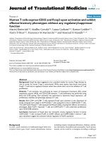
Foxp3 expression following T cell activationFigure 1
Foxp3 expression following T cell activation. T cells were isolated from healthy volunteers and split into two groups.
Control group remained unactivated and cultured in the presence of IL-2 for 3 days (A; top panel) and another group was acti-
vated with B/I for 16 h and cultured in the presence of IL-2 for 3 days (A; middle panel) or 6 days (A; bottom panel). Absolute
numbers of CD4+ and CD8+ T cells on pooled samples were determined on days 0, 3, and 6 post-culture by flow cytometry
analysis (B). Expression of FoxP3 and CD25 were determined in freshly isolated CD4+ T cells (day 0) and after a 3-day stimu-
lation with anti-CD3/CD28 Abs (C). Freshly isolated and B/I-activated T cells were subjected to flow cytometry to determine
T cell phenotypes (D; top panel); Foxp3+ effector and memory T cells were determined in gated CD4+Foxp3+ cells or gated
CD8+Foxp3+ cells (D; bottom panel). Representative data are shown from two donors in duplicate experiments.
Journal of Translational Medicine 2009, 7:89 />Page 4 of 7
(page number not for citation purposes)
CD62L+: 2% to 12%). In addition, both CD4+ and CD8+
T cells showed FoxP3
high
expression following activation
compared to FoxP3
low
expression on day 0 (Fig. 1D, mid-
dle and bottom panels). All CD4+Foxp3+ T cells
expressed CD44 among which 80% also expressed CD62L
(Fig. 1D, middle panel, far right). These data show that
20% of CD4+Foxp3+ T cells are effector and 80% are
memory phenotypes. A similar phenotypic trend was
detected for CD8+Foxp3+ T cells, showing 100% CD44+
of which 67% were CD62L+ T cells (Fig. 1D, bottom
panel, far right). These results show that 33% of
CD8+Foxp3+ T cells are effector and 67% are memory
phenotypes. Data presented in Figs. 1A-D suggest that
increased expression of FoxP3
high
in effector T cells was
due to the cell differentiation rather than cell prolifera-
tion, because relative percent of CD44+CD62L- effector T
cells decreased after B/I activation. Similar mechanism
may exist in memory T cells because of the expression of
FoxP3
high
after activation compared to FoxP3
low
on day 0.
Activation-induced FoxP3 expression in CD4+ T cells fails
to convey regulatory function in vitro
T cells were labeled with CFSE and stimulated with anti-
CD3 (1 ug/ml) and anti-CD28 (1 ug/ml) Abs in the pres-
ence or absence of the B/I-activated CD4+CD25+FoxP3+
T cells (2:1 and 20:1 responder:suppressor ratios) for 3
days. Flow cytometry analysis showed similar rates of pro-
liferation of gated CD8+ T cells in the absence or presence
of inducible FoxP3+ T cells (Fig. 2A, 60% vs. 61% and
65%). The CD3/CD28 activation also induced FoxP3
expression in responder CD4+ T cells. Gated
CD4+FpxP3+ T cells also showed 70-75% proliferation
upon activation (Fig. 2A). Analysis of T cell apoptosis
revealed similar rates of apoptosis in responder T cells in
the absence or presence of CD4+FoxP3+ T cells (Fig. 2B,
57% vs. 57 and 59%). Majority of the B/I-activated
CD4+FoxP3+ T cells (74-76%) were found to be apoptotic
during anti-CD3/CD28 activation in co-culture with
responder T cells.
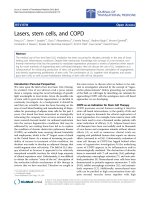
Figure 2
T cell proliferation in the presence of inducible CD4+FoxP3+ T cellsFigure 2
T cell proliferation in the presence of inducible
CD4+FoxP3+ T cells. To perform a co-culture suppres-
sion assay, responder T cells were labeled with CFSE and cul-
tured in the absence or presence of different ratios of
inducible FoxP3+ T cells (20:1 and 2:1) for 3 days in the pres-
ence of anti-CD3/CD28 Abs. Gated CD8+ T cells showed
CFSE dilution (A, left panel). Responder CD4+ T cells that
expressed FoxP3 due to a 3-day activation were also gated
and analyzed for CFSE dilution (A, right panel). Cells
obtained from a co-culture suppression assay (A, left panel)
were also stained for Annexin V in order to determine apop-
tosis in responder CD8+ T cells (B, left panel) and the B/I-
activated CD4+FoxP3+ T cells (B, right panel).
Journal of Translational Medicine 2009, 7:89 />Page 5 of 7
(page number not for citation purposes)
Allogeneic activation of T cells during MLR induces Foxp3
expression in CD4+CD25+ T cells associated with effector/
memory phenotype
We performed an 8-day allogenic MLR to determine
whether induction of Foxp3 expression in T cells was sta-
ble during MLR and whether such an induced Foxp3+
expression might inhibit T cell proliferation. Responder
and stimulator cells were obtained from different healthy
donors. Stimulator cells were irradiated (5000 rad) and
cultured with responder cells for 8 days in the presence of
10 M BrdU (BD Pharmingen). Cells were then stained
with relevant Abs and subjected to flow cytometry analy-
sis. As shown in Fig. 3A (top panel) 86% of CD4+CD25+
T cells and 93% of CD8+CD25+ T cells showed BrdU
incorporation as a result of cell proliferation. No prolifer-
ation was detected in the responder or stimulator cells
alone (data not shown). Such allogenic proliferation took
place in the presence of an activation-induced Foxp3
expression in CD4+ T cells such that 8% of CD4+ T cells
were CD25+Foxp3+ (Fig. 3A, bottom panel).
CD8+CD25+ T cells, on the other hand, did not show sta-
ble expression of Foxp3. These results are consistent with
our observation in Fig. 1 showing that expression of
Foxp3 in CD4+ T cells is more stable than that in CD8+ T
cells 6-8 days following T cell activation. In previous
reports, suppressive assays in vitro were conducted in the
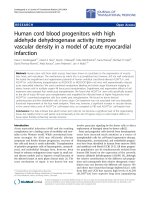
Foxp3 expression following allogeneic MLRFigure 3
Foxp3 expression following allogeneic MLR. Cells were analyzed by flow cytometry after an 8-day MLR. BrdU incorpora-
tion was determined on gated CD4+CD25+ or CD8+CD25+ T cells (A; top panel). Gated CD4+ or CD8+ T cells were ana-
lyzed for the detection of CD25+Foxp3+ cells (A; bottom panel). Gated CD4+ T cells (B; top panel) or CD8+ T cells (B;
bottom panel) were analyzed for the expression of CD44, CD62L, Foxp3. The CD44+ and CD62L+ T cells were determined
by gating on CD4+Foxp3+ or CD8+Foxp3+ T cells. Representative data are shown from two donors in duplicate experiments.
Journal of Translational Medicine 2009, 7:89 />Page 6 of 7
(page number not for citation purposes)
presence of high ratios of CD4+CD25+ T cells (Tregs) to
responder cells, to determine the suppressive function on
T cell activation and proliferation. Such artificial increases
in the ratio of CD4+CD25+ T cells to responder cells
would reduce in vivo validity of the observation. The fre-
quency of CD4+CD25+Foxp3+ T cells induced during
MLR was 8% which is considered to be within the physi-
ologically relevant range as reported by other groups [21-
24]. Frequency of naturally occurring Tregs in mouse is
also around this range, yet having regulatory effects for the
inhibition of autoimmunity. If Foxp3 expressing CD4+ T
cells had any regulatory function, it should have inhibited
cell proliferation during the culture in vitro. Similar to B/I-
induced T cell activation, T cell phenotypes in a MLR
included CD44+ effector (16%) and CD44+CD62L+
memory T cells (84%) (Fig. 3B). Again, all CD4+Foxp3+ T
cells expressed CD44 among which 90% also expressed
CD62L (Fig. 2B). These data show that 10% of
CD4+Foxp3+ T cells are effector and 90% are memory
phenotypes. A similar phenotypic trend was detected for
CD8+Foxp3+ T cells, showing 100% CD44+ of which
76% were CD62L+ T cells. These results show that 24% of
CD8+Foxp3+ T cells are effector and 76% are memory
phenotypes. Lack of regulatory function in these Foxp3+ T
cells may be because of their effector/memory phenotype
since it has been reported that expression of Foxp3 in
human memory T cells resulted in diminished suppressor
activity [25]. In addition, Treg type 1 (Tr1) cells confer
suppressor function in the absence of FoxP3 expression
[26]. Given the role of Foxp3 as master regulator of Treg
lineage commitment and maintenance in mouse [27], it
does not seem to have such bona fide regulatory function
for Treg lineage commitment in human T cells.
Conclusion
In conclusion, the present study shows that Foxp3 expres-
sion is not a reliable marker for human Tregs. T cell acti-
vation, CD4+ T cells in particular, is associated with the
expression of Foxp3 in effector/memory T cells without
detectable regulatory function when present at physiolog-
ically relevant ratios.
Abbreviations
PBMC: peripheral blood mononuclear cells; AICD: activa-
tion induced cell death; MLR: mixed lymphocyte reaction;
T regs: regulatory T cells.
Competing interests
The authors declare that they have no competing interests.
Authors' contributions
MK performed B/I activation of T cells, flow cytometry,
MLR, and BrdU proliferation assays; MG performed flow
cytometry; LG performed B/I activation of T cells; KG par-
ticipated in study design; HDB participated in study
design and manuscript preparation; FMM participated in
study design and data analysis; MHM designed the exper-
iments, analyzed data, and prepared the manuscript.
All authors read and approved the final manuscript.
Acknowledgements
This work was supported by NIH R01 CA104757 grant (M. H. Manjili) and
Massey Cancer Center Pilot Project Program, 646564. We gratefully
acknowledge the support of VCU Massey Cancer Centre and the Com-
monwealth Foundation for Cancer Research.
References
1. Brunkow ME, Jeffery EW, Hjerrild KA, Paeper B, Clark LB, Yasayko
SA, Wilkinson JE, Galas D, Ziegler SF, Ramsdell F: Disruption of a
new forkhead/winged-helix protein, scurfin, results in the
fatal lymphoproliferative disorder of the scurfy mouse. Nat
Genet 2001, 27:68-73.
2. Fontenot JD, Gavin MA, Rudensky AY: Foxp3 programs the
development and function of CD4+CD25+ regulatory T
cells. Nat Immunol 2003, 4:330-336.
3. Wildin RS, Ramsdell F, Peake J, Faravelli F, Casanova JL, Buist N, Levy-
Lahad E, Mazzella M, Goulet O, Perroni L, Bricarelli FD, Byrne G,
McEuen M, Proll S, Appleby M, Brunkow ME: X-linked neonatal
diabetes mellitus, enteropathy and endocrinopathy syn-
drome is the human equivalent of mouse scurfy. Nat Genet
2001, 27:18-20.
4. Chatila TA, Blaeser F, Ho N, Lederman HM, Voulgaropoulos C,
Helms C, Bowcock AM: JM2, encoding a fork head-related pro-
tein, is mutated in X-linked autoimmunity-allergic disregula-
tion syndrome. J Clin Invest 2000, 106:R75-R81.
5. Walker MR, Kasprowicz DJ, Gersuk VH, Benard A, Van Landeghen M,
Buckner JH, Ziegler SF: Induction of Foxp3 and acquisition of T
regulatory activity by stimulated human CD4+CD25-T cells.
J Clin Invest 2003, 112:1437-1443.
6. Morgan ME, van Bilsen JH, Bakker AM, Heemskerk B, Schilham MW,
Hartgers FC, Elferink BG, Zanden L van der, de Vries RR, Huizinga
TW, Ottenhoff TH, Toes RE: Expression of FOXP3 mRNA is not
confined to CD4+CD25+ T regulatory cells in humans. Hum
Immunol 2005, 66:13-20.
7. Roncador G, Brown PJ, Maestre L, Hue S, Martínez-Torrecuadrada JL,
Ling KL, Pratap S, Toms C, Fox BC, Cerundolo V, Powrie F, Banham
AH: Analysis of FOXP3 protein expression in human
CD4+CD25+ regulatory T cells at the single-cell level. Eur J
Immunol 2005, 35:1681-1691.
8. Gavin MA, Torgerson TR, Houston E, DeRoos P, Ho WY, Stray-Ped-
ersen A, Ocheltree EL, Greenberg PD, Ochs HD, Rudensky AY: Sin-
gle-cell analysis of normal and FOXP3-mutant human T
cells: FOXP3 expression without regulatory T cell develop-
ment. Proc Natl Acad Sci USA 2006, 103:6659-6664.
9. Pillai V, Ortega SB, Wang CK, Karandikar NJ: Transient regulatory
T-cells: A state attained by all activated human T-cells.
Clin
Immunol 2007, 123:18-29.
10. Wang J, Ioan-Facsinay A, Voort EI van der, Huizinga TW, Toes RE:
Transient expression of FOXP3 in human activated nonreg-
ulatory CD4+ T cells. Eur J Immunol 2007, 37:129-138.
11. Allan SE, Crome SQ, Crellin NK, Passerini L, Steiner TS, Bacchetta R,
Roncarolo MG, Levings MK: Activation-induced FOXP3 in
human T effector cells does not suppress proliferation or
cytokine production. Int Immunol 2007, 19:345-354.
12. Tran DQ, Ramsey H, Shevach EM: Induction of FOXP3 expres-
sion in naive human CD4+FOXP3 T cells by T-cell receptor
stimulation is transforming growth factor-beta dependent
but does not confer a regulatory phenotype. Blood 2007,
110:2983-2990.
13. Michaëlsson J, Mold JE, McCune JM, Nixon DF: Regulation of T cell
responses in the developing human fetus. J Immunol 2006,
176:5741-5748.
14. Hueman MT, Stojadinovic A, Storrer CE, Foley RJ, Gurney JM, Shriver
CD, Ponniah S, Peoples GE: Levels of circulating regulatory
CD4+CD25+ T cells are decreased in breast cancer patients
Publish with BioMed Central and every
scientist can read your work free of charge
"BioMed Central will be the most significant development for
disseminating the results of biomedical research in our lifetime."
Sir Paul Nurse, Cancer Research UK
Your research papers will be:
available free of charge to the entire biomedical community
peer reviewed and published immediately upon acceptance
cited in PubMed and archived on PubMed Central
yours — you keep the copyright
Submit your manuscript here:
/>BioMedcentral
Journal of Translational Medicine 2009, 7:89 />Page 7 of 7
(page number not for citation purposes)
after vaccination with a HER2/neu peptide (E75) and GM-
CSF vaccine. Breast Cancer Res Treat 2006, 98:17-29.
15. Okita R, Saeki T, Takashima S, Yamaguchi Y, Toge T: CD4+CD25+
regulatory T cells in the peripheral blood of patients with
breast cancer and non-small cell lung cancer. Oncol Rep 2005,
14:1269-1273.
16. Mold JE, Michaëlsson J, Burt TD, Muench MO, Beckerman KP, Busch
MP, Lee TH, Nixon DF, McCune JM: Maternal alloantigens pro-
mote the development of tolerogenic fetal regulatory T cells
in utero. Science 2008, 322:1562-1565.
17. Morales JK, Kmieciak M, Graham L, Feldmesser M, Bear HD, Manjili
MH: Adoptive transfer of HER2/neu-specific T cells expanded
with alternating gamma chain cytokines mediate tumor
regression when combined with the depletion of myeloid-
derived suppressor cells. Cancer Immunol Immunother 2009,
58:941-953.
18. Cantrell D: T cell antigen receptor signal transduction path-
ways. Annu Rev Immunol 1996, 14:259-274.
19. Chatila T, Silverman L, Miller R, Geha R: Mechanisms of T cell acti-
vation by the calcium ionophore ionomycin. J Immunol 1989,
143:1283-1289.
20. Bear HD, Roberts J, Cornell D, Tombes MB, Kyle B: Adoptive
immunotherapy of cancer with pharmacologically activated
lymph node lymphocytes: a pilot clinical trial. Cancer Immunol
Immunother 2001, 50:269-274.
21. Toulza F, Nosaka K, Takiguchi M, Pagliuca A, Mitsuya H, Tanaka Y,
Taylor GP, Bangham CR: Foxp3(+) regulatory T cells are dis-
tinct from leukaemia cells in HTLV-1 associated adult T-cell
leukaemia. Int J Cancer 2009, 125:2375-2382.
22. Card CM, McLaren PJ, Wachihi C, Kimani J, Plummer FA, Fowke KR:
Decreased immune activation in resistance to HIV-1 infec-
tion is associated with an elevated frequency of
CD4(+)CD25(+)FOXP3(+) regulatory T cells. J Infect Dis 2009,
199:1318-1322.
23. Feyler S, von Lilienfeld-Toal M, Jarmin S, Marles L, Rawstron A, Ash-
croft AJ, Owen RG, Selby PJ, Cook G: CD4(+)CD25(+)Foxp3(+)
regulatory T cells are increased whilst CD3(+)CD4(-)CD8(-
)alphabetaTCR(+) Double Negative T cells are decreased in
the peripheral blood of patients with multiple myeloma
which correlates with disease burden.
Br J Haematol 2009,
144:686-695.
24. Bi X, Suzuki Y, Gatanaga H, Oka S: High frequency and prolifera-
tion of CD4+ FOXP3+ Treg in HIV-1-infected patients with
low CD4 counts. Eur J Immunol 2009, 39:301-309.
25. Oswald-Richter K, Grill SM, Shariat N, Leelawong M, Sundrud MS,
Haas DW, Unutmaz D: HIV infection of naturally occurring and
genetically reprogrammed human regulatory T-cells. PLoS
Biol 2004, 2:E198.
26. Roncarolo MG, Gregori S: Is FOXP3 a bona fide marker for
human regulatory T cells? Eur J Immunol 2008, 38:925-927.
27. Josefowicz SZ, Rudensky A: Control of regulatory T cell lineage
commitment and maintenance. Immunity 2009, 30:616-625.