báo cáo hóa học:" Plasma cytokines in women with chronic fatigue syndrome" doc
Bạn đang xem bản rút gọn của tài liệu. Xem và tải ngay bản đầy đủ của tài liệu tại đây (298.01 KB, 8 trang )
BioMed Central
Page 1 of 8
(page number not for citation purposes)
Journal of Translational Medicine
Open Access
Research
Plasma cytokines in women with chronic fatigue syndrome
Mary Ann Fletcher*
†1,2
, Xiao Rong Zeng
1,2
, Zachary Barnes
1
, Silvina Levis
1,2
and Nancy G Klimas
†1,2
Address:
1
Department of Medicine, University of Miami Miller School of Medicine, 1600 NW 10th Ave, Miami, FL USA and
2
Miami Veterans
Health Care Center, 1201 NW 16th St, Miami, FL USA
Email: Mary Ann Fletcher* - ; Xiao Rong Zeng - ; Zachary Barnes - ;
Silvina Levis - ; Nancy G Klimas -
* Corresponding author †Equal contributors
Abstract
Background: Chronic Fatigue Syndrome (CFS) studies from our laboratory and others have
described cytokine abnormalities. Other studies reported no difference between CFS and controls.
However, methodologies varied widely and few studies measured more than 4 or 5 cytokines.
Multiplex technology permits the determination of cytokines for a large panel of cytokines
simultaneously with high sensitivity and with only 30 ul of plasma per sample. No widely accepted
laboratory test or marker is available for the diagnosis or prognosis of CFS. This study screened
plasma factors to identify circulating biomarkers associated with CFS.
Methods: Cytokines were measured in plasma from female CFS cases and female healthy controls.
Multiplex technology provided profiles of 16 plasma factors including the pro -inflammatory
cytokines: tumor necrosis factor α (TNFα), lymphotoxin α (LTα), interleukin (IL) - IL-Iα, IL-1β, IL-
6; T
H
1 cytokines: interferon γ (IFNγ), IL-12p70, IL-2, IL-15; T
H
2: IL-4, IL-5; T
H
17 cytokines, IL-17
and IL-23; anti-inflammatory cytokines IL-10, IL-13; the inflammatory mediator and neutrophil
attracting chemokine IL-8 (CXCL8). Analysis by receiver operating characteristic (ROC) curve
assessed the biomarker potential of each cytokine.
Results: The following cytokines were elevated in CFS compared to controls: LTα, IL-1α, IL-1β,
IL-4, IL-5, IL-6 and IL-12. The following cytokines were decreased in CFS: IL-8, IL-13 and IL-15. The
following cytokines were not different: TNFα, IFNγ, IL-2, IL-10, IL-23 and IL-17. Applying (ROC)
curve analyses, areas under the curves (AUC) for IL-5 (0. 84), LTα (0.77), IL-4 (0.77), IL-12 (0.76)
indicated good biomarker potential. The AUC of IL-6 (0.73), IL-15 (0.73), IL-8 (0.69), IL-13 (0.68)
IL-1α (0.62), IL-1β (0.62) showed fair potential as biomarkers.
Conclusion: Cytokine abnormalities are common in CFS. In this study, 10 of 16 cytokines
examined showed good to fair promise as biomarkers. However, the cytokine changes observed
are likely to more indicative of immune activation and inflammation, rather than specific for CFS.
As such, they are targets for herapeutic strategies. Newer techniques allow evaluation of large
panels of cytokines in a cost effective fashion.
Published: 12 November 2009
Journal of Translational Medicine 2009, 7:96 doi:10.1186/1479-5876-7-96
Received: 27 June 2009
Accepted: 12 November 2009
This article is available from: />© 2009 Fletcher et al; licensee BioMed Central Ltd.
This is an Open Access article distributed under the terms of the Creative Commons Attribution License ( />),
which permits unrestricted use, distribution, and reproduction in any medium, provided the original work is properly cited.
Journal of Translational Medicine 2009, 7:96 />Page 2 of 8
(page number not for citation purposes)
Background
According to a Centers for Disease Control (CDC) report
[1] the overall prevalence in the USA of Chronic Fatigue
Syndrome (CFS), is 235 per 100,000 persons (95% confi-
dence interval, 142-327 per 100,000 persons). Up to 80%
of those affected are women [2]. These individuals suffer
from severe fatigue that impairs daily activity, diminishes
quality of life for years and has no known cure [3]. CFS
represents an economic burden for society (e.g., high rates
of unemployment due to disability) and healthcare insti-
tutions [4]. Hypothetical initiating events for CFS include
infections, psychiatric trauma and exposure to toxins.
Many of the symptoms are inflammatory in nature (myal-
gia, arthralgia, sore throat, tender lymphadenopathy),
and have prompted a theory of infection induced illness
[5,6]. In 60 to 80% of published samples, CFS presents
with acute onset of illness, with systemic symptoms simi-
lar to influenza infection that do not subside [7]. These
observations have led to reports of associated microbial
infections or reactivation of latent viral infections [5,8-
13]. However, there is no consensus as to etiology.
There is a considerable literature describing immune dys-
function in CFS [14,15]. Elevation of pro-inflammatory
cytokines [16,17] and evidence of T
H
2 (T helper cell type
2) cytokine activation [15,18] were reported. Other stud-
ies reported no difference between CFS and controls.
However, methodologies varied widely and few studies
measured more than four or five cytokines. Lack of sensi-
tivity of standard ELISA (enzyme-linked immunosorbent
assay) technology limited use of plasma for the detection
of case/control differences.
Despite evidences of immunological and molecular medi-
ators, no individual marker or combination of markers
has been sufficiently associated with CFS to enable its use
as a biomarker for the diagnosis or management of CFS.
The goal of this study was to determine if, using new tech-
nology, plasma cytokines had sufficient sensitivity and
specificity to distinguish CFS cases from age-matched
healthy controls. Using a multiplex assay, 16 cytokines
(T
H
1, T
H
2, T
H
17, pro-inflammatory, anti-inflammatory)
were compared among cases and controls. Because of the
strong gender bias in CFS (80% female), only women
were included in the study.
Methods
Patients
Female CFS patients (n = 40; mean age 50) were from the
CFS and Related Disorders Clinic at the University of
Miami. A diagnosis of CFS was made using the Interna-
tional Case Definition [19,20]. Female healthy controls (n
= 59; mean age 53) were from a NIH funded study. All
subjects signed an informed consent approved by the
Institutional Review Board of the University of Miami. All
CFS study subjects had a SF-36 summary physical score
(PCS) below the 50
th
percentile, based on population
norms. Exclusion criteria for CFS included all of those
listed in the current Centers for Disease Control (CDC)
CFS case definition, including the listed psychiatric exclu-
sions, as clarified in the International CFS Working Group
[20]. All CFS subjects were assessed for psychiatric diagno-
sis at the time of recruitment with the Composite Interna-
tional Diagnostic Instrument [21]. Based on this
assessment, we excluded subjects with DSM IV diagnoses
for psychotic or melancholic depression, panic attacks,
substance dependency, or psychoses as well as any sub-
jects currently suicidal. We also excluded subjects with
Borderline or Antisocial Personality Disorder. Subjects
had no history of heart disease, COPD, malignancy, or
other systemic disorders that would be exclusionary, as
clarified by Reeves et al. [20]. Subjects were also excluded
for the following reasons: less than 18 yrs of age, active
smoking or alcohol history, history of significant inability
to keep scheduled clinic appointments in past.
Ethical Issues
This study was approved by the institutional review board
and all patients gave written, informed consent.
Blood Collection
Morning blood samples were collected into ethylene
diamine tetra acetic acid. Plasma was separated within 2
hours of collection and stored at -80°C until assayed.
Cytokine Array System
We measured 16 cytokines in plasma using Quansys rea-
gents and instrument (Quansys Biosciences, Logan,
Utah). The Quansys Imager, driven by an 8.4 megapixel
Canon 20D digital SLR camera, supports 96 well plate
based chemiluminescent imaging. The Q-Plex™ Human
Cytokine - Screen (16-plex) is a quantitative ELISA-based
test where sixteen distinct capture antibodies have been
absorbed to each well of a 96-well plate in a defined array.
Manipulation of the range of the standard curves and
exposure time allowed reliable co mparisons between CFS
patients and controls of both low and high level cytokine
concentrations in plasma. For the standard curves, we
used the second order (k = 2) polynomial regression
model (parabolic curve), Y = b
0
+b
1
X+b
2
X
2
+b
k
X
k
, where
Y caret is the predicted outcome value for the polynomial
model with regression coefficients b
1
to k for each degree
and y intercept b
0
. Quadruplicate determinations were
made, i.e., each sample was run in duplicate in two sepa-
rate assays.
Statistical Analysis
The cytokine measurements were not normally distrib-
uted. Since the sample sizes between control and test
groups were also different, the nonparametric Kruskal-
Journal of Translational Medicine 2009, 7:96 />Page 3 of 8
(page number not for citation purposes)
Wallis one-way analysis based on rank sums was used to
determine the magnitudes of between-group differences.
Values of p < 0.05 were considered statistically significant.
The diagnostic accuracy of those cytokines significantly
different among cases and controls was analyzed by
receiver operating characteristics (ROC) curve analyses
[22] using the Statistical Package for Social Sciences
(SPSS) version 16 for Windows.
Results
We clustered the results of the cytokine assays into 5
groups according to the cytokine literature. The results of
the individual Kruskal-Wallis analyses are shown in Table
1.
Pro- inflammatory cytokines
A significant elevation in the relative amounts of 4 of 5
pro-inflammatory cytokines in peripheral blood plasma
of patients with CFS was found when compared with the
controls Only tumor necrosis factor (TNF)α was
unchanged. In cases, lymphotoxin (LT)α was elevated by
257% and IL-6 by 100% over the controls.
T
H
2 cytokines
Both interleukin (IL)-4 and IL-5 were elevated in CFS,
with the median of IL-4 240% and of IL-5 95% higher in
cases over controls.
Table 1: Cytokines in Plasma of Female CFS Patients Compared to Female Healthy Controls
CYTOKINE
B
TYPE CFS CASES
N = 40
CONTROLS
N = 59
% DIFFERENCE IN MEDIAN VAL-
UES
C
KRUSKAL-WALLIS
χ
2
P
TNFα Pro-inflammatory 7.3 (3.4 - 22.6) 6.4 (4.5 - 38.3) + 14 0.0 .949
LTα Pro-inflammatory 7.5 (4.5 - 38.3) 2.1 (4.5 - 12.4) + 257 20.4 .000
IL-6 Pro-inflammatory 6.4 (3.8 - 14.4) 3.2 (2.1 - 5.9) +100 15.1 .000
IL-1α Pro-inflammatory 3.2 (1.7 - 4.4) 2.3 (0.9 - 3.9) + 39 4.1 .044
IL-1β Pro-inflammatory 13.4 (4.5 - 38.3) 6.2 (4.2 - 38.3) + 100 4.2 .041
IFNγ T
H
1 3.1 (0.1 - 11.8) 2.6 (1.2 - 10.6) + 19 0.5 .467
IL-2 T
H
1 2.3 (1.4 - 5.4) 2.5 (2.1 - 3.5) - 8 0.6 .420
IL-12 T
H
1 4.4 (2.4 - 7.3) 2.0 (1.7 - 2.5) + 120 18.8 .000
IL-15 T
H
1 13.5 (7.0 - 23.6) 27.4 (19.7 - 49.4) - 51 15.0 .000
IL-17 T
H
17 3.8 (0.8 - 7.2) 2.9 (1.9 - 6.7) + 31 0.1 .785
IL-23 T
H
17 82.(70.3 - 113) 101.7 (45.0 - 375.6) - 16 0.8 .814
IL-4 T
H
2 1.7 (0.9 - 4.3) 0.5 (.03 - 1.1) + 240 20.7 .000
IL-5 T
H
2 7.4 (6.3 - 10.0) 3.8 (3.2 - 5.6) + 95 33.6 .000
IL-10 Anti-inflammatory 3.3 (2.1 - 5.6) 3.6 (2.2 - 6.4) - 9 0.1 .748
IL-13 Anti-inflammatory 1.7 (1.2 - 2.1) 2.0 (1.9 - 2.1) -15 9.6 .002
IL-8
(CXCL8)
NK cell attracting 9. (5.0 - 15.8) 15.4 (11.5 - 22.2) - 42 9.7 .002
a
Values are expressed as medians. Values in parentheses are 25
th
and 75
th
percentiles.
b
Cytokines determined as pg/ml.
c
Percent differences were calculated by using the normal controls as a reference; the + or - sign indicates the direction of change.
Journal of Translational Medicine 2009, 7:96 />Page 4 of 8
(page number not for citation purposes)
Anti-inflammatory cytokines
IL-13 was significantly lower (!5%) in CFS patients while
IL-10 was not different.
T
H
1 cytokines
Median plasma levels of IL-2 and IFNγ in CFS were similar
to those in controls. However, IL-12 was significantly ele-
vated (120%) and IL-15 decreased 15% in cases compared
to controls.
IL-8 (CXCL8)
This chemokine was 42% lower in the CFS patients.
T
H
17 cytokines
IL-17 and IL-23 were not significantly different in CFS
cases compared to controls.
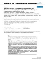
ROC curve analyses
Results for those cytokines that were significantly higher
in the case/control comparison are shown in Figure 1 and
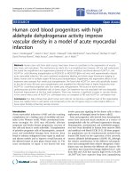
Table 2. Those for cytokines that were lower in CFS than
controls are shown in Figure 2 and Table 3. Area under the
curve (AUC) for IL-5 (0. 84), LTα (0.77), IL-4 (0.77), IL-
12 (0.76) indicated good biomarker potential. Coordi-
nates of the curves for these 4 cytokines are in Additional
File 1. The AUC of IL-6 (0.73), IL-15 (0.73), IL-8 (0.69),
IL-13 (0.68) IL-1α (0.62), IL-1β (0.62) showed fair poten-
tial as biomarkers (Tables 2 and 3).
Discussion
Several studies report cytokine abnormalities in CFS; how-
ever, the findings are mixed. Differences between reports
may be largely due to differences in methodologies [14].
Amounts of cytokines in plasma or serum are often below
the level of detection in traditional ELISA assays. In addi-
tion to assay sensitivity, results using the direct approach
are influenced by length of time following blood draw to
separation of serum or plasma, temperature of storage
and repeated thawing and freezing. In vitro stimulation
whole blood or peripheral blood mononuclear cells
(PBMC) is another approach to study cytokines. ELISA is
then used to measure cytokine content of supernatants of
culture fluids. Obviously, results depend on culture con-
ditions and stimulants used. Other techniques include
either in unstimulated or stimulated PBMC. Results
obtained with these methodologies are not directly com-
parable.
The availability of sensitive multiplex technology permit-
ted the determination of 16 cytokines simultaneously on
plasma samples from female CFS patients and age and
gender matched healthy controls. In the CFS cases, we
found an unusual pattern of the cytokines that define the
CD4 T cell. Dendritic cell derived IL-12, the main T
H
1-
inducing cytokine leading to production of IFNγ, IL-2 and
TNFα, was elevated. However, IFNγ, IL-2 and TNFα were
unchanged in plasma of CFS cases compared to controls.
Another dendritic cell derived cytokine, IL-15, was
decreased. IL-2 and IL-15 are key participants in CD8 T
cell and NK cell activation and function. Sharing the beta
and gamma receptor subunits results in several common
functions: e.g. cytotoxicity. On the other hand, due to
their distinct alpha receptor subunits, they play opposing
roles in immune processes such as activation induced cell
death (IL-2) and immunological memory (IL-15) [23]. IL-
23 (unchanged between controls and cases) stimulates
the differentiation and function of the T
H
17 subset of
CD4 T cells, a relatively newly described immune defense.
The T
H
17 CD4 cell produces IL-17, protects surfaces (e.g.,
skin, lining of the intestine) against bacteria, and plays a
critical role in chronic intestinal inflammation [24,25].
The unchanged IL-17 and IL-23 levels in CFS noted in this
study would argue against bacterial gastrointestinal infec-
tions as playing an important role in persistent illness.
Along with the T
H
1 abnormalities, we found up regula-
tion of T
H
2 associated cytokines, IL-4 and IL-5, in the CFS
subjects. Allergy is common in CFS cases. Years ago, Straus
et al, reported >50% atopy in 24 CFS patients [26]. The
elevation of these two cytokines implies a type 2 shift -
and diminished stimulus for cytotoxic lymphocyte func-
tion.
Table 2: AUC for Plasma Cytokines Significantly Higher in CFS Cases vs. Controls
Cytokines Area Std. Error
a
Asymptotic Sig.
b
Asymptotic 95% Confidence Interval
Lower Boundary Upper Boundary
LTα .769 .049 .000 .673 .865
IL-6 .731 .050 .000 .633 .828
IL-1α .620 .056 .044 .509 .730
IL-1 β .621 .062 .041 .499 .744
IL-5 .844 .041 .000 .764 .925
IL-4 .770 .048 .000 .676 .864
IL-12 .758 .054 .000 .653 .863
a
Under the nonparametric assumption
b
Null hypothesis: true area = 0.5
Journal of Translational Medicine 2009, 7:96 />Page 5 of 8
(page number not for citation purposes)
The probability of chronic inflammation [17] in CFS is
supported by the elevation of four members of the pro-
inflammatory cytokine cascade [27], LTα, IL-1α, IL-1β,
and IL-6, in the CFS samples compared to controls. The
exception was TNFα, although the median value for cases
was 14% higher than controls and about 1/4 of CFS
patients in other studies had elevated TNFα [15,17]. Inter-
leukin-13, associated with inhibitory effects on inflamma-
tory cytokine production, was lower in cases compared to
controls. The anti-inflammatory cytokine, IL10, was not
different. The inflammatory mediator IL-8 (a chemokine
known as CXCL8) known to be responsible for the migra-
tion and activation of neutrophils and NK cells [28] was
decreased in plasma of CFS patients.
The observations of abnormal cytokine patterns in CFS
patients support the reports of retrovirus infections and
reactivation of latent herpes virus infections. DeFreitas, et
al found HTLV-II- like gag sequences by polymerase chain
reaction and in situ hybridization as well as antibodies
reactive with human T- lymphotropic virus (HTLV) in a
majority of 30 CFS cases. Twenty healthy controls were
negative for the three assays [11]. Holmes, et al, reported
that structures consistent with stages of a Lentivirus repli-
cative cycle were observed by electron microscopy in 12-
day PBMC cultures from 10 of 17 CFS patients and not in
controls [12]. Recently, DNA from a human gammaretro-
virus, xenotropic murine leukemia virus-related virus
(XMRV), was found in the PBMC of 68 of 101 patients
compared to 8 of 218 healthy controls. Patient-derived,
activated PBMC produced infectious XMRV in vitro. Both
cell associated and cell-free transmission of the virus to
Table 3: AUC for Plasma Cytokines Significantly Lower in CFS Cases vs. Controls
Cytokines Area Std. Error
a
Asymptotic Sig.
b
Asymptotic 95% Confidence Interval
Lower Boundary Upper Boundary
IL-8 .685 .062 .002 .564 .806
IL-15 .731 .056 .000 .620 .841
IL-13 .682 .064 .002 .556 .808
a
Under the nonparametric assumption
b
Null hypothesis: true area = 0.5
ROC curves shows the classification performance of plasma cytokines from CFS cases and healthy controlsFigure 1
ROC curves shows the classification performance of
plasma cytokines from CFS cases and healthy con-
trols. Curves are for the 7 cytokines significantly elevated (p
< .05) in cases compared to controls (IL-4, IL-5, IL-12, LTα,
IL-1α, IL-1β, and IL-6).
ROC curves show the classification performance of plasma cytokines from CFS cases and healthy controlsFigure 2
ROC curves show the classification performance of
plasma cytokines from CFS cases and healthy con-
trols. Curves are for the 3 cytokines significantly lower (p <
.05) in cases compared to controls (IL-8, IL-13 and IL-15).
Journal of Translational Medicine 2009, 7:96 />Page 6 of 8
(page number not for citation purposes)
uninfected primary lymphocytes and indicator cell lines
was possible [13]. The XMRV gag and env sequences dis-
covered in CFS cases were more than 99% similar to those
previously reported for prostate tumor-associated strains
of XMRV [29].
Latent herpes virus infections are likely to be important in
CFS. Immunologic effects of persistent herpetic infections
do not require of virus DNA synthesis. For example,
Glazer and colleagues [9] reported that EBV encoded
deoxyuridine triphosphate nucleotidohydrolase (dUT-
Pase) upregulated the production of proinflammatory
cytokines, including IL-1β and IL-6. Also, dUTPase
administered to mice, produced sickness behaviors
known to be induced by some of the cytokines we showed
to be upregulated. A subsequent paper showed that EBV-
encoded dUTPase can enhance production of proinflam-
matory cytokines by monocytes/macrophages in contact
with endothelial cells of blood vessels [30]. In addition,
Ariza, et al demonstrated that the purified EBV-encoded
dUTPase activated NFkappaB in a dose-dependent
through Toll Like Receptor 2 (TLR2). Treatment of human
monocyte-derived macrophages with an anti-EBV-
encoded dUTPase or with an anti-TLR2 blocked the pro-
duction of IL-6 [31]. Iwakiri, et al reported that EBV-
encoded small RNA (EBER), which is released from EBV-
infected cells, was responsible for immune activation by
EBV, including release of proinflammatory cytokines [32].
A recent study (M Vera, MA Fletcher, C Cuba, L Garcia, N
Klimas, presented to the International Association for
Chronic Fatigue Syndrome/Myalgic Encephalitis, Reno,
NV, March, 2009) reported that the anti-viral and
immuno-modulatory drug, inosine pranobex, led to sig-
nificant improvement in the clinical scores of 61 patients
treated for 6 months. Immune activation was decreased,
NK cell activity was improved and titers of anti-Epstein
Barr Virus Viral Capsid Antigen IgG were significant
decreased. Antibody titers to Human Herpes Virus 6 were
unchanged. A larger randomized trial would seem appro-
priate.
According to ROC analysis, plasma IL-5 was best at distin-
guishing CFS cases from controls, with the highest per-
centage difference from the median of normal and the
largest AUC. We recently reported elevation of IL-5 in the
supernatants of mitogen-stimulated cultured lym-
phocytes from Gulf War Illness (GWI) cases compared to
controls [33]. The symptoms of GWI are similar to those
reported in CFS. Three other cytokines with AUC values
consistent with good potential as biomarkers were LTα,
IL-4 and IL-12. Less promising as systemic markers of CFS,
but with AUC significantly different in cases compared to
controls, were IL-6, IL-15, IL-13, IL-1α and IL-1β.
The cytokine changes observed between CFS patients and
healthy, matched controls are likely to be indicative of
immune activation and inflammation. Fibromyalgia,
GWI, rheumatologic disorders and multiple sclerosis may
have similar cytokine patterns. Future research will be
required to determine if the cytokine patterns associated
with CFS cases are similar or distinct from other complex,
chronic and poorly understood illnesses.
Obvious limitations of this study are that the samples rep-
resent a single point in time and a single gender. The par-
ent protocol, from which the CFS samples were gathered,
is a larger longitudinal study. Subjects are followed over
18 months and sample collection includes times of rela-
tive symptom remission or exacerbation. Completion of
the study will allow the correlation of CFS related symp-
toms and other immune markers with the cytokine pat-
terns. CFS is a condition that affects women in
disproportionate numbers. The larger study will have suf-
ficient power to allow the study of cytokine patterns in
men with CFS. As Broderick and colleagues have pointed
out, markers of immune status tend to be highly variable
and context-specific leading to inconsistent biomarker
lists [34]. These indicators are parts of a complex and inte-
grated system and their inter-dependency must be
addressed. Accordingly, we are currently engaged in com-
bining the proteomic and genomic data on cytokines with
other immunologic and neuroendocrine markers, both
proteomic and genomic, in order to map the network
structure of neuroendocrine-immune interaction in CFS.
We will focus on identifying associations between nodes
that are differentially expressed across disease group and
controls.
The finding of cytokine imbalances in the peripheral
blood compartment has implications for physiological
and psychological function changes. The decreased natu-
ral killer (NK) cell cytotoxic and lymphoproliferative
activities and increased allergic and autoimmune manifes-
tations in CFS would be compatible with the hypothesis
that the immune system of affected individuals is biased
towards a T- helper (T
H
) 2 type, or humoral immunity-ori-
ented cytokine pattern. The elevations in LTα, IL-1α, IL1β
and IL-6 indicate inflammation, likely to be accompanied
by autoantibody production, inappropriate fatigue, myal-
gia and arthralgia, as well as changes in mood and sleep
patterns.
Conclusion
This is study is among the first in the CFS literature to
report the plasma profiles of a reasonably large panel of
cytokines assessed simultaneously by multiplex tech-
nique. Cytokine abnormalities appear to be common in
CFS. Several showed promise as potential biomarkers. The
changes from the normal condition indicate immune acti-
Journal of Translational Medicine 2009, 7:96 />Page 7 of 8
(page number not for citation purposes)
vation and inflammation - and point to potential thera-
peutic strategies. The results imply a disorganized
regulatory pattern of T
H
1 function, critical to antiviral
defense. The data from this study support a T
H
2 shift, pro-
inflammatory cytokine up regulation and down regula-
tion of important mediators of cytotoxic cell function.
Competing interests
The authors declare that they have no competing interests.
Authors' contributions
MAF and NGK conceived of the study, participated in its
design, coordination, performed the statistical analysis
and drafted the manuscript; NGK and SL participated in
patients' diagnosis and assessment; ZB participated in
subject recruitment and data management; XRZ carried
out the immunoassays. All authors read and approved the
final manuscript.
Additional material
Acknowledgements
This work was supported by grants from the NIAAA: R21AA016635 (PI
MA Fletcher); NIAID: R01AI065723 (PI MA Fletcher); CFIDS Assoc. of
America: (PI N Klimas); NIAID: UO1 AI459940 (PI N Klimas); NIAMS
AR048932 (PI S Levis)
References
1. Reyes M, Nisenbaum R, Hoaglin DC, Unger ER, Emmons C, Randall
B, Stewart JA, Abbey S, Jones JF, Gantz N, Minden S, Reeves WC:
Prevalence and incidence of chronic fatigue syndrome in
Wichita, Kansas. Arch Intern Med 2003, 163:1530-1536.
2. Jason LA, Richman JA, Rademaker AW, Jordan KM, Plioplys AV, Tay-
lor RR, McCready W, Huang CF, Plioplys S: A community-based
study of chronic fatigue syndrome. Arch Intern Med 1999,
159:2129-2137.
3. Bombardier C, Buchwald D: Outcome and prognosis of patients
with chronic fatigue vs. chronic fatigue syndrome. Arch Intern
Med 1995, 155:2105-2110.
4. Bombardier C, Buchwald D: Chronic Fatigue, Chronic Fatigue
Syndrome, and Fibromyalgia. Disability and Health-Care
Use. Med Care 1996, 34:924-930.
5. Klimas NG, Morgan R, Salvado F, Fletcher MA: Immunologic
abnormalities of chronic fatigue syndrome. J Clin Microbiol
1990, 28:1403-1410.
6. Evengård B, Klimas N: Chronic fatigue syndrome: Probable
pathogenesis and possible treatments. Drugs 2002,
62:2433-2446.
7. Evengård B, Jonzon E, Sandberg A, Theorell T, Lindh G: Differences
between patients with chronic fatigue syndrome and with
chronic fatigue at an infectious disease clinic in Stockholm,
Sweden. Psychiatry Clin Neurosci 2003, 57:361-368.
8. Straus SE, Tosato G, Armstrong G, Lawley T, Preble OT, Henle W,
Davey R, Pearson G, Epstein , Brus I: Persisting illness and fatigue
in adults with evidence of Epstein-Barr virus infection. Ann
Intern Med 1985, 102:7-16.
9. Glaser R, Padgett DA, Litsky ML, Baiocchi RA, Yang EV, Chen M, Yeh
PE, Klimas NG, Marshall GD, Whiteside T, Herberman R, Kiecolt-
Glaser J, Williams MV: Stress-associated changes in the steady-
state expression of latent Epstein-Barr virus: implications for
chronic fatigue syndrome and cancer. Brain Behav Immun 2005,
19:91-103.
10. Ledina D, Bradari( N, Milas I, Ivi( I, Brnci( N, Kuzmici( N: Chronic
fatigue syndrome after Q fever. Med Sci Monit 2007,
13:CS88-92.
11. DeFreitas E, Hilliard B, Cheney PR, Bell DS, Kiggundu E, Sankey D,
Wroblewska Z, Palladino M, Woodward JP, Koprowski H: Retrovi-
ral sequences related to human T-lymphotropic virus type II
in patients with chronic fatigue immune dysfunction syn-
drome. Proc Natl Acad Sci USA 1991, 88:2922-6.
12. Holmes MJ, Diack DS, Easingwood RA, Cross JP, Carlisle B: Electron
microscopic immunocytological profiles in chronic fatigue
syndrome. J Psychiatr Res 1997, 31:115-22.
13. Lombardi VC, Ruscetti FW, Gupta JD, Pfost MA, Hagen KS, Peterson
DL, Ruscetti SK, Bagni RK, Petrow-Sadowski C, Gold B, Dean M, Sil-
verman RH, Mikovits JA: Detection of infectious retrovirus,
XMRV, in blood cells of patients with chronic fatigue syn-
drome. Science 2009, 326:585-589.
14. Maher K, Klimas NG, Fletcher MA: Immunology. In Handbook of
Chronic Fatigue Edited by: Jason LA, Fennell PA, Taylor RR. Hoboken,
NJ: John Wiley & Sons; 2003:124-151.
15. Patarca-Montero R, Antoni M, Fletcher MA, Klimas NG: Cytokine
and other immunologic markers in chronic fatigue syn-
drome and their relation to neuropsychological factors. Appl
Neuropsych 2001, 8:51-6.
16. Gupta S, Aggarwal S, See D, Starr A: Cytokine production by
adherent and non-adherent mononuclear cells in chronic
fatigue syndrome. J Psych Res 1997, 31:149-56.
17. Patarca R: Cytokines and Chronic Fatigue Syndrome. Ann NY
Acad Sci 2001, 933:185-200.
18. Skowera A, Cleare A, Blair D, Bevis L, Wessely S, Peakman M: High
levels of type 2 cytokine-producing cells in chronic fatigue
syndrome. Clin Exp Immunol 2004, 135:294-302.
19. Fukuda K, Straus SE, Hickie I, Sharpe MC, Dobbins JG, Komaroff A:
The chronic fatigue syndrome: a comprehensive approach
to its definition and study. International Chronic Fatigue
Syndrome Study Group. Ann Intern Med 1994, 121:953-9.
20. Reeves WC, Lloyd A, Vernon SD, Klimas N, Jason LA, Bleijenberg G,
Evengard B, White PD, Nisenbaum R, Unger ER, International
Chronic Fatigue Syndrome Study Group: Identification of ambigu-
ities in the 1994 chronic fatigue syndrome research case def-
inition and recommendations for resolution. BMC Health
Services Res 2003, 3:25.
21. World Health Organization, Composite International Diag-
nostic Instrument [ />instruments_download.php]
22. Zweig MH, Campbell G: Receiver-Operating Characteristic
(ROC) plots: A fundamental evaluation tool in Clinical Med-
icine. Clin Chem 1993, 39:561-577.
23. Waldmann TA: The biology of interleukin-2 and interleukin-
15: implications for cancer therapy and vaccine design.
Nature Rev Immun 2006, 6:595-601.
24. Boniface K, Blom B, Liu YJ, de Waal Malefyt R: From interleukin-
23 to T-helper 17 cells: human T-helper cell differentiation
revisited. Immunol Rev 2008, 226:132-46.
25. Iwakura Y, Ishigame H: The IL-23/IL-17 axis in inflammation J.
Clin Invest 2006, 116:1218-1222.
26. Straus SE, Dale JK, Wright R, Metcalfe DD: Allergy and the
chronic fatigue syndrome. J Allergy Clin Immunol 1988, 81(5 Pt
1):791-5.
27. Goldberg RB: Cytokine and Cytokine-like Inflammation Mark-
ers, Endothelial Dysfunction and Imbalanced Coagulation in
Development of Diabetes and Its Complications. J Clin Endo-
crinol Metab 2009, 94:3171-82.
28. Lin F, Nguyen CM, Wang SJ, Saadi W, Gross SP, Jeon NL: Effective
neutrophil chemotaxis is strongly influenced by mean IL-8
concentration. Biochem Biophys Res Commun 2004, 319:576-81.
29. Urisman A, Molinaro RJ, Fischer N, Plummer SJ, Casey G, Klein EA,
Malathi K, Magi-Galluzzi C, Tubbs RR, Ganem D, Silverman RH,
DeRisi JL: Identification of a novel Gammaretrovirus in pros-
Additional file 1
Coordinates of the curves for those cytokines with AUC that indicated
good biomarker material.
Click here for file
[ />5876-7-96-S1.doc]
Publish with BioMed Central and every
scientist can read your work free of charge
"BioMed Central will be the most significant development for
disseminating the results of biomedical research in our lifetime."
Sir Paul Nurse, Cancer Research UK
Your research papers will be:
available free of charge to the entire biomedical community
peer reviewed and published immediately upon acceptance
cited in PubMed and archived on PubMed Central
yours — you keep the copyright
Submit your manuscript here:
/>BioMedcentral
Journal of Translational Medicine 2009, 7:96 />Page 8 of 8
(page number not for citation purposes)
tate tumors of patients homozygous for R462Q RNASEL
variant. PLoS Pathog 2006, 2:e25.
30. Waldman WJ, Williams MV Jr, Lemeshow S, Binkley P, Guttridge D,
Kiecolt-Glaser JK, Knight DA, Ladner KJ, Glaser R: Epstein-Barr
virus-encoded dUTPase enhances proinflammatory
cytokine production by macrophages in contact with
endothelial cells: evidence for depression-induced athero-
sclerotic risk. Brain Behav Immun 2008, 2:215-23.
31. Ariza ME, Glaser R, Kaumaya PT, Jones C, Williams MV: The EBV-
encoded dUTPase activates NF-kappa B through the TLR2
and MyD88-dependent signaling pathway. J Immunol 2009,
182:851-9.
32. Iwakiri D, Zhou L, Samanta M, Matsumoto M, Ebihara T, Seya T, Imai
S, Fujieda M, Kawa K, Takada K: Epstein-Barr virus (EBV)-
encoded small RNA is released from EBV-infected cells and
activates signaling from toll-like receptor 3. J Exp Med 2009,
206:2091-9.
33. Whistler T, Fletcher MA, Lonergan W, Zeng XR, Lin JM, Laperriere
A, Vernon SD, Klimas NG: Impaired immune function in Gulf
War Illness. BMC Med Genomic 2009, 5:12.
34. Fuite J, Vernon SD, Broderick G: Neuroendocrine and immune
network re-modeling in chronic fatigue syndrome: an
exploratory analysis. Genomics 2008, 92:393-9.