báo cáo hóa học:" Correlation between expression of p53, p21/WAF1, and MDM2 proteins and their prognostic significance in primary hepatocellular carcinoma" docx
Bạn đang xem bản rút gọn của tài liệu. Xem và tải ngay bản đầy đủ của tài liệu tại đây (723 KB, 8 trang )
BioMed Central
Page 1 of 8
(page number not for citation purposes)
Journal of Translational Medicine
Open Access
Research
Correlation between expression of p53, p21/WAF1, and MDM2
proteins and their prognostic significance in primary hepatocellular
carcinoma
Mei-Fang Zhang
1,2
, Zhi-Yi Zhang
1,2
, Jia Fu
1,2
, Yu-Feng Yang
1,2
and
Jing-Ping Yun*
1,2
Address:
1
State Key Laboratory of Oncology in Southern China, Cancer Center of Sun Yat-Sen University, Guangzhou, China and
2
Department of
Pathology, Cancer Center, Sun Yat-Sen University, Guangzhou 510060, China
Email: Mei-Fang Zhang - ; Zhi-Yi Zhang - ; Jia Fu - ; Yu-
Feng Yang - ; Jing-Ping Yun* -
* Corresponding author
Abstract
Background: Tumor Protein p53 (p53), cyclin-dependent kinase inhibitor 1A (p21/WAF1), and
murine double minute 2 (MDM2) participate in the regulation of cell growth. Altered expression
of these gene products has been found in malignant tumors and has been associated with poor
prognosis. Our aim was to investigate the expression of the 3 proteins in hepatocellular carcinoma
(HCC) and their prognostic significance.
Methods: We examined p53, p21/WAF1, and MDM2 expression in 181 pairs of HCC tissues and
the adjacent hepatic tissues by performing immunohistochemistry and examined the expression of
the 3 proteins in 7 pairs of HCC tissues and the adjacent hepatic tissues by using western blot
analysis.
Results: The expression of p53, p21/WAF1, and MDM2 in the HCC tissues was significantly higher
than those in the adjacent hepatic tissues (P < 0.05). A statistical correlation was observed between
p53 and p21/WAF1 expression in HCC tissues (R = 0.195, P = 0.008). A statistical correlation was
observed between expression of p53 and p21/WAF1 (R = 0.380, P = 0.000), p53 and MDM2 (R =
0.299, P = 0.000), p21/WAF1 and MDM2 (R = 0.285, P = 0.000) in 181 liver tissues adjacent to the
tumor. Patients with a low pathologic grade HCC (I+II) had a higher tendency to express p53 on
tumor cells than the patients with high pathologic grade HCC (III+IV) (P = 0.007). Survival analysis
showed that positive p21/WAF1 expression or/and negative MDM2 expression in HCC was a
predictor of better survival of patients after tumor resection (P < 0.05).
Conclusions: The proteins p53, p21/WAF1, and MDM2 were overexpressed in all the HCC cases
in this study, and p53 and p21/WAF1 overexpression were positively correlated. The expression
of p21/WAF1 and MDM2 can be considered as 2 useful indicators for predicting the prognosis of
HCC.
Published: 22 December 2009
Journal of Translational Medicine 2009, 7:110 doi:10.1186/1479-5876-7-110
Received: 9 October 2009
Accepted: 22 December 2009
This article is available from: />© 2009 Zhang et al; licensee BioMed Central Ltd.
This is an Open Access article distributed under the terms of the Creative Commons Attribution License ( />),
which permits unrestricted use, distribution, and reproduction in any medium, provided the original work is properly cited.
Journal of Translational Medicine 2009, 7:110 />Page 2 of 8
(page number not for citation purposes)
Background
Hepatocellular carcinoma (HCC) is the fifth most com-
mon malignancy worldwide and is the third most com-
mon cause of cancer-related deaths [1]. HCC develops in
patients with chronic liver diseases, and its etiopathogen-
esis includes viral infection (hepatitis B and C), alcohol,
and aflatoxin B1 consumption. The majority of HCC
patients have associated cirrhosis and impaired liver func-
tion, making the treatment of HCC more difficult than
that of many other cancers. Surgery, including transplan-
tation, remains the only potential curative modality for
HCC.
Prognosis of HCC remains unsatisfactory even after surgi-
cal resection and liver transplantation. Considerable
interest has been generated in identifying factors that
influence the prognosis of HCC. Several staging systems
have been developed to predict survival period after the
diagnosis of HCC [2]. The most widely studied prognostic
factors are related to the pathological characteristics of the
neoplasm, including tumor size, grade, stage, and vascular
invasion. However, several biological molecules that can
predict the survival period of HCC patients have been
reported in recent years; however, the results are contro-
versial.
Previous studies have explored the molecular alterations
in HCC, including changes in the expression of p53, cyc-
lin-dependent kinase inhibitor 1A (p21/WAF1), and
murine double minute 2 (MDM2). The tumor suppressor
gene p53 plays a key role in regulating the cell cycle and
serves as a principal mediator of growth arrest, senes-
cence, and apoptosis in response to a broad array of cellu-
lar damage [3]. The p21/WAF1 protein is encoded by the
human WAF1/CIP1 gene and its expression is directly
induced by the wild-type p53 protein [4]. This protein
binds to a variety of cyclin-dependent kinases and inhibits
their activity, regulates DNA repair, and directly blocks
DNA replication by inhibiting the proliferating cell
nuclear antigen [5], thus inhibiting cell-cycle progression
and decreasing cell growth. MDM2 is the product of a
p53-inducible gene and inhibits the p53 activity by ubiq-
uitinating p53 and creating a negative-feedback loop [5-
8]. Altered expression of these gene products has been
found in malignant tumors including HCC and correlated
with poor prognosis. In HCC, the prognostic value of p53
is controversial, since several studies show an association
of p53 with patient survival [9-12], while other investiga-
tions report no association [13,14]. The predictive value
of the p21/WAF1 expression level in HCC is also ambigu-
ous [10,11,15]. However, few studies pertaining to the
expression of the 3 proteins p53, p21/WAF1, and MDM2
in HCC cases have reported different results [11,16].
We determined the expression of p53, p21/WAF1, and
MDM2 in a relatively large sample size of 181 pairs of
human HCC tissues and the corresponding adjacent
hepatic tissues obtained after resection by performing
immunohistochemistry (IHC). In addition, we performed
western blot analysis in 7 such pairs. Further, we
attempted to address the correlation among their expres-
sion and the relationship between their expression and
the clinical parameters, including overall survival.
Methods
Clinical samples
Samples from 181 Chinese patients with HCC and their
clinical records from 1997 to 2007 were collected from
the Cancer Center of Sun Yat-Sen University, Guangzhou,
China. Tissue blocks prepared from HCC tissues and the
adjacent liver tissues were sectioned for performing IHC
of p53, p21/WAF1, and MDM2; in addition, for 7 cases,
we collected the tissue samples inclusive of the HCC and
its adjacent tissues from the tissue bank department of
this cancer center and subjected these samples to western
blot analyses. The collection of the human specimens in
the study was approved by the Independent Ethics Com-
mittee of the Cancer Center of Sun Yat-Sen University.
Western blot analysis
For immunolabeling, lysates were prepared from the tis-
sues as described previously [17,18]. We separated 100 μg
of each lysate by sodium dodecyl sulfate-polyacrylamide
gel electrophoresis (SDS-PAGE). The proteins were trans-
ferred onto blotting membranes. After blocking, the
membranes were incubated overnight with rabbit poly-
clonal antibody against p53 (Clone: FL-393; Cat No. sc-
6243; Santa Cruz, CA); mouse monoclonal antibody
against p21/WAF1 (Clone: SX118; Cat No. 556430; BD
Pharmigen, CA) and MDM2 (Clone: N-20; Cat No. sc-
813; Santa Cruz, CA); and mouse monoclonal antibody
against glyceraldehydes 3-phosphate dehydrogenase
(GAPDH) (Kangchen Biotech; Shanghai, China) (p53,
1:500; p21/WAF1, 1:250; MDM2, 1:2000; and GAPDH,
1:1000), followed by incubation with horseradish perox-
idase-conjugated immunoglobulin G (IgG). The blots
were then visualized by using an ECL kit (Amersham Life
Science; Piscataway, NH, USA) and exposed for 1 min to
an X-ray film.
Immunohistochemistry
For immunohistochemistry studies, a labeled-streptavi-
din-biotin (LAB-SA) method was performed with Histo-
stain
®
-Plus Bulk Kit Zymed
®
2
nd
generation LAB-SA
detection system (CAT. NO. 85-9043, Zymed Laborato-
ries, CA) as previously described [18,19]. All the primary
antibodies (p53, p21/WAF1, and MDM2; mouse mono-
clonal antibody, Cat No. ZM-0408, ZM-0206, and ZM-
0425, respectively, Zymed, CA) were ready to use without
dilution. Each paraffin-embedded tissue section (4 μm in
thickness) was deparaffinized, hydrated, and incubated in
3% H
2
O
2
and microwaved for 3 minutes to block endog-
Journal of Translational Medicine 2009, 7:110 />Page 3 of 8
(page number not for citation purposes)
enous peroxidase activity. The tissue sections were sub-
jected to antigen retrieval by microwaving in 10 mM
citrate buffer for 30 min. The sections were incubated with
serum blocking solution (Reagent A) for 10 minutes to
block nonspecific binding and then with the primary anti-
bodies in moist chamber for 60 minutes. After rinsed with
PBS for 2 minutes, the sections were incubated with the
biotinylated secondary antibody (Reagent B) for 10 min-
utes and rinsed with PBS. The sections followed by incu-
bation with enzyme conjugate (Reagent C) for 10
minutes. Subsequently, the sections were stained with
DAB and counterstained with hematoxylin. Serum block-
ing solution (Reagent A) in place of the primary antibody
was used as a negative control. A brown particle in nuclei
was considered as positive labeling. Immunostaining
labeling intensities were defined as: +, less than 10% of
the tumor cells were positive; ++, 10%-50% of the tumor
cells were positive; +++, more than 50% the tumor cells
were positive; -, negative staining. These sections were
observed under light microscopy and the staining intensi-
ties were assessed by 2 pathologists Dr JP Yun and Dr MF
Zhang.
Statistical analysis
Statistical analysis was performed to determine the rela-
tionship between the clinical parameters of gender, age,
tumor size, number of tumors, hepatitis B surface antigen
(HBsAg), pathologic grade, serum level of alpha-fetal pro-
tein (AFP), and the 3 immunohistochemical markers by
Peason's chi-square test. The Spearman correlation was
employed to examine the relationship between the
expression of p53, p21/WAF1, and MDM2. Survival was
assessed by the Kaplan-Meier method, and log-rank test
was used to analyze survival curves. Statistical significance
was initially set at P < 0.05. All statistical analysis was per-
formed using the SPSS 13.0 software for Windows.
Results
Increase in the expression of p53, p21/WAF1, and MDM2
in HCC
The expression of p53 and MDM2 in the 7 pairs was
higher in the HCC tissues than in the adjacent hepatic tis-
sues (tissues 1-7), as determined by western blot (Figure
1). In 6 out of 7 pairs, p21/WAF1 expression was higher in
the HCC tissues than in the adjacent hepatic tissues (tis-
sues 1-3 and 5-7). In 1 case, the expression of p21/WAF1
in the HCC tissue was lower than that in adjacent hepatic
tissue (tissue 4). These results indicated that the expres-
sion levels of p53, p21/WAF1, and MDM2 were higher in
the HCC tissues than those in the adjacent hepatic tissues.
The expression of p53, p21/WAF1, and MDM2 in the 181
pairs of tissues was analyzed by IHC. As shown in Figure
2, p53, p21/WAF1, and MDM2 were mainly located in the
nuclei of the cancer cells and highly expressed in the HCC
tissue. Statistical analysis showed that positive propor-
tions of p53, p21/WAF1, and MDM2 expression in HCC
tissues were 70.7% (128/181), 33.1% (60/181), and
52.5% (95/181), respectively. Positive proportions of
p53, p21/WAF1, and MDM2 expression in the corre-
sponding adjacent hepatic tissues were 16.6% (30/175),
15.5% (28/178), and 32.6% (59/179), respectively. The
expression of p53, p21/WAF1, and MDM2 in HCC was
significantly higher than that in adjacent hepatic tissues (P
< 0.05 for each protein).
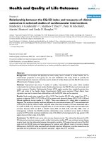
Expression of p53, p21/WAF1 and MDM2 in HCC by West-ern blotFigure 1
Expression of p53, p21/WAF1 and MDM2 in HCC by
Western blot. The expression of p53, p21/WAF1, and
MDM2 was detected in hepatocellular carcinoma (HCC) tis-
sues by western blot analysis. We used 7 pairs of HCC tis-
sues and the adjacent hepatic tissues. Tissues T1-7 were
HCC tissues and N1-7 were the adjacent hepatic tissues. The
expression of the housekeeping gene, glyceraldehydes 3-
phosphate dehydrogenase (GAPDH), served as a control.
The expression of p53 was higher in the HCC tissues (T1-7)
than in the adjacent hepatic tissues (N1-7). The MDM2
expression followed a similar trend in both the tissues. The
expression of p21/WAF1 was higher in HCC tissues (T1-3,
T5-7) than the adjacent hepatic tissues (N1-3, N5-7).
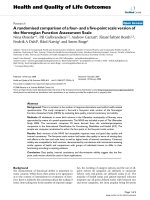
Expression of p53, p21/WAF1 and MDM2 in HCC by IHCFigure 2
Expression of p53, p21/WAF1 and MDM2 in HCC by
IHC. The hematoxylin and eosin (H&E) stained sections
show a solid area of hepatocellular carcinoma (HCC) (A).
Immunohistochemical staining for p53 (B), p21/WAF1 (C),
and MDM2 (D) in HCC. (Mag. ×400).
Journal of Translational Medicine 2009, 7:110 />Page 4 of 8
(page number not for citation purposes)
Statistically significant correlation between p53, p21/
WAF1, and MDM2 expression in HCC tissues
We calculated the correlation between p53, p21/WAF1,
and MDM2 expression in 181 HCC tissues by Spearman
correlation analysis (Table 1). Statistical correlation was
observed between p53 and p21/WAF1 expression in HCC
(R = 0.195, P = 0.008). No statistical correlations were
observed between p53 and MDM2 expression in HCC (P
= 0.058) and between p21/WAF1 and MDM2 expression
in HCC (P = 0.431). Interestingly, statistical correlations
were observed between the expressions of p53 and p21/
WAF1 (R = 0.380, P = 0.000), p53 and MDM2 (R = 0.299,
P = 0.000), p21/WAF1 and MDM2 (R = 0.285, P = 0.000)
in 181 liver tissues adjacent to the tumor (Table 2).
We further investigated the differences between the
expression of p53, p21/WAF1, and MDM2 in 181 pairs of
HCC on the basis of different clinical parameters, includ-
ing the gender, age, tumor size, number of tumors,
HBsAg, pathologic grade, and serum level of AFP of the
patient. We observed a statistical correlation between p53
and the pathologic grade in HCC tissues (P = 0.007).
Patients with a low pathologic grade (I+II) had a higher
tendency to express p53 on tumor cells than patients with
high pathologic grade (III+IV). No statistical significance
was found between p53, p21/WAF1, and MDM2 expres-
sion and the other clinical parameters (Table 3).
Positive p21/WAF1 expression or/and negative MDM2
expression in HCC tissues associated with better survival in
patients
The associations between survival time and the 3 immu-
nohistochemical markers (p53, p21/WAF1, and MDM2)
in HCC were analyzed with Kaplan-Meier survival analy-
sis (Figure 3). The survival curve for p21/WAF1-positive
patients tended to be better than that for p21/WAF1-neg-
ative patients (P = 0.026). The survival curve for MDM2-
negative patients tended to be better than that for MDM2-
positive patients (P = 0.043). There was no significant cor-
relation between p53 expression and the survival time of
the patients (P = 0.275). Further analysis of the prognostic
value of combining p21 and MDM2 expression in HCC
was undertaken. It can be divided into 4 groups: p21+/
MDM2-, p21+/MDM2+, p21-/MDM2- and p21-/MDM2+.
The survival curve for p21+/MDM2- patients tended to be
better than that for p21-/MDM2+ patients (P = 0.012),
and there was no significant difference between the other
groups. These results indicated that the expression of p21/
WAF1 and MDM2 were associated with survival in
patients with HCC.
Discussion
The results from our study revealed a significant increase
in the expression of p53, p21/WAF1, and MDM2 in HCC
tissues than the corresponding adjacent hepatic tissues;
the expression levels of the 3 proteins in the former was
70.7%, 33.1%, and 52.5%, respectively and those in the
later were 16.6%, 15.5%, and 32.6%, respectively. These
results indicated that these proteins play important roles
in hepatocarcinogenesis.
Several IHC-based studies have reported the proportion of
p53-positive HCC cases to vary in the range of 22% to
81% [20]. The cause for the variation in p53 expression
can be partly attributed to the lack of a consistent cutoff
value among different studies for determining positive
p53 expression. In some studies, the HCC was regarded as
p53-positive when ≥10% of the tumor cells expressed
p53, while in others, this cutoff value was defined as ≥5%
of the tumor cells being positive for p53; further, the
majority of studies have not defined the lower limit for
p53-positive tumor cells. Another cause of the discrepancy
in the reported percentage of p53-positive tumors is the
differences in the p53 expression with the prevalent carci-
nogenic factors and certain unknown molecular mecha-
nisms. The tumor suppressor gene, p53, has been reported
to be mutated in 24-69% of HCCs. Mutations of p53
result in unregulated replication of defective DNA,
Table 1: Correlation among p53, p21/WAF1, and MDM2 expression in HCC tissues
n p53 positive p21/WAF1 positive MDM2 positive
n P value n P value n P value
p53 181
positive 128 50 0.008* 73 0.058
negative 53 10 22
p21/WAF1 181
positive 60 50 0.008* 34 0.431
negative 121 78 61
MDM2 181
positive 95 73 0.058 24 0.431
negative 86 55 18
*Statistically significant (P < 0.05, 2-sided probability)
Journal of Translational Medicine 2009, 7:110 />Page 5 of 8
(page number not for citation purposes)
genomic instability, and cancer progression because of the
loss of the tumor-suppressive activity of the wild-type p53
gene. Wild-type p53 has a short half-life and is therefore
undetectable by IHC. Mutations in the p53 gene result in
stabilization of the protein, permitting nuclear accumula-
tion, and immunohistochemical detection. A number of
previous studies have focused on the incidence of p53
gene mutations or p53 protein expression in HCC and
have reported that there is a large variation among geo-
graphical areas because of the differences in the prevalent
carcinogenic factors and some unknown molecular mech-
anisms. However, few of studies have investigated the p53
protein expression in the liver tissues adjacent to the
tumor in the same group of HCC patients. On the basis of
our results, the comparison between p53 expression in
HCC tissues and the corresponding adjacent liver tissues
indicate that IHC can be used to assess the status of p53
expression in HCC and that p53 plays important roles in
hepatocarcinogenesis.
The protein p21/WAF1 plays a key role in the p53-medi-
ated cell cycle arrest in response to DNA damage [5,21-
23]. Its expression varies in different malignancies; it is
overexpressed in non-small cell lung carcinoma [24] and
cutaneous squamous cell carcinoma [25], but is decreased
in colorectal carcinoma [26] and ovarian carcinoma [27].
Table 2: Correlation among p53, p21/WAF1, and MDM2 expression in the adjacent hepatic tissues
n p53 positive p21/WAF1 positive MDM2 positive
n P value n P value n P value
P53 175
positive 30 14 0.000* 19 0.000
negative 145 14 40
p21/WAF1 178
positive 28 13 0.000* 18 0.000
negative 150 17 41
MDM2 179
positive 59 18 0.000 18 0.000
negative 120 12 10
*Statistically significant (P < 0.05, 2-sided probability)
Table 3: The expression of p53, p21/WAF1, and MDM2 in HCC tissues and clinical parameters
Cases(n) p53 positive p21/WAF1 positive MDM2 positive
n (%) P value n (%) P value n (%) P value
Sex 181
Male 165 117(70.9) 0.857 53(32.1) 0.348 84(50.9) 0.174
Female 16 11(68.8) 7(43.8) 11(68.8)
Age 181
<45 y 75 55(73.3) 0.518 23(30.7) 0.553 38(50.7) 0.682
≥45 y 106 73(68.9) 37(34.9) 57(53.8)
Tumor size 181
<5 cm 61 45(73.8) 0.523 21(34.4) 0.796 33(54.1) 0.758
≥5 cm 120 83(69.2) 39(32.5) 62(51.7)
Tumor amount 181
1 145 100(69.0) 0.301 46(31.7) 0.417 74(51.0) 0.435
≥2 36 28(77.8) 14(38.9) 21(58.3)
HbsAg 181
Positive 161 116(72.0) 0.267 53(32.9) 0.853 85(52.8) 0.815
Negative 20 12(60.0) 7(35.0) 10(50.0)
Histological grade
Δ
181
I+II 143 108(75.5) 0.007* 49(34.3) 0.627 75(52.4) 0.940
III+IV 38 20(52.6) 11(28.9) 206(52.6)
Serum AFP 181
<20 ng/ml 52 37(71.2) 0.935 22(42.3) 0.098 26(50.0) 0.673
≥20 ng/ml 129 91(70.5) 38(29.5) 69(53.5)
Δ
Histological grade was with reference to the World Health Organization classification published in 2002.
*Statistically significant (P < 0.05, 2-sided probability)
Journal of Translational Medicine 2009, 7:110 />Page 6 of 8
(page number not for citation purposes)
Previous studies have suggested that p21/WAF1 mRNA
expression in nontumor liver tissues is significantly higher
than that in HCC tissues, indicating that its expression
might represent a form of cyclin dependent kinase (CDK)
inhibitor dysfunction involved in tumorigenesis. How-
ever, Qin [28] reported much higher expression of p21/
WAF1 in HCC tissues (64.9%) than in the corresponding
adjacent liver tissues (30.9%) by IHC. In another report,
more than 90% cases showed negative staining for p21/
WAF1 in nontumor liver tissues [15]. Similar to Qin's
observation, we observed a significant difference of p21/
WAF1 expression between the HCC tissues and the corre-
sponding adjacent liver tissues. Overexpression of p21/
WAF1 in HCC tissues cannot be attributed to its mutation
since no mutant forms of p21/WAF1 have been detected
thus far. A possible explanation for the overexpression of
p21/WAF1 is that aberrant CDK-inhibitory regulation
leads to incomplete inhibition of CDK activity and sup-
presses tumor cell growth, which probably results in
increased expression of the protein so as to control the
abnormal cell-cycle progression and suppress the replica-
tion of tumor cells [28]. Overexpression of p21/WAF1 can
be considered to be a late-stage molecular event in hepa-
tocarcinogenesis.
Contrary to some previous reports [15,28], our data
showed that p53 expression correlated with p21/WAF1
expression either in HCC tissues or in corresponding adja-
cent liver tissues, indicating that both p53 and p21/WAF1
may play a role in hepatocarcinogenesis. Correlation
between the expression of p21/WAF1 and p53 in nontu-
mor liver tissues is expected because p21/WAF1 activation
in nontumor hepatic areas occurs in a p53-dependent
manner [29-31]. However, the correlation between their
expression in HCC tissues is unexpected. We hypothesize
that the expression of the protein p21/WAF1 in HCC tis-
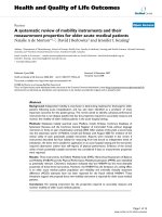
The Kaplan-Meier survival curvesFigure 3
The Kaplan-Meier survival curves. The Kaplan-Meier survival of p53, p21/WAF1 and MDM2 in HCC. Positive or negative
p53 expression did not correlate with the survival of patients (P = 0.275) (A). There was a significant difference in survival
between patients with positive p21/WAF1 expression and negative p21/WAF1 expression (P = 0.026) (B) and between patients
with negative MDM2 expression and positive MDM2 expression (P = 0.043) (C). There was a significant difference in survival
between patients with p21+/MDM2- expression and p21-/MDM2+ expression (P = 0.012) (D).
Journal of Translational Medicine 2009, 7:110 />Page 7 of 8
(page number not for citation purposes)
sues may also be activated in a p53-dependent manner;
however, additional experiments should be performed to
confirm this hypothesis. MDM2 and p53 are linked to
each other through an autoregulatory negative feedback
loop aimed at maintaining low cellular p53 levels in the
absence of stress [8]. Mutant p53 proteins in tumor cells
are stable because they are deficient in transactivating
MDM2 hence they have a defective negative feedback
loop [8]. These can explain our results that p53 expression
correlates with MDM2 expression in corresponding adja-
cent liver tissues but not in HCC tissues.
The results from our study showed that the expression of
p21/WAF1 and MDM2 in HCC was associated with sur-
vival in patients with HCC, indicating that p21/WAF1 and
MDM2 can be considered as predictors of the prognosis of
HCC patients. Previous studies have reported conflicting
results on the prognostic value of p53, p21/WAF1, and
MDM2 expression in HCC. Naka [9] reported that posi-
tive immunostaining for p53 protein expression was a sig-
nificant indicator of poor prognosis in 126 HCC patients.
Further, Sung [12] reported that p53 overexpression was a
poor prognostic factor of survival in 105 HCC patients,
and Schoniger-Hekele [11] reported that the survival of
patients overexpressing p53 among 81 HCC patients was
poorer than that of those who did not express p53. Hu
[32] reported that patients overexpressing p53 among 124
HCC patients had shorter survival periods and higher
recurrence rates than the p53-negative patients. In a study
conducted using samples collected from 122 HCC
patients after tumor resection, Kao [15] reported that
HCC patients with negative expression of p21/WAF1
exhibited a poorer survival rate than the HCC patients
with positive expression of p21/WAF1, suggesting that the
expression of p21/WAF1 in HCC was a survival prognostic
factor. MDM2 was rarely reported to be a prognostic factor
in HCC, but was often reported in the maxillary sinus
squamous cell carcinoma [33], in Egyptian esophageal
carcinoma [34], in breast carcinoma [35], prostate adeno-
carcinoma [36], gastric cancer [37,38], and epithelial
ovarian cancer [39,40].
Conclusion
In summary, we provided evidence for the significant
higher expression of p53, p21/WAF1, and MDM2 in HCC
tissues than in the corresponding adjacent liver tissues;
p53 expression correlated with p21/WAF1 expression
either in HCC tissues or in corresponding adjacent liver
tissues. Further, we determined that p21/WAF1 and
MDM2 expression in HCC was associated with survival in
patients with HCC, indicating that p21/WAF1 and MDM2
can be used to predict the prognosis of patients with HCC.
Competing interests
The authors declare that they have no competing interests.
Authors' contributions
JPY carried out and coordinated the study, immunohisto-
chemical examinations of tumor specimens and data
analysis, and drafted the manuscript. ZYZ and JF partici-
pated in the interpretation of data, conducted immuno-
histochemistry, and western blot analysis. All authors read
and approved the final manuscript.
Acknowledgements
The study was supported in part by the grant from the National Natural
Science Foundation of China (No.30471960), Guangdong Natural Science
Foundation (No. 06021198 and No. 8151008901000057), and the Ministry
of Health of China (2008ZX10002-019).
References
1. Parkin DM: Global cancer statistics in the year 2000. Lancet
Oncol 2001, 2:533-543.
2. Okuda K, Ohtsuki T, Obata H, Tomimatsu M, Okazaki N, Hasegawa
H, Nakajima Y, Ohnishi K: Natural history of hepatocellular car-
cinoma and prognosis in relation to treatment. Study of 850
patients. Cancer 1985, 56:918-928.
3. Levine AJ: p53, the cellular gatekeeper for growth and divi-
sion. Cell 1997, 88:323-331.
4. Michieli P, Chedid M, Lin D, Pierce JH, Mercer WE, Givol D: Induc-
tion of WAF1/CIP1 by a p53-independent pathway. Cancer
Res 1994, 54:3391-3395.
5. el-Deiry WS, Tokino T, Velculescu VE, Levy DB, Parsons R, Trent JM,
Lin D, Mercer WE, Kinzler KW, Vogelstein B: WAF1, a potential
mediator of p53 tumor suppression. Cell 1993, 75:817-825.
6. Chen J, Marechal V, Levine AJ: Mapping of the p53 and mdm-2
interaction domains. Mol Cell Biol 1993, 13:4107-4114.
7. Kubbutat MH, Ludwig RL, Ashcroft M, Vousden KH: Regulation of
Mdm2-directed degradation by the C terminus of p53. Mol
Cell Biol 1998, 18:5690-5698.
8. Moll UM, Petrenko O: The MDM2-p53 interaction. Mol Cancer
Res 2003, 1:1001-1008.
9. Naka T, Toyota N, Kaneko T, Kaibara N: Protein expression of
p53, p21WAF1, and Rb as prognostic indicators in patients
with surgically treated hepatocellular carcinoma. Anticancer
Res 1998, 18:555-564.
10. Qin LF, Ng IO: Expression of p27(KIP1) and p21(WAF1/CIP1)
in primary hepatocellular carcinoma: clinicopathologic cor-
relation and survival analysis. Hum Pathol 2001, 32:778-784.
11. Schoniger-Hekele M, Hanel S, Wrba F, Muller C: Hepatocellular
carcinoma survival and clinical characteristics in relation to
various histologic molecular markers in Western patients.
Liver Int 2005, 25:62-69.
12. Sung CO, Yoo BC, Koh KC, Cho JW, Park CK: Prognostic signifi-
cance of p53 overexpression after hepatic resection of hepa-
tocellular carcinoma. Korean J Gastroenterol 2005, 45:425-430.
13. Terris B, Laurent-Puig P, Belghitti J, Degott C, Henin D, Flejou JF:
Prognostic influence of clinicopathologic features, DNA-
ploidy, CD44H and p53 expression in a large series of
resected hepatocellular carcinoma in France. Int J Cancer
1997, 74:614-619.
14. Tseng PL, Tai MH, Huang CC, Wang CC, Lin JW, Hung CH, Chen
CH, Wang JH, Lu SN, Lee CM, et al.: Overexpression of VEGF is
associated with positive p53 immunostaining in hepatocellu-
lar carcinoma (HCC) and adverse outcome of HCC patients.
J Surg Oncol 2008, 98(5):349-57.
15. Kao JT, Chuah SK, Huang CC, Chen CL, Wang CC, Hung CH, Chen
CH, Wang JH, Lu SN, Lee CM, et al.: P21/WAF1 is an independ-
ent survival prognostic factor for patients with hepatocellu-
lar carcinoma after resection. Liver Int 2007, 27:772-781.
16. Bahnassi AA, Zekri AR, El-Houssini S, Mokhtar NM, Abdel-Aziz AO,
Sherif GM, El-Mishad AM, Khaled HM: Hepatitis C virus-NS3P in
relation to p53, p21waf, mdm2, p21-ras and c-erbB2 in hepa-
tocarcinogenesis. J Gastroenterol Hepatol 2005, 20:1731-1740.
17. Miao J, Chen GG, Chun SY, Lai PP: Hepatitis B virus X protein
induces apoptosis in hepatoma cells through inhibiting Bcl-
xL expression. Cancer Lett 2006, 236:115-124.
Publish with BioMed Central and every
scientist can read your work free of charge
"BioMed Central will be the most significant development for
disseminating the results of biomedical research in our lifetime."
Sir Paul Nurse, Cancer Research UK
Your research papers will be:
available free of charge to the entire biomedical community
peer reviewed and published immediately upon acceptance
cited in PubMed and archived on PubMed Central
yours — you keep the copyright
Submit your manuscript here:
/>BioMedcentral
Journal of Translational Medicine 2009, 7:110 />Page 8 of 8
(page number not for citation purposes)
18. Yun JP, Chew EC, Liew CT, Chan JY, Jin ML, Ding MX, Fai YH, Li HK,
Liang XM, Wu QL: Nucleophosmin/B23 is a proliferate shuttle
protein associated with nuclear matrix. J Cell Biochem 2003,
90:1140-1148.
19. Yun JP, Zhang MF, Hou JH, Tian QH, Fu J, Liang XM, Wu QL, Rong
TH: Primary small cell carcinoma of the esophagus: clinico-
pathological and immunohistochemical features of 21 cases.
BMC Cancer 2007, 7:38.
20. Mann CD, Neal CP, Garcea G, Manson MM, Dennison AR, Berry DP:
Prognostic molecular markers in hepatocellular carcinoma:
a systematic review. Eur J Cancer 2007, 43:979-992.
21. el-Deiry WS, Harper JW, O'Connor PM, Velculescu VE, Canman CE,
Jackman J, Pietenpol JA, Burrell M, Hill DE, Wang Y, et al.: WAF1/
CIP1 is induced in p53-mediated G1 arrest and apoptosis.
Cancer Res 1994, 54:1169-1174.
22. Xiong Y, Hannon GJ, Zhang H, Casso D, Kobayashi R, Beach D: p21
is a universal inhibitor of cyclin kinases. Nature 1993,
366:701-704.
23. Dulic V, Kaufmann WK, Wilson SJ, Tlsty TD, Lees E, Harper JW,
Elledge SJ, Reed SI: p53-dependent inhibition of cyclin-depend-
ent kinase activities in human fibroblasts during radiation-
induced G1 arrest. Cell 1994, 76:1013-1023.
24. Marchetti A, Doglioni C, Barbareschi M, Buttitta F, Pellegrini S, Bert-
acca G, Chella A, Merlo G, Angeletti CA, Dalla Palma P, et al.: p21
RNA and protein expression in non-small cell lung carcino-
mas: evidence of p53-independent expression and associa-
tion with tumoral differentiation. Oncogene 1996,
12:1319-1324.
25. Tron VA, Tang L, Yong WP, Trotter MJ: Differentiation-associ-
ated overexpression of the cyclin-dependent kinase inhibitor
p21waf-1 in human cutaneous squamous cell carcinoma. Am
J Pathol 1996, 149:1139-1146.
26. el-Deiry WS, Tokino T, Waldman T, Oliner JD, Velculescu VE, Burrell
M, Hill DE, Healy E, Rees JL, Hamilton SR, et al.: Topological con-
trol of p21WAF1/CIP1 expression in normal and neoplastic
tissues. Cancer Res 1995, 55:2910-2919.
27. Lukas J, Groshen S, Saffari B, Niu N, Reles A, Wen WH, Felix J, Jones
LA, Hall FL, Press MF: WAF1/Cip1 gene polymorphism and
expression in carcinomas of the breast, ovary, and
endometrium. Am J Pathol 1997, 150:167-175.
28. Qin LF, Ng IO, Fan ST, Ng M: p21/WAF1, p53 and PCNA expres-
sion and p53 mutation status in hepatocellular carcinoma.
Int J Cancer 1998, 79:424-428.
29. Albrecht JH, Meyer AH, Hu MY: Regulation of cyclin-dependent
kinase inhibitor p21(WAF1/Cip1/Sdi1) gene expression in
hepatic regeneration. Hepatology 1997, 25:557-563.
30. Wagayama H, Shiraki K, Yamanaka T, Sugimoto K, Ito T, Fujikawa K,
Takase K, Nakano T: p21WAF1/CTP1 expression and hepatitis
virus type. Dig Dis Sci 2001, 46:2074-2079.
31. Wagayama H, Shiraki K, Sugimoto K, Ito T, Fujikawa K, Yamanaka T,
Takase K, Nakano T: High expression of p21WAF1/CIP1 is cor-
related with human hepatocellular carcinoma in patients
with hepatitis C virus-associated chronic liver diseases. Hum
Pathol 2002, 33:429-434.
32. Hu TH, Wang CC, Huang CC, Chen CL, Hung CH, Chen CH, Wang
JH, Lu SN, Lee CM, Changchien CS, et al.: Down-regulation of
tumor suppressor gene PTEN, overexpression of p53, plus
high proliferating cell nuclear antigen index predict poor
patient outcome of hepatocellular carcinoma after resec-
tion. Oncol Rep 2007, 18:1417-1426.
33. Bandoh N, Hayashi T, Takahara M, Kishibe K, Ogino T, Katayama A,
Imada M, Nonaka S, Harabuchi Y: Loss of p21 expression is asso-
ciated with p53 mutations and increased cell proliferation
and p27 expression is associated with apoptosis in maxillary
sinus squamous cell carcinoma. Acta Otolaryngol 2005,
125:779-785.
34. Bahnassy AA, Zekri AR, Abdallah S, El-Shehaby AM, Sherif GM:
Human papillomavirus infection in Egyptian esophageal car-
cinoma: correlation with p53, p21, mdm2, C-erbB2 and
impact on survival. Pathol Int 2005, 55:53-62.
35. Turbin DA, Cheang MC, Bajdik CD, Gelmon KA, Yorida E, De Luca
A, Nielsen TO, Huntsman DG, Gilks CB: MDM2 protein expres-
sion is a negative prognostic marker in breast carcinoma.
Mod Pathol 2006, 19:69-74.
36. Doganavsargil B, Simsir A, Boyacioglu H, Cal C, Hekimgil M: A com-
parison of p21 and p27 immunoexpression in benign glands,
prostatic intraepithelial neoplasia and prostate adenocarci-
noma. BJU Int 2006, 97:644-648.
37. Drebber U, Baldus SE, Nolden B, Grass G, Bollschweiler E, Dienes
HP, Holscher AH, Monig SP: The overexpression of c-met as a
prognostic indicator for gastric carcinoma compared to p53
and p21 nuclear accumulation. Oncol Rep 2008, 19:1477-1483.
38. Seo YH, Joo YE, Choi SK, Rew JS, Park CS, Kim SJ: Prognostic sig-
nificance of p21 and p53 expression in gastric cancer. Korean
J Intern Med 2003, 18:98-103.
39. Schmider A, Gee C, Friedmann W, Lukas JJ, Press MF, Lichtenegger
W, Reles A: p21 (WAF1/CIP1) protein expression is associ-
ated with prolonged survival but not with p53 expression in
epithelial ovarian carcinoma. Gynecol Oncol 2000, 77:237-242.
40. Dogan E, Saygili U, Tuna B, Gol M, Gurel D, Acar B, Koyuncuoglu M:
p53 and mdm2 as prognostic indicators in patients with epi-
thelial ovarian cancer: a multivariate analysis. Gynecol Oncol
2005, 97:46-52.