Báo cáo hóa học: " Boosting high-intensity focused ultrasoundinduced anti-tumor immunity using a sparse-scan strategy that can more effectively promote dendritic cell maturation" ppt
Bạn đang xem bản rút gọn của tài liệu. Xem và tải ngay bản đầy đủ của tài liệu tại đây (2.59 MB, 12 trang )
RESEA R C H Open Access
Boosting high-intensity focused ultrasound-
induced anti-tumor immunity using a sparse-scan
strategy that can more effectively promote
dendritic cell maturation
Fang Liu
1†
, Zhenlin Hu
1†
, Lei Qiu
1
, Chun Hui
2
, Chao Li
2
, Pei Zhong
3*
, Junping Zhang
1*
Abstract
Background: The conventional treatment protocol in high-intensity focused ultrasound (HIFU) therapy utilizes a
dense-scan strategy to produce closely packed thermal lesions aiming at eradicating as much tumor mass as
possible. However, this strategy is not most effective in terms of inducing a systemic anti-tumor immunity so that
it cannot provide efficient micro-metastatic control and long-term tumor resistance. We have previously provided
evidence that HIFU may enhance systemic anti-tumor immu nity by in situ activation of dendritic cells (DCs) inside
HIFU-treated tumor tissue. The present study was conducted to test the feasibility of a sparse-scan strategy to
boost HIFU-induced anti-tumor immune response by more effectively promoting DC maturation.
Methods: An experimental HIFU system was set up to perform tumor ablation experiments in subcutaneous
implanted MC-38 and B16 tumor with dense- or sparse-scan strategy to produce closely-packed or separated
thermal lesions. DCs infiltration into HIFU-treated tumor tissues was detected by immunohistochemistry and flow
cytometry. DCs maturation was evaluated by IL-12/IL-10 production and CD80/CD86 expression after co-culture
with tumor cells treated with different HIFU. HIFU-induced anti-tumor immune response was evaluated by
detecting growth-retarding effects on distant re-challenged tumor and tumor-specific IFN-g-secreting cells in HIFU-
treated mice.
Results: HIFU exposure raised temperature up to 80 degrees centigrade at beam focus within 4 s in experimental
tumors and led to formation of a well-defined thermal lesion. The infiltrated DCs were recruited to the periphery of
lesion, where the peak temperature was only 55 degrees centigrade during HIFU exposure. Tumor cells heated to
55 degrees centigrade in 4-s HIFU exposure were more effective to stimulate co-cultured DCs to mature. Sparse-
scan HIFU, which can reserve 55 degrees-heated tumor cells surrounding the separated lesions, elicited an
enhanced anti-tumor immune response than dense-scan HIFU, while their suppressive effects on the treated
primary tumor were maintained at the same level. Flow cytometry analysis showed that sparse-scan HIFU was
more effective than dense-scan HIFU in enhancing DC infiltration into tumor tissues and promoting their
maturation in situ.
Conclusion: Optimizing scan strategy is a feasible way to boost HIFU-induced anti-tumor immunity by more
effectively promoting DC maturation.
* Correspondence: ;
† Contributed equally
1
Department of Biochemical Pharmacy, School of Pharmacy, Second Military
Medical University, Shanghai 200433, China
3
Department of Mechanical Engineering and Materials Science, Duke
University, Box 90300, Durham, NC 27708-0300, USA
Liu et al. Journal of Translational Medicine 2010, 8:7
/>© 2010 Liu et al; license e BioMed Central Ltd. This is an Open Access article dis tributed under the terms of the Creative Commons
Attribution License ( which perm its unrestricted use, distribution, and reproduction in
any medium, provided the original work is properly cited.
Introduction
In recent years, high-intensity focused ultrasound
(HIFU) has emerged as a new and promising treatment
modality for a variety of cancers, including breast[1],
prostate[2], kidney, liver[3], bone[4], uterus and pan-
creas cancers[5,6]. By f ocusing acousti c energy in a
small cigar-shaped volume inside the tumor, HIFU can
rapidlyraisethetissuetemperatureatitsbeamfocus
above 65°C, leading to cellular coagulative necrosis and
thermal lesion formation in a well-defined region. In
principle, HIFU can be applied to most internal organs
with an appropriate acoustic window for ultrasound
transmission except those with a ir-filled viscera such as
lung or bowel. In particular, HIFU is advantageous in
treating patients with unresectable cancers, such as pan-
creatic carcinoma, or with poor physical condition for
surgery. Unlike radiati on and ch emothera py, HIFU can
be applied repetitiv ely without the apprehension of
accumulating systemic toxicity. This unique feature
allows multiple HIFU sessions to be performed if local
rec urrence occurs. Clinical studies have already demon-
strated promising outcome of HIFU treatment for sev-
eral types of malignances, including prostate cancer,
breast cancer, uterine fibroids, hepatocellular carcino-
mas, and bone malignances [7,8]. Although some ther-
mal (skin burn, damage to adjacent bones or nerves)
and non-therma l (pain, fever, local infection, and bowe l
perforation) complications of HIFU treatment have been
reported, most of the complications were minor and
without severe adverse consequences[8,9].
At present, the primary drawback of HIFU is that it
cannot be used to kill micro-metastases outside the pri-
mary tumor s ite. In fact, distant metastasis is a major
cause of mortality following clinical HIFU therapy[1 0].
Lengthy treatment time also represents a limitation.
Because each HIFU pulse generally creates an ablated
spot of ~10 × 3 × 3 mm in size, up to 1000 lesions may
need to be packed closely together during HIFU treat-
ment by scanning the beam f ocus in a matrix of posi-
tions to cover entire tumor volume. With current
treatment algorithms, t his may translate into a proce-
dure time exceeding 4 hours. Currently, the conven-
tional HIFU treatment protocol in clinic utilizes a dens e
scanning pattern to erad icate as much tumor mass as
possible. Nevertheless, local recurrence of the tumor,
duetoincompletetissuenecrosis,isstillfrequently
observed following HIFU therapy[10,11]. Clearly, the
quality and effectiveness of HIFU cancer therapy need
further improvement.
In addition to direct localized destruction of tumor
tissues, preliminary evidence from several recent studies
has suggested that HIFU may enhance host systemic
anti-tumor immunity[12,13]. Although the underlying
mechanism is still largely unknown, the potential for a
HIFU-elicited anti-tumor immunity is attractive and
may help to control local recurrenc e and distant metas-
tasis following thermal ablation of the p rimary tumor.
On the other hand, the anti-tumor immune response
reported in previous studies was not strong enough to
achieve a therapeutic gain. As mentioned above, local
tumor recurrence and distant metastasis are often the
cause of failure for HIFU therapy[10,12], indicating the
need to augment the host ant i-tumor immunity. The re-
fore, the optimized strategies that can reduce the pri-
mary tumor mass and elicit simultaneously a strong
anti-tumor immune response are highly desirable.
The induction and maintenance of an effective antitu-
mor immune response is critically dependent on dendri-
tic cells (DCs), the most effective antigen-presenting
cells (APCs) that capture antigens in peripheral tumor
tissues and migrate to secondary lymphoid organs,
wheretheycross-presentthecapturedantigenstoT
cells and activate them[14]. To act as potent APCs, DCs
must undergo maturation, a state characterized by the
upregulation of MHC and costimulatory molecules and
the production of cytokines such as IL-12. However, the
requisite signals for DC maturationareoftenabsent
from the bed of poorly immunogenic tumors, and many
tumor cells even actively produce immunosuppressive
cytokinessuchasVEGFtosuppressDCfunction[15].
Thus, DCs infi ltrated in tumor tissues typically exhibit a
‘’suppressed’’ phenotype, and show significantly reduced
ability to stimulate allogeneic T cells when compared
with normal DCs. Such alterations in DCs development
and function are associated with tumor escape from
immune-mediated s urveillance[16,17]. On the other
hand, several studies have demonstrated that dying
tumor cells responding to chemotherapy or radiotherapy
can express ‘danger’ and ‘eat me’ signals s uch as heat-
shock proteins (HSPs) on the cell surface or release
intracellular HSP molecules to stimulate DCs to mature
and elicit a strong anti-tumor immu ne response[18]. In
the setting of HIFU therapy, we have demonstrated in
vitro that HIFU treatment results in the release endo-
genous immunostimulatory factors from tumor cells and
stimulates DCs to mature[19]. We have further provided
evidence that HIFU can stimulate DCs to infiltrate into
tumor tissues, migrate to d raining lymph nodes after
being activated, and subsequently elicit tumor-specific
CTL responses[20]. Based on these observations, we
have postulated that in situ activation of DCs inside
HIFU-treated tumor tissue may constitute an important
mechanism for HIFU-induced anti-tumor immunity.
Given t he central role of DCs maturation i n the devel-
opment of anti-tumor immune response, it is reasonable
to speculate that an optimized HIFU strategy that can
Liu et al. Journal of Translational Medicine 2010, 8:7
/>Page 2 of 12
more effectively activate DCs to mature should have
potential to elicit a stronger anti-tumor immunity.
The present study was conducted to search for a feasi-
ble way to boost HIFU-induced anti-tumor immunity by
more effectively stimulating DCs to mature. To this end,
we set up an experimental HIFU system and performed
a series o f tumor ablation experiments in subcutaneous
implanted MC-38 and B16 tumor models. We found
that the infiltrated DCs were mostly recruited to the
periphery of thermal lesions after HIFU exposure and
the tumor cells at the periphery of HIFU-induced ther-
mal lesions could more effectively stimulated DCs to
mature. Based on these finding, we further hypothesize
a sparse-scan strategy that can produce separated ther-
mal lesions and reserve surrounding peripheral tumor
tissuemayprovidemorestimuliforDCmaturation
than currently used dense-scan strategy, and finally
enhance the strength of HIFU-induced systemic anti-
tumor immune response. By comparing the tumor abla-
tion efficiency and anti-tumor immune response elicited
by two different HIFU treatment strategies, i.e., spare vs.
dense scan, in well-controlled animal experiments, we
demonstrated that it is actually feasible to boost HIFU-
induc ed anti-tumor immunity through optimizing HIFU
scan strategy. Finally, we did ex vivo experiments to
assess the number of tumor-infiltrating DCs and their
maturation status in HIFU-treated tumor tissues and
found that sparse-scan HIFU was more effective than
dense-scan HIFU in enhancing infiltration of DCs into
tumor tissues and promoting their maturation in situ.
Materials and methods
Cell culture
MC-38 mouse colon adenocarcinoma tumor cell line
was kindly provided by Dr. Timothy M. Clay of Duke
Comprehensive Cancer Center, Duke University (Dur-
ham, NC, USA). B16 mouse melanoma cell line and EL4
mouse lymphoma cell line were obtained from Shanghai
Institute of Cell Biology and Biochemistry (Shanghai,
China). All of cell lines were maintained in complete
Dulbeco’s modified eagle medium (DMEM), supplemen-
ted with 10% fetal bovine serum (FBS) (Gibco, USA) at
37°C and 5% CO
2
.
Experimental animals and Tumor Model
C57BL/6 female mice, 5-8 weeks old, were purchased from
Shanghai SLAC Laboratory Animal CO. LTD (Shanghai ,
China). Tumor models were prepared by subcutaneous ly
injecting 5 × 10
5
MC-38 or B16 tumor cells suspended in
50 μl of PBS in the left hindlimb of the C57BL/6 mice.
The tumor was allowed to grow for 8 days to reach a dia-
meter of 8-10 mm before HIFU treatment. All procedures
involving animal treatment and their care in this study
were approved by the animal care committee of the
Second Military Medical University in Shanghai in accor-
dance with institutional and Chinese government guide-
lines for animal experiments.
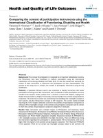
HIFU Exposure System
In vivo HIFU treatment of tumor was carried out utiliz-
ing a B-mode ultrasound imaging-guided HIFU expo-
sure system as reported in our previous study [20]
(Figure 1A). A HIFU transducer (provided by Shanghai
A&S Science Technology Development CO., LTD,
Shanghai, China) with a focal length of 63 mm, operated
at 3 .3 MHz was mounted at the bottom of a tank filled
with degassed water. The transducer was driven by sinu-
soidal signals produced by a function generator con-
nected in series with a 55-dB power amplifier (DF 5857,
Ningbo Zhongce Dftek Electronics Co. Ltd, Ningbo,
China). The operation and exposure parameters of the
HIFU system were controlled by LabView programs via
a GP IB board installed in a PC. During the experiment,
the anesthetized animal was placed in a custom-
designed holder (Figure 1B and 1C) co nnected to a 3-D
positioning system d riven by computer-contr olled step
motors (provided by Shanghai A&S Science Technology
Development CO., LTD, Shanghai, China). To facilitate
alignment of the tumor to the HIFU focus, a portable
ultrasound imaging system (Terason 2000, Terason, Inc.,
Burlington, MA) with a 5/10 MHz probe was used to
prov ide B-mode images of the tumor cross section. The
medial plane of the tumor was aligned with the focus of
the HIFU transducer. Figure 1D shows an example of
the B-mode ultrasound images of the tumor grown in
the hindlimb of the mouse. As shown in the figure, the
tumor outli ne was clearly defined, with the focus of the
HIFU transducer highlighted with a cross -hair indicator.
Treatment of the tumor was accomplis hed through pro-
gressive scanning of the whole tumor volume point-by-
point, translating the tumor-bearing mouse incremen-
tally with the 3-D step motor positioning system.
In vitro HIFU treatment of tumor cells was performed
inaHIFUexposuresystemshowninFigure1E.The
HIFU transducer was mounted horizontally inside a
water tank filled with degassed water. 1 × 10
5
tumor
cells suspended in 20 μl DMEM were loaded in a 0.2 ml
PCR thin-walled tube, which was placed vertically with
its conical bottom aligned within beam focus of the
HIFU transducer.
Measurement of temperature profile
The temperature profile at the HIFU focus was mea-
sured by using a Digital Thermometor (MC3000-000,
Shanghai DAHUA-CHINO Instrument Co, Ltd, Shang-
hai, China) with 0.1 mm bare-wire thermocouple
inserted into the tumor tissue or the cell suspension.
The thermocouple embedded in the t umor or cell
Liu et al. Journal of Translational Medicine 2010, 8:7
/>Page 3 of 12
suspension was first aligned to the HIFU focus then
temperature elevations and dist ributions around the
center of focus during HIFU exposures were recorded.
Assay of DC infiltration inside tumor tissue by
immunohistochemistry
One day after the HIFU treatment, tumors were surgi-
cally excised, freshly frozen in Tissue-Tek O.C.T. com-
pound (Sakura Finetek, Torrance, CA USA), and
sectioned at 6 μm thickness. The cryostat sections were
then fixed in acetone and immunostained with hamster
anti-mous e CD11c mAb (clone HL3, PharMingen). Sub-
sequently, the antibody was visualized using an anti-
hamster Ig H RP detection kit (Pharm ingen) following
the manufacturer’s protocol. Finally, sections were coun-
terstained with hematoxylin and evaluated by light
microscopy.
Generation of bone marrow-derived DC [19]
Bone marrow cells were flushed from the femurs and
tibiae of female C57BL/6 mice, filtere d through a Falcon
100-μm nylon cell strainer (BD Labware), and depleted
of red blood cells by five minute incubation in ACK
lysis buffer (0.15 M NH4Cl, 1.0 mM KHCO3, 0.1 mM
Na2EDTA, pH 7.4). Whole bone marrow cells were pla-
ted in six-well plates (BD Labware) in RPMI-1640 sup-
plemented with 10% FCS (GIBCO-BRL, USA), GM-CSF
(10 ng/ml), and IL-4 (10 ng /ml) (BD Biosciences Phar-
mingen, USA), and i ncubated at 37°C and 5% CO2.
Three days later, the floating cells (mostly granulocytes)
were removed, and the adherent cells w ere replenished
with fresh medium containing GM-CS F and IL-4. Non-
adherent and loosely adherent cells were harvested on
day 6 as immature DC (typically contained >90% cells
expressing CD11c and MHC class II on the surface, as
determined by flow cytometry).
In vitro stimulation of DCs with HIFU-treated tumor cells
and assay for their maturation status
5×10
5
immature DCs generated from mouse bone
marrow cells were co-cultured with HIFU-treated B16
tumor cells at ratio of 1:1 in 1 ml of culture for 2 days
at 37°C with 5% CO
2
. DC alone, DC stimulated with
CpG-ODN1826 (5’ -TCCATGACGTTCCTGACGTT-3’,
Coley Pharmaceutical, Wellesley, MA), which is a
known potent DC stimulator, and DC co-cultured with
non-HIFU treated B16 tumor cells were used as control.
After incubation, supernatants were harvested and
assayed for secreted IL-12 and IL-10 by commercial
ELISA kits (Biosource International, CA, USA). To ana-
lyze the expressi on levels of co-stimulat ory molecules,
DCs were collected into cold PBS plus 1% dialyzed
bovine serum albumin, the n washed and stained on ice
for 30 min with a combination of the following mono-
clonal antibodies: FITC-Conjugated Anti-Mouse CD11c,
PE-Conjugated Anti-Mouse CD86, and PE-CY5-Conju-
gated Anti-Mouse CD80 (BD Biosciences Pharminge n,
USA). Subsequently, the cells were washed again and
analyzed using a FACSCalibur flow cytometer (Bec ton-
Dickinson).
Tumor growth regression assay
Following HIFU treatment, Mice were thereafter moni-
tored daily for tu mor growth. Mean tumor area for each
group was calculated as the product of bisecting tumor
diameters obtained from caliper measurements. Mea-
surements were terminated and mice were sacrificed
when tumors reached 20 mm in their largest dimension,
or when mice became visibly unwell, or when the tumor
became ulcerated.
ELISPOT Assay [20]
Spleens were harvested from euthanized tumor-bearing
mice 14 days after HIFU treatment. Splenocytes from
mice bearing MC-38 tumors in each group were resti-
mulated in vitro by culture with mitomycin-pretreated
MC-38 (specific) or EL4 (irrelevant) tumor cells at 20:1
responder-to-stimulator ratios for 24 h. Splenocytes
from mice bearing B16 tumors were stimulated with 1
μg/ml of relevant peptides mouse TRP2
181-188
(VYDFFVWL, purchased from Dalton Chemical Labora-
tories Inc. Toronto, ON, Canada), or irrelevant control
Figure 1 The experimental HIFU system. (A) Diagram of the in vivo HIFU exposure setup. (B) A tumor-bearing mouse. (C) The way the mouse
was fixed during HIFU exposure. (D) The B-mode ultrasound image of the tumor. (E) Diagram of the in vitro HIFU exposure setup.
Liu et al. Journal of Translational Medicine 2010, 8:7
/>Page 4 of 12
peptide (OVA
257-264
: SIINFEKL) for 24 h. Re-stimulate d
splenocytes (1 × 10
6
cells in 100 μl medium) were then
plated in 96-well nitrocellulose filter plates pre-coated
with anti-mouse interferon-g antibody (Pharmingen, San
Diego, CA). After incubation for 24 h at 37°C and 5%
CO2, the plates were washed with PBS, and “spots,” cor-
responding to cytokine-producing cells, were visualized
by incubation with 100 μl per well of biotinylated anti-
mouse IFN-g Ab (Pharmingen) overnight at 4°C. After
washing with PBS/0.5% Tween, 1.25 μg/ml avidin alka-
line phosphatase (Sigma) was added to the well in 100
μl PBS for 1 hour at room temperature. The develop-
ment of the assay was then performed with l00 μlof5-
bromo-4-chloro-3-indolylphosphate/nitro blue tetrazo-
lium (BCIP/NBT tablets, Sigma) for 10 minutes. The
reaction is stopped by the addition of water and the
plates allowed drying before counting individual spots
with a Zeiss automated ELISPOT reader. The results
were exp ressed as the number of spot-forming cells per
10
6
input cells. Overall, three independent experiments
were performed with six replicate wells included in each
treatment.
Assay of DC infiltration inside tumor tissue by flow
cytometry
One day after the HIFU treatment, tumors were surgi-
cally excised. Single cell suspensions were generated
from resected tumors as previously described[21].
Briefly, tumors were diced in Ca
2+
- and Mg
2+
-free HBSS
after resection, and incubated with 1 mg/ml type IV col-
lagenase(Sigma-Aldrich)for90minatroomtempera-
ture and under constant stirring. EDTA (2 mM) was
added to the mixture for 30 additional min before filtra-
tion of the cell suspension on 70-μm cell strainers (BD
Biosciences). The cell suspension was finally washed
twice in HBSS before analysis. For flow cytometry, the
following fluorochrome-conjugated antibodies (all pur-
chased from BD PharMingen) were used for staining:
CD45-FITC, CD11c-PE, I-A-PE-CY5, CD80-PE-CY5,
CD-80-PE-CY5. After adding the appropriate antibo dy,
the cells were incubated a t 4°C for 30 min in PBS plus
1% of dialyzed bovine serum albumin and washed twice
by centrifugation using fluorescence-activated cell sort-
ing (FACS) buffer. Fluorescence was analyzed with a
FACSCalibur flow cytometer and the CellQuest software
(Becton-Dickinson).
Results and Discussion
HIFU system could produce a typical thermal effect on
experimental tumors
In clinical HIFU therapy, tumor tissue was ablated pre-
dominantly by therm al effect which is dependent on the
temperature elevation achieved at beam focus during
HIFU exposure. If the temperature is raised to 56°C or
higher in the tissue, thermal lesion will form within a
few seconds as a result of cellular coagulative necrosis.
In fact, the temperature within the focal volume may
rise rapidly above 80°C duringHIFUtreatments[22].In
the present study, we at first calibrated our HIFU sys-
tem t o achieve a typical thermal effect on experimental
tumors. By adjusting output pr essure level and exposure
duration, we found that, when the transducer was run
in continuous wave (CW) mode at a pressure level of P
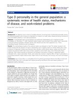
+
= 19.5/P
-
= -7.2 (MPa), an elevated temperature was
achieved up to 80°C within 4 s at the beam focus in
bot h MC-38 and B16 tumor (Figure 2A). This tempera-
ture profile is a representat ive of the clinical HIFU
dosage used in cancer therapy. Under this condition,
one HIFU exposure could generate a typical thermal
lesion with a w ell-defined size of 1 × 5 mm (transverse
× longitudinal direction) in the treatment region (Figure
2C and 2D). The peripheral tissue around thermal lesion
was also heated but with a lower peak temperat ure
(around 55°C) (Figure 2B).
The infiltrated DCs were mostly recruited to the
periphery of thermal lesions after hifu exposure
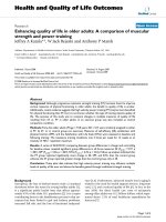
We next investigated whether HIFU can enhance infil-
tration of DCs into treated tumor tissues. Tumor sam-
ples were collected 1 day after HIFU treatment, and 6-
μm cryostat sections were cut a nd stained with anti-
CD11c Abs. Figure 3 showed the results of a representa-
tive experiment. In the untreated tumor, only a small
amount of DC infiltration was observed. In contrast, DC
infiltration was enhanced in HIFU-treated tumor tissues.
Most interestingly, it was noted that the infiltrated DC
was recruited to the periphery of thermal lesion (Figure
3).
Tumor cells at the periphery of HIFU-Induced thermal
lesion may possess a stronger immunostimulatory
property for DCs maturation
A prior study has documented a s ignificant up-regula-
tion of HSPs at the bo rder zone of HIFU-induced ther-
mal lesion in patients with benign prostatic hyperplasia
[23]. HSPs have been shown to interact with a number
of receptors present on the surface of DCs and promote
their maturation[24]. These findings imply the possibi-
lity that tumor cells at the periphery of HIFU-induced
thermal lesion may possess a stronger immunosimula-
tory property for DCs maturation. The finding in this
study that the infiltrated DCs were mostly recruited to
the periphery of thermal lesions after HIFU exposure
further raises the possibility that tumor cells withi n this
specific zone may have distinct impacts on infiltrated
DCs . To provide experimental evidence, we co-cultured
immature DCs generated from mouse bone marrow
cells with different HIFU-treated tumor cells and
Liu et al. Journal of Translational Medicine 2010, 8:7
/>Page 5 of 12
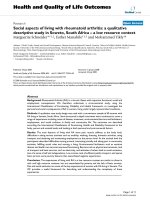
assessed their maturing status by assay of IL-12p70/IL-
10 production and CD80/86 expression on DCs. We at
first determined two different in vitro HIFU exposure
conditions, under which the temperature in the cell sus-
pension could reach a peak value of 55°C and 80°C
respectively within a 4-s exposure duration. Figure 4A
showed the distinct temperature profiles in tumor cell
suspensions produced by the two different HIFU expo-
sure conditions, which correspond to those produced in
vivo by HIFU at the periphery and the center of thermal
lesion, respectively. For convenience, these exposure
conditions were referred to hereafter as “55°C-HIFU”
and “80°C-HIFU” , respectively. After HIFU treatment,
B16 tumor cells were co-culture with immature DC for
2 days, and the release of IL-12p70 and IL-10 and sur-
face expression of maturation markers (CD80 and
CD86) o n DCs were assayed. DC alone, DC stimulated
with CpG-ODN, and DC c o-cultured with non-HIFU
treated B16 tumor cells were used as control. The
results were shown in figure 4B-D. DCs did not sponta-
neously secrete IL-12p70 and IL-10 when cultured in
the absence of exogenous stimuli. CpG-ODN, a known
potent DC stimulator, induced the highest level of IL-
12p70 production w hile only moderately increasing IL-
10 production, and significantly enhanced the expression
of CD80 and CD86, indicating CpG-ODN induced
immature DC towards a mature phenotype. Normal B16
tumor cells shown no effects on IL-12 p70 production
but markedly increased IL-10 production, and signifi-
cantly decreased the expressions of CD80 and CD86.
Since IL-12p70 and IL-10 are reported as immunosti-
mulatory versus immunosuppressive DC-produced cyto-
kines that may differentially affect the functional
outcome of T-cell cross-pr iming[25,26], this result
Figure 2 Thermal effects of HIFU treatment. (A) Temperature profiles at the beam focus in MC-38 and B16 tumors when the transducer was
run in continuous wave (CW) mode at a pressure level of P
+
= 19.5/P
-
= -7.2 MPa. Representative data of three independent experiments with
consistent results are shown. (B) Lateral distribution of peak temperature in tumors produced by HIFU during 4-s exposures. Results are
expressed as means ± SD out of four independent experiments. (C) Transversal and (D) longitudinal views of thermal lesions produced by HIFU
with different treatment duration (4, 3, and 2 s) at above pressure level. The representative section from four treated mice with similar results is
shown.
Liu et al. Journal of Translational Medicine 2010, 8:7
/>Page 6 of 12
confirmed previous finding that normal tumors could
induce or restrict tumor-infiltrating DCs towards an
immature phenotype [16,27]. After HIFU-treatment,
however, tumor cells became effective in inducing IL-
12p70 production while their effects on IL-10 produc-
tion markedly reduced. Furthermore, bot h 55°C-HIFU-
and 80°C-HIFU-treated tumor cells significantly
enhanced surface expressions of CD80 and CD86 on co-
cultured DCs. More importantly, 55°C-HIFU-treated
tumor cells showed much more potent immunostimula-
tory activities than 80°C-HIFU-treated ones, both in the
induction of IL-12p70 production and in the upregula-
tion of CD80 and CD86 expression.
Similar results were obtained with the other cell line
MC-38 (data not shown). These results demonstrated
that HIFU-treatment can change tumor cells from
immunosuppressive to immunostimulator y for DCs
maturation. More importantly, tumor cells exposed to
‘55°C-HIFU’ , which produced a temperature elevation
similar to that at the periphery of thermal lesion, exhib-
ited a markedly stronger immunostimulatory poten cy
than those exposed to ‘80°C-HIFU’ , which produced a
temperature elevation similar to that at the center of
thermal lesion. These data therefore provide evidence
that tumor cells at the periphery of thermal lesions can
more effectively activate DCs to mature than those
within the lesions.
We speculated that intracellular HSP molecules
release or their membrane exposure induced by HIFU
treatments may be the keyno te mechanism responsible
for the stimulatory activities of DC maturation provided
byHIFU-treatedtumorcells.Wehavedonesomepilot
experiments to compare the effects of different HIFU
treatments on the expression of HSPs in tumor cells.
Our preliminary results suggested the HIFU treatments
caused significant up-regulations of HSP70 and HSP90
expression in tumor cells, in which 55°C-HIFU was
more effective than 80°C-HIFU (Data not shown).
Further studies are underway to determine whether
these up-regulated HSPs are released in the extracellular
milieu or translocated to cell surface to investigate more
deeply the mechanisms of DC activation by HIFU-trea-
ted tumor cells.
It is feasible to boost HIFU-induced anti-tumor immunity
through optimizing scan strategy
A dense-scan strategy is usually used in clinical HIFU
therapy to produce closely packed or even overlapped
thermal lesions to achieve a complete tumor ablation
bec ause tumor cells at the board zone of thermal lesion
are used to be considered to be heated only sub-lethally
and may survive HIFU treatment. However, our data
suggest that the presence of such cells in situ may lea d
to “clinical benefit’ by potently activating infiltrated DCs
to mature. Since the maturation of tumor-infiltrating
DCs will lead to the development of strong anti-tumor
immunity, an optimized strategy that can reserve these
cells in HIFU-treated tumor may have a potential to
Figure 3 HIFU-induced DC infiltration surrounding the thermal lesion. Tumor tissue samples were collected 1 day after HIFU treatment. 6-
μm cryostat sections were cut and stained with anti-CD11c Abs. Then the antibody was visualized using the Anti-Hamster Ig HRP detection kit.
The sections were counterstained with hematoxylin. Representative sections from each group of four mice are shown.
Liu et al. Journal of Translational Medicine 2010, 8:7
/>Page 7 of 12
elicit a stronger anti-tumor immune response. In the
clinical setting, the simplest way to achieve this goal is
to adjusting the scan strategy, e.g. using a sparse-scan
strategy to produce separated rather than closely packed
thermal lesions. Hence, we further propose d a sparse-
scan strategy may elicit a stronger systemic anti-tumor
immune response than currently used dense-scan strat-
egy. To test this hypothesis, we compared the tumor
ablati on efficiency and anti-tumor immune response eli-
cited by two different HIFU treatment strategies, i.e.,
sparse v s. dense scan, in well-controlled animal experi-
ments. Because our HIFU system can produced a ther-
mal lesion with a well define size of 1 × 5 mm in the
experimental tumor by one pulse of HIFU exposure, a
step size of 1 mm was used in dense-scan strategy
which can produce closely packed thermal lesions and
Figure 4 DC maturation stimulated by HIFU-treated tumor cells.(A)Temperatureprofilesproducedby55°C-HIFUand80°C-HIFU.(B)
Immature DCs were incubated for 2 days in the presence of CpG-ODN, normal B16 cells, 55°C-HIFU and 80°C-HIFU treated B16 cells. Levels of
IL-12 p70 and IL-10 in the culture supernatants were measured by ELISA. (C) Expression of CD80 and CD86 on the surface of DC (thick line) was
assayed by Flow cytometry. Solid thin line represents the expression of these markers on surface of non-stimulated DC. Representative data out
of three separate experiments are shown. (D) The expression levels of CD80 and CD86 on DCs were presented as mean fluorescence intensity.
Results in panels B and D are expressed as means ± SD out of three independent experiments. * p < 0.05 compared with ‘DC Alone’,
#
p < 0.05
compared with ‘DC+normal B16’,
!
p < 0.05 compared with ‘DC+80°C-HIFU’ by Student’s t test.
Liu et al. Journal of Translational Medicine 2010, 8:7
/>Page 8 of 12
well mimic the conventional treatment protocol in clini-
cal HIFU therapy. In sparse-scan strategy, the step size
was increased to 2 mm to produce a cluster of separated
lesions with inter-lesion spacing of 1 mm. Figure 5A
showed the closely packed and separated lesions in MC-
38 tumor pro duced by the dense- and sparse-scan strat-
egy, respectively. Tumor growth regression assay
revealed HIFU treatment with the sparse- and dense-
scan strategies have similar retarding effects on growth
of treated tumors (Figure 5B and 5C), even though the
total number of thermal lesions produced by sparse
scan strategy is much less than that in dense scan strat-
egy. To further assess whether HIFU treatments could
induce a systemic anti-tumor immune response in vivo,
tumor challenge experiments were performed one day
following HIFU treatment by injecting 1 × 10
6
MC-38
or B16 cells subcutaneously in the contra lateral hin-
dlimb. As expected, the sparse-scan HIFU was found to
have a stronger retarding effect on challenged tumor
growth (Figure 5D and 5E). To further quantify the
anti-tumor immune response, we eva luated whether
HIFU treatment could elicit tumor-specific IFN-g-secret-
ing cells using ELISPOT assay. Consistent with finding
in tumor challenge experiments, splenocytes retrieved
on day 14 after tumor ino culation in HIFU-treated mice
contained significantly moretumor-specificIFN-g-
secreting cells than that from the control group (Figure
5F and 5G). Taken all together, these results demon-
strated that optimization of scan strategy in HIFU treat-
ment can indeed induce a more p owerful anti-tumor
effect and immune response. He re we only focused on
proof of principle, so we did not further optimize the
inter-lesion spacing or the total number of lesions for
the most effective treatment outcome in the present
study.
Sparse-scan HIFU was more effective than dense-scan
HIFU in enhancing infiltration of DCs into tumor tissues
and promoting their maturation in situ
In order to provide more experimental evidence that the
boosted antitumor immune response by sparse-scan
HIFU is associated with the stage of the maturation of
DCs recruited to the treated tumor, we next determined
whether different HIFU treatment could differentially
alter DC numbers in the tumor tissues and their func-
tional status. We t reated C57BL/6 mice bearing B16 or
MC-38 tumors in the left hindlimb with HIFU under
sparse- or dense-scan strategy. On the day following
HIFU treatment, mice were sacrificed. Upon tumor dis-
sociation, single cell suspensions were generated from
Figure 5 Comparison of tumor ablation and systemic immune response induced by two different scan strategies. (A) Thermal lesions
produced by dense- and sparse-scan strategies in MC-38 tumors. (B-C) The suppressive effects of different scan strategies on the growth of
treated primary tumors. (D-E) The retarding effects on the growth of distant re-challenged tumors. (F-G) Tumor-specific IFN-g-secreting cells
detected in the splenocytes of HIFU-treated mice. C57BL/6 mice were inoculated s.c. on right hind leg with 5 × 10
5
MC-38 or B16 tumor cells
and treated with different HIFU on day 9 of tumor inoculation. Mice were challenged with 1 × 10
6
MC-38 or B16 tumor cells by s.c. inoculation
on the left hind leg one day after HIFU treatment. Both primary and challenged tumor growth was monitored daily. Tumor-specific IFN-g-
secreting cells were detected in splenocytes by ELISPOTS assays. Results were expressed as mean ± SD for each group (n = 8 per group). *P <
0.05; **P < 0.001 versus non-treatment control by Student’s t test. This experiment is representative of three experiments with consistent results.
Liu et al. Journal of Translational Medicine 2010, 8:7
/>Page 9 of 12
resected tumors. The presence of cells with a DC phe-
notype and their surface expression of the activation
markers MHC class II (MHC II), CD80, and CD86 were
analyzed by flow cytometry after immunostainning. Leu-
kocytic cells (CD45
+
) could be distinguished by FACS
analysis from malignant ce lls by their size (FSC-H) and
morphology (SSC-H) (Figure 6A). In the leukocytic cell
population, CD11c
+
/MHC II
+
,CD11c
+
/CD86
+
,and
CD11c
+
/CD80
+
cells were vi sualized (Figure 6B), indi-
cating the presence of cells with a DC phenotype. Nota-
bly, higher proportion of tumor-infiltrating DCs (CD 11c
+
/MHC II
+
cells) were recovered from HIFU-treated
tumors, as compared with non-treatment control (Figure
6C and 6B), indicating that HIFU-treatment enhanced
DC infiltration into tumor tissues. Furthermore, up-
regulated levels of t he maturation markers CD80 and
CD86 were found in CD11c+ sub population recovered
from HIFU-treated tumors (Figure 6B, D, and 6E), indi-
cating HIFU-treatment induced infiltrated D Cs towards
a mature phenotype. Most importantly, Sparse-scan
HIFU was found to be more effective than dense-scan
HIFU in enhancing infiltration of DCs into tumor tis-
sues and promoting their maturation in situ,asevi-
denced by higher proportion of tumor-infiltrating DCs
Figure 6 DCs were recruited into tumor tissues one day after HIFU treatment and exhibited the surface phenotype of maturation.(A)
The presence of CD45
+
tumor-infiltrating leukocytes in tumor tissues was identified in the gate indicated. (B) CD11c
+
cells in the gate defined in
A were analyzed for the expression of MHC II, CD80, and CD86. Representative data of six independent experiments with consistent results are
shown. (C) The proportion of tumor-infiltrating DC (CD11c
+
/MHC II
+
) (expressed in percentage of total cells) was investigated for the indicated
tumors one day after different HIFU-treatment. (D) The expression levels of CD86 (presented as mean fluorescence intensity) were analyzed in
CD11c+ cells infiltrating B16 or MC-38 tumor one day after different HIFU-treatment. (E) The expression levels of CD80 (presented as mean
fluorescence intensity) were analyzed in CD11c
+
cells infiltrating B16 or MC-38 tumor one day after different HIFU-treatment. (C-E) Results were
expressed as mean ± SD for each group (n = 6 per group). *P < 0.05 versus non-treatment control;
#
P < 0.05 versus Dense-scan HIFU by
Student’s t test. This experiment is representative of three experiments with consistent results.
Liu et al. Journal of Translational Medicine 2010, 8:7
/>Page 10 of 12
and their higher levels of surface maturation markers
(Figure 6B-E). These results suggest that the enhanced
antitumor immune response induced by sparse-scan
HIFU may be associated with more DC infiltration into
treated tumors and effective DC maturation in situ.
Conclusion
In the present study, our results showed that a sparse-
scan strategy in HIFU ablation of tumor pro duced sepa-
rated thermal lesions with a proper intra-lesion space
and elicited a stronger systemic anti-tumor immune
response than currently used dense-scan HIFU strategy,
which is associated with more effective promotion of
DC maturation b y tumor c ells at the periphery of ther-
mal lesions.
These preliminary f indings have significant implica-
tions for improving HIFU treatment of cancer in clinic.
The conventional treatment protocol in clinical HIFU
therapy utilizes a dense-scan strategy to produce closely
packed or overlapped thermal lesions aiming at eradicat-
ing as much tumor mass as possible. Although effective
in reducing the primary tumor mass, this strategy is
time consuming and is not most effective in terms of
induction of systemic anti-tumor immunity, so that it
can not provide efficient micro-metastatic control and
long-term tumor resistance for cancer patients. Here we
proposed that, by simply adjusting scan strategy to pro-
duce separated thermal lesions with a proper intra-
lesion space, a stronger systemic anti-tumor immune
response may be elicited while not impair its tumor
ablation efficiency, so that the overall quality and effec-
tiveness of HIFU cancer therapy can be improved and
the treatment time can be significantly shorten. Future
clinical studies will tell whether this promise comes true.
Acknowledgements
This work was supported in part by National Natural Science Foundation of
China No 30772072 and Shanghai Pujiang Program No 08PJ1400200.
Author details
1
Department of Biochemical Pharmacy, School of Pharmacy, Second Military
Medical University, Shanghai 200433, China.
2
School of Life Sciences &
Biotechnology, Shanghai Jiao Tong University,China.
3
Department of
Mechanical Engineering and Materials Science, Duke University, Box 90300,
Durham, NC 27708-0300, USA.
Authors’ contributions
FL and ZH conducted the study, participated in data interpretation,
performed the statistical analysis, and drafted the manuscript. LQ
participated in the in vitro studies. CH and CL participated in the in vivo
studies. PZ and JP participated in design, coordination, and data
interpretation and drafted the manuscript. All authors read and approved
the final manuscript.
Competing interests
The authors declare that they have no competing interests.
Received: 29 September 2009
Accepted: 27 January 2010 Published: 27 January 2010
References
1. Hynynen K, Pomeroy O, Smith DN, Huber PE, McDannold NJ, Kettenbach J,
Baum J, Singer S, Jolesz FA: MR imaging-guided focused ultrasound
surgery of fibroadenomas in the breast: a feasibility study. Radiology
2001, 219:176-185.
2. Chaussy C, Thuroff S: The status of high-intensity focused ultrasound in
the treatment of localized prostate cancer and the impact of a
combined resection. Curr Urol Rep 2003, 4:248-252.
3. Kennedy JE, Wu F, ter Haar GR, Gleeson FV, Phillips RR, Middleton MR,
Cranston D: High-intensity focused ultrasound for the treatment of liver
tumours. Ultrasonics 2004, 42:931-935.
4. Chen W, Wang Z, Wu F, Zhu H, Zou J, Bai J, Li K, Xie F: [High intensity
focused ultrasound in the treatment of primary malignant bone tumor].
Zhonghua Zhong Liu Za Zhi 2002, 24:612-615.
5. Zhang L, Chen WZ, Liu YJ, Hu X, Zhou K, Chen L, Peng S, Zhu H, Zou HL,
Bai J, Wang ZB: Feasibility of magnetic resonance imaging-guided high
intensity focused ultrasound therapy for ablating uterine fibroids in
patients with bowel lies anterior to uterus. Eur J Radiol 2008.
6. Xiong LL, Hwang JH, Huang XB, Yao SS, He CJ, Ge XH, Ge HY, Wang XF:
Early clinical experience using high intensity focused ultrasound for
palliation of inoperable pancreatic cancer. Jop 2009, 10:123-129.
7. Thuroff S, Chaussy C, Vallancien G, Wieland W, Kiel HJ, Le Duc A,
Desgrandchamps F, De La Rosette JJ, Gelet A: High-intensity focused
ultrasound and localized prostate cancer: efficacy results from the
European multicentric study. J Endourol 2003, 17:673-677.
8. Wu F, Wang ZB, Chen WZ, Wang W, Gui Y, Zhang M, Zheng G, Zhou Y,
Xu G, Li M, et al: Extracorporeal high intensity focused ultrasound
ablation in the treatment of 1038 patients with solid carcinomas in
China: an overview. Ultrason Sonochem 2004, 11:149-154.
9. Visioli AG, Rivens IH, ter Haar GR, Horwich A, Huddart RA, Moskovic E,
Padhani A, Glees J: Preliminary results of a phase I dose escalation
clinical trial using focused ultrasound in the treatment of localised
tumours. Eur J Ultrasound 1999, 9:11-18.
10. Wu F, Wang ZB, Chen WZ, Zou JZ, Bai J, Zhu H, Li KQ, Xie FL, Jin CB, Su HB,
Gao GW: Extracorporeal focused ultrasound surgery for treatment of
human solid carcinomas: early Chinese clinical experience. Ultrasound
Med Biol 2004, 30:245-260.
11. Van Leenders GJ, Beerlage HP, Ruijter ET, de la Rosette JJ, Kaa van de CA:
Histopathological changes associated with high intensity focused
ultrasound (HIFU) treatment for localised adenocarcinoma of the
prostate. J Clin Pathol 2000, 53:391-394.
12. Wu F, Wang ZB, Lu P, Xu ZL, Chen WZ, Zhu H, Jin CB: Activated anti-
tumor immunity in cancer patients after high intensity focused
ultrasound ablation. Ultrasound Med Biol 2004, 30:1217-1222.
13. Zhou Q, Zhu XQ, Zhang J, Xu ZL, Lu P, Wu F: Changes in circulating
immunosuppressive cytokine levels of cancer patients after high
intensity focused ultrasound treatment. Ultrasound Med Biol 2008,
34:81-87.
14. Banchereau J, Steinman RM: Dendritic cells and the control of immunity.
Nature 1998, 392:245-252.
15. Takahashi A, Kono K, Ichihara F, Sugai H, Fujii H, Matsumoto Y: Vascular
endothelial growth factor inhibits maturation of dendritic cells induced
by lipopolysaccharide, but not by proinflammatory cytokines. Cancer
Immunol Immunother 2004, 53:543-550.
16. Almand B, Clark JI, Nikitina E, van Beynen J, English NR, Knight SC,
Carbone DP, Gabrilovich DI: Increased production of immature myeloid
cells in cancer patients: a mechanism of immunosuppression in cancer.
J Immunol 2001, 166:678-689.
17. Satthaporn S, Robins A, Vassanasiri W, El-Sheemy M, Jibril JA, Clark D,
Valerio D, Eremin O: Dendritic cells are dysfunctional in patients with
operable breast cancer. Cancer Immunol Immunother 2004, 53:510-518.
18. Tesniere A, Panaretakis T, Kepp O, Apetoh L, Ghiringhelli F, Zitvogel L,
Kroemer G: Molecular characteristics of immunogenic cancer cell death.
Cell Death Differ 2008, 15:3-12.
19. Hu Z, Yang XY, Liu Y, Morse MA, Lyerly HK, Clay TM, Zhong P: Release of
endogenous danger signals from HIFU-treated tumor cells and their
stimulatory effects on APCs. Biochem Biophys Res Commun 2005,
335:124-131.
20. Hu Z, Yang XY, Liu Y, Sankin GN, Pua EC, Morse MA, Lyerly HK, Clay TM,
Zhong P: Investigation of HIFU-induced anti-tumor immunity in a
murine tumor model. J Transl Med 2007, 5:34.
Liu et al. Journal of Translational Medicine 2010, 8:7
/>Page 11 of 12
21. Preynat-Seauve O, Schuler P, Contassot E, Beermann F, Huard B, French LE:
Tumor-infiltrating dendritic cells are potent antigen-presenting cells able
to activate T cells and mediate tumor rejection. J Immunol 2006,
176:61-67.
22. Hill CR, Rivens I, Vaughan MG, ter Haar GR: Lesion development in focused
ultrasound surgery: a general model. Ultrasound Med Biol 1994,
20:259-269.
23. Kramer G, Steiner GE, Grobl M, Hrachowitz K, Reithmayr F, Paucz L,
Newman M, Madersbacher S, Gruber D, Susani M, Marberger M: Response
to sublethal heat treatment of prostatic tumor cells and of prostatic
tumor infiltrating T-cells. Prostate 2004, 58:109-120.
24. Tsan MF, Gao B: Heat shock proteins and immune system. J Leukoc Biol
2009, 85:905-910.
25. Trinchieri G: Interleukin-12 and the regulation of innate resistance and
adaptive immunity. Nat Rev Immunol 2003, 3:133-146.
26. Vicari AP, Trinchieri G: Interleukin-10 in viral diseases and cancer: exiting
the labyrinth. Immunol Rev 2004, 202:223-236.
27. Gabrilovich DI, Ciernik IF, Carbone DP: Dendritic cells in antitumor
immune responses. I. Defective antigen presentation in tumor-bearing
hosts. Cell Immunol 1996, 170:101-110.
doi:10.1186/1479-5876-8-7
Cite this article as: Liu et al.: Boosting high-intensity focused
ultrasound-induced anti-tumor immunity using a sparse-scan strategy
that can more effectively promote dendritic cell maturation. Journal of
Translational Medicine 2010 8:7.
Submit your next manuscript to BioMed Central
and take full advantage of:
• Convenient online submission
• Thorough peer review
• No space constraints or color figure charges
• Immediate publication on acceptance
• Inclusion in PubMed, CAS, Scopus and Google Scholar
• Research which is freely available for redistribution
Submit your manuscript at
www.biomedcentral.com/submit
Liu et al. Journal of Translational Medicine 2010, 8:7
/>Page 12 of 12