Báo cáo hóa học: " Increased shedding of HU177 correlates with worse prognosis in primary melanoma" pdf
Bạn đang xem bản rút gọn của tài liệu. Xem và tải ngay bản đầy đủ của tài liệu tại đây (312.88 KB, 9 trang )
RESEARC H Open Access
Increased shedding of HU177 correlates with
worse prognosis in primary melanoma
Heather K Hamilton
1†
, Amy E Rose
1†
, Paul J Christos
2
, Richard L Shapiro
3
, Russell S Berman
3
, Madhu Mazumdar
2
,
Michelle W Ma
1
, Daniel Krich
1
, Leonard Liebes
4
, Peter C Brooks
5,6
, Iman Osman
1*
Abstract
Background: Increased levels of cryptic collagen epitope HU177 in the sera of melanoma patients have been
shown to be associated with thicker primary melanomas and with the nodular histologic subtype. In this study, we
investigate the association between HU177 shedding in the sera and clinical outcome in terms of disease-free
survival (DFS) and overall survival (OS).
Methods: Serum samples from 209 patients with primary melanoma prospectively enrolled in the Interdisciplinary
Melanoma Cooperative Group at the New York University Langone Medical Center (mean age = 58, mean
thickness = 2.09 mm, stage I = 136, stage II = 41, stage III = 32, median follow-up = 54.9 months) were analyzed
for HU177 concentration using a validated ELISA assay. HU177 serum levels at the time of diagnosis were used to
divide the study cohort into two groups: low and high HU177. DFS and OS were estimated by Kaplan-Meier
survival analysis, and the log-rank test was used to compare DFS and OS between the two HU177 groups.
Multivariate Cox proportional hazards regression models were employed to examine the independent effect of
HU177 category on DFS and OS.
Results: HU177 sera concentrations ranged from 0-139.8 ng/ml (mean and median of 6.2 ng/ml and 3.7 ng/ml,
respectively). Thirty-eight of the 209 (18%) patients developed recurrences, and 34 of the 209 (16%) patients died
during follow-up. Higher HU177 serum level was associated with an increased rate of melanoma recurrence (p =
0.04) and with increasing mortality (p = 0.01). The association with overall survival remained statistically significant
after controlling for thickness and histologic subtype in a multivariate model (p = 0.035).
Conclusions: Increased shedding of HU177 in the serum of primary melanoma patients is associated with poor
prognosis. Further studies are warranted to determine the clinical utility of HU177 in risk stratification compared to
the current standard of care.
Background
Limitations of the current melanoma staging paradigm
beget limitations in o ur ability to det ermine the most
appropriate treatment for primary melanoma patients
with regard to maximizing therapeutic benefit and mini-
mizing morbidity. Well-characterized clinical prognostic
markers such as tumor thickness and ulceration only
partly explain the variability in the clinical course of
melanoma. Patients with thin melanoma <1 mm, char-
acteriz ed as having a favorable prognosis, have reported
rates of metastasis ranging f rom 3-22% [1]. Conversely,
patients with thicker lesions not uncommonly have
extended periods of disease-free survival. Although sen-
tinel lymph node biopsy has improved our ability to pre-
dict prognosis for patie nts with intermediate thickness
lesions, further markers are needed to determine which
of these patients are most likely to develop metastases
and thus are most likely to benefit from post-surgical
adjuvant therapy.
There is a ne ed for development of new biomarkers
that reflect the underlying melanoma biology. Mitotic
rate has recently become part of t he American Joint
Committee on Cancer staging criteria based on studies
demonstrating that its addition to a morphologically-
based classification system improved risk stratification
for patients with thin primary melanoma [2]. Advances
* Correspondence:
† Contributed equally
1
Department of Dermatology, New York University School of Medicine, New
York, NY, USA
Hamilton et al. Journal of Translational Medicine 2010, 8:19
/>© 2010 Hamilton et al; licensee BioMed Central Ltd. This is an Open Acces s article distributed under the t erms of the Creative
Commons Attribution License ( which permits unrestricted use, distribution, and
reproduction in any medium, provi ded the original work is properly cited.
in the understanding of melanoma biology have
resulted in the discover y of other promising protein
biomarkers that are predictive of melanoma-specific
mortality and reflective of varying aspects of tumori-
genesis including resistance to antigrowth signals (p16/
INK4a), limitless replicative potential (Ki-67), tissue
invasion (matrix metalloproteinase-2), and sustained
angiogenesis (iNOS) [3]. None of these b iomarkers,
however, have been adopted into clinical practice
which may be attributable to several reasons i ncluding
lack of multiva riate analyses with subsequen t overesti-
mation o f prognostic ut ility [3].
Recent efforts in genomi cs research have focused on
the development of tu mor specific and pa tient specific
gene expression signatures that are predictive of clini-
cal outcome or respo nse to treatment. Even in large
scale studies, however, the prognostic accuracy of gene
classifiers has not yet proven to be superior to thick-
ness and ulceration in predicting metastasis [4].
Furthermore, gene expression profiling typically
requires fresh frozen tissue from the surgical resection,
and studies of the effect of sampling melanocytic
lesions for research have raised concerns about the
possibility of compromising the accuracy of the patho-
logic diagnosis and subsequent staging [5 ]. At present,
the emerging technology is labor-intensive an d likely
prohibitively expensive for integration into the com-
mon clinical practice for melanoma patients. Immuno-
histochemistry-based biomarkers are also limited by
experimental variability, lack of reproducibility, and
inter-observer variation in the classification of staining
intensities [6]. By contrast, serum-based biomarkers
are non-invasive, relatively low cost, and can easily be
incorporated into clinical practice as a way to monitor
disease progression over time.
It is known that cellular interactions with the extracel-
lular matrix (ECM) can regulate a wide range of biologic
functions including adhesion, migration, proliferation,
and angiogenesis [7]. Previous studies have identified
cryptic regulatory epitopes that, under normal physiolo-
gic conditions, are hidden within the 3-dimensional
structure of the ECM protein collagen [8,9]. Following
proteolytic remodeling of the collagenous ECM during
tumor growth and invasion, however, these unique cryp-
tic epitopes are exposed and shed into the serum. Cryp-
tic collagen epitope HU177 has been specifically
associated with increased angiogenesis and tumor
growth in vivo [9]. We have successfully developed an
ELISA assay to detect and quantify levels of cryptic epi-
tope HU177 in the serum of melanoma patients and
demonstrated that the level of HU177 correlated with
tumor thickness and with the nodular histologic subtype
[10]. In the current study, we sought to determine the
prognostic relevance of HU177 serum levels. We
demonstrate that HU177 shedding in the sera is asso-
ciated with increa sed recurrence and decreased overall
survival independent of tumor thickness suggesting that
it may have potential as a biomarker of aggressive dis-
ease in primary melanoma. Additionally, HU177 serum
levels may be useful in the stratification of patients for
inclusion in clinical trials of anti-angiogenesis based
chemotherapeutics.
Methods
The study cohort consisted of 209 primary melanoma
patients prospectively enrolled in the Interdisciplinary
Melanoma Cooperative Group (IMCG) at the New York
University (NYU) Langone Medical Center between Sep-
tember 2002 and November 2006. Demographic and
clinicopathologic data were recorded prospectively for
all patients, and patients were follow ed through July
2008. Follow-up ended in July 2008 to allow sufficient
time for da ta auditing, which was complet ed by Decem-
ber 2008. The NYU Institutional Review Board approved
this study and informed consent was obtained from all
patients at the time of enrollment.
All blood samples were collected at the time of pri-
mary melanoma diagnosis in 10 ml BD serum tubes,
stored immediately at 4°C, and then centrifuged at 10°
C for 10 minu tes at 1,500 × g. In 178 patients, serum
was collected after surgery. In 29 patients, serum was
collected on the day of surgery, and in 2 patients,
serum was collected before surgery. Previously pub-
lished results demonstrated that time of collection
does not influence the relationship between HU177
level and tumor characteristics [10]. The supernatant
serum was aliquoted into 1.5 ml cryovials and stored
at -80°C until further use. All samples studied with the
ELISA assay were subjected to only one freeze-thaw
cycle.
HU177 cryptic epitope concentration (ng/ml) was
quantified by a capture assay d escribed in detail pre-
viously [10]. Briefly, 96-well microtiter plates were
coated with a monoclonal antibody to HU177. Patient
samples and denatured collagen IV standards were
incubated in each well in triplicate, followed by incu-
bation with biotinylated anti-collagen IV antibody
(Southern Biotech, Birmingham, Alabama), subse-
quently with anti-biot in monoclonal antibody conju-
gated to horseradish peroxidase (Sigma Aldrich, St.
Louis, Missouri), and lastly with 3, 3’,5,5’ -tetramethyl-
benzidine (TMB) substrate. Substrate absorbance was
measured at 400 nm using a model 680 Bio-Rad
microplate reader (Bio-Rad Laboratories, Hercules,
California). Although thereisnotruepositiveornega-
tive with which to determine the sensitivity and the
specificity of the a ssay, the a ccuracy of the levels was
determined using a standard curve of known
Hamilton et al. Journal of Translational Medicine 2010, 8:19
/>Page 2 of 9
concentrations of denatured collagen t hat ranged from
0-40 ng/ml and fit with either a linear or a second
degree polynomi al equation (r
2
≥ 0.993) from which
the concentration of cryptic epitope in patient samples
was extrapolated [ 10]. Random samples wer e also s ub-
jected to additions of 100 ng denatured collagen a nd
recoveries were equal to the endogenous level plus the
external spike. Investigators performing the HU177
ELISA assay were blinded to clinicopathologic data.
Descriptive statistics were calculated for baseline
demographic and clinicopathologic characteristi cs.
HU177 values were dichotomi zed into two groups using
the mean (6.2 ng/ml) and median (3.7 ng/ml) values
determined previously in this cohort [10]. The chi-
square test or Fisher’s exact test, as appropriate, was
used to compare recurrence and mortality proportions
between the two HU177 categories. Disease-free survival
(DFS) and overall survival (OS) were estimated by
Kaplan-Meier survival analysis and the log-rank test was
used to compare DFS and OS between the two HU177
groups. Multivariate Cox proportional hazards regres-
sion models were employed to examine the effect of
HU177 category (e.g. ≤ 3.7 ng/ml vs. >3.7 ng/ml) o n
DFS and OS, adj usting for tumor thickne ss (continu-
ous), histologic subtype (nodular/other melanoma vs.
superficial spreading melanoma), and ulceration status.
The proportional hazards assumption was evaluated by
statistically assessing t he interaction of each predictor
variable with time in the model. In addition, Schoenfeld
residuals for each predictor variable in the model were
examined when evaluating the proportional hazards
assumption. All p-values were two-sided with statistical
significance evaluated at the 0.05 alpha level. Ninety-five
percent confidence intervals (95% CI) were calculated to
assess the precision of the o btained estimates. All ana-
lyses were performed in SAS Version 9.1 (SAS Institute
Inc., Cary, North Carolina) and Stata Version 10.0 (Stata
Corporation, College Station, Texas).
Results
Clinical and pathologic characteristics of the 209 patients
in the study population are presented in Table 1. The
median follow-up time for survivors was 54.9 months.
Follow-up ranged from 2 months to 81 months, with
the low er end resulting from loss to follow-up or study
withdrawal prior to the end of the study period. Thirty-
eight of the 209 (18% ) patients developed recurre nces
and/or metastases (13 skin, 8 lymph node, 17 visceral),
and 34 of the 209 (16%) pati ents died during follow-up .
The mean and median HU177 levels (ng/ml) for the
entire cohort were 6.2 and 3.7 (range 0.003-139.8),
respectively. The number of recurrences, deaths, and
median HU177 levels by melanoma stage are displayed
in Table 2.
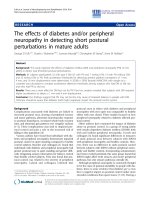
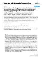
The HU177 level was greater than the mean HU177
level of the cohort (6.2 ng/ml) in 59 pat ients (28%) and
greater than the median concentration (3.7 ng/ml) in
106 p atients (51%) (Figure 1). Because the distribution
of HU177 levels was positively skewed, we analyzed the
data using the median in addition to the mean. Analyses
based o n both mean and median HU177 concentration
are provided to allow for a comparison of the two dis-
tinct cut points. However, the use of the median HU177
value as a categorical cut point is emphasized in our
results.
Table 1 Baseline characteristics of 209 primary
melanoma patients
Variable Patients (n = 209)
Number (%)
Gender
Male 124 (59.3)
Female 85 (40.7)
Age (years)
Mean ± SD; Median 58.3 ± 16.9; 58
Primary tumor histologic subtype
Superficial spreading melanoma 123 (58.9)
Nodular melanoma 52 (24.9)
Acral lentiginous melanoma 6 (2.9)
Desmoplastic melanoma 6 (2.9)
Lentigo maligna melanoma 7 (3.3)
Other melanoma 10 (4.8)
Unknown 5 (2.4)
Thickness (mm)
Mean ± SD; Median 2.09 ± 3.83; 0.95
Ulceration
Absent 169 (80.9)
Present 35 (16.7)
Unknown 5 (2.4)
AJCC stage
Stage I 136 (65.0)
Stage II 41 (20.0)
Stage III 32 (15.0)
Abbreviations used: SD, standard deviation; AJCC, American Joint Committee
on Cance r.
Table 2 Recurrences, deaths, and median HU177 levels
by stage
Stage Recurrences Deaths Median HU177 (ng/ml)
I (n = 136) 9 (7%) 9 (7%) 3.66
II (n = 41) 11 (27%) 10 (24%) 3.91
III (n = 32) 18 (56%) 15 (47%) 3.89
Hamilton et al. Journal of Translational Medicine 2010, 8:19
/>Page 3 of 9
Elevated HU177 concentration is associated with
increased melanoma recurrence
HU177 sera concentration greater than the median
(3.7 ng/ml) was associated with a higher recurrence rate
compared to HU177 sera concentration less than or
equal to the median (23.6% vs. 12.6%; p = 0.04). This
association remained statistically significant when the
mean (6.2 ng/ml) was used to dichotomize the HU177
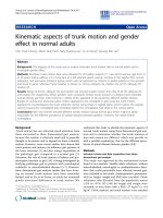
distribution (27.1% vs. 14.7%; p = 0.04). Kaplan-Meier
survival analysis demonstrated improved DFS for
patients with HU177 sera concentration less than or
equal to the median compared to patients with sera
concentration greater than the median (p = 0.04 by
log-rank test) (Figure 2).
Elevated HU177 concentration is associated with
increasing mortality
HU177 sera concentration greater than the median (3.7
ng/ml ) was associated with a higher mortality rate com-
pared to HU177 sera concentration less than or equal to
the median (22.6% vs. 9.7%; p = 0.01). The observed
association remained statistically significant when the
mean HU177 level was used to dichotomize the HU177
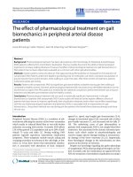
distribution (28.8% vs. 11.3%; p = 0 .002). Kaplan-Meier
survival analysis demonstrated improved OS for patients
with HU177 sera concentration less than or equal to
the median c ompared to patients with sera concentra-
tion greater than the median (p = 0.01 by log-rank test)
(Figure 3).
HU177 concentration is associated with disease-free and
overall survival after adjustment for tumor thickness and
histologic subtype
Because the number of recurrences in the cohort was
relatively low (n = 38), the mo st balanced multivariate
model included 3 variables inclusive of the epito pe
concentration. Variables t hat were most strongly corre-
lated with epitope concentration in the univariate ana-
lyses (histologic subtype a nd thickness) were included
in the multivariate model. High levels of HU177
remained an independent prognostic fac tor for DFS
and OS when controlling for tumor thickness and for
10 20 30 40 50
HU177 concentration (ng/ml)
Number of Patients
0
20
40
60
80
0
Figure 1 Histogram of HU177 sera concentration in 209 patients with primary melanoma. Median = 3.7 ng/ml, Mean = 6.2 ng/ml, SD =
11.5 ng/ml, Min = 0.003 ng/ml, Max = 139.8 ng/ml. One patient with a HU177 concentration of 139.8 ng/ml is not shown.
Hamilton et al. Journal of Translational Medicine 2010, 8:19
/>Page 4 of 9
histologic subtype. In the DFS hazard model control-
ling for tumor thickness and histology, the hazard
ratio for HU177 >3.7 ng/ml (the median) was 2.01
(95% CI = 1.002, 4.04; p = 0.049) (Table 3). In the OS
hazard model controlling for tumor thickness and his-
tology, the hazard ratio f or HU177 >3.7 ng/ml (the
median) was 2.23 (95% CI = 1.06, 4.70; p = 0.035)
(Table 3). The proportional hazards assumption was
not violated for any of the predictor variables in the
DFS and OS models.
If ulceration is included in the multivariate model
(instead of histologic subtype), the independent prog-
nostic value of HU177 level r emains statistically signifi-
cant (DFS, p = 0.048; OS, p = 0.048), and tumor
thickness loses its predictive significance (DFS, p =
0.257; OS, p = 0.199) (not s hown). This suggests that
the variables are collinear and thus only one should be
added to the model. Because thickness was more closely
associated with epitope concentration than ulceration i n
the univariate analysis, it was entered into the multivari-
ate model along with histologic subtype.
Regarding the impact of sentinel lymph node (SLN)
data, only 100/209 (48%) patients had S LN biopsies per-
formed, thus its influence on survival could only be
meaningfully assessed on a univariate analysis. A subset
analysis, however, of the 100 patients who underwent
SLN biopsy showed that SLN status was a significant
predictor of both DFS (HR 3.73, 95% CI = 1.75-7.94;
p = 0.000 6) an d OS (HR 2.58, 95% CI = 1.00-6.68;
p = 0.05) on univariate analysis.
Discussion
Our result s sugg est that pro-angiogenic crypt ic collagen
epitopeHU177mayhaveprognosticsignificanceasa
biomarker of poor outcome in primary melanoma.
Higher levels of HU177 were associated with an
increased rate of recurrence and increasing mortality.
Clinical decision making in the care of melanoma
patients is based primarily on tumor morphology as
thickness and ulceration consistent ly prove to be the
most accurate predictors of survival [11]. Sentinel lymph
node biopsy has been shown to be predictive of
106 81 58 0
103 68 21 1
Number at risk
0 20 40 60 80
Time (months)
3.7 ng/ml >3.7 ng/ml
Disease-Free Survival
n=103, 13 recurrences
n=106, 25 recurrences
1.00
0.75
0.50
0.25
0.00
p=0.04 by log-rank test
85
19
>3.7 ng/ml
3.7 ng/ml
Figure 2 Kaplan-Meier analysis for dise ase-free survival by median epitope concentration. Patients with elevated HU177 concentrations
above the median value demonstrated a reduced disease-free survival probability compared to patients with HU177 concentrations below the
median (HU177 >3.7 ng/ml: n = 106 patients, 25 recurrences; HU177 = 3.7 ng/ml: n = 103 patients, 13 recurrences; p = 0.04 by log-rank test).
Hamilton et al. Journal of Translational Medicine 2010, 8:19
/>Page 5 of 9
recurrence, but it is typically only considered standard
of care for patients with intermediate thickness lesions.
Our previously reported meta-analysis demonstrated
that few patients with thin melanoma have a positive
SLN, and there are no clinical or histopathologic criteria
that can reliably identify thin melanoma patients who
might benefit from this intervention [12]. As reflected in
our cohort in which 51% of patients have melanomas <1
mm thick, trends in downward stage migration mean
that a larger percentage of newly diagnosed melanoma
patients will not be considered for SLN biopsy but
could nonetheless benefit from non-invasive serologic
prognostic markers.
A number of sera markers have been evaluated for
their prognostic significance in primary melanoma with
limited success. For example, angiogenic factors vascular
endothelial growth factor (VEGF), basic fibroblast
growth factor (bFGF), interleukin-8 (IL-8), and
106 89 70 0
>3.7 ng/ml
103 90 73 26
3.7 ng/ml
Number at risk
Time (months)
p=0.01 by log-rank test
Overall Survival
n=103, 10 deaths
n=106, 24 deaths
1.00
0.75
0.50
0.25
0.00
3.7 ng/ml >3.7 ng/ml
020406080
23
1
Figure 3 Kaplan-Meier analysis for overall survival by med ian epitope concentration. Patients with elevated HU177 concentrations above
the median value demonstrated a reduced overall survival probability compared to patients with HU177 concentrations below the median
(HU177 >3.7 ng/ml: n = 106 patients, 24 deaths; HU177 = 3.7 ng/ml: n = 103 patients, 10 deaths; p = 0.01 by log-rank test).
Table 3 Association between HU177 concentration and
DFS/OS, controlling for tumor thickness and histologic
subtype
Variable P-value Hazard ratio (95% CI)
Disease-free survival
Epitope concentration
a
0.049 2.01 (1.002, 4.04)
Tumor thickness
b
0.065 1.05 (1.00, 1.10)
Histology
c
0.001 3.66 (1.70, 7.86)
Overall survival
Epitope concentration
a
0.035 2.23 (1.06, 4.70)
Tumor thickness
b
0.004 1.08 (1.02, 1.13)
Histology
c
0.363 1.41 (0.67, 2.98)
Abbreviations used: DFS, disease-free survival; OS, overall survival; CI,
confidence interval.
a
>3.7 ng/ml vs. ≤ 3.7 ng/ml (referent).
b
Treated as a continuous variable in the model.
c
Nodular/other melanoma vs. superficial spreading melanoma (referent).
Hamilton et al. Journal of Translational Medicine 2010, 8:19
/>Page 6 of 9
ang iogenin have been studied for their value in predict-
ing outcome. One study reported that elevated concen-
trations of VEGF independently correlated with poor
overall survival [13]. The results, however, have not
been replicated by other investigators [14,15]. Similarly,
IL-8 and bFGF were found to be independent predictors
of overall survival [13], but additional studies to validate
their fi ndings are pending. Angiogenin showed less pro-
mise: serum levels were not found to correlate with out-
come [13]. Other candidates such as S-100 beta, a well-
established diagnostic marker for melanoma by immu-
nohistochemistry, have been found to have limited prog-
nostic relevance in early stage melanoma [16].
A serum-based marker of aggressive biology such as
HU177 has the potential to identify primary melanoma
patients at high ris k for the development of distant
metastases who s hould be treated in the post-surgical
adjuvant period. Even if the appropriate risk stratifica-
tion tools were developed, however, current data suggest
that adjuvant therapy with interferon fails to confer a
survival advantage [17]. Thus, it is im perative that the
development of prognostic biomarkers and the develop-
ment of novel molecularly targete d therapy occur simul-
taneously. Our results showing a correlation between
pro-angiogenic collagen epitope HU177 and worse over-
all survival suggest that targeting angiogenesis in the
post-surgical adjuvant period may be a rational
approach for patients with primary melanoma. A shift in
the balance between pro- and anti-angiogenic peptides
towardsangiogenesispromotesneovascularization,
which is essential for tumor progression among other
processes. Angiogenesis has been successfully targeted
in other malignancies, resulting in the FDA approval of
anti-VEGF agent bevacizumab for use as combination
therapy in the treatment of metastatic colorectal and
non-small cell lung cancer [18, 19]. The ut ility of anti-
angiogenic therapy in melanoma , however , has not been
clearly defined. Since metastatic melanoma has a poor
prognosis, anti-angiogenic treatments would delay mela-
noma progression and have a great impact on cancer-
specific mortality. We have already shown the potential
utility o f HU177 in pro gnosis but it may also serve as a
therapeutic target, similar to bevacizumab but with its
effect prior to metastasis. Metastasis requires changes in
the vascular basement membrane, of which type IV col-
lagen is a part. Both the pro-angiogenic factor HU177
and the angiogenesis inhibitor tumstatin are type IV col-
lagen cleavage pro ducts. Disruption of this balance
between pro- and anti-angiogenic peptides promotes
neov ascularization. Treatm ents targeting pro-angiogenic
factors, such as HU177, appear to be more c linically
relevant. A recent study demonstrated that tumstatin
slows tumor growth in renal cell carcinoma and colorec-
tal cancer cell lines, but all tumors eventually escaped
tumstatin-induced growth inhibition and entered into
an exponential growth phase. This rapid growth was
shown to result from an up-regulation of genes encod-
ing pro-angiogenic peptides, possibly in response to
hypoxic conditions. Genes encoding anti-angiogenic fa c-
tors were not silenced [20]. Another study investigating
carboplatin/paclitaxel/bevacizu mab combination therapy
in stage IV melanoma demonstr ated that the addition of
bevacizumab w as well tolerated and the median overall
survival was higher than in previous reports of single
agent treatment with dacarbazine (52 weeks vs. 25.6
weeks) [21]. Although limited conclusions can be drawn
from this uncontrolled trial, the results do suggest that
targeting angio genesis, in particular pro-angiogenic fac-
tors, as part of a combination chemotherapy regimen
may be a useful strategy.
The association between pro-angiogenic HU177 and
poor prognosis in our study is consistent with other
serum biomarker studies that have identified VEGF and
serum angiopoietin-2 (sAng-2) as useful predictors of
response to therapy. In a study of 59 patients with meta-
static melanoma o r renal c ell carcinoma receiving high
dose recombinant interleukin-2 (IL-2), serum was col-
lected and analyzed for potential biomarkers of response
using a customized protein array platform. Serum VEGF
and fibronectin were shown to be independently predic-
tive of response to IL-2 [22]. Another serum bio marker
study of 98 patients with stage I-IV melanoma identified
an increase in sAng-2 levels by 50-400% in 90% of
patients during progression from stage III to IV mela-
noma leading authors to conclude that sAng-2 levels are
associated with disease progression in metastatic mela-
noma [23]. Both of these studies, however, are focused
on biomarkers of advanced disease. A notable advantage
of our study is that 65% of patients included had stage I
melanoma, and the level of HU177 shedding in the
serum was predictive of decreased overall surviv al inde-
pendent of tumor thickness. Because HU177 has poten-
tial as a biomarker that can be utilized early in the
disease course, there is perhaps a g reater chance that it
will influence the cli nical decisions that alter the disease
course and ultimately impact outcomes.
Our f indings emphasize the role of interactions with
the cellular microenv ironment as potential targets for
therapy and bioma rker development. A key limitation of
current in vitro and in vi vo models is that they often
overlook the contribution of the ECM and the tumor
microenvironment toward the initiation and progress ion
of tumorigenesis. Increasing evidence, however, supports
the notion that melanoma cells interact with the adja-
cent microenvironment in a bi-directional manner
through molecular signals that c an modulate the malig-
nant phenotype [24]. Previous in vivo studies of HU177
demonstrated that cleavage of type IV collagen during
Hamilton et al. Journal of Translational Medicine 2010, 8:19
/>Page 7 of 9
ECM remodeling led to exposure of cryptic regulatory
sites, such as HU177, and that an antibody directed at
the HU177 cryptic site inhibited cell adhesion, migra-
tion, and proliferation on denatured collagen type IV
[25]. It is thought that the HU177 measured in sera is
shed not from the tumor but from the tumor microen-
vironment. Thus, while current efforts to target VEGF
and other pro-angiogenic factors whose expression is
regulated by the melanoma cell have thus far been
unsuccessful, our approach focus ed on non-cellular epi-
topes as new targets for biomarkers and treatment is
novel and highly selective. Preliminary data from pre-
clinical trials demonstrate that anti-HU177 mAB
TRC093 significantly enhances the anti-tumor activity of
bevacizumab in a melanoma mouse xenograft model
demonstrating the potential utility of monitoring HU177
as part of an anti-angiogenic therapeutic strategy [26].
We demonstrate that HU177 levels are associated with
worse outcome independent of tumor thickness. These
results emphasize that, while the shedding of HU177 is
associated with tumor remodeling and invasion, it is not
merely a surrogate read-out of thickness. In the multi-
variate model, although the p-value for tumor thickness
is lower than that for epitope concentration, the hazard
ratio for the epitope concentration is more than double
that of thickness (Table 3). Because thic kness was an a-
lyzed as a continuous variable and HU177 epitope con-
centration was evaluated as a categorical variable (high
vs.low),atruecomparisonbetweenthestrengthof
these two prognostic factors cannot be undertaken. The
analysis demonstrates, however, that HU177 maintains
its prognostic value independent of well-characterized
prognostic variables that constitu te the current standard
of care.
Conclusions
High levels of cryptic collagen epitope HU177 are asso-
ciated with higher recurrence rates and increasing mor-
tality. HU177 shows promise as a serum biomarker that
is reflective of melanoma biology, that can be easily inte-
grated into the clinical management of melano ma
patients, and which may have potential a s a molecular
target for adjuvant therapy. These data justify further
validation studies in a larger, independent cohort.
Acknowledgements
Written informed consent was obtained from all patients at the time of
enrollment. Study findings were, in part, supported by the National Institute
of Health (2R01CA91645, Brooks), the Chemotherapy Foundation (Osman),
Varian Medical Systems, Inc. (Liebes), the NYU Cancer Institute Cancer Center
Support Grant (5P30CA016087-27, Osman and Liebes), and the Marc Jacobs
Campaign to support the Interdisciplinary Melanoma Cooperative Group.
Author details
1
Department of Dermatology, New York University School of Medicine, New
York, NY, USA.
2
Division of Biostatistics and Epidemiology, Weill Medical
College of Cornell University, New York, NY, USA.
3
Department of Surgery,
New York University School of Medicine, New York, NY, USA.
4
Department of
Medicine, New York University School of Medicine, New York, NY, USA.
5
Departments of Radiation Oncology and Cell Biology, New York University
School of Medicine, New York, NY, USA.
6
Maine Medical Center Research
Institute, Center for Molecular Medicine, 81 Research Drive Scarborough, ME
04074, USA.
Authors’ contributions
HKH participated in study design, coordination, data collection, data analysis,
and drafting of the manuscript. AER participated in data collection, analysis,
drafting, and finalizing the manuscript text. PJC performed the statistical
design and analysis under the guidance of MM. RLS recruited patients for
the study, provided input on study design, and helped to draft the
manuscript. RSB recruited patients for the study, provided input on study
design, and helped to draft the manuscript. MM was the principal statistician
for the study, providing guidance on statistical design and data analysis.
MWM participated in data analysis and manuscript drafting/finalization. DK
collected and helped analyze clinical data extracted from the melanoma
database. LL participated in the conceptual study design and provided
guidance regarding interpretation of results. PCB provided guidance in study
design and interpretation of results. IO served as the principal investigator
for the project, overseeing the study design, analysis of data, interpretation
of results, and writing of the manuscript. All authors read and approved the
final manuscript.
Competing interests
PCB serves as a consultant to TRACON Pharma and has received honoraria
from TRACON Pharma in the last two years.
Received: 13 November 2009
Accepted: 23 February 2010 Published: 23 February 2010
References
1. Kalady MF, White RR, Johnson JL, Tyler DS, Seigler HF: Thin melanomas:
predictive lethal characteristics from a 30-year clinical experience. Ann
Surg 2003, 238(4):528-535.
2. Gimotty PA, Elder DE, Fraker DL, Botbyl J, Sellers K, Elenitsas R, Ming ME,
Schuchter L, Spitz FR, Czerniecki BJ, Guerry D: Identification of high-risk
patients among those diagnosed with thin cutaneous melanomas. J Clin
Oncol 2007, 25(9):1129-1134.
3. Gould Rothberg BE, Bracken MB, Rimm DL: Tissue biomarkers for
prognosis in cutaneous melanoma: a systematic review and meta-
analysis. J Natl Cancer Inst 2009, 101(7):452-474.
4. Winnepenninckx V, Lazar V, Michiels S, Dessen P, Stas M, Alonso SR,
Avril MF, Ortiz Romero PL, Robert T, Balacescu O, Eggermont AM, Lenoir G,
Sarasin A, Tursz T, Oord van den JJ, Spatz A, Melanoma Group of the
European Organization for Research and Treatment of Cancer: Gene
expression profiling of primary cutaneous melanoma and clinical
outcome. J Natl Cancer Inst 2006, 98(7):472-482.
5. Florell SR, Smoller BR, Boucher KM, Grossman D, Harris RM, Bowen GM,
Leachman SA: Sampling of melanocytic nevi for research purposes: a
prospective, pilot study to determine effect on diagnosis. J Am Acad
Dermatol 2008, 59(5):814-821.
6. Becker D, Mihm MC, Hewitt SM, Sondak VK, Fountain JW, Thurin M:
Markers and tissue resources for melanoma: meeting report. Cancer Res
2006, 66(22):10652-10657.
7. Schittny JC, Yurchenco PD: Basement membranes: molecular organization
and function in development and disease. Curr Opin Cell Biol 1989,
1(5):983-988.
8. Sternlicht MD, Werb Z: How matrix metalloproteinases regulate cell
behavior. Annu Rev Cell Dev Biol 2001, 17:463-516.
9. Xu J, Rodriguez D, Petitclerc E, Kim JJ, Hangai M, Moon YS, Davis GE,
Brooks PC: Proteolytic exposure of a cryptic site within collagen type IV
is required for angiogenesis and tumor growth in vivo. J Cell Biol 2001,
154(5):1069-1079.
10. Ng B, Zakrzewski J, Warycha M, Christos PJ, Bajorin DF, Shapiro RL,
Berman RS, Pavlick AC, Polsky D, Mazumdar M, Montgomery A, Liebes L,
Brooks PC, Osman I: Shedding of distinct cryptic collagen epitope
(HU177) in sera of melanoma patients. Clin Cancer Res 2008,
14(19):6253-6258.
Hamilton et al. Journal of Translational Medicine 2010, 8:19
/>Page 8 of 9
11. Balch CM, Soong SJ, Gershenwald JE, Thompson JF, Reintgen DS,
Cascinelli N, Urist M, McMasters KM, Ross MI, Kirkwood JM, Atkins MB,
Thompson JA, Coit DG, Byrd D, Desmond R, Zhang Y, Liu PY, Lyman GH,
Morabito A: Prognostic factors analysis of 17,600 melanoma patients:
validation of the American Joint Committee on Cancer melanoma
staging system. J Clin Oncol 2001, 19(16):3622-3634.
12. Warycha MA, Christos PJ, Mazumdar M, Darvishian F, Shapiro RL,
Berman RS, Pavlick AC, Kopf AW, Polsky D, Osman I: Changes in the
presentation of nodular and superficial spreading melanomas over 35
years. Cancer 2008, 113(12):3341-3348.
13. Ugurel S, Rappl G, Tilgen W, Reinhold U: Increased serum concentration of
angiogenic factors in malignant melanoma patients correlates with
tumor progression and survival. J Clin Oncol 2001, 19(2):577-583.
14. Osella-Abate S, Quaglino P, Savoia P, Leporati C, Comessatti A,
Bernengo MG: VEGF-165 serum levels and tyrosinase expression in
melanoma patients: correlation with the clinical course. Melanoma Res
2002, 12(4):325-334.
15. Ascierto PA, Leonardi E, Ottaiano A, Napolitano M, Scala S, Castello G:
Prognostic value of serum VEGF in melanoma patients: a pilot study.
Anticancer Res 2004, 24(6):4255-4258.
16. Utikal J, Schadendorf D, Ugurel S: Serologic and immunohistochemical
prognostic biomarkers of cutaneous malignancies. Arch Dermatol Res
2007, 298(10):469-477.
17. Kirkwood JM, Manola J, Ibrahim J, Sondak V, Ernstoff MS, Rao U: A pooled
analysis of eastern cooperative oncology group and intergroup trials of
adjuvant high-dose interferon for melanoma. Clin Cancer Res 2004,
10(5):1670-1677.
18. Hurwitz H, Fehrenbacher L, Novotny W, Cartwright T, Hainsworth J,
Heim W, Berlin J, Baron A, Griffing S, Holmgren E, Ferrara N, Fyfe G,
Rogers B, Ross R, Kabbinavar F: Bevacizumab plus irinotecan, fluorouracil,
and leucovorin for metastatic colorectal cancer. N Engl J Med 2004,
350(23):2335-2242.
19. Sandler A, Gray R, Perry MC, Brahmer J, Schiller JH, Dowlati A, Lilenbaum R,
Johnson PH: Paclitaxel-carboplatin alone or with bevacizumab for non-
small-cell lung cancer. N Engl J Med 2006, 355(24):2542-2550.
20. Fernando NT, Koch M, Rothrock C, Gollogly LK, D’Amore PA, Ryeom S,
Yoon SS: Tumor escape from endogenous, extracellular matrix-associated
angiogenesis inhibitors by up-regulation of multiple proangiogenic
factors. Clin Cancer Res 2008, 14(5):1529-1539.
21. Perez DG, Suman V J, Fitch TR, Amatruda T, Morton RF, Jilani SZ,
Constantinou CL, Egner JR, Kottschade LA, Markovic SN: Phase 2 trial of
carboplatin, weekly paclitaxel, and biweekly bevacizumab in patients
with unresectable stage IV melanoma: a North Central Cancer Treatment
Group study, N047A. Cancer 2009, 115(1):119-127.
22. Sabatino M, Kim-Schulze S, Panelli MC, Stroncek D, Wang E, Taback B,
Kim DW, Deraffele G, Pos Z, Marincola FM, Kaufman HL: Serum vascular
endothelial growth factor and fibronectin predict clinical response to
high-dose interleukin-2 therapy. J Clin Oncol 2009, 27(16):2645-2652.
23. Helfrich I, Edler L, Sucker A, Thomas M, Christian S, Schadendorf D,
Augustin HG: Angiopoietin-2 levels are associated with disease
progression in metastatic malignant melanoma.
Clin Cancer Res 2009,
15(4):1384-1392.
24. Stevens AP, Spangler B, Wallner S, Kreutz M, Dettmer K, Oefner PJ,
Bosserhoff AK: Direct and tumor microenvironment mediated influences
of 5’-deoxy-5’-(methylthio)adenosine on tumor progression of malignant
melanoma. J Cell Biochem 2009, 106(2):210-219.
25. Cretu A, Roth JM, Caunt M, Akalu A, Policarpio D, Formenti S, Gagne P,
Liebes L, Brooks PC: Disruption of endothelial cell interactions with the
novel HU177 cryptic collagen epitope inhibits angiogenesis. Clin Cancer
Res 2007, 13(10):3068-3078.
26. Roth JM, Akalu A, Vary C: Targeting the HU177 cryptic collagen epitope
with humanized antibody TRC093 functions cooperatively with anti-
VEGF therapy to inhibit tumor growth [abstract]. Proceedings of the 100th
Annual Meeting of the American Association for Cancer Research 2009, , 1:
317.
doi:10.1186/1479-5876-8-19
Cite this article as: Hamilton et al.: Increased shedding of HU177
correlates with worse prognos is in primary melanoma. Journal of
Translational Medicine 2010 8:19.
Submit your next manuscript to BioMed Central
and take full advantage of:
• Convenient online submission
• Thorough peer review
• No space constraints or color figure charges
• Immediate publication on acceptance
• Inclusion in PubMed, CAS, Scopus and Google Scholar
• Research which is freely available for redistribution
Submit your manuscript at
www.biomedcentral.com/submit
Hamilton et al. Journal of Translational Medicine 2010, 8:19
/>Page 9 of 9