Báo cáo hóa học: "Microrna profiling analysis of differences between the melanoma of young adults and older adults" pot
Bạn đang xem bản rút gọn của tài liệu. Xem và tải ngay bản đầy đủ của tài liệu tại đây (1.3 MB, 23 trang )
RESEARC H Open Access
Microrna profiling analysis of differences between
the melanoma of young adults and older adults
Drazen M Jukic
1,2†
, Uma NM Rao
2
, Lori Kelly
2†
, Jihad S Skaf
3
, Laura M Drogowski
1
, John M Kirkwood
4
,
Monica C Panelli
4*
Abstract
Background: This study represents the first attempt to perform a profiling analysis of the intergenerational
differences in the microRNAs (miRNAs) of primary cutaneous melanocytic neoplasms in young adult and older age
groups. The data emphasize the importance of these master regulators in the transcriptional machinery of
melanocytic neoplasms and suggest that differential levels of expressions of these miRs may contribute to
differences in phenotypic and pathologic presentation of melanocytic neoplasms at different ages.
Methods: An exploratory miRNA analysis of 666 miRs by low density microRNA arrays was conducted on formalin
fixed and paraffin embedde d tissues (FFPE) from 10 older adults and 10 young adults including conventional
melanoma and melanocytic neoplasms of uncertain biological significance. Age-matched benign melanocytic nevi
were used as controls.
Results: Primary melanoma in patients greater than 60 years old was characterized by the increased expression of
miRs regulating TLR-MyD88-NF-kappaB pathway (hsa-miR-199a), RAS/RAB22A pathway (hsa-miR-204); growth
differentiation and migration (hsa-miR337), epithelial mesenchymal transition (EMT) (let-7b, hsa-miR-10b/10b*),
invasion and metastasis (hsa-miR-10b/10b*), hsa-miR-30a/e*, hsa-miR-29c*; cellular matrix components (hsa-miR-
29c*); invasion-cytokinesis (hsa-miR-99b*) compared to melanoma of younger patients. MiR-211 was dramatically
downregulated compared to nevi controls, decreased with increasing age and was among the miRs linked to
metastatic processes. Melanoma in young adult patients had increased expression of hsa-miR-449a and decreased
expression of hsa-miR-146b, hsa-miR-214*. MiR-30a* in clinical stages I-II adult and pediatric melanoma could
predict classification of melanoma tissue in the two extremes of age groups. Although the number of cases is
small, positive lymph node status in the two age groups was characterized by the statistically significant expression
of hsa-miR-30a* and hsa-miR-204 (F-test, p-value < 0.001).
Conclusions: Our findings, although preliminary, support the notion that the differential biology of melanoma at
the extremes of age is driven, in part, by deregulation of microRNA expression and by fine tuning of miRs that are
already known to regulate cell cycle, inflammation, Epithelial-Mesenchymal Transition (EMT)/stroma and more
specifically genes known to be altered in melanoma. Our analysis reveals that miR expression differences create
unique patterns of frequently affected biological processes that clearly distinguish old age from young age
melanomas. This is a novel characterization of the miRnomes of melanocytic neoplasms at two extremes of age
and identifies potential diagnostic and clinico-pathologic biomarkers that may serve as novel miR-based targeted
modalities in melanoma diagnosis and treatment.
* Correspondence:
† Contributed equally
4
University of Pittsburgh Cancer Institute, Division of Hematology-Oncology
Hillman Cancer Center, Pittsburgh, Pennsylvania, USA
Jukic et al. Journal of Translational Medicine 2010, 8:27
/>© 2010 J ukic et al; li censee BioMed Central Ltd. This is an Open Access article distributed under the terms of the Creative Commons
Attribution License ( which permits unrestricted use, distribution, and reproduction in
any medium, provide d the origin al work is properly cited.
Background
The incidence of melanoma dramatically increases with
age, and accounts for 7% of all malignancies seen in
patients between the ages of 15-29 years [1,2]. Despite
thefactthatalmost450newpatientswithmelanoma
under the age of 20 are diagnosed with melanoma each
year in the U nited States, published reports of this dis-
ease in young people have usually been restricted in
number and often constitute series from single institu-
tions. Two recently published large studies from the
Surveillance Epidemiology and End Results (SEER) and
National Cancer Database (NCDB) dat abases confir med
and expanded previous observations that pediatric/
young adult melanoma ma y be clinically similar to adult
melanoma; howe ver some differences in clinical presen-
tation and outcome such as the higher incidence of
nodal metastases in children and adolescents with
localized dise ase are evident, particularly in younger
patients [1-6].
The outcome of melanoma in the younger, as com-
pared to the older, populations has been shown to differ
quite substantially. In the young adult and pediatric
population the issue is complicated because of inability
even amongst experts to identify conventional melano-
mas from certain melanocytic neoplasms of uncertain
biologic behavior because of subtle overlapping histo-
morphological features. Notably in Spitzoid nevi, this
subject has been debated since the entity was first
described by Sophie Spitz in 1948 [7] beca use some of
these neoplasm have metastasized to regional lymph
nodes [8,9]. It has also been recently suggested that the
Spitzoid melanocytic neoplasms with nodal metastases
mayhaveabetterprognosisinyoung/pediatricage
group [10]. In many of the cases, these lesions have
been treated as malignant melanomas [11].
The aim of this study was to identify the differences
between melanoma in young and o lder adult popula-
tions with the ultimate goal of finding useful biomarkers
of etiology and outcome at different ages. Therefore we
have included some of the Spitzoid melanocytic neo-
plasms (as a part of the group of patients age less than
30 years old/Mel 30) that have documented sentinel
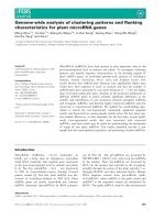
lymph node metastases. (Figure 1).
As Chen summarized [12], the use of DNA microar-
rays to monitor tumor RNA profiles has defined a mole-
cular taxonomy of cancer, which can be used to identify
new drugs and better define prognosis, with the ultimate
potential to predict patterns of drug resistance. Cellular
behavior is also governed by transla tional and posttr an-
slational control mechanisms that are not reflected in
mRNA profiles of tumor specimens. Since microRNAs
regulate gene expression at the post-transcriptional
level, the availability of a comprehensive microRNA
(miRNAs/miR) expression profile can provide informa-
tion that is complementary to that derived from mRNA
transcriptional profiling. Thus, comprehensive micro-
RNA expression profiling can help to unravel these mas-
ter regulators of gene expression, which represent a
Figure 1 Atypical Spitz. Example of atypical Spitz neoplasm of uncertain biological significance.
Jukic et al. Journal of Translational Medicine 2010, 8:27
/>Page 2 of 23
pivotal regulatory network in the t ranscriptional cell
machinery and have been associated with deregulation
of immune and cell cycle processes in cancer [13].
MiRNAs are a family of endogenous, small (18-25
nucleotides in length), noncoding, functional RNAs. It is
estimated that there may be 1000 miRNA genes in the
human genome (Internet address: g er.a c.
uk/Software/Rfam/mirna/). The latest update of miR-
Base (Internet address: rele ase 13 Ma rch 2009, h ttp://
microrna.sanger.ac.uk/sequences/index.shtml) includes
more than 1900 annotated miR sequences.
MiRNAs are transcribed by RNA polymerase II or III
as longer primary-miRNA molecules, which are subse-
quently processed in the nucleus by the RNase III endo-
nuclease Drosha and DGCR8 (the “ microprocessor
complex” ) to form approximately 7 0 nucleotide-long
intermediate stem-loop structures called “ precursor
miRNAs” (pre-miRNAs). These pre-miRNAs are trans-
ported from the nucleus to the cytoplasm, where they
are further processed by the endonuclease Dicer. Dicer
produces an imperfect duplex composed of the mature
miRNA sequence and a fragment of similar size
(miRNA*), which is derived from the opposing arm of
the pre-miRNA [14].
Only the mature-miRNA remains stable on the RNA-
induced silencing complex (R ISC) and induces post-
transcriptional silencing of one or more target genes by
binding with imperfect complementarity to a target
sequence in the 3’ -UTR of the target RNA with respect
to a set of general rules that are only incompletely
deter mined experimentally and bioinformatically to date
[15]. Identification of miRNA targets has been difficult
because only the seed sequence, about 6-8 bases of the
approximately 22 nucleotides, aligns perfectly with the
targe t mRNA’s3’ untranslated region. The remainder of
the miRNA may bind perfectly to the target mRNA, but
more often it does not [14]. RNA interference and
related small RNA mediated pathways are central in the
silencing of gene expression, and at least 30% of human
genes are thought to be regulated by microRNAs [16].
MiRNAs are expressed in a tissue-specific manner, and
can contribute to cancer development and progre ssion.
They are differentially expressed in normal tissues and
both hematological and solid tumors. In human solid
tumors such as hepatocellular carcinoma [17] and ovar-
ian cancer [18], the miRNA expression signature defines
neoplasm-specific dys-regulation of specific gene targets.
Despite the hundreds of miRs discovered to date, their
biological functions are incompletely understood.
Increasing evidence suggests that the expression of miR-
NAs (miRs) is deregulated in many cancers, and miRs
can control cell proliferation, differentiation and apopto-
sis [19]. The alteration of miR expression may contri-
bute to the initiation and manintanance of tumors as
their abnormal levels have important pathogenic conse-
quences: miR overexpression in tumors usually contri-
butes to oncogenesis by downregulating tumor
suppressors. For example, the mir-17-miR 92 cluster
reduces the transcription factor E2F1 in lymphomas and
miR -21 represses the tumor suppressor PTEN in hepa-
tocellular carcinoma. MiRs lost by tumors lead to onco-
gene overexpression (let -7 loss leads to expression of
KRAS, NRAS in lung carcinoma, while miR15a and 16-1
loss leads to expression of BCL-2 i n CLL and cyclinD1
in prostate carcinoma [20].
The significance of microRNA differential modulation
in the diagnostic and progno stic workup of melanocytic
neoplasms, especially in relationship to the age-stratified
groups, has not, to our knowledge, been investigated.
In this article, we present profiling results in regard to
666 microRNAs evaluated in melanocytic neoplasms of
pediatric and young adults compared with o lder adults;
the results of which emphasize the importance of these
master regulators in the transcriptional machinery of
melanocytic neoplasms and support the notion that dif-
ferential levels of expressions of these miRs may contri-
bute to differences in phenotypic and pathologic
presentation of melanocytic neoplasms at different ages.
We performed an exploratory analysis of 666 miR on
formalin-fixed paraffin-embedded (FFPE)-primary mela-
noma tissue using the Taqman ®TLDA miRNA arrays
platform A and B (Appl ied Biosystems, Foster City, CA,
) to investigate
whether there were different ially expressed miRs
between young adult and adult melanoma specimens
(including melanocytic neoplasms of uncertain biological
potential). The comparativeprofilingwaspurposively
conducted at extremes of age, <30 and >60 years, to
clearly define age groups. Our study represents the first
attempt to perform a true intergenerational and com-
parative microRNA profiling of the primary melanocytic
neoplasms of adults and young adults.
We observed distinct miRNA profiles in the primary
melanocytic neoplasms of adults and young adults that
could also potentially be associated with the clinical
parameters of stage and noda l involvement. Our obser-
vations represent an important basis for expanded analy-
sis of the etiology and clinico-pathologic spectrum of
this disease.
Materials and methods
Patient Selection
This study included the utilization of archiv al melanoma
specimens obtaine d and was approved by the University
of Pittsburgh Cancer Institute (UPCI) Internal Review
Board (IRB): UPCI reference IRB#: PRO07120294.
Archival paraffin blocks of melanocytic neoplasms stu-
died at the UPCI were retrieved from the files of the
Jukic et al. Journal of Translational Medicine 2010, 8:27
/>Page 3 of 23
Health Sciences Tissue Bank (HSTB) database and dis-
bursed by UPCI HSTB according to UPCI-IRB regula-
tions. Ten primary FFPE-tissues (including melanocytic
neoplasms of uncertain biological potential) were
obtained from two cohorts of patients respecti vely seg-
regated according to age: Cohort A - > 60 years and
Cohort B - <30 years and util ized for microRNA profil-
ing. These two case cohorts were separated by at least
30 years, thereby representing an adequate basis for an
intergenerational study.
Additionally, 6 benign nevi were used as homologous
controls (3 from adults and 3 from young adult patients,
respectively). A total of 26 lesions (20 test specimens +
6 controls) were analyzed. Primary diagnostic workup
and verification of the diagnosis of primary neoplasms
was performed by two independent reference
pathologists.
Total RNA was isolated from all lesions from (at aver-
age) 30 5 μm sections obtained specifically from areas
that contained at least 70% viable tumor (identifi ed by a
pathologist). RNA quality was assessed using Nanodrop
(OD 260/280 and 260/230 (Table 1)). The overall micro-
RNA profiling of these two groups (adult and young
adult) included a total of 56 Taqman ® microRNA Low
density arrays (TLDAs). Each group included 10 mela-
nocytic neoplasm samples (older adult melanoma, AM,
pediatric and young adult melanoma PM) and 3 control
nevi specimens (adult nevi, AN, pediatric nev i, PN). The
assays were run in 3 batches for processing and a cali-
brator RNA was included in each batch f or normaliza-
tion. For each specimen, 2 TLDA were run, TLDA
panel A and TLDA panel B.
Patient characteristics of specimen groups utilized for
class comparison analyses are summarized in Table 2.
The pediatric and young adult melanoma (PM) speci-
mens were obtained from 5 males and 5 females, and
the 3 control nevi (PN) from 1 male and 2 females.
Patient PM8 had a Spitzoid neoplasm of uncertain
Table 1 Summary Of RNAs Extracted From FFPE Melanoma And Nevus (Control) Specimens Obtained From Pediatric
Or Young Adults < 30 Years Of Age And Older Adults > 60 Years Of Age
Sample ID Sample
Name
FFPE Tissue
Type
Percentage Tumor or
Nevus
Total RNA yield
(ug)
ng/ul
RNA
OD 260/
280
OD 260/
230
TB08-190A PM7 Mel 80% 2.26 251 1.98 2.02
TB08-192 1H PM2 Mel 90% 0.45 50.1 1.79 1.47
TB08-239 B PM3 Mel 80% 0.72 79.61 1.87 1.23
TB09-044B PM6 Mel 75% 2.03 226 1.94 1.59
TB08-243A PM8 Mel 85% 1.85 205 1.94 1.95
TB08-231 A PM4 Mel 75% 0.31 34.97 1.81 1.35
TB08-199D PM11112 Mel 75% 1.24 103 1.9 1.65
TB08-195 2A PM5 Mel 80% 0.17 18.69 1.76 1.23
TB08-245D PM9 Mel 100% 2.37 263 1.94 1.83
TB08-477-
478C
PM10 Mel 90% 4.59 255 1.88 1.72
TB08-242A PN1 Nevus 100% 0.77 85.89 1.86 1.41
TB08-232 2A PN2 Nevus 100% 2.71 226 1.86 1.56
TB08-188A PN3 Nevus 100% 0.30 25 1.84 1.45
TB08-236 1L AM1 Mel 100% 0.93 103.09 1.88 1.6
TB08-180P 1H AM2 Mel 100% 3.23 269 2 1.86
TB08-217 1D AM3 Mel 75% 1.42 158.07 1.97 1.64
TB08-223 C AM10 Mel 70% 0.57 63 1.88 1.72
TB08-181 B AM4 Mel 95% 11.29 941 1.84 1.35
TB08-211 1J AM5 Mel 90% 0.66 55 1.89 1.66
TB08-216 F AM6 Mel 80% 0.46 51.37 1.93 1.59
TB08-219 1G AM9 Mel 75% 0.47 52 1.89 1.86
TB08-237 1G AM7 Mel 70% 1.23 136.28 1.85 1.63
TB09-043B AM8 Mel 90% 2.72 302 1.87 1.17
TB09-003 A AN1 Nevus 100% 0.90 100 1.99 1.71
TB08-233D AN2 Nevus 100% 0.36 30 1.93 1.68
TB08-234A AN3 Nevus 100% 0.12 10.4 1.8 1.22
Top group (PM/PN): young adults <30 yrs old; lower group (AM/AN): adults >60; PM = pediatric and young adult melanoma (<30 yrs); AM = adult melanoma
(>60 yrs);PN = pediatric and young adult nevus (<30 yrs); AN = adult nevus (>60 yrs); % tumor refers to the percentage of tumor in the area that was ID &
scraped for RNA isolation. Quality of RNA was established by Nanodrop OD reading.
Jukic et al. Journal of Translational Medicine 2010, 8:27
/>Page 4 of 23
Table 2 Patients Characteristics
Sample
name
Mel 60/
30 or
Nevus
60/30
Age Age
range
Gender Diagnosis Site T
Stage
N
Stage
M
Stage
Stage Group
at Diagnosis-
AJCC 6th Ed.
PM7 Mel 30 21 20-29 M Melanoma, invasive and insitu, arising in
association with a nevus
Trunk cT1* pN0 cM0 Unknown
PM2 Mel 30 26 20-29 M Superficial spreading melanoma, invasive and in
situ
Back pT1b pN1a cM0 3B
PM3 Mel 30 26 20-29 F Melanoma, superficial spreading in radial growth
phase & vertical, epithelioid, nevoid and balloon
cell
Scapula pT2b pN0 cM0 2A
PM6 Mel 30 28 20-29 F Superficial spreading melanoma, invasive Thigh pT1b pN0 cM0 1B
PM8 Mel 30 28 20-29 M Highly atypical spitzoid neoplasm Arm n/a n/a n/a n/a
PM4 Mel 30 28 20-29 F Superficial spreading melanoma, invasive Shin pT1a pN0 cM0 1A
PM11112 Mel 30 29 20-29 F Superficial spreading (Spitzoid) melanoma, insitu &
invasive
Thigh pT1a pN0 cM0 1A
PM5 Mel 30 29 20-29 M Melanoma in situ (arising in compound
melanocytic nevus)
Abdomen pTis cN0 cM0 0
PM9 Mel 30 29 20-29 F Invasive and in situ melanoma, nodular. Note:
Description of superficial spreading also in
synopsis but registry only codes final diagnoses.
Buttock pT4b pN3 cM1c 4
PM10 Mel 30 29 20-29 M Superficial spreading melanoma, insitu and
invasive
Scalp pT1a cN0 cM0 1A
PN1 Nevus 30 12 10-19 F Compound, predominantly intradermal
melanocytic nevus
Forehead n/a n/a n/a n/a
PN2 Nevus 30 14 10-19 M Compound predominantly intradermal
melanocytic nevus with architectural features of
congenital onset
Scalp n/a n/a n/a n/a
PN3 Nevus 30 26 20-29 F Compound melanocytic nevus with features of a
congenital nevus, architectural disorder and mild
cytologic atypia (aka Clark’s nevus with features of
congenital onset).
Back n/a n/a n/a Unknown
AM1 Mel 60 64 60-69 F Melanoma, invasive, nevoid type. Leg pT2a pN0 cM0 1B
AM2 Mel 60 69 60-69 M Superficial spreading (outside path) and Nevoid
Melanoma, invasive
Ear pT4b pN3 cM0 3C
AM3 Mel 60 69 60-69 M Desmoplastic melanoma, invasive Forehead pT3a pN0 cM0 2A
AM10 Mel 60 72 70-79 M Malignant melanoma in situ arising in a
compound dysplastic nevus
Back pTis cN0 cM0 0
AM4 Mel 60 73 70-79 M Nodular melanoma, invasive and insitu Calf pT4b pN3 cM0 3C
AM5 Mel 60 78 70-79 F Melanoma, insitu and invasive Foot pT2b pN2c cM0 3B
AM6 Mel 60 79 70-79 M Lentingo malignant melanoma in situ with focus
invasive melanoma
Back pT1a cN0 cM0 1A
AM9 Mel 60 79 70-79 M Invasive melanoma (&Melanoma in Situ arising in
a background of dysplastic nevus
Back pT1a cN0 cM0 1A
AM7 Mel 60 82 80-89 F Desmoplastic melanoma with associated
lentiginous component
Arm pT4a pN0 cM0 2B
AM8 Mel 60 86 80-89 M Nodular melanoma (3% in situ) Flank pT2a cN0 cM0 1B
AN1 Nevus 60 62 60-69 F Compound, predominantly intradermal
melanocytic nevus with architectural features of
congenital onset
Back n/a n/a n/a n/a
AN2 Nevus 60 63 60-69 M Compound predominantly intradermal
melanocytic nevus with architectural features of
congenital onset
Flank n/a n/a n/a n/a
AN3 Nevus 60 68 60-69 M Compound melanocytic nevus with moderate
cytological atypia and congenital features.
Deltoid n/a n/a n/a n/a
PM = pediatric and young adult melanoma (<30 yrs);AM = adult melanoma (>60 yrs);PN = pediatric and young adult nevus(<30 yrs); AN = adult nevus(>60 yrs);
Mel 60: adult melanoma (>60 yrs); Mel 30: pediatric and young adult melanoma (<30 yrs); Nevus 60: adult nevus(>60 yrs); Nevus 30: pediatric and young adult
nevus(<30 yrs). TNM Staging:regardless of year of diagnosis, all cases staged according to AJCC 6th Edition. P:pathologic staging; c: clinical staging. * Not able to
stage T further as Clarks level missing in original path report.
Jukic et al. Journal of Translational Medicine 2010, 8:27
/>Page 5 of 23
malignant potential, PM5 was classified as stage 0, 6 PM
patients were classified as Stage I or II (PMs 11112, 3, 4,
6, 7
(Tstage)
, 10), PM2 was classified as Stage III and PM9
as Stage IV.
The adult melanomas (AM) were obtained from 3
female patients and 7 male patient s, the nevi (AN) were
obt ained from 1 female and 2 male patients. AM10 was
classified as stage 0 (AM10), 6 AM patients as Stage I
or II (AM1, 3, 6, 7, 8, 9) and 3 AM patients as Stage III
(AM2, 4, 5).
Two patients PM patients (PM2 and PM9) and 3
patients AM patients (AM2, AM4, AM5) had melanoma
which spread to the lymph nodes.
Taqman® microRNA Low density arrays (TLDA)
The ABI Taqman® microRNA Low density arrays
(TLDA, Applied Biosystems, Foster City, CA, http://
www.appliedbiosystems.com) were selected as the plat-
form for microRNA melanoma profiling (additional file
1). This platform consists of 2 arrays: TLDA panel A
(377 functionally defined microRNAs) and TLDA panel
B (289 microRNAs whose function is not yet completely
defined) for a total of 666 microRNA assays. Each array/
panel includes, among other endogenous controls, the
mammalian U6 (MammU6) assay that is repeated four
times on each card as a positive control as well as an
assay u nrelated to mammalian species, ath-miR159a, as
negative control (Figure 2). This platform represented
the most comprehensive Taqman Low Density Array
(TLDA) for global screening of miRs for which commer-
cially available primer-probe sets existed that were
extensively validated.
Isolation of RNA, Reverse Transcription, Preamplification
and Taqman PCR
Total RNA was isolated from FFPE-tissue utilizing a
modified RecoverALL (Recover All Ambion #AM1975)
protocol for isolation of RNA from paraffin slide sec-
tions. In brief, using a scalpel blade (#15) wetted in
xylene, areas containing >70% tumor were excised from
thirty 5 um paraffin tissue sections and placed in an
microcentrifuge tube containing 1 ml of xylene, vor-
texed and incubated at 50°C for 3 minutes to melt the
paraffin. The material was then centrifuged at 14,000
Figure 2 Engogenous Control Profiles. A: endogenous controls of TLDA panel A profiled a cross all specimens. B: endogenous controls of
TLDA panel B profiled across all specimens. The Mammalian U6 assay was selected for data normalization. Endogenous controls in panel A
included MammU6-4395470, RNU44-4373384, RNU48-4373383. Endogenous control in panel B included MammU6-4395470, RNU44-4373384,
RNU48-4373383, RNU244373379, RNU434373375, RNU6B-4373381
Jukic et al. Journal of Translational Medicine 2010, 8:27
/>Page 6 of 23
rpm for 5-10 min at room temperature. The xylene was
then removed using a 1 ml pipette and the pellet was
washed 3 times with 1 ml of 100% room temperature-
ethanol. The pellet was then air-dried at room tempera-
ture for 15 minutes. Following deparaffinization, tiss ue
was protease digested by incubating the pellet in 400 ul
digestion buffer and 4 ul protease a t 50°C for 3 hours.
For RNA isolation, 480 ul o f isolation additive was
added to the sample, followed by vortexing and addition
of 1.1 ml of 100% ethanol. The mixture was then loaded
onto a prepared filter and collection tube according to
the manufacturer-supplied procedure. Flow through was
discarded and filter washed with wash buffer. Nuclease
digest ion and fi nal RNA purification was carried over as
follows. Sixty ul DNase master mix (containing 6 ul 10×
DNase buffer, 4 ul DNase, 50 ul nuclease free water)
was added to the center of the filter and incubated for
30 minutes at r oom temperature. The filter was subse-
quently washed according to the manufacturer’sproto-
col, and RNA was eluted twice with 30 ul preheated
nuclease-free water. RNA quality and quantity was mea-
sured by Nanodrop technology.
RNA was further purified and concentrated by preci-
pitation for 1 hour at -70°C using 1/10 volume ammo-
nium acetate, 1 ul glycogen (5 ug/ul) an d 2.5 volume
100% ethanol. RNA was then washed, dried and r esus-
pended in 12-15 ul nuclease-free water.
RNA reverse transcription was accomplished accord-
ing to the ABI microRNA TLDA Reverse Transcription
Reaction protocol. In brief, the Megaplex RT Primers,
TaqMan® MicroRNA Reverse Transcription Kit compo-
nents and MgCl
2
were thawed on ice. Two master
mixes per specim en, one for each TLDA panel (panel A
and panel B) consisting of 0.80 ul MegaPlex RT primers
(10×), 0.20 ul dNTPs with dTTP (100 mM), 1.50 ul
MultiScribe™ ReverseTranscriptase (50 U/μL), 0.80 ul
10 × RT Buf fer, 0.90 ul MgCl
2
(25mM),0.10ulRNase
Inhibitor, 0.20 ul nuclease- free water (20 U/μL) were
prepared. Three μL (30 ng) total RNA (or 3 uL of water
for the No Template Control reactions) were loaded
into appropriate wells of a 96-well plate containing
4.5 uL RT reaction mix and incubated on ice for 5 min.
The following thermal cycling conditions were used in
the ABI 9700 thermal cycler: standard or max ramp
speed, 16°C 2 min, 42°C 1 min 40 cycles, 50°C 1 sec,
hold 85°C 5 min, hold 4°C.
The cDNA product (2.5 ul per specimen) was pream-
plified according to the ABI TLDA preamplification pro-
tocol. A total of 22.5 ul of pre-amplification reaction
mix consisting of 12.5 ul TaqMan® PreAmp Master Mix
(2×); 2.5 ul Megaplex™ PreAmp Primers (10×); 7.5 ul
nuclease-free water was pre pared and ad ded to the
cDNA product in a 96-well optical plate sealed with
MicroAmp® Clear Adhesive Film (ABI PN #4306311).
The plate was spun briefly and incubated on ice for
5 min. The preamplifcation was conducted in the ABI
9700 thermal cycler using stand ard ramp speed and the
following thermal cycling conditions: hold 95°C10 min;
hold 55°C 2 min; hold 72°C 2 min; 12 cycle at 95°C 15
sec and 60°C 4 min; hold 4°C forever.
The preamplified product was diluted with 75 uL of
0.1× TE pH 8.0 mixed, briefly centrifuged and stored at
-25°C before TaqMan Real Time assay.
TLDA TaqMan Real Time Assay was set up for each
sample as follows: 450 μlofTaqMan®UniversalPCR
Master Mix-No AmpErase® UNG (2×) were added to
9 μl of diluted PreAmp product in a 1.5-mL microcen-
trifuge tube containing 441 ul of nuclease-free water.
The reac tion was mixed six times by inverting the tube
and then briefly centrifuged.
One hundred ul of the PCR reaction mix were loaded
into each port of the TLDA array.
The TLDA plate was centrifuged with 9 up and down
ramp rates at 1200 rpm for 1 min and loaded into the
7900 HT Sequence Detection System using the 384-well
TaqMan Low Density Array default thermal-cycling
conditions.
Data Analysis
TLDA were run in the 7900 HT Sequence Detection
system. The ABI TaqMan S DS v2.3 software wa s uti-
lized to obtain raw C
T
values. To review results, the raw
C
T
data (SDS file format) were exported from t he Plate
Centric View into the ABI TaqMan RQ manager soft-
ware. Automatic baseline and manual CT were set to
0.2 for all samples.
The data discussed in this publication have been
deposited in NCBI’s Gene Expression Omnibus (GEO)
and are accessible through GEO Series accession num-
ber G SE192 29 (Internet address: .
nih.gov/geo/query/acc.cgi?acc=GSE19229).
Statistical analysis of TLDA
The global data set of 666 miRs was used for analysis.
Data analysis used two different methods. The first
method (Analysis I) utilized ABqPCR package (kindly
provided and supported by Dr. Jihad S. Skaf, SOLiD
Next Generation Sequencing Specialist Applied Biosys-
tems. This software utilizes values obtained from relative
quantification of miRs for class comparisons and genera-
tion of fold changes (FC values).
The cutoff P value for the Student T test performed in
ABqPCR was set at < 0.05 level of significance.
MammU6 was used as an endogenous control (Figure
2). Fold changes (FC values) were calculated from the
raw Cycle Threshold (C
T
) values by the DataShop soft-
ware according to the following formula:
Jukic et al. Journal of Translational Medicine 2010, 8:27
/>Page 7 of 23
FC = 2 - (delta delta C
T
)
[delta][delta] C
T
=[delta]C
T
, sample - [delta] C
T
,
reference
delta delta C
T
=[C
T
Mel - C
T
MammU6] - [C
T
Nevus -
C
T
MammU6]
In which” [delta] C
T
, sample” is the C
T
value for a ny
specimen normalized to the endogenous housekeeping
MammU6, and “ [delta] C
T
, reference” is the C
T
value
for the calibrator (TB-08-242A, PN1), also n ormalized
to the endogenous housekeeping miR. PN1 was chosen
as calibrator for all samples.
The second method (Analysis II) utilized BR B Tools
[21]. Input data for class comparison, permutations and
prediction analysis consisted of the miR expression C
T
values normalized to the endogenous housekeeping
MammU6 (C
T
, sample - C
T
, MammU6).
Class comparison univariate and multivariate analysis
Class comparison between the various groups (Mel 60,
Mel 30, Nevus 60, Nevus 30) was performed along with
univariate Two-sample T-test. The nominal significance
level of each univariate test was 0.05. The global data
set of 666 miRs was used for analysis. MiRs were con-
sidered statistically significant if their p-value was
≤ 0.05. A stringent signific ance threshold was used to
limit the number of false positive findings.
We also performed a global test of whether the
expression profiles differed between the classes by per-
muting the labels of which arrays corresponded to
which classes. For each permutation, the p-values were
re-computed and the number of genes significant at the
0.001 level was noted. The significance level of the glo-
bal test was the proportion of the permutations that
gave at least as many significant miRs as were given
with the actual data.
We identified miRs that were differentially expressed
among the two classes using a multivariate permutation
test [22,23]. We used the multivariate permutation test
to provide 90% confidence that the false discovery rate
was less than 10%. The false discovery rate is the pro-
portion of the list of miRs claimed to be differentially
expressed that are false positives. The test statis tics used
are random variance t-statistics for each miR [24].
Although t-statistics were used, the multivariat e permu-
tation test is non-parametric and does not require the
assumption of Gaussian distributions.
Multidimensional scaling/PCA analysis
BRB-ArrayTools was use d to perform multi-dimensional
scaling analysis (MDA) of the miRs expressed in mela-
noma and nevi samples. In a 3-dimensional representa-
tion, the samples with very similar expression profiles
are displayed close together. The MDA was computed
using Euclidean distance, hence it was equivalent to a
principal component analysis (PCA). BRB-ArrayTools
utilized the first three principal component s as the axes
for the multi-dimensional scaling representation. The
principal components are orthogonal linear comb ina-
tions of the miRs. That is, they represent independent
perpendicular dimensions that are rotations of the miR
axes . The first principal comp onent is the linear combi-
nation of the miRs with the largest variance over the
samples of all such linear combin ations. The second
principal component is the linear combination of the
miRs t hat is orth ogona l (perpendicular) to the firs t and
has the largest variance over the samples of all such
orthogonal linear combinations, and so on. The samples
were first centered by their means and standardized by
their norms, and then the multi-dimensional scaling
components were computed using a Euclidean distance
on the resulting centered and scaled sample data. The
statistical significance test was based on a null hypoth-
esis that the e xpression profiles came from the same
multivariate Gaussian (normal) distribution. A multivari-
ate Gaussian distribution is a unimodal distribution that
represents a single cluster.
Class Prediction
We developed models for utilizing the miR expression
profiles to predict the class of future samples. We devel-
oped models based on the Compound Covariate Predic-
tor [25], Diagonal Linear Discriminant Analysis, Nearest
Neighbor Classification [26], and Support Vector
Machines with linear kernel [27]. The models incorpo-
rated genes that were differentially expressed among
genes a t the 0.001 significance level, as assessed by the
random variance t-test [24]. We estimated the predic-
tion error of each model using leave-one-out cross-vali-
dation (LOOCV) as described by Simon et al. [28].
For each LOOCV training set, the entire model-buil d-
ing process was repeated, including the gene s election
process. We also evaluated whether the cross-validated
error rate estimate for a model was significantly less
than one would expect from random prediction. The
class labels were randomly permuted and the entire
LOOCV process was repeated. The signif icance level is
the proportion of the random permutations that gave a
cross-vali dated error rate no greater than the cross-vali-
dated e rror rate obtained with the real data. A total of
1000 random permutations were used.
Hierarchical clustering analysis
The log (base 2) transformed FC expression values or
the MammU6 normalized C
T
values were used to visua-
lize modulation of miRs in heat maps by hierarchical
clustering analysis according to Eisen [29].
Mining analysis was conducted util izing the following
open access microRNA data bases with the following
internet addresses:
Jukic et al. Journal of Translational Medicine 2010, 8:27
/>Page 8 of 23
Mirdata base [30]: ger.a c.uk/
sequences/
MicroCosm Targets Version 5 />enright-srv/microcosm/htdocs/targets/v5/
Entrez c ross data base search: h ttp://www.ncbi.nlm.
nih.gov/sites/gquery;
Entrez Gene: />gquery
Gene Cards: />Pic Tar data base: />PicTar_vertebrate.cgi was used to for identification of
predicted miR target
Mir2Disease database [31]: is a manually curated
database for microRNA deregulation in human disease
and was used to identify the deregulation of specific
miRs across different diseases 2disease.
org/
The Melanoma Molecular Map project http://www.
mmmp.org/MMMP/ is a multiinteractive data base for
research on melanoma biology and treatment. It was
used to mine the miRNAs reported to date to be differ-
entially modulated in melanoma co mpared to normal
tissue.
Results
Primary melanoma lesions, separated according to two
age groups (< 30 and > 60 years old), were utilized for
microRNA profiling. Each group included 10 samples of
melanoma (older adult melanoma, AMs, and pediatric
to young adult melanoma, PMs) and 3 each control nevi
specimens (adult nev i, ANs, and pediatric-young adult
nevi, PNs, respectively). For each specimen 2 TLDA
were run, TLDA panel A and TLDA panel B. Patient
characteristics are displayed in Table 2, which defines
the groups of specimens utilized for the class compari-
son analyses.
Multidimensional Scaling Analysis was performed on
the global miR data set utilized in analysis II of 666
miRs across all samples to visualize similarities and dis-
similarities between AMs, PMs and respective con trol
nevi. (Figure 3a and 3b). The majority of PMs clustered
in space in c lose proximity to the nevi controls (PNs
and also ANs) (Figure 3b). Interestingly three adult mel-
anomas (AM 6, 9, 10) grouped closely to the young
adult cases and nevi; AM9 and AM10 both developed
from dysplastic nevi. Furthermore, 3 young a dult cases
(PM 3, 9, 10) grouped with the adult cases. All three
cases were characterized by superficial spreading. PM9,
the case with the highest st age (Stage IV), grou ped
further away not only from the other young adult but
also from the adult cases.
Class comparison analyses were conducted between
the two major groups of 10 primary melanomas each
and the respective nevi controls: 10 AM, 3 AN, 10 PM
and 3 PN. Utilizing the first of the two approaches
described in the analysis section (relative quantification
method), 35 miRs were found to be differentially
expressed between AMs and PMs (Mel 60 vs Mel 30),
(Table 3); 36 miRs were significantly differentially
expressedbetweenANsandAMs(Nevus60vsMel60,
Table 4); 39 miRs between PNs and PMs (Nevus 30 vs
Mel 30, Table 5); 2 differentially expressed between ANs
vs PNs (Nevus 60 vs Nevus 30, Table 6) at the p < 0.05
level of significance. Results from the relative quantifica-
tion approach were compared with those obtained from
normalized-absolute quantification values of miR
expression. Twenty miRs were identified by both meth-
ods to be differentially expressed between Nevus 60 vs
Mel 60, 17 between Nevus 60 vs. Mel 60, 10 between
Nevus 30 vs Mel 30 and 1 between Ne vus 60 vs Nevus
30 (Table 7).
Differences in miR profiles between Mel 60 and Mel
30 were visualized by Hierarchi cal Clustering analysis
(Figure 4) and by Multidimensional Scaling (MDS) ana-
lysis (Figure 5a).
Interestingly, PM8a young adult, highly a typical Spit-
zoid neoplasm, clustered by both methods with the
adult melanoma cases.
Primary melanoma in patients greater than 60 years
old (Mel 60 or AMs) was characterized by the increased
expression of miRs which regulate: TLR-MyD88-NF-
kappaB pathway (hsa-miR-199a), RAS/RAB22A pathway
(hsa-miR-204); growth differentiation and migration
(hsa-miR337), epithelial Mesenchymal Transition EMT
(let-7b), hsa-miR 489, invasion and metastasis (h sa-miR-
10b/10bSTAR(*), hsa-miR-30a/e*, hsa-miR-29c); regul a-
tion of cellular mat rix components (hsa-miR-29c*);
expressed in stem cells and still of unknown function
(hsa- miR-505 *); invasion an d cytokinesis (hsa-miR 99b*)
compared to melanoma of younger patients. In addition,
as shown by H ierarchical Clustering, these miRs
grouped together in signature nodes (hsa-miR -199a,
let-7b, Figure 4a) (hsa-miR-30a/e* ; hsa-miR-29c*, Figure
4b), indicating similar regulation and as we later con-
firmed from the literature, similar biological functions
(see discussion-invasion and metastasis).
Interestingly the highest expression of miR-10b was
observed in nodu lar melanoma (AM8), invasive melano-
mas (AM6, AM9) and desmoplastic m elanoma (AM7)
(see raw CT data GEO Series accession number
GSE19229 (Internet address: .
nih.gov/geo/query/acc.cgi?acc=GSE19229).AlsomiR-
30a* was 1 of 4 miRs significantly different ially
expressed at the p-value of 0.001 betwee n stage I-II
young adult and a dult melanoma (Table 8); it was 1 of
the 2 miRs differentially expressed among node-positive/
node-negative adults a nd node-positive/node-negative
young adult melanomas (Table 9), and was the only miR
Jukic et al. Journal of Translational Medicine 2010, 8:27
/>Page 9 of 23
of the 666 tested that can a ccurately predict classifica-
tion of melanoma tissue into the young adult-pediatric
vs adult groups (Tables 10 and 11).
On the contrary, other well known miRs were found to
be downregulated in the older age group melanomas com-
pared to younger age group melanomas: hsa-miR-211;
hsa-miR 455-5p, hsa-miR-24; hsa-miR944. It is interesting
that expression of miR 211 is dramatically downregulated
in primary melanomas compared to nevi control and
decreases with increasing age (Table 3, 4 and Figure 4).
Primary melanoma in young adult patients (Table 3, 5
and Figure 4) was characterized by the increased expres-
sion of hsa-miR 449 a (Mel 60< Mel 30> Nevus 30) and
decreased expression of hsa-miR146b (Mel 60> Nevus
60 and >Mel 30) hsa-miR 214* (Mel 60>Mel 30 Mel 30 >
Nevus 30).
Among the miRs expressed at higher levels in the con-
trol nevi compared to adult or young adult melanoma
was hsa-miR 574-3p (Nevus 60> Mel 60> Mel 30).
Only 2 miRs distinguished adult from young adult-
pediatric nevi, hsa-miR374a* and has-miR-566 (Table 6).
ThelattermiRwasexpressedat8-foldhigherlevelsin
the adult nevi than in the adult melanoma (Table 4).
To analyze similarities and dissimilarities between pri-
mary melanomas and nevi in miR profiles relative to
clinical and pathological diagnosis, we performed a class
compa rison analysis by two-sample t-test between Stage
I-II adult and young adult-pediatric melanoma. Four
miRs: hsa-miR 30 a*/e*, hsa-miR -10b*, hsa-miR- 337-5p
were found to be significantly differentially expressed
between the t wo groups, composed of 6 patients each
(Tables 2, 8). Multidimensional Scaling Analysis was uti-
lized to visualize the striking miR profiling that clearly
segregated adult from young adult cases and nevi con-
trols (Figure 5b).
To investigate whether nodal involvement (related to
age) could be correlated with the expression of a specific
set of miRs, we conducted a univariate F-test among
Mel 30
Mel 60
Nevus 30
Nevus 60
a
b
PN2
PN3
PN1
AM9
AM10
AM6
PM10
PM3
PM9
AN1
AN2
AN3
AM4
AM8
AM2
AM5
AM7
PM11112
PM8
PM7
PM4
PM5
PM2
PM6
AM3
Figure 3 Multidimensional scaling analysis based on 666 miRs across all samples. a) Multidimensional scaling analysis (MSA) based on the
666 miRs across all samples by analysis II (BRB tools/MDS b) MSA represented in a) rotated in space to enhance the visualization of melanomas
and nevi controls.
Jukic et al. Journal of Translational Medicine 2010, 8:27
/>Page 10 of 23
four groups consisting of node positive adult, node
negative adult, node positive young adult-pediatric, node
negative young adult-pediatric.
Two miRs were found to be significantly differentially
expressed among the 4 classes: hsa-miR-204 and hsa-
miR-30a* (Table 9).
In order to explore the possibility that a set of miRs
could aid in the classification of young adults vs. adult
melanoma, Class P rediction analysis was computed
using BRB ArrayTools between Mel 30 (10 specimens)
and Mel 60 (10 specimens) across the global data set of
666 MammU6 normalize d miRs (Analysis II). MiRs that
significantly differed between the classes at 0.001 signifi-
cance level were used for class prediction classification.
Hsa-miR 30a* (Tables 10 and 11) was found to be a
potential candidate predictor.
Table 3 Mirs Significantly Differentially Expressed Between Older Adult Melanoma (Mel 60) And Pediatric And Young
Adult Melanoma (Mel 30)
Array A Hsa-miR Name-Assay# FC (MEL60/MEL30) Log2(FC) p value FDR (BH) FC Bin
hsa-miR-204-4373094 34.6805 5.1161 0.0007 0.1571 FC > 4
hsa-miR-199a-5p-4373272 4.3354 2.1162 0.0024 0.2701 FC > 4
hsa-miR-211-4373088 0.2785 -1.8441 0.0044 0.2701 FC 2.0-4.0
hsa-miR-574-3p-4395460 1.8143 0.8594 0.0053 0.2701 FC 1.6-2.0
hsa-miR-449a-4373207 0.3750 -1.4150 0.0057 0.2701 FC 2.0-4.0
hsa-miR-455-5p-4378098 0.4594 -1.1221 0.0070 0.2788 FC 2.0-4.0
hsa-miR-337-5p-4395267 2.6855 1.4252 0.0167 0.4867 FC 2.0-4.0
hsa-let-7b-4395446 1.9118 0.9349 0.0212 0.4867 FC 1.6-2.0
hsa-miR-140-3p-4395345 1.6343 0.7087 0.0221 0.4867 FC 1.6-2.0
hsa-miR-330-3p-4373047 1.9706 0.9786 0.0229 0.4867 FC 1.6-2.0
hsa-miR-489-4395469 1.8103 0.8563 0.0251 0.4867 FC 1.6-2.0
hsa-miR-24-4373072 0.6601 -0.5992 0.0264 0.4867 FC 1.2-1.6
hsa-miR-146b-3p-4395472 2.6336 1.3970 0.0283 0.4867 FC 2.0-4.0
hsa-miR-125b-4373148 1.8045 0.8516 0.0292 0.4867 FC 1.6-2.0
hsa-miR-192-4373108 0.6908 -0.5336 0.0334 0.4867 FC 1.2-1.6
hsa-miR-10b-4395329 2.2070 1.1421 0.0341 0.4867 FC 2.0-4.0
hsa-miR-199b-5p-4373100 2.3762 1.2486 0.0348 0.4867 FC 2.0-4.0
hsa-miR-19b-4373098 0.5745 -0.7996 0.0369 0.4873 FC 1.6-2.0
hsa-miR-423-5p-4395451 2.0952 1.0671 0.0398 0.4909 FC 2.0-4.0
hsa-miR-20a-4373286 0.5834 -0.7775 0.0421 0.4909 FC 1.6-2.0
hsa-miR-9-4373285 3.4546 1.7885 0.0433 0.4909 FC 2.0-4.0
Array B Hsa-miR Name-Assay# FC (MEL60/MEL30) Log2(FC) p value FDR (BH) FC Bin
hsa-miR-30aSTAR-4373062 2.2183 1.1494 0.0000 0.0021 FC 2.0-4.0
hsa-miR-10bSTAR-4395426 1.7444 0.8027 0.0022 0.0739 FC 1.6-2.0
hsa-miR-30eSTAR-4373057 1.6826 0.7507 0.0026 0.0739 FC 1.6-2.0
hsa-miR-409-3p-4395443 2.1484 1.1032 0.0049 0.1038 FC 2.0-4.0
hsa-miR-29cSTAR-4381131 2.2418 1.1647 0.0069 0.1151 FC 2.0-4.0
hsa-miR-125b-1STAR-4395489 2.7217 1.4445 0.0096 0.1341 FC 2.0-4.0
hsa-miR-432-4373280 2.6512 1.4066 0.0157 0.1808 FC 2.0-4.0
hsa-miR-505STAR-4395198 2.2251 1.1539 0.0193 0.1808 FC 2.0-4.0
hsa-miR-944-4395300 0.4042 -1.3068 0.0204 0.1808 FC 2.0-4.0
hsa-miR-766-4395177 2.6347 1.3976 0.0215 0.1808 FC 2.0-4.0
hsa-miR-214STAR-4395404 1.7814 0.8330 0.0252 0.1926 FC 1.6-2.0
hsa-miR-99bSTAR-4395307 1.4101 0.4958 0.0285 0.1993 FC 1.2-1.6
hsa-miR-572-4381017 0.4892 -1.0314 0.0411 0.2653 FC 2.0-4.0
hsa-miR-768-3p-4395188 1.2722 0.3474 0.0483 0.2896 FC 1.2-1.6
Array A: TLDA panel A (377 functionally defined microRNAs) array B: TLDA panel B (290 MicroRNAs whose function is not yet completely defined) TLDA A and B
totaled 667 microRNA assay s. FC: fold change; Pvalue student T test ≤ 0.05; FDR: false discovery rate; FC bin: Range of fold change. MirRs in bo ld font were found
to be significantly differentially expressed between the two groups by the relative quantification (ABqPCR software-Analysis I) based method and by Class
Comparison (BRB tools-Analysis II) based on absolute CT values normalized to endogenous control MammU6 (see materials and methods). N/A: not applicable.
Jukic et al. Journal of Translational Medicine 2010, 8:27
/>Page 11 of 23
Discussion
A limited number of miRs has been discovered
expressed in melanoma and correlated with dysregulated
pathways of growth and metastasis [15,32-38](miR
modulated in melanoma -Melanoma Molecular Map
project />Only two studies to date have addressed the impor-
tance o f characterizing melanoma tissue (as opposed to
cell lines) by miR profiling. Schultz et al. reported on a
new r egulatory mechanism of early melanoma develop-
ment [35]. These authors analyzed 157 miRs in laser-
microdissected tissues from benign melanocytic nevi
and primary malignant melanomas using quantitative
real-time PCR and found 72 microRNAs differentially
expressed between melanoma and nevus tissue. Mem-
bers of the let-7 family of microRNAs were significantly
downregulated in primary melanomas as compared with
benign nevi, suggesting a possible role of these
Table 4 Mirs Significantly Differentially Expressed Between Adult Nevus (Nevus 60) And Adult Melanoma (Mel 60)
Array A Hsa-miR Name-Assay# FC (NEVUS60/MEL60) Log2(FC) p value FDR (BH) FC Bin
hsa-miR-211-4373088 23.2024 4.5362 0.0000 0.0009 FC > 4
hsa-miR-455-5p-4378098 4.0390 2.0140 0.0001 0.0099 FC > 4
hsa-miR-891a-4395302 11.9232 3.5757 0.0010 0.0768 FC > 4
hsa-miR-532-3p-4395466 2.0532 1.0379 0.0017 0.0997 FC 2.0-4.0
hsa-miR-888-4395323 9.6379 3.2687 0.0023 0.1103 FC > 4
hsa-miR-574-3p-4395460 1.7254 0.7869 0.0037 0.1287 FC 1.6-2.0
hsa-miR-510-4395352 11.7097 3.5496 0.0038 0.1287 FC > 4
hsa-miR-382-4373019 0.0794 -3.6541 0.0049 0.1454 FC > 4
hsa-miR-98-4373009 0.0532 -4.2327 0.0099 0.2571 FC > 4
hsa-miR-576-3p-4395462 0.2275 -2.1362 0.0109 0.2571 FC > 4
hsa-miR-539-4378103 0.2609 -1.9384 0.0118 0.2571 FC 2.0-4.0
hsa-miR-509-5p-4395346 5.3581 2.4217 0.0173 0.3251 FC > 4
hsa-miR-424-4373201 0.2554 -1.9691 0.0177 0.3251 FC 2.0-4.0
hsa-miR-513-5p-4395201 3.7696 1.9144 0.0208 0.3553 FC 2.0-4.0
hsa-miR-493-4395475 0.1723 -2.5369 0.0270 0.4147 FC > 4
hsa-miR-197-4373102 2.5425 1.3462 0.0290 0.4147 FC 2.0-4.0
hsa-miR-508-3p-4373233 3.3230 1.7325 0.0295 0.4147 FC 2.0-4.0
hsa-miR-146b-5p-4373178 0.3392 -1.5599 0.0382 0.5068 FC 2.0-4.0
hsa-miR-23b-4373073 3.4283 1.7775 0.0414 0.5208 FC 2.0-4.0
hsa-miR-362-5p-4378092 0.5702 -0.8104 0.0442 0.5208 FC 1.6-2.0
hsa-miR-223-4395406 0.3426 -1.5453 0.0458 0.5208 FC 2.0-4.0
Array B Hsa-miR Name-Assay# FC (NEVUS60/MEL60) Log2(FC) p value FDR (BH) FC Bin
hsa-miR-7-4378130 0.3368 -1.5701 0.0014 0.1379 FC 2.0-4.0
hsa-miR-223STAR-4395209 0.0939 -3.4130 0.0045 0.1753 FC > 4
hsa-miR-566-4380943 8.3006 3.0532 0.0054 0.1753 FC > 4
hsa-miR-409-3p-4395443 0.1789 -2.4824 0.0160 0.2391 FC > 4
hsa-miR-632-4380977 1.7186 0.7812 0.0168 0.2391 FC 1.6-2.0
hsa-miR-650-4381006 0.1692 -2.5635 0.0173 0.2391 FC > 4
hsa-miR-181a-2STAR-4395428 1.7991 0.8473 0.0225 0.2391 FC 1.6-2.0
hsa-miR-432-4373280 0.0997 -3.3257 0.0233 0.2391 FC > 4
hsa-miR-571-4381016 0.3030 -1.7224 0.0237 0.2391 FC 2.0-4.0
hsa-miR-193bSTAR-4395477 3.7280 1.8984 0.0281 0.2391 FC 2.0-4.0
hsa-miR-604-4380973 0.4573 -1.1288 0.0288 0.2391 FC 2.0-4.0
hsa-miR-513-3p-4395202 3.2062 1.6809 0.0293 0.2391 FC 2.0-4.0
hsa-miR-22STAR-4395412 0.1556 -2.6844 0.0347 0.2495 FC > 4
hsa-miR-801-4395183 0.1982 -2.3350 0.0356 0.2495 FC > 4
hsa-miR-20aSTAR-4395548 2.5320 1.3403 0.0465 0.3040 FC 2.0-4.0
Array A: TLDA panel A (377 functionally defined microRNAs) array B: TLDA panel B (290 MicroRNAs whose function is not yet completely defined) TLDA A and B
totaled 667 microRNA assay s. FC: fold change; Pvalue student T test ≤ 0.05; FDR: false discovery rate; FC bin: Range of fold change. MirRs in bo ld font were found
to be significantly differentially expressed between the two groups by the relative quantification (ABqPCR software-Analysis I) based method and by Class
Comparison (BRB tools-Analysis II) based on absolute CT values normalized to endogenous control MammU6 (see materials and methods). N/A: not applicable.
Jukic et al. Journal of Translational Medicine 2010, 8:27
/>Page 12 of 23
moleculesastumorsuppressorsinmelanoma.Let-7b
inhibited cell cycle progression and ancho rage-indepen-
dent growth of melanoma cells.
The second study [36] investigated t he value of
miRNA expression patterns in predicting metastatic risk
in uveal melanoma, previously desc ribed to consist o f
two distinct subtypes: high- and low-risk of metastatic
death. After screening 470 human miRs, Worley et al.
found that miR-let-7b a nd miR-199 were the most sig-
nificant predictors for the two classes.
Table 5 Mirs Significantly Differentially Expressed Between Pediatric And Young Adult Nevus (Nevus 30) Vs Pediatric
And Young Adult Melanoma (Mel 30)
Array A Hsa-miR Name-Assay# FC (NEVUS30/MEL30) Log2(FC) p value FDR (BH) FC Bin
hsa-miR-886-3p-4395305 0.4464 -1.1637 0.0001 0.0289 FC 2.0-4.0
hsa-miR-449a-4373207 0.2143 -2.2223 0.0006 0.0541 FC > 4
hsa-miR-124-4373295 0.2453 -2.0273 0.0011 0.0541 FC > 4
hsa-miR-382-4373019 0.1211 -3.0453 0.0011 0.0541 FC > 4
hsa-miR-301b-4395503 0.2264 -2.1432 0.0012 0.0541 FC > 4
hsa-miR-363-4378090 0.1417 -2.8193 0.0015 0.0577 FC > 4
hsa-miR-22-4373079 0.1349 -2.8895 0.0019 0.0635 FC > 4
hsa-miR-505-4395200 0.2482 -2.0105 0.0028 0.0749 FC > 4
hsa-miR-135a-4373140 0.3156 -1.6640 0.0031 0.0749 FC 2.0-4.0
hsa-miR-125b-4373148 2.0505 1.0360 0.0032 0.0749 FC 2.0-4.0
hsa-miR-518f-4395499 0.3908 -1.3554 0.0193 0.3107 FC 2.0-4.0
hsa-miR-886-5p-4395304 0.3436 -1.5412 0.0212 0.3107 FC 2.0-4.0
hsa-miR-517c-4373264 0.2671 -1.9043 0.0229 0.3107 FC 2.0-4.0
hsa-miR-31-4395390 0.1841 -2.4418 0.0247 0.3107 FC > 4
hsa-miR-542-3p-4378101 0.4443 -1.1704 0.0251 0.3107 FC 2.0-4.0
hsa-miR-449b-4381011 0.4818 -1.0536 0.0251 0.3107 FC 2.0-4.0
hsa-miR-135b-4395372 0.1699 -2.5570 0.0273 0.3107 FC > 4
hsa-miR-212-4373087 0.3822 -1.3875 0.0279 0.3107 FC 2.0-4.0
hsa-miR-15a-4373123 0.2598 -1.9443 0.0281 0.3107 FC 2.0-4.0
hsa-miR-362-3p-4395228 2.5474 1.3490 0.0301 0.3107 FC 2.0-4.0
hsa-miR-21-4373090 0.3731 -1.4224 0.0302 0.3107 FC 2.0-4.0
hsa-miR-134-4373299 0.4606 -1.1185 0.0305 0.3107 FC 2.0-4.0
hsa-miR-379-4373349 0.5984 -0.7408 0.0318 0.3107 FC 1.6-2.0
hsa-miR-301a-4373064 0.4202 -1.2510 0.0319 0.3107 FC 2.0-4.0
hsa-miR-424-4373201 0.2091 -2.2578 0.0332 0.3107 FC > 4
hsa-miR-548b-5p-4395519 0.5227 -0.9359 0.0382 0.3442 FC 1.6-2.0
hsa-miR-211-4373088 5.3696 2.4248 0.0400 0.3443 FC > 4
hsa-miR-494-4395476 0.2695 -1.8915 0.0412 0.3443 FC 2.0-4.0
hsa-miR-519a-4395526 0.4132 -1.2752 0.0458 0.3697 FC 2.0-4.0
Array B Hsa-miR Name-Assay# FC (NEVUS30/MEL30) Log2(FC) p value FDR (BH) FC Bin
hsa-miR-650-4381006 0.1393 -2.8436 0.0000 0.0036 FC > 4
hsa-let-7iSTAR-4395283 4.3578 2.1236 0.0111 0.2768 FC > 4
hsa-miR-572-4381017 0.3278 -1.6091 0.0117 0.2768 FC 2.0-4.0
hsa-miR-135aSTAR-4395343 0.4993 -1.0021 0.0175 0.2768 FC 2.0-4.0
hsa-miR-768-3p-4395188 1.5165 0.6008 0.0181 0.2768 FC 1.2-1.6
hsa-miR-604-4380973 0.3778 -1.4043 0.0188 0.2768 FC 2.0-4.0
hsa-miR-223STAR-4395209 0.1451 -2.7853 0.0200 0.2768 FC > 4
hsa-miR-639-4380987 0.5274 -0.9230 0.0284 0.3442 FC 1.6-2.0
hsa-miR-214STAR-4395404 0.6008 -0.7349 0.0438 0.4602 FC 1.6-2.0
hsa-miR-409-3p-4395443 0.5134 -0.9619 0.0474 0.4602 FC 1.6-2.0
Array A: TLDA panel A (377 functionally defined microRNAs) array B: TLDA panel B (290 MicroRNAs whose function is not yet completely defined) TLDA A and B
totaled 667 microRNA assay s. FC: fold change; Pvalue student T test ≤ 0.05; FDR: false discovery rate; FC bin: Range of fold change. MirRs in bo ld font were found
to be significantly differentially expressed between the two groups by the relative quantification (ABqPCR software-Analysis I) based method and by Class
Comparison (BRB tools-Analysis II) based on absolute CT values normalized to endogenous control MammU6 (see materials and methods). N/A: not applicable.
Jukic et al. Journal of Translational Medicine 2010, 8:27
/>Page 13 of 23
Table 6 Mirs Significantly Differentially Expressed Between Adult Nevus (Nevus 60) And Young Adult/Pediatric Nevus
(Nevus 30)
Array A Hsa-miR Name-Assay# FC (NEVUS60/NEVUS30) Log2(FC) p value FDR (BH) FC Bin
None significant N/A N/A N/A
Array B Hsa-miR Name-Assay# FC (NEVUS60/NEVUS30) Log2(FC) p value FDR (BH) FC Bin
hsa-miR-566-4380943 5.3288 2.4138 0.0359 0.9974 FC > 4
hsa-miR-374aSTAR-4395236 7.9972 2.9995 0.0371 0.9974 FC > 4
Array A: TLDA panel A (377 functionally defined microRNAs) array B: TLDA panel B (290 MicroRNAs whose function is not yet completely defined) TLDA A and B
totaled 667 microRNA assay s. FC: fold change; Pvalue student T test ≤ 0.05; FDR: false discovery rate; FC bin: Range of fold change. MirRs in bo ld font were found
to be significantly differentially expressed between the two groups by the relative quantification (ABqPCR software-Analysis I) based method and by Class
Comparison (BRB tools-Analysis II) based on absolute CT values normalized to endogenous control MammU6 (see materials and methods). N/A: not applicable.
Table 7 Summary Of Number Of Mirs Identified By Class Comparison Analysis I and II
Class
Comparison
Array
A
a
Array
B
a
Total # of significant MiRs Array A+B
Analysis I
a
Total # of significant MiRs Array A+B
Analysis II
b
MiRs common in Analysis
I and II
Mel 60 vs Mel 30 21 14 35 23 20
Nevus 60 vs Mel
60
21 15 36 35 17
Nevus 30 vs Mel
30
29 10 39 29 10
Nevus 60 vs
Nevus 30
022 2 1
a
Number of MirRs that were found to be significantly differentially expressed at p = 0.05 level between the two grou ps by the relative quantification (ABqPCR
software) based method.
b
Number of MiRs identified by Class Comparison (BRB tools) based on absolute C
T
values normalized to endogenous control MammU6
(see materials and methods).
Figure 4 Unsupervised Hierarchical clustering of miRs significantly differentially expressed between Mel 60 and Mel 30 groups
(p ≤ 0.05); a) TLDA A; b) TLDA B. (for MiRs statistics refer to Table 3).
Jukic et al. Journal of Translational Medicine 2010, 8:27
/>Page 14 of 23
Our miRNA profiling of FFPE-primary melanomas
obtained from older adults and pediatric or young adult
patients in relation t o age-matche d nevus controls
represents the f irst intergenerational study to analyze
expression of 666 miR in primary melanomas and con-
trol nevi. Although we acknowledge that our finding s
need to be further validated on an inde pendent set of
adult and young adult/pediatric fresh frozen specimens,
the descriptive mining analysis we conducted (summar-
ized in Additional file 2) reveals the specific gene
expression regulation of the melanoma tumor types in
the two groups of patients, which are separated by at
Figure 5 Multidimensional scaling analysis based on 23 differentially expressed miRs between Mel 60 and Mel 30. a) MSA based on the
23 miRs that by analysis II (BRB tools) differentiate Mel 60 from Mel 30 p 0.005; b) MSA across all stages of all samples and based on the 4 miRs
(hsa-miR30a/e*, hsa-miR10b*, hsa-miR-337p) that differentiate Mel 60 stage 1-2 from Mel 30 Stage 1-2.
Table 8 MiRs Significantly Differentially Expressed Between Stage I-II Adult Melanoma (Mel 60) And Stage I-II Young
Adult-Pediatric Melanoma (Mel 30)
MiR Parametric
p-value
FDR Permutation
p-value
Geom mean of intensities
in class 1
Geom mean of intensities
in class 2
Fold-
change
hsa-miR-30aSTAR-
4373062
0.0001 0.0733 0.0022 7.7570 6.1934 1.2525
hsa-miR-30eSTAR-
4373057
0.0003 0.1046 0.0022 6.8663 5.7540 1.1933
hsa-miR-10bSTAR-
4395426
0.0007 0.1507 0.0022 10.5589 9.4540 1.1169
hsa-miR-337-5p-
4395267
0.0009 0.1524 0.0022 17.2304 14.8781 1.1581
Stage I-II Adult melanoma were compared with stage I-II pediatric melanoma by Two-sample T-test on the global data set of 666 miRs C
T
values normalized to
MammU6 endogenous control (see analysis II). Class 1: Mel 30 Stage I-II; Class 2: Mel 60 Stage I-II. Exact permutatio n p-values for significant genes were
computed based on 462 available permutations. Nominal significance level of each univariate test: 0.001. Global test: probability of getting at least 4 genes
significant by chance (at the 0.001 level) if there are no real differences between the classes: 0.02597.
Jukic et al. Journal of Translational Medicine 2010, 8:27
/>Page 15 of 23
least 30 years in age. We report several miRs with
expression profiles paralleling those described in the lit-
erature for melanoma and other cancers (ovarian, breast,
lungs, pancreas) and miRs with expression modulated in
the opposite direction. This is not surprising since, as
Nicoloso et al.,[20], miRs are tissue- and tumor-specific;
there seems to be a tumor-specific pattern of miR gene
modulation [13].
Hierarchical Clustering and MDS analysis substan-
tiated the clinical observations that melanoma in the
older population studied here differs significantly from
the melanoma of younger patien ts. It is of particular
interest that the only young adult female lesion classified
as an atypical Spitzoid neoplasm (PM8) clustered with
the adult melanoma cases. This finding provides us with
additional information about the the still-puzzling and
complex pathological diagnosis of Spitzoid neoplasms
[39-42].
Barnhill et al. report on the need to perform a sys-
tematic and rigorous evaluation of Spitzoid lesions u ti-
lizing all histopathological, clinical, and ancillary
information [43] Although our report includes only one
such lesion, it suggests that miR profiling of Spitzoid
lesion s may provide that ancillary molecular data, whi ch
could be of aid in the formulation of the pathological
evaluation and in risk assessment and stratification.
Primary melanoma in patients older than 60 was char-
acterized, in particular, by the increased expression
of hsa-miR-204, hsa-miR-199a, hsa-miR337, let-7b,
hsa-miR-489, hsa-miR-10b/10b*; hsa-miR-30a/e*; hsa-
miR-29c*; hsa-miR-505*; and hsa-miR 99b* compared
to melanoma of younger patients (<30), indicating
similar regulation, and as we later confirmed from the
literature, similar biological functions (see discussion-
invasion and metastasis).
MiR-204 was significantly (34 fold) upregulated in
older adult versus younger adult/pediatric melanomas.
This miR is normally expressed in the choroid plexus,
retinal pigment epithelium, and ciliary body [44]. Its
expression is reported in insulinomas and directly corre-
lates with immunohistochemical expression of insulin
[45]. In acute myeloid leukemia, miR-204 targets
HOXA10 and M EIS1, two members of the homeobox
family of transcription f actors involved in leukemia
development [46]. Wu et al. reported that miR-204,
miR-99b, and miR-193b were great ly downregulated in
adenocarcinoma tissues while miR-205, miR-449,and
miR-429 were greatly enriched [47].
Comparative genomic hybridization (CGH) studies of
DNA copy number abnormalities in genomic regions
containing known miRNA genes showed that miR-204
is downregulated in a minority of melanoma cell lines
[48]. Schultz et al. reported down-regulation of miR-204
in primary malignant melanomas compared to benign
nevi [35]. In contrast to this data, we are the first to
report that miR-204 expression is greatly increased in
primary melanomas of patients older than 60 compared
to melanomas of younger adults and pediatric patients
younger than 30. The biological significance of this find-
ing in melanoma represents a compelling subject for
future investigation considering that, in addition to the
targ ets cited above (HOXA 10 and MEIS1), another pre-
dicted target of miR-204 is RAB22A, a member of
the RAS oncogene family, which is involved in the
Table 9 Mirs Differentially Expressed Between Node Positive And Node Negative Adult (Mel 60) And Young
Adult-Pediatric (Mel 30)
MiR Parametric
p-value
FDR Permutation
p-value
Geom mean of
intensities in class 1
Geom mean of
intensities in class 2
Geom mean of
intensities in class 3
Geom mean of
intensities in class 4
hsa-miR-204-
4373094
0.00004 0.02784 < 1e-07 15.74986 11.70222 14.82001 6.94659
hsa-miR-
30aSTAR-
4373062
0.00035 0.11658 0.00010 7.67985 6.27768 7.25131 6.95430
The univariate F-test at the nominal significance level of 0.001 was performed among 4 classes: Class 1: Node-negative-Mel 30; Class 2: Node-negative-Mel 60;
Class 3: Node-positive-Mel30; Class 4: Node-positive-Mel60. Permutation p-values for significant MiRs were computed based on 10000 random permutations. The
Global test: probability of getting at least 2 genes significant by chance (at the 0.001 level) if there are no real differences between the classes was 0.137.
Table 10 Class Prediction Analysis: Young Adult-Pediatric (Mel 30) vs Adult Melanoma (Mel 60)
Parametric
p-value
t-value % CV
support
Geom mean of intensities
in class 1
Geom mean of intensities
in class 2
Fold-
change
MiR
0.00008 5.05700 100.00000 7.60029 6.47345 1.17407 hsa-miR-30aSTAR-
4373062
Class prediction analysis was computed using BRB tools between Class 1: Mel 30 (10 specimens) and Class 2: Mel 60 (10 specimens) across the global data set of
666 MammU6 normalized MiRs (Analysis II). MiRs significantly different between the classes at 0.001 significance level were used for class prediction.
Jukic et al. Journal of Translational Medicine 2010, 8:27
/>Page 16 of 23
trafficking from endosomes to the Golgi apparatus
(Internet address: />PicTar_vertebrate.cgi algorithm for the identification
of miR target). RAB22A was found to reside in regions
of chromosomal breakpoints and has altered/increased
expression in melanoma [49](Additional file 3).
Hsa-miR-199a was m ore than 4 fold upregulated in
adult melanomas (>60 years) compared to youn g mela-
nomas (<30 years). This miR may not only be a critical
biomarker of differentiation between adult and young
adult melanomas but may also play an important role in
the tumor microenvironment and provide a potential
target for tumor treatment. Chen et al. recently identi-
fied hsa-miR-199a as a regulator of IKKbeta expression
[50]. High miR-199a expression leads to inhibition of
IKKbeta, and these autho rs showed that IKKbeta is a
major factor pro moting a functional TLR-MyD88-NF-
kappaB pathway, which is associated with the capacity
to constitutively secrete proinflammatory/protumor
cytokines in ovarian cancer, whereby promoting tumor
progression and chemoresistance. Chen et al. report that
Type I epithelial ovarian cancer (EOC) cells have high
levels of IKKb expression due to low hsa-miR-199a;
therefore, when stimulated, nuclear factor-kB (NF-kB)
activation leads to cytokine production, cell p rolifera-
tion and induction of antiapoptotic proteins. In Type
II EOC, cell expression of IKKb is low due to high
hsa-miR-199a expression, which blocks the TLR4-
MyD88-NF-kB pathway response to ligands and inhi-
bits cytokine production, resulting in chemosensitivity.
IKKb is highly active in many other different types of
cancer including melanoma [51].
It is possible that melanomas in older patients (>60)
with high levels of hsa-miR-199a are similar to Type II
EOC, have low NFKB expression levels and a less
inflammatory microenvironment. By contrast, melanoma
in the younger age group would appear similar to Type
I EOC cells, with high levels of IKKb expression due to
low hsa-miR-199a that, when stimulated by nuclear
factor-kB (NF-kB) activation, would lead to cytokine
production, cell proliferation and induction of anti-
apoptotic proteins as a result of the expression of an
active IKKbeta pathway. It remains to be evaluated and
it is the obj ect of our future studies, whether the tumor
inflammatory cytokine profile in adult melanomas is
downregulated with respect to young adult-pediatric
melanomas as a consequence of differential NFKB
activation.
There is clear evidence that lymph node metastases
are more prevalent among younger patients with mela-
noma compared to the adult population, suggesting that
melanoma cells in the young are more prone to progres-
sion and to subsequent invasion and metastasis [52]
Sondak et al. reviewed 419 patients who underwent sen-
tinel lymph node (SLN) biopsy for melanoma from a
prospectively collected melanoma database and reported
that high mitotic rate and younger age are predictors of
SLN positivity [53].
Interestingly, the finding that high miR-199a expres-
sion leads to inhibition of IKKbeta and downregulation
Table 11 Performance Of Classification Methods used for
Class Prediction Analysis
Performance of the Compound Covariate Predictor Classifier:
Class Sensitivity Specificity PPV NPV
Mel 30 0.8 0.9 0.889 0.818
Mel 60 0.9 0.8 0.818 0.889
Performance of the Diagonal Linear Discriminant Analysis Classifier:
Class Sensitivity Specificity PPV NPV
Mel 30 0.8 0.9 0.889 0.818
Mel 60 0.9 0.8 0.818 0.889
Performance of the 1-Nearest Neighbor Classifier:
Class Sensitivity Specificity PPV NPV
Mel 30 0.8 0.8 0.8 0.8
Mel 60 0.8 0.8 0.8 0.8
Performance of the 3-Nearest Neighbor Classifier:
Class Sensitivity Specificity PPV NPV
Mel 30 0.7 0.9 0.875 0.75
Mel 60 0.9 0.7 0.75 0.875
Performance of the Nearest Centroid Classifier:
Class Sensitivity Specificity PPV NPV
Mel 30 0.8 0.9 0.889 0.818
Mel 60 0.9 0.8 0.818 0.889
Performance of the Support Vector Machine Classifier:
Class Sensitivity Specificity PPV NPV
Mel 30 0000
Mel 60 0000
Performance of the Bayesian Compound Covariate Classifier:
Class Sensitivity Specificity PPV NPV
Mel 30 0.7 0.5 0.583 0.625
Mel 60 0.5 0.7 0.625 0.583
The performance of classification methods used for class prediction analysis in
Table 10 was conducted as follows: the Leave-one-out cross-validation
method was used to compute mis-classi fication rate. Based on 100 random
permutations, compound covariate predictor p-value = 0.04, diagonal linear
discriminant analysis classifier p-value = 0.04, 1-nearest neighbor classifier p-
value = 0.02, 3-nearest neighbors classifier p-value = 0.0 3, nearest centroid
classifier p-value = 0.04, support vector machines classifier p-value = 0.72,
Bayesian compound covariate classifier p-value = 0.05. For each classification
method and each class: Sensitivity = the probability for a class A sample to be
correctly predicted as class A, Specificity = probability for a non class A
sample to be correctly predicted as non-A, PPV = probability that a sample
predicted as class A actually belongs to class A, NPV = probability that a
sample predicted as non class A actually does not belong to class A.
T-values used for the (Bayesian) compound covariate predictor were truncated
at abs(t) = 10 level. Equal class prevalence was used in the Bayesian
compound covariate predictor. Threshold of predicted probability for a
sample being predicted to a class from the Bayesian compound covariate
predictor was 0.8. % CV support proportion of the cross-validation loops that
contained each MiR in the classifiers. T value = ratio of the estimate divided
by the standard error.
Jukic et al. Journal of Translational Medicine 2010, 8:27
/>Page 17 of 23
of the TLR-MyD88-NF-kappaB pathway is consistent
with other lines of evidence that suggests that miR-
199a/a* is indeed a putative tumor suppressor. Expres-
sion of miR-199a/a* is silenced in all proliferating cell
lines tested except fibroblasts; introducti on of miR-
199a/a* caused a poptosis in cancer cells; miR-199a*
down-regulates MET proto-oncogene and also down-
regulates ERK2, an effector downstream of MET (Addi-
tional file 2 and 3) [54].
The o bservation that hsa-miR-337-5p i s differentially
upregulated in melanomas developing in older com-
pared with younger patients is a novel finding. Not
much is known to date in regard to the role of hsa-
miR-337-5p in cancer. It appears that this miR may be
involved in regulation of cell growth, differentiation and
migration. Hussein et al. reported that over-expression
of Lyn tyrosine kinase, a marker of leukemic cell growth
in B-CLL, was associated with a significant down-regula-
tion of microRNA-337-5p [55]. Palmieri et al. found that
miR-337 was upregulated in osteoblast-promoting bone
formation and in turn regulated the expression of genes
related to receptors (growth hormone releasing hor-
mone receptor, GHRHR) and extracellular matrix pro-
teins (cartilage oligomeric matrix protein, COMP) [56].
The upregulation of miR-let-7b in the adult co mpared
to the pediatric and young adult group is intriguing, in
view of the finding of Schultz whereby forced overex-
pression of let-7b in melanoma cells in vitro downregu-
lates the expression of cyclin-D1, D3, A, and cyclin
dependent kinase (CDK4), all of which have been
described to play a role in melanoma development [35]
(Additional file 3). Consistent with its down-modulating
effects on cell cycle regulators, overexpression of let-7b
inhibited cell cycle progression and ancho rage-indepen-
dent growth of melanoma cells.
Furthermore, Lee at al.,[57] showed that there is a
direct linkage between let-7b and the high-mobility
groupproteinandoncogene(HMGA2).HMGA2isa
non-histone chromatin factor that is primarily expressed
in undifferentiated tissues, tumors of mesenchymal ori-
gin and lung cancer. In pancreatic cancer cells, this pro-
tein maintains Epithelial Mesenchymal Transition
(EMT) [58] Let-7b negatively regulates HMGA2 and, b y
repressing this oncogenic target, acts as growth suppres-
sor [57].
MiR let-7b is expressed 2-fold higher in the melanoma
of older patients (Mel 60 group) compared to younger
patients (Mel30) we studied, which is of interest consid-
ering the function of this inhibitor of cell cycle progres-
sion and EMT (Add itional file 2). This is then similar to
the case we made for miR-199a. The fact that lymph
node metastases are more prevalent in young people
with melanoma compared to adults [52] suggests that
melanoma cells in the young are more prone to EMT
progression and subsequent invasion and metastasis,
compared with melanoma cells of older populations.
Expression of cyclins-D1, D3 A and CDK4, as well as
HMGA2 in adult and young adult-pediatric melanomas
represents a central and future focus for our comparison
of transgenerational melanoma specimens.
We found statistically significant changes in the same
2 miRs, let-7b and with miR-199a, previously reported
by Worley et al. as important biomarkers of melanoma.
Expression of miR-let-7b and miR-199a differentiate
ocular melanoma o f high- and low risk fo r metastasis
[36]. It is notable that in ocular melanoma the upregula-
tion of these two miRs denoted high metastatic potential
while in cutaneous melanoma upregulation was linked
to inhibition of growth and EMT.
The significance of differential upregulation of hsa-
miR-489 is elusive. This miR is essential for the regula-
tion of osteogenesis by down-regulating differentiation
of mesenchymal stem cells [59].
The two-fol d upregulation of hsa-m iR-10b/10b(*)
expression in adult m elanoma, compounded with the
observation that expression of this miR is significantly
differentially expressed between adult and young
patients wit h stage I-II melanoma (Table 8) is of parti-
cular importance, because miR-10b and, its less predo-
minant form miR-10b*, have been reported to be
upregulated in prostate cancer [31,60] pancreatic cancer
[61] ovarian cancer [62] glioblastoma [63] metastatic
breast cancer [64] chronic lymphocytic leukemia [65]
and melanoma cell lines [48].
More specif ically, miR-10b appears to be a key onco-
miR associated with metastasis: it is induced by Twist
and proceeds to inhibit translation of the messenger
RNA encoding homeobox D10, which results in
increased expression of the well-characterized pro-meta-
static gene RHOC. Ove rexpression of miR-10b in oth er-
wise non-metastatic b reast tumors initiates robust
invasion and metastasis. Thus miR-10b positively regu-
lates cell migration and invasion, and its high expression
correlates with clinical progression in breast cancer [64].
Furthermore, Hutchison et al. recently demonstrated
that RhoC has a distinct a nd specific function in the
process of epithelial-to-mesenchymal transition (EMT)
in renal proximal tubular cells. RhoC is the isoform
solely responsible for stress fiber formation, and inhibit-
ing its expression reduces EMT-induced migration by
50% [66].
The specimens with highest expression of miR-10b
were an adult nodular melanoma (AM8, Stage 1B), 2
invasive thinner adult melanomas (AM6, AM9 Stage IA)
and a deeper desmoplastic melanoma (AM7, Stage IIB).
These observation s suggests that miR-10 is a candidate
biomarker for metastatic potential of localized early
stagemelanoma(StageI-II).Whileourstudyincluded
Jukic et al. Journal of Translational Medicine 2010, 8:27
/>Page 18 of 23
diverse morphotypes, a larger study to evaluate morpho-
types is required to validate the predictive value of this
molecule.
Similar to hsa-miR10b, hsa-miR-30a*/e*, which was
upregulated in the melanoma of older adults compared
to the young, is a biomarker of metastasis in liver cancer
[67]. MiR-30a is part of a 20-miRNA metastasis signa-
ture that may distinguish p rimary hepatocellular carci-
noma (HCC) tissues with venous metastases from
metastasis-free solitary tumors with 10-fold cross-valida-
tion. The 20-miRNA tumor signature including miR-30a
was validated as a significant, independent predictor of
survival and relapse [67].
It i s not su rprising that among miR-30a-predicted tar-
gets are molecules directly related to cell proliferation
and inflammation: mitogen activated protein kinase 5
(MAP3K5), the RAS related protein RAB32 and the sup-
pressor of cytokine signaling, SOC1 (Internet address:
. uk/enright-srv/microcosm/cgi-bin/tar-
gets/v5/search.pl
Important in the characterization of primary mela-
noma and its metastatic potential, we repo rt that miR-
30a* is 1 of 4 miRs significantly differentially expressed
at the p-value of 0.001 between stage I and II young
adult and adult m elanomas (Table 8); it is 1 of the 2
miRs differentially expressed among node-positive and
node-negative adult melanomas as well as between
node-positive and node-n egative young adult melano-
mas ( Table 9); and it is the only miR out of 666 tested
that can accurately predict classification of melanoma
tissue into the young adult-pediatric vs. adult groups
(Tables 10 and 11).
Although hsa-miR-29c* was found to be down-regu-
lated in nasopharynge al carcinoma (NPC), ovarian, lym-
phoma and other cancers [62,68,69], we report that this
miR was 2 fold higher in ad ult melanomas compared to
young adult-pediat ric melanomas. We hypothesize that
that miR-29c could have an important regulatory func-
tion in the stroma surrounding the tumor microenviron-
ment, given the critical cancer role of its predicted
targets (Internet address: />srv/microcosm/cgi-bin/targets/v5/search.pl, http://pictar.
mdc-berlin.de/cgi-bin/PicTar_vertebra te.cgi) encoding
extracellular matrix proteins associated with cellular
matrix, migration and metastasis, several collagen alpha-
chain precursors, disintegrin and metalloproteinase pre-
cursors (ADAMS), and TNF related proteins (Additional
file 2). Further investigations focused on the regulatory
mechanism of these predicted targets are undoubtedly
necessary to support this hypothesis.
Several of the miRs we report as upregulated in this
study among adult melanomas have recently been
described collectively as under-expressed in renal acute
rejection biopsies compared to normal allograft biops ies
[70](let-7c, miR-10b, miR-30a-3p, miR30e-3p) .This
makes sense biologically, that a group of miR-regulators
of cell growth, proliferation, invasion, and survival
woul d be upregulated in a persisti ng, progressing tumor
and downregulated in tissue being rejected. Further-
more, our current observations are concordant with the
similarity in mRNA transcripts expression between renal
allograft rejection and melanoma that we previously
described [71].
We a cknowledge the necessity of testing the effect of
silencing these miRs and assessing their modulation in a
setting of mixed responses, in areas of ongoing tumor
rejection vs. tumor progression (by FNA) [71]. These
experiments would help to establish whether this group
of miRs does, in fact, constitute candidates for targeted
therapies.
Hsa-miR-505*; is a relatively newly discovered miR
that has been recently found to be among the 10% more
significantly d ifferentially expressed in undifferentiated
human Embryonic Stem Cells (hESC) [72]. We are the
first to report the modulation of this miR in the context
of melanoma. It is possible that the upregulation of this
miR in the adult melanoma indicates the activation of
cancer stem cells, but this hypothesis would need to be
tested.
Hsa miR 99b* along with miR-10, miR-125b and miR-
30, are upregulated in adult compared to young age
melanomas. This observation overlaps with the findings
of Prueitt et al., [60] in prostate cancer. The authors
showed that these same microRNAs were greater than 2
fold upregulated in prostate cancer with perineural inva-
sion (PNI), the dominant pathway for local invasion in
prostate cancer vs. prostate cancer without PNI. Pre-
dicted PIC Tar targets for miR-99b include calmodulin
2 (CALM2), which mediates the control of several pro-
tein kinases and phosphatases and is involved in the
pathway that regulate the centrosome cycle and progres-
sion through cytokinesis.
Among the miRs that we found were downregulated
in older age melanomas compared to younger mela-
noma, were hsa-miR-211, hsa-miR-455-5p, hsa-miR-24
and hsa-miR944. The expression of hsa-miR-211 is dra-
matically downregulated in primary melanoma com-
pared to nevi control and decreases with increasing age
(Table 3, 4 and Figure 4). Very little is known about the
function and targets of this miR. Our observation is in
contrast to the 1.4 fold upregulation of this miR in pri-
mary melanoma, compared to benign nevi reported by
Schultz, et al., [35]. It is also contrary to the upregula-
tion of miR-211 in oral carcinoma, which was associated
with the most advanced nodal metastasis, vascular inva-
sion, and poor prognosis [73].
It is very intriguing that among the miRbase predicted
target genes (Internet address: />Jukic et al. Journal of Translational Medicine 2010, 8:27
/>Page 19 of 23
enright-srv/microcosm/cgi-bin/targets/v5/search.pl)
of miR-211 is the CC-Chemokine receptor 10 (CCR10)
(Additional file 2) which is expressed in melanocytes,
dermal fibroblasts, dermal microvascular endothelial
cells, T-cells, and skin-derived Langerhans cells. CCR10
binds the inflammatory chemokines MCP-1, MCP-3
MCP-4, RANTES and CTACK-CCL27 which selectively
attracts circulating memory T-cells that specifically
express the cutaneous lymphocyte-associated antigen
CLA (internet address: ewithcyto-
kines.de/cope.cgi?key=CCR10)
The progressive age dependent-down-regulation of
miR-211 observed in melanoma, compared to a benign
nevus microenvironment, may therefore underlie the
importance of further studying what appears to be a
master immuno-regulatory role of this miR in the mela-
noma tumor microenvironm ent, EMT and invasion. As
discussed adult melanomas invasive capacity maybe
related more to the de-regulated activity of miRs
impacting on EMT, stromal components, cell cycle and
growth differentiation and on the reduction of inflam-
matory pathways (upregulated miR-199a, miR-let 7b,
miR-10b, miR30a, miR99b); whereas young melanomas
seemstobedrivenbyregulatorymoleculesmoretar-
geted at increasing inflammation in the tumor microen-
viroment (low miR-199, miR-211, miR-944).
Co-downregulation of miR-455, -24 and -944 in adult
melanoma, compared to young adults, is certainly of
biological significance since these miRs are involved in
metabolic (miR-455), and cell repair mechanisms (miR-
24),aswellasinflammation-immunity-differentiation
and cell growth (miR-944). Hsa-miR-455-5p-increasing
expression correlates with the differentiation process of
brown adipocytes, while decreased expression of miR-
455occursinmuscletissuewherelargechangesin
metabolic capacity take place [74] MiR-24-mediated
downregulation of H2AX suppresses DNA repair in
terminally differentiated blood cells [75]. Hsa-miR944 is
a novel miR [76] that has amo ng its predicted targets
(internet address: />microcosm/cgi-bin/targets/v5/search.pl),theC-Rel
proto-oncogene, one of the five transactivator members
of the REL/NFkb family [77], suggesting a role for miR-
944 in the regulation of NFKb.
It is conceivable that these miRs, including miR-24,
would be in the group of candidate miR biomarkers pre-
viously discussed, that partially explain the ability of the
young adult melanomas to metastasize more frequently
to the lymph nodes (low miR-199). Our observations,
corroborated by similar findings in other cancers, sug-
gest that adult melanomas may rely on different path-
ways of invasion than young adult melanomas.
Regarding the characterization of nevus tissue, we are
the first to report that only 2 miRs distinguished adult
from young adult-pediatric nevi: hsa-miR374a* and has-
miR-566. The MiR-374a* predicted targets FL cytokine
receptor precursor (FLT3); BRCA2 and CDKN1A-inter-
acting protein (BCCIP); CD9 antigen (p24, Leukocyte
antigen MIC3, Motility-related protein, MRP-1)(In ter-
net a ddress: />cosm/cgi-bin/targets/v5/search.pl), seem to suggest a
possible regulatory role of this miR in immune regula-
tion, DNA repair and cell cycle.
The expression of hsa-miR-566 was 8 fold higher in
adultnevicomparedtoadultmelanomasand5fold
higher compared to the young adult nevi. While to our
knowledge, the regulatory function of this miR has not
yet b een elucidated, our observation suggests that
marked upregulation of hsa-miR 566 expression level
maybe considered a distinguishing feature of normal
nevus tissue compared to melanoma and dysregulation/
downregulation o f miR 566 expression could be consid-
ered a putative marker of the malignant melanoma phe-
notype in advanced age.
Particularly puzzling was the expression of hsa-miR-
449a across the miRnome of the adult and young adult/
pediatric melanomas and nevi. Hsa-miR-449a downregu-
lation in adult melanomas is consistent with the down-
regulation of miR-449a found in prostate cancer tissues
and the recent discovery that histone deacetylase 1
(HDAC-1) is a target of miR-499 [78]. HDAC i s fre-
quently over-expressed in a broad range of cancer types
where it alters cellular epigenetic programming to pro-
mote cell proliferation and survival. High miR-499
expression allows repression of HDAC expression and
consequent inhibition of cell proliferation, while down-
regulation of miR-499 promotes cell growth. It remains
unexplained why miR 499 is downregulated in young
adult nevi compared to young adult melanomas.
Finally, hsa-miR-146b and hsa-miR-214* were both
found to be upregulated in adult compared to young adult
melanomas and down-regulated in the age-matched nevi
tissue. Hsa-miR-146b upregulation in melanoma confirms
the data of Igoucheva et al.,[32] that reports upregulation
of miR-146b with vertical growth pattern a nd metastatic
melanoma compared to normal melanocytes.
The expression of miR-214* was similarly upregulated
in adult melanomas compared to young melanomas, but
downregulated in young adult nevi compared to young
adult melanomas. This miR has been reported to be
upregulated in lung, pancreatic, gastric cancer and
down-regulated in hepatocellular carcinoma [Internet
address: />Interestingly while there are no reports to our knowl-
edge on the e xpression of miR-214 in melanoma, miR-
214 is a miR predicted to target the tumor suppressor
gene PTEN, which is absent or significantly reduced in
melanoma (Additional file 3).
Jukic et al. Journal of Translational Medicine 2010, 8:27
/>Page 20 of 23
Conclusions
Our analysis of t he miRnome of pediatric and young
adult melanomas in relation to older adult melanomas
provides a new basis for characterization of melanoma
at the extremes of age. Our findings, although prelimin-
ary and obtained from a relatively small number of
FFPE specimens, support the notion that the differential
biology of this disease at the extremes of ag e is driven,
in part, by deregulation of microRNA expression and by
fine tuning of miRs tha t are already known to regulate
cell cycle, inflammation, EMT/stroma and more s pecifi-
cally genes known to be altered in melanoma. Further-
more, our analysis reveals that miR expression
differences create unique patterns of frequently affected
biologic al processes that clearly distinguish old age from
young age melanomas.
Additional file 1: Supplemental file. Study Schema
Additional file 2: Supplemental table. Summary Of MiRs Characteristic
Of Adult And Young Adult-Pediatric Melanoma And Their Predicted
Gene Targets
Additional file 3: Supplemental table. Genes deregulated in melanoma
and miRs predicted to target these genes [79]
Acknowledgements
The project described was supported by Grant Number 5 UL1 RR024153
from the National Center for Research Resources (NCRR), a component of
the National Institutes of Health (NIH) and NIH Roadmap for Medical
Research, and its contents are solely the responsibility of the authors and do
not necessarily represent the official view of NCRR or NIH. Information on
NCRR is available at Information on Re-engineering
the Clinical Research Enterprise can be obtained from .
gov/clinicalresearch/overview-translational.asp.
A special grant from the Office of the Senior Vice Chancellor for the Health
Sciences, University of Pittsburgh, and the P50 121973 (SPORE in Skin
Cancer) also contributed to the support for this study.
We thank Alberto Pappo (Texas Children’s Hospital, Baylor College of
Medicine, Baylor TX) and Bruce Jeffrey Averbook (Division of Surgical
Oncology, Metro Health Medical Center Cleveland, OH) for helpful
comments and discussions on pediatric melanoma. Further gratitude is
extended to Sharon Winters, Althea Schneider, Mary Beth Miller for their
help and dedication in retrieving clinical data from the UPCI Cancer Registry.
We thank Michelle Bisceglia, Patricia Clark, Marianne Notaro, Tina Tomko and
Lindsay Mock for retrieving the archival specimens used in this study from
the University of Pittsburgh Health Sciences Tissue Bank.
Author details
1
Department of Dermatology, University of Pittsburgh School of Medicine,
Pittsburgh, Pennsylvania, USA.
2
Department of Pathology, University of
Pittsburgh School of Medicine, Pittsburgh, Pennsylvania, USA.
3
Life
Technologies, Carlsbad, California, USA.
4
University of Pittsburgh Cancer
Institute, Division of Hematology-Oncology Hillman Cancer Center,
Pittsburgh, Pennsylvania, USA.
Authors’ contributions
DMJ was project co-PI and reference pathologist, selected FFPE of adult and
pediatric melanoma and control lesions, reviewed the manuscript. UNMR
was responsible for original collection of melanoma specimens, reference
pathologist for primary evaluation of adult and pediatric melanoma cases,
provided advice and assisted with the writing of the manuscript. LK assisted
with specimen retrieval and selection from Health Sciences Tissue Bank,
isolated RNA, conducted TLDA assays and organized raw data, equal
contribution as first author. JSS carried out microRNA analysis and assisted in
interpreting the data (using ABqPCR software). LMD assisted in the retrieval
of the FFPE specimens, selection of cases and editing of the manuscript.
JMK performed the original clinical evaluation of the patients from whom
the archived lesions were obtained, provided advice on the project and
manuscript. MCP was project PI, designed the study, carried out microRNA
analysis (using BRB tools), and wrote the manuscript.
All authors read and approved the final manuscript.
Competing interests
The authors declare that they have no competing interests.
Received: 7 December 2009 Accepted: 19 March 2010
Published: 19 March 2010
References
1. Bleyer A, OlM , Barr R, Ries LAG: Cancer epidemiology in older
adolescents and young adults 15 to 29 years of age, including SEER
incidence and survival:1975-2000. Bethesda, MD: NIH Pub No 06-5767.
2. Bleyer A, Viny A, Barr R: Cancer in 15- to 29-year-olds by primary site. The
Oncologist 2006, 11(6):590-601.
3. Bleyer WA: Cancer in older adolescents and young adults: epidemiology,
diagnosis, treatment, survival, and importance of clinical trials. Med
Pediatr Oncol 2002, 38(1):1-10.
4. Pappo AS: Melanoma in children and adolescents. Eur J Cancer 2003,
39(18):2651-2661.
5. Lange JR, Palis BE, Chang DC, Soong S-J, Balch CM: Melanoma in Children
and Teenagers: An Analysis of Patients From the National Cancer Data
Base. J Clin Oncol 2007, 25(11):1363-1368.
6. Strouse JJ, Fears TR, Tucker MA, Wayne AS: Pediatric melanoma: risk factor
and survival analysis of the surveillance, epidemiology and end results
database. J Clin Oncol 2005, 23(21):4735-4741.
7. Spitz S: Melanomas of childhood. 1948. CA Cancer J Clin 1991, 41(1):40-51.
8. Kayton ML, La Quaglia MP: Sentinel node biopsy for melanocytic tumors
in children. Semin Diagn Pathol 2008, 25(2):95-9.
9. Murali R, Sharma RN, Thompson JF, Stretch JR, Lee CS, McCarthy SW,
Scolyer RA: Sentinel lymph node biopsy in histologically ambiguous
melanocytic tumors with spitzoid features (so-called atypical spitzoid
tumors). Annals of Surgical Oncology 2008, 15(1):302-309.
10. Gerami P, Wass A, Mafee M, Fang Y, Pulitzer MP, Busam KJ: Fluorescence in
Situ Hybridization for Distinguishing Nevoid Melanomas From Mitotically
Active Nevi. Am J Surg Pathol 2009, 33(12):1783-8.
11. Kirkwood JM, Jukic DM, Averbook BJ, Sender LS: Melanoma in pediatric,
adolescent, and young adult patients. Semin Oncol 2009, 36(5):419-431.
12. Chen CZ: MicroRNAs as oncogenes and tumor suppressors. N Engl J Med
2005, 353(17):1768-1771.
13. Deng S, GA Calin, Croce CM, Coukos G, Zhang L: Mechanisms of
microRNA deregulation in human cancer. Cell Cycle 2008, 7(17):2643-2646.
14. Bartels CL, Tsongalis GJ: MicroRNAs Novel Biomarkers for Human Cancer.
Clin Chem 2009, 55(4):623-631.
15. Mueller DW, Rehli M, Bosserhoff AK: miRNA: Expression Profiling in
Melanocytes and Melanoma Cell Lines Reveals miRNAs Associated with
Formation and Progression of Malignant Melanoma. J Invest Dermatol
2009,
129(7):1740-1751.
16. Lewis BP, Burge CB, Bartel DP: Conserved Seed Pairing, Often Flanked by
Adenosines, Indicates that Thousands of Human Genes are MicroRNA
Targets. Cell 2005, 120(1):15-20.
17. Wang Y, Lee ATC, Ma JZI, Wang J, Ren J, Yang Y, Tantoso E, Li KB, Ooi LLPJ,
Tan Patrick, Lee CGL: Profiling microRNA expression in hepatocellular
carcinoma reveals microRNA-224 up-regulation and apoptosis inhibitor-5
as a microRNA-224-specific target. The Journal of biological chemistry 2008,
283:13205-13215.
18. Zhang L, Volinia S, Bonome T, Calin GA, Greshock J, Yang N, Liu CG,
Giannakakis A, Alexiou P, Hasegawa K, Johnstone CN, Megraw MS, Adams S,
Lassus H, Huang J, Kaur S, Liang S, Sethupathy P, Leminen A, Simossis VA,
Sandaltzopoulos R, Naomoto Y, Katsaros D, Gimotty PA, DeMichele A,
Huang Q, Bützow R, Rustgi AK, Weber BL, Birrer MJ, Hatzigeorgiou AG,
Croce CM, Coukos G: Genomic and epigenetic alterations deregulate
microRNA expression in human epithelial ovarian cancer. Proc Natl Acad
Sci USA 2008, 105(19):7004-7009.
Jukic et al. Journal of Translational Medicine 2010, 8:27
/>Page 21 of 23
19. Calin GA, Croce CM: MicroRNA Signatures in human cancer. Nat Rev
Cancer 2006, 6:857-866.
20. Nicoloso MS, Spizzo R, Shimizu M, Rossi S, Calin GA: MicroRNAs – the
micro steering wheel of tumour metastases. Nature Reviews Cancer 2009,
9:293-301.
21. Simon R, Lam A: BRB-ArrayTools User Guide, version 3.2. Biometric
Research Branch, National Cancer Institute [ />22. Simon R, Korn E, McShane L, Radmacher M, Wright G, Zhao Y: Design and
Analysis of DNA Microarray Investigations. Springer-Verlag New York 2003.
23. Korn EL, Troendle JF, McShane LM, Simon R: Controlling the number of
false discoveries: Application to high-dimensional genomic data. Journal
of Statistical Planning and Inference 2004, 124:379-398.
24. Wright GW, Simon R: A random variance model for detection of
differential gene expression in small microarray experiments.
Bioinformatics 2003, 19:2448-2455.
25. Radmacher MD, McShane LM, Simon R: A paradigm for class prediction
using gene expression profiles. Journal of Computational Biology 2002,
9:505-511.
26. Dudoit S, Fridlyand F, Speed TP: Comparison of discrimination methods
for classification of tumors using DNA microarrays. Journal of the
American Statistical Association 2002, 97:77-87.
27. Ramaswamy S, Tamayo P, Rifkin R, Mukherjee S, Yeang CH, Angelo M,
Ladd C, Reich M, Latulippe E, Mesirov JP, Poggio T, Gerald W, Loda M,
Lander ES, Golub TR: Multiclass cancer diagnosis using tumor gene-
expression signatures. Proc Nat Acad Sci USA 2001, 98:15149-15154.
28. Simon R, Radmacher MD, Dobbin K, McShane LM: Pitfalls in the analysis of
DNA microarray data: Class prediction methods. Journal of the National
Cancer Institute 2003, 95:14-18.
29. Eisen MB, Spellman PT, Brown PO, Botstein D: Cluster analysis and display
of genome-wide expression patterns. Proc Natl Acad Sci USA 1998,
95(25):14863-14868.
30. Griffiths-Jones S, Saini HK, van Dongen S, Enright AJ: miRBase: tools for
microRNA genomics. Nucleic Acids Res 2008, 36:D154-D158.
31. Jiang Q, Wang Y, Hao Y, Juan L, Teng M, Zhang X, Li M, Wang G, Liu Y:
miR2Disease: a manually curated database for microRNA deregulation in
human disease. Nucleic Acids Res 2009, 37:D98-104.
32. Igoucheva O, Alexeev V: MicroRNA-dependent regulation of cKit in
cutaneous melanoma. Biochem Biophys Res Commun 2009, 379(3):790-794.
33. Segura MF, Hanniford D, Menendez S, Reavie L, Zou X, Alvarez-Diaz S,
Zakrzewski J, Blochin E, Rose A, Bogunovic D, Polsky D, Wei J, Lee P,
Belitskaya-Levy I, Bhardwaj N, Osman I, Hernando E: Aberrant miR-182
expression promotes melanoma metastasis by repressing FOXO3 and
microphthalmia-associated transcription factor. Proc Natl Acad Sci USA
2009, 106(6):1814-1819.
34. Migliore C, Petrelli A, Ghiso E, Corso S, Capparuccia L, Eramo A,
Comoglio PM, Giordano S: MicroRNAs impair MET-mediated invasive
growth. Cancer Res 2008, 68(24):10128-10136.
35. Schultz J, Lorenz P, Gross G, Ibrahim S, Kunz M: MicroRNA let-7b targets
important cell cycle molecules in malignant melanoma cells and
interferes with anchorage independent growth. Cell Research 2008,
18:549-557.
36. Worley LA, Long MD, Onken MD, Harbour JW: Micro-RNAs associated with
metastasis in uveal melanoma identified by multiplexed microarray
profiling. Melanoma Res 2008, 18(3):184-190.
37. Yan D, Zhou X, Chen X, Hu DN, Dong XD, Wang J, Lu F, Tu L, Qu J:
MicroRNA-34a inhibits uveal melanoma cell proliferation and migration
through downregulation of c-Met. Invest Ophthalmol Vis Sci 2009,
50(4):1559-1565.
38. Glud M, Klausen M, Gniadecki R, Rossing M, Hastrup N, Nielsen FC,
Drzewiecki KT: MicroRNA Expression in Melanocytic Nevi: The Usefulness
of Formalin-Fixed, Paraffin-Embedded Material for miRNA Microarray
Profiling. J Invest Dermato 2009, 129(5):1219-1224.
39. Barnhill RL, Argenyi ZB, From L, Glass LF, Maize JC, Mihm MC Jr, Rabkin MS,
Ronan SG, White WL, Piepkorn M: Atypical Spitz nevi/tumors: lack of
consensus for diagnosis, discrimination from melanoma, and prediction
of outcome. Hum Pathol 1999, 30(5):513-520.
40. Barnhill RL, Flotte TJ, Fleischli M, Perez-Atayde A: Cutaneous melanoma
and atypical Spitz tumors in childhood. Cancer 1995, 76(10):1833-1845.
41. Mones JM, Ackerman AB: “Atypical” Spitz’s nevus, “malignant” Spitz’s
nevus, and “metastasizing” Spitz’s nevus: a critique in historical
perspective of three concepts flawed fatally. Am J Dermatopathol 2004,
26(4):310-333.
42. Busam KJ, Murali R, Pulitzer M, McCarthy SW, Thompson JF, Shaw HM,
Brady MS, Coit DG, Dusza S, Wilmott J, Kayton M, Laquaglia M, Scolyer RA:
Atypical spitzoid melanocytic tumors with positive sentinel lymph nodes
in children and teenagers, and comparison with histologically
unambiguous and lethal melanomas. Am J Surg Pathol 2009,
33(9):1386-1395.
43. Barnhill RL: The spitzoid lesion: the importance of atypical variants and
risk assessment. Am J Dermatopathol 2006, 28(1):75-83.
44. Deo M, Yu JY, Chung KH, Tippens M, Turner DL: Detection of mammalian
microRNA expression by in situ hybridization with RNA oligonucleotides.
Dev Dyn 2007, 235(9)
:2538-48, Erratum in: Dev Dyn 236(3):912.
45. Roldo C, Missiaglia E, Hagan JP, Falconi M, Capelli P, Bersani S, Calin GA,
Volinia S, Liu CG, Scarpa A, Croce CM: MicroRNA expression abnormalities
in pancreatic endocrine and acinar tumors are associated with
distinctive pathologic features and clinical behavior. J Clin Oncol 2006,
24(29):4677-4684.
46. Garzon R, Garofalo M, Martelli MP, Briesewitz R, Wang L, Fernandez-
Cymering C, Volinia S, Liu CG, Schnittger S, Haferlach T, Liso A, Diverio D,
Mancini M, Meloni G, Foa R, Martelli MF, Mecucci C, Croce CM, Falini B:
Distinctive microRNA signature of acute myeloid leukemia bearing
cytoplasmic mutated nucleophosmin. Proc Natl Acad Sci USA 2008,
105(10):3945-3950.
47. Wu W, Lin Z, Zhuang Z, Liang X: Expression profile of mammalian
microRNAs in endometrioid adenocarcinoma. Eur J Cancer Prev 2009,
18(1):50-55.
48. Zhang L, Huang J, Yang N, Greshock J, Megraw MS, Giannakakis A, Liang S,
Naylor TL, Barchetti A, Ward MR, Yao G, Medina A, O’brien-Jenkins A,
Katsaros D, Hatzigeorgiou A, Gimotty PA, Weber BL, Coukos : MicroRNAs
exhibit high frequency genomic alterations in human cancer. Proc Natl
Acad Sci USA 2006, 103(24):9136-9141.
49. Okamoto I, Pirker C, Bilban M, Berger W, Losert D, Marosi C, Haas OA,
Wolff K, Pehamberger H: Seven novel and stable translocations
associated with oncogenic gene expression in malignant melanoma.
Neoplasia 2005, 7(4):303-311.
50. Chen R, Alvero AB, Silasi DA, Kelly MG, Fest S, Visintin I, Leiser A,
Schwartz PE, Rutherford T, Mor G: Regulation of IKKbeta by miR-199a
affects NF-kappaB activity in ovarian cancer cells. Oncogene 2008,
27(34):4712-4723.
51. Yang J, Richmond A: Constitutive IkappaB kinase activity correlates with
nuclear factor-kappaB activation in human melanoma cells. Cancer Res
2001, 61(12):4901-4909.
52. Livestro DP, Kaine EM, Michaelson JS, Mihm MC, Haluska FG, Muzikansky A,
Sober AJ, Tanabe KK: Melanoma in the young: Differences and similarities
with adult melanoma: a case-matched controlled analysis. Cancer 2007,
110(3):614-624.
53. Sondak VK, Taylor JM, Sabel MS, Wang Y, Lowe L, Grover AC, Chang AE,
Yahanda AM, Moon J, Johnson TM: Mitotic rate and younger age are
predictors of sentinel lymph node positivity: lessons learned from the
generation of a probabilistic model. Ann Surg Oncol 2004, 11(3):247-258.
54. Kim S, Lee UJ, Kim MN, Lee EJ, Kim JY, Lee MY, Choung S, Kim YJ, Choi YC:
MicroRNA miR-199a* Regulates the MET Proto-oncogene and the
Downstream Extracellular Signal-regulated Kinase 2 (ERK2)*. J Biol Chem
2008, 283(26):18158-18166.
55. Hussein K, von Neuhoff N, Büsche G, Buhr T, Kreipe H, Bock O: Opposite
expression pattern of Src kinase Lyn in acute and chronic
haematological malignancies. Ann Hematol 88(11):1059-1067.
56. Palmieri A, Pezzetti F, Spinelli G, Arlotti M, Avantaggiato A, Scarano A,
Scapoli L, Zollino I, Carinci F: PerioGlas regulates osteoblast RNA
interfering. J Prosthodont 2008, 17(7):522-526.
57. Lee YS, Dutta A: The tumor suppressor microRNA let 7 represses the
HMGA2 oncogene. Genes Dev 2007, 21(9):1025-1030.
58. Watanabe S, Ueda Y, Akaboshi S, Hino Y, Sekita Y, Nakao M: HMGA2
maintains oncogenic RAS-induced epithelial-mesenchymal transition in
human pancreatic cancer cells. Am J Pathol 2009, 174(3):854-868.
59. Schoolmeesters A, Eklund T, Leake D, Vermeulen A, Smith Q, Force
Aldred S, Fedorov Y: Functional Profiling Reveals Critical Role for miRNA
in Differentiation of Human Mesenchymal Stem Cells. PLoS ONE 4(5):
e5605.
Jukic et al. Journal of Translational Medicine 2010, 8:27
/>Page 22 of 23
60. Prueitt RL, Yi M, Hudson RS, Wallace TA, Howe TM, Yfantis HG, Lee DH,
Stephens RM, Liu CG, Calin GA, Croce CM, Ambs S: Expression of
microRNAs and protein-coding genes associated with perineural
invasion in prostate cancer. Prostate 2008, 68(11):1152-1164.
61. Bloomston M, Frankel WL, Petrocca F, Volinia S, Alder H, Hagan JP, Liu CG,
Bhatt D, Taccioli C, Croce CM: MicroRNA expression patterns to
differentiate pancreatic adenocarcinoma from normal pancreas and
chronic pancreatitis. JAMA 2007, 297(17):1901-1908.
62. Dahiya N, Sherman-Baust CA, Wang TL, Davidson B, Shih IeM, Zhang Y,
Wood W, Becker KG, Morin PJ: MicroRNA expression and identification of
putative miRNA targets in ovarian cancer. PLoS ONE 2008, 3(6):e2436.
63. Ciafre SA, Galardi S, Mangiola A, Ferracin M, Liu CG, Sabatino G, Negrini M,
Maira G, Croce CM, Farace MG: Extensive modulation of a set of
microRNAs in primary glioblastoma. Biochem Biophys Res Commun 2005,
334(4):1351-1358.
64. Ma L, Teruya-Feldstein J, Weinberg RA: Tumour invasion and metastasis
initiated by microRNA-10b in breast cancer. Nature 2007,
449(7163):682-688.
65. Calin GA, Liu CG, Sevignani C, Ferracin M, Felli N, Dumitru CD, Shimizu M,
Cimmino A, Zupo S, Dono M, Dell’Aquila ML, Alder H, Rassenti L, Kipps TJ,
Bullrich F, Negrini M, Croce CM: MicroRNA profiling reveals distinct
signatures in B cell chronic lymphocytic leukemias. Proc Natl Acad Sci
USA 2004, 101(32):11755-11760.
66. Hutchison N, Hendry BM, Sharpe CC: Rho isoforms have distinct and
specific functions in the process of epithelial to mesenchymal transition
in renal proximal tubular cells. Cell Signal 2009, 21(10):1522-1531.
67. Budhu A, Jia HL, Forgues M, Liu CG, Goldstein D, Lam A, Zanetti KA, Ye QH,
Qin LX, Croce CM, Tang ZY, Wang XW: Identification of metastasis-related
microRNAs in hepatocellular carcinoma. Hepatology 2008, 47(3):897-907.
68. Stamatopoulos B, Meuleman N, Haibe-Kains B, Saussoy P, Neste Van den E,
Michaux L, Heimann P, Martiat P, Bron D, Lagneaux L: MicroRNA-29c and
microRNA-223 downregulation has in vivo significance in chronic
lymphocytic leukemia and improves disease risk stratification. Blood
2009, 113:5237-5245.
69. Srikumar Sengupta, Johan A, den Boon, I-How Chen, Michael A, Newton,
Stephen A, Stanhope, Yu-Juen Cheng, Chien-Jen Chen, Allan Hildesheim,
Bill Sugden, Paul Ahlquist: MicroRNA 29c is down-regulated in
nasopharyngeal carcinomas, up-regulating mRNAs encoding
extracellular matrix proteins. Proc Natl Acad Sci USA 2008,
105(15):5874-5878.
70. Sharma VK, Ding R, Hummel A, Snopkowski C, Dadhania D, Seshan SV,
Suthanthiran M, Anglicheau D: MicroRNA expression profiles predictive of
human renal allograft status. Proc Natl Acad Sci USA 2009,
106(13):5330-5335.
71. Panelli MC, Wang E, Phan G, Puhlmann M, Miller L, Ohnmacht GA, Klein HG,
Marincola FM: Gene-expression profiling of the response of peripheral
blood mononuclear cells and melanoma metastases to systemic IL-2
administration. Genome Biol 2002, 3(7):RESEARCH0035.
72. Tzur G, Levy A, Meiri E, Barad O, Spector Y, Bentwich Z, Mizrahi L,
Katzenellenbogen M, Etti Ben-Shushan E, Reubinoff BE, Galun E: MicroRNA
Expression Patterns and Function in Endodermal Differentiation of
Human Embryonic Stem Cells. PLoS ONE 2008, 3(11):e3726.
73. Chang KW, Liu CJ, Chu TH, Cheng HW, Hung PS, Hu WY, Lin SC:
Association between high miR-211 microRNA expression and the poor
prognosis of oral carcinoma. J Dent Res 2008, 87(11):1063-1068.
74. Walden TB, Timmons JA, Keller P, Nedergaard J, Cannon B: Distinct
expression of muscle-specific microRNAs (myomirs) in brown
adipocytes. J Cell Physiol 2009, 218(2):444-449.
75. Lal A, Pan Y, Navarro F, Dykxhoorn DM, Moreau L, Meire E, Bentwich Z,
Lieberman J, Chowdhury D: miR-24-mediated downregulation of H2AX
suppresses DNA repair in terminally differentiated blood cells. Nat Struct
Mol Biol 2009, 16(5):492-498.
76. Lui WO, Pourmand N, Patterson BK, Fire A: Patterns of known and novel
small RNAs in human cervical cancer. Cancer Res 2007, 67(13):6031-6043.
77. De Siervi A, De Luca P, Moiola C, Gueron G, Tongbai R, Chandramouli GV,
Haggerty C, Dzekunova I, Petersen D, Kawasaki E, Kil WJ, Camphausen K,
Longo D, Gardner K: Identification of new Rel/NFkappaB regulatory
networks by focused genome location analysis. Cell Cycle 2009,
8(13):2093-20100.
78. Noonan EJ, Place RF, Pookot D, Basak S, Whitson JM, Hirata H, Giardina C,
Dahiya R: miR-449a targets HDAC-1 and induces growth arrest in
prostate cancer. Oncogene 2009, 28(14):1714-1724.
79. Maldonado JL, Fridlyand J, Patel H, Jain AN, Busam K, Kageshita T, Ono T,
Albertson DG, Pinkel D, Bastian BC: Determinants of BRAF Mutations in
Primary Melanomas. JNCI Journal of the National Cancer Institute 2003,
95(24):1878-1890.
doi:10.1186/1479-5876-8-27
Cite this article as: Jukic et al.: Microrna profiling analysis of differences
between the melanoma of young adults and older adults. Journal of
Translational Medicine 2010 8:27.
Submit your next manuscript to BioMed Central
and take full advantage of:
• Convenient online submission
• Thorough peer review
• No space constraints or color figure charges
• Immediate publication on acceptance
• Inclusion in PubMed, CAS, Scopus and Google Scholar
• Research which is freely available for redistribution
Submit your manuscript at
www.biomedcentral.com/submit
Jukic et al. Journal of Translational Medicine 2010, 8:27
/>Page 23 of 23