Báo cáo hóa học: " Abdominal irradiation modulates 5-Fluorouracil pharmacokinetics" doc
Bạn đang xem bản rút gọn của tài liệu. Xem và tải ngay bản đầy đủ của tài liệu tại đây (4.64 MB, 8 trang )
Hsieh et al. Journal of Translational Medicine 2010, 8:29
/>Open Access
RESEARCH
© 2010 Hsieh et al; licensee BioMed Central Ltd. This is an Open Access article distributed under the terms of the Creative Commons
Attribution License ( which permits unrestricted use, distribution, and reproduction in
any medium, provided the original work is properly cited.
Research
Abdominal irradiation modulates 5-Fluorouracil
pharmacokinetics
Chen-Hsi Hsieh†1,2, Yen-Ju Hsieh
†1
, Chia-Yuan Liu
1,4
, Hung-Chi Tai
3
, Yu-Chuen Huang
7,8
, Pei-Wei Shueng
2,9
, Le-
Jung Wu
2
, Li-Ying Wang
10
, Tung-Hu Tsai*
1,6
and Yu-Jen Chen*
1,3,5
Abstract
Background: Concurrent chemoradiation with 5-fluorouracil (5-FU) is widely accepted for treatment of abdominal
malignancy. Nonetheless, the interactions between radiation and 5-FU remain unclear. We evaluated the influence of
abdominal irradiation on the pharmacokinetics of 5-FU in rats.
Methods: The radiation dose distributions of cholangiocarcinoma patients were determined for the low dose areas,
which are generously deposited around the intrahepatic target volume. Then, corresponding single-fraction radiation
was delivered to the whole abdomen of Sprague-Dawley rats from a linear accelerator after computerized
tomography-based planning. 5-FU at 100 mg/kg was intravenously infused 24 hours after radiation. A high-
performance liquid chromatography system equipped with a UV detector was used to measure 5-FU in the blood.
Ultrafiltration was used to measure protein-unbound 5-FU.
Results: Radiation at 2 Gy, simulating the daily human treatment dose, reduced the area under the plasma
concentration vs. time curve (AUC) of 5-FU by 31.7% compared to non-irradiated controls. This was accompanied by a
reduction in mean residence time and incremental total plasma clearance values, and volume of distribution at steady
state. Intriguingly, low dose radiation at 0.5 Gy, representing a dose deposited in the generous, off-target area in clinical
practice, resulted in a similar pharmacokinetic profile, with a 21.4% reduction in the AUC. This effect was independent
of protein binding capacity.
Conclusions: Abdominal irradiation appears to significantly modulate the systemic pharmacokinetics of 5-FU at both
the dose level for target treatment and off-target areas. This unexpected and unwanted influence is worthy of further
investigation and might need to be considered in clinical practice.
Background
Concurrent use of chemotherapy and radiation therapy
(CCRT) is becoming the standard treatment for various
malignancies, especially locally advanced cancers. 5-Fluo-
rouracil (5-FU) is one of the most commonly used and clas-
sical chemotherapeutic agents of CCRT. It is used as a
neoadjuvant, definitive, or adjuvant treatment for cancers
arising from the esophagus [1], biliary tract [2], pancreas
[3], stomach [4], rectum [5], and bladder [6], in combina-
tion with RT.
Pharmacokinetics is the study of a drug and/or its metab-
olite kinetics in the body and what the body does to the
drugs [7]. Pharmacokinetic properties of drugs are affected
by elements such as the site of administration and the con-
centration at which the drug is administered. Modulation of
pharmacokinetics of anti-cancer drugs, such as 5-FU, is
reportedly influential on disease-free survival (DFS) rates
for colorectal cancer [8].
Three-dimensional conformal radiotherapy (3DCRT),
intensity-modulated radiotherapy (IMRT), and tomotherapy
are currently used for cancer treatment worldwide. These
therapies are supposed to produce greater target dose con-
formity and better critical organ sparing effects, allowing
target dose escalation, with lower toxicity to normal tissues
[9-12]. Nonetheless, each is usually accompanied by gen-
eral, low-dose distribution to the torso. Yet, no comprehen-
* Correspondence: ,
1
Institute of Traditional Medicine, School of Medicine, National Yang-Ming
University, Taipei, Taiwan
1
Institute of Traditional Medicine, School of Medicine, National Yang-Ming
University, Taipei, Taiwan
†
Contributed equally
Hsieh et al. Journal of Translational Medicine 2010, 8:29
/>Page 2 of 8
sive understanding regarding the biological effects of this
general, low-dose distribution is established.
With abdominal RT, including intent-to-treat hepatic
lesions, it is usually inevitable to irradiate part of the liver,
the largest organ occupying at least one third of the upper
abdomen. Since the liver is the major site of metabolism for
the majority of chemotherapeutic agents, it is rational to
hypothesize that RT could influence the pharmacokinetics
of anti-cancer drugs. However, no data regarding to the
interaction of RT and pharmacokinetics is published. In the
present study, we investigated the effect of RT, including
therapeutic fraction size and off-target dose, on the pharma-
cokinetics of 5-FU in rats. The conceptual correlation to
clinical practice in humans is drawn from point of view of
the radiation oncologist.
Materials and methods
Treatment planning selection
Prior to the pharmacokinetic analysis in rats, we demon-
strated the concept that low dose radiation distribution areas
are generously deposited around the intrahepatic target vol-
ume in cholangiocarcinoma patients. From 1 January 2008
through 30 September 2008, treatment plans of four cholan-
giocarcinoma patients receiving CCRT were retrospectively
reviewed and various treatment planning results were com-
pared. Approval for the study was obtained from the Insti-
tutional Review Board of Far Eastern Memorial Hospital.
All patients had American Joint Committee on Cancer
Stage IIIA.
Target and treatment planning
Although patients were treated by only one mode of RT,
four sets of radiation plans were made for each patient
including that for conventional radiotherapy (2DRT),
3DCRT, IMRT, and tomotherapy. The PINNACLE
3
version
7.6c planning system for the former three modes and the Hi
Art Planning system for tomotherapy (Tomotherapy, Inc.,
Madison, Wisconsin, USA) were used. Normal liver was
defined as the total liver volume minus the gross tumor vol-
ume. The treatment fields for 2DRT, 3DCRT, and IMRT
were 2, 4, and 7, respectively. The field width, pitch, and
modulation factor (MF) used in tomotherapy were 2.5 cm,
0.32, and 3.5, respectively. A fraction size of 2 Gy was cho-
sen as the daily dose. For the radiation dose to the normal
liver, an isodose line of 0.5 Gy was designed to represent
the off-target, general low-dose area during daily treatment.
Materials and reagents
The 5-FU and high-performance liquid chromatography
(HPLC)-grade methanol were purchased from Sigma
Chemicals (St. Louis, MO, USA) and Tedia Company, Inc.
(Fairfield, OH, USA), respectively. Milli-Q grade (Milli-
pore, Bedford, MA, USA) water was used for the prepara-
tion of solutions and mobile phases.
Animals and sample preparation
Adult, male Sprague-Dawley rats (300 ± 20 g body weight)
were provided by the Laboratory Animal Center at National
Yang-Ming University (Taipei, Taiwan). They were housed
in a specific pathogen-free environment and had free access
to food (Laboratory Rodent Diet 5001, PMI Nutrition Inter-
national LLC, MO, USA) and water. All experimental ani-
mal surgery procedures were reviewed and approved by the
animal ethics committee of Mackay Memorial Hospital,
Taipei, Taiwan (MMH-A-S-98011).
The rats were anesthetized with urethane 1 g/ml and α-
chloralose 0.1 g/ml (1 ml/kg, intraperitoneal injection), and
were immobilized on a board to undergo computed tomog-
raphy for simulation of the whole abdominal field. The cra-
nial margin was set at 5 mm above the diaphragm. 2DRT
was used to deliver the radiation dose. The experimental
animals were randomized to control (0 Gy), 0.5, and 2 Gy
groups. Each group's data was collected from 6 to 8 rats per
group (6 for controls, 8 for 0.5 Gy, and 7 for 2 Gy).
Allometric scaling of the radiation doses (0.5 and 2 Gy)
between humans and rats, respectively, was an important
consideration in this study. In a literature review, we found
no direct comparison of allometric scaling using abdominal
irradiation. Thus, we compared the scaling data from total-
body irradiation of rats and humans instead. The lethal dose
(LD50) is defined as the dose of any agent or material that
causes a mortality rate of 50% in an experimental group
within a specified period of time. The allometric scaling of
LD50 (Gy) of total-body irradiation for human and rat is 4
Gy and 6.75 Gy, respectively [13]. Given that this differ-
ence is moderate, we decided to use 0.5 and 2 Gy for rats to
simulate the relevant dose range for daily treatment of
human torso.
Ambre et al. [14] studied the elimination of 5-FU and its
metabolites after intravenous administration of 5-FU at 15
and 150 mg/kg to rats. The results of that study suggested
that saturation of the catabolic pathway occurred after the
higher dose. Jarugula et al. [15] proved that the dose-nor-
malized area under the curve (AUC) was significantly
higher after administration of 100 mg/kg (mean ± standard
deviation, SD, 1.14 ± 0.55 mg· h/L/mg) than after 50 mg/kg
(mean ± SD, 0.50 ± 0.16 mg· h/L/mg) or 10 mg/kg (mean ±
SD, 0.43 ± 0.11 mg· h/L/mg). Based on these studies, we
chose 100 mg/kg as a feasible 5-FU dose in rats for exami-
nation of 5-FU pharmacokinetic parameters.
Twenty hours after RT, the rats were administered 100
mg/kg 5-FU in 2 mL of normal saline by intravenous infu-
sion into the femoral vein over a 2-min period [15]. A 150-
μL blood sample was withdrawn from the jugular vein with
a fraction collector according to a programmed schedule at
5, 15, 30, 45, and 60 min, and 1.5, 2, 2.5, and 3 h following
drug administration. The blood samples were immediately
centrifuged at 3300 × g for 10 min. The resulting plasma
(50 μL) was added to 1 mL of ethyl acetate a clean tube,
Hsieh et al. Journal of Translational Medicine 2010, 8:29
/>Page 3 of 8
vortexed for 5 min, and centrifuged at 5900 × g for 10 min.
After centrifugation, the upper organic layer containing the
ethyl acetate was transferred to a new tube and evaporated
to dryness under flowing nitrogen. The dried residue was
reconstituted with 50 μL of Milli-Q water (Millipore). A
20-μL aliquot of the solution was injected to the high per-
formance liquid chromatography-ultraviolet (HPLC-UV)
detection system.
Liquid chromatography
Chromatographic analysis was performed on a Model LC-
20AT HPLC system (Shimadzu, Tokyo, Japan) equipped
with a Model SPD-20A wavelength UV detector, SIL-
20AC autosampler, and an LC Solution data processing
system. A LiChroCART RP-18e column (Purospher, 250
mm, 5 μm, Merck, Darmstadt, Germany) with a LiChro-
CART 4-4 guard column was used for separation. The
mobile phase comprised 10 μM potassium phosphate-meth-
anol (99: 1, v/v, pH 4.5 adjusted by 85% phosphoric acid),
and the flow rate of the mobile phase was 1 ml/min. The
detection wavelength was set at 266 nm.
Protein binding
The protein binding of 5-FU was determined by ultrafiltra-
tion. The 150 μL of plasma was divided into two parts; 50
μL of plasma was used to measure the total concentration of
5-FU, while the remaining plasma was transferred to an
ultrafiltration tube (Centrifugal, Millipore, Bedford, MA,
USA) for measurement of free 5-FU.
Pharmacokinetics and data analysis
Pharmacokinetic parameters such as the AUC for concen-
tration vs. time, terminal elimination phase half-life (t
1/2
),
maximum observed plasma concentration (Cmax), mean
residence time (MRT), total plasma clearance (CL), volume
of distribution at steady state (Vss), and the elimination
constant (Kel) were calculated by the pharmacokinetics cal-
culation software WinNonlin Standard Edition, Version 1.1
(Scientific Consulting, Apex, NC, USA) using a compart-
mental method.
Statistical methods
The results are presented as means ± standard deviations.
Differences in actuarial outcomes between the groups were
calculated using one-way analysis of variance (ANOVA),
with post hoc multiple comparisons. All analyses were per-
formed using the Statistical Package for the Social Sci-
ences, version 12.0 (SPSS, Chicago, IL, USA).
Results
Comparison of treatment plans for different radiation
dosing techniques
In the clinical setting, the liver volumes of the cholangio-
carcinoma patients receiving 0.5 Gy in daily 2 Gy doses
were estimated using a dose-volume histogram for 2DRT,
3DCRT, IMRT, and tomotherapy. The mean ± SD of the
liver volumes of the four patients was 1394 ± 94 cc. The
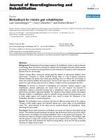
liver volumes receiving 0.5 Gy were 32.5%, 53.5%, 57.9%,
and 66.1%, respectively (Figure 1). A representative exam-
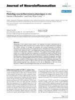
ple of isodose distribution with 2 Gy to the targets using the
different techniques is illustrated in Figure 2. It suggests
that the low-dose radiation area generously deposits around
the intrahepatic target volume, especially when advanced,
conformal radiation techniques are used.
Chromatographic analysis and method validation
Under the conditions described above, the retention time of
5-FU was 5.4 min. The linearity of calibration curves was
demonstrated by the good determination coefficients (r
2
)
obtained for the regression line. Good linearity was
achieved over the range 0.01-5 μg/ml, with all coefficients
of correlation greater than 0.998. All samples were freshly
prepared, including the standard solutions, from the same
stock solution (5 mg/mL). The 0.01-μg/mL limit of quanti-
fication was defined the lowest concentration on the cali-
bration curve that could be measured routinely with
acceptable bias and relative SD.
The overall mean precision, defined by the relative SD,
ranged from 0.2% to 11.0%. Analytical accuracy was
expressed as the percentage difference of the mean
observed values compared to known concentrations varying
from -10.0% to 14.0%. The recovery results for concentra-
tions of 0.1- 10 μg/mL were 92.0%- 94.0%.
Pharmacokinetics of 5-FU
To verify that local RT modulated the systemic pharma-
cokinetics of 5-FU, we established an experimental model
using CT-based planning and whole abdominal irradiation
in rats, and merged it to our pharmacokinetics assay system.
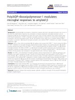
Intriguingly, we found that irradiation markedly reduced the
AUC of 5-FU in rats by 21.4% at 0.5 Gy (p = 0.007) and
31.7% at 2 Gy (p < 0.001), respectively (Figure 3). Of spe-
cial interest, the radiation at 2 Gy to the rat abdomen simu-
lated the daily treatment dose to a human, approximating
the low-dose radiation (0.5 Gy) deposited in the generous,
off-target area in clinical practice. Irradiation significantly
decreased T
1/2
and MRT (p = 0.02 for the 0.5-Gy group and
p < 0.001 for 2-Gy group), and by contrast, increased the
CL (p = 0.03 for the 0.5-Gy group and p < 0.001 for the 2-
Gy group), and Vss (p = 0.05 for the 0.5-Gy and for the 2-
Gy groups, respectively) of 5-FU when compared to non-
irradiated controls (Table 1). There was no significant dif-
ference in the values of Cmax and Kel within any group.
Protein binding
We next examined whether the differences involved protein
binding of 5-FU in plasma. Protein binding of 5-FU in rat
plasma ranged from 62% to 66% among the different
Hsieh et al. Journal of Translational Medicine 2010, 8:29
/>Page 4 of 8
groups. Protein bound/unbound ratios of 5-FU did not differ
by radiation dose or post-radiation interval.
Discussion
Advances in radiation technology have provided better con-
formal dose distribution to simultaneously hit the target
lesions and spare critical organs [9-12]. Nonetheless, areas
other than the target area are exposed to significant low
dose radiation, making radiation oncologists uncomfortable
with this uncertainty in daily practice. Most of this concern
comes from a deficiency of knowledge about the biological
effects of exposure to radiation within the general, low-dose
volumes, especially those exposures produced by the latest
advanced technologies. In the clinical cases treated with
different techniques, we noted that more than 50% of the
normal liver was exposed to 0.5 Gy during daily 2-Gy radi-
ation treatments, except when using 2DRT to treat cholang-
iocarcinoma patients. In the corresponding animal model,
we found, for the first time, after an extensive literature
review, that local RT, not only at the therapeutic 2-Gy frac-
tion, but also at 0.5 Gy (representing a dose deposited in the
general, off-target area in clinical practice), modulated sys-
temic 5-FU pharmacokinetics. Paolo et al. reported that col-
orectal cancer patients given radiation doses resulting in
lower 5-FU AUC had reportedly lower DFS rates [8]. Thus,
the reduction of the 5-FU AUC caused by RT could influ-
ence the outcomes of cancer patients receiving abdominal
CCRT to an extent that demands our consideration and is
not negligible. Therefore, the pharmacokinetics of 5-FU
during CCRT should be rechecked and the optimal 5-FU
dose should be reevaluated, and adjusted if necessary, dur-
ing CCRT.
The liver catabolyzes about 80% of 5-FU via the dihydro-
pyrimidine dehydrogenase (DPD) pathway to generate
Figure 1 The dose-volume histogram of the normal liver under different modalities. The average dose-volume curve of the normal liver under
different modalities with 2 Gy to the tumor bed using the dose-volume histogram evaluation for the four patients. The transverse axis illustrates de-
livered dose in cGy and the vertical axis represents the percentage of liver's volume.
Hsieh et al. Journal of Translational Medicine 2010, 8:29
/>Page 5 of 8
toxic 5-fluoro-5,6-dihydro-uracil (5-FDH2), whereas the
anabolic pathway, via orotate phosphoribosyl transferase
(OPRT), produces active metabolites including 5-fluorouri-
dine-5'-monophosphate (FUMP), 5-fluorouridine (5-FUrd),
and 5-fluoro-2'-deoxyuridine (5-FdUrd) [16,17]. To eluci-
date which pathway was involved or was affected by RT-
induced pharmacokinetic alteration, further assays for the
activities of DPD and OPRT are of importance.
It is possible that metabolic and excretory systems dys-
function in such radiation-induced reductions of 5-FU
AUC. Since the liver falls into the irradiated volume, DPD,
a rate limiting step in the catabolism of 5-FU [18], may be
affected by radiation injury to liver. About 80% of the
administered 5-FU is degraded by DPD [19]. Because 5-FU
has a relatively narrow therapeutic index, a strong correla-
tion is described between exposure to 5-FU and both hema-
tologic and gastrointestinal toxicity [20]. The biochemical
basis of severe 5-FU toxicity is attributed to impaired drug
catabolism, resulting in a markedly prolonged 5-FU plasma
t
1/2
and almost complete absence of drug catabolites [21].
Additionally, there is ample evidence to suggest that sys-
temic low DPD activity is associated with an increased risk
of development of severe 5-FU-associated toxicity. The
overall toxicity was twice as high in patients with profound
DPD deficiencies (< 45% of the mean DPD activity of a
control population) when compared to patients with moder-
ate DPD deficiencies (between 45% and 70% of the mean
DPD activity of a control population), as reported by
Milano et al. [22]. In addition, mutations and single nucle-
otide polymorphisms (SNPs) can cause deficiencies in DPD
enzymatic activity, and patients with DPD deficiencies have
a reduced capacity to metabolize 5-FU and are at risk of
developing severe toxic reactions [23-25].
Figure 2 Isodose distribution by different irradiation techniques. An example of isodose distribution using different irradiation techniques deliv-
ering 2 Gy to the tumor bed for one cholangiocarcinoma patient. A) The conventional radiation therapy (2DRT). B) Three-dimensional conformal ra-
diotherapy (3DCRT). C) Intensive modulated radiotherapy (IMRT). D) Tomotherapy. Orange line, liver; green line, stomach; bright orange line, planning
target volume; purple line, clinical target volume for 2DRT and 3DCRT; light green line, IMRT and tomotherapy. The areas for 2 Gy and 0.5 Gy were
contoured with red and blue color lines for 2DRT, 3DCRT and IMRT, respectively. The areas for 2 Gy and 0.5 Gy are red and blue, respectively, for to-
motherapy.
Hsieh et al. Journal of Translational Medicine 2010, 8:29
/>Page 6 of 8
The kidney is another organ located within the irradiated
volume in the current study. From 10% to 20% of 5-FU is
excreted unchanged in the urine [26]. For patients with
renal dysfunction, the plasma concentration of 5-FU on
nondialysis days is significantly higher than on dialysis
days, and this may be due to the removal of some factors
from plasma by hemodialysis, which inhibit DPD activity
[27]. Because the therapeutic index for 5-FU is relatively
narrow and correlated with hematologic and gastrointesti-
nal toxicity [20], decreased renal function may lead to
increased systemic exposure and increased toxicity. There-
fore, possible renal dysfunction induced by radiation could
have influenced the PK of 5-FU in the current study.
However, the radiation doses used in this study were
much less than the tolerable doses to the liver, which in
humans is defined as the radiation dose to normal tissue that
results in a complication probability of 5% within 5 years
after radiotherapy (TD5/5) [28]; the TD5/5 for the human
liver is 30 Gy, and for kidneys, it is 23 Gy. The consensus
for TD5/5 of liver and kidney in rat is lacking. But the dose
could produce detectable hepatic and renal injury has been
reported. Whole-liver irradiation of 15-Gy in a single-expo-
sure dose would produce detectable hepatic injury in rats
[29] and 25 Gy showed significant histological abnormali-
ties and liver injury, as measured by increased rose bengal
retention and liver enzymes [30]. Sharma et al. [31] demon-
strated that non lethal doses (10 Gy) cause subtle but imme-
diate changes in renal function and structure in rats. Thus,
the possibility that dysfunction of metabolic and excretory
systems take place in such radiation-induced reduction of
AUC might not be great enough to compromise our find-
ings.
CCRT with 5-FU-based regimens are validated as benefi-
cial for controlling many kinds of cancer, such as those aris-
ing from the biliary tract [2], stomach [4], pancreas [3], and
rectum [5]. The favorable effects are thought to be mediated
through the mechanisms of radiosensitization and com-
bined cytotoxicity and synergy. Our results raise the possi-
bility that RT-modulated 5-FU pharmacokinetics could be
one of the mechanisms of action for better tumor control, or
for the opposite, for greater complications of CCRT. These
possibilities remain to be validated in the clinical setting.
Table 1: 5-Fluorouracil (100 mg/kg, i.v.) pharmacokinetics in rats after irradiation with and without 0.5 and 2 Gy.
Parameters Controls Whole abdomen irradiation
0 Gy 0.5 Gy 2 Gy
AUC (min μg/mL) 4641 ± 414 3647 ± 726* 3168 ± 270*
†
t
1/2
(min) 32.3 ± 10 30.3 ± 2.5 26.9 ± 4.0*
Cmax (μg/mL) 160.0 ± 33 131 ± 19 146 ± 27
MRT (min) 36.0 ± 2.7 31 ± 4.2* 25 ± 1.5*
†
CL (mL/kg/min) 21.0 ± 1.9 28.5 ± 7.3* 31.7 ± 2.6*
†
Vss (mL/kg) 798.0 ± 89 885 ± 96* 824 ± 89*
Kelgo1/minp 0.026 ± 0.001 0.031 ± 0.004 0.037 ± 0.001
AUC: area under the plasma concentration vs. time curve; t
1/2
: terminal elimination phase half-life; Cmax: maximum observed plasma
concentration; MRT: mean residence time; CL: total plasma clearance; Vss: volume of distribution at steady state; Kel: elimination constant.
*The mean difference is significant at the 0.05 level in comparison to the control group.
†
The mean difference is significant at the 0.05 level between the 0.5 and 2 Gy groups.
Figure 3 The area under the curve (AUC) for plasma concentra-
tion versus time of 5-FU. The AUC of 5-FU 100 mg/kg to rats in the
control, 0.5-, and 2-Gy groups. The transverse axis illustrates time in
minutes and the vertical axis represents the concentration of 5-FU in
the plasma.
Hsieh et al. Journal of Translational Medicine 2010, 8:29
/>Page 7 of 8
Conclusions
To our best knowledge, this is the first study proving
abdominal irradiation significantly modulates the systemic
pharmacokinetics of 5-FU at dosage levels for both the tar-
get and off-target areas. For abdominal irradiation with con-
current 5-FU therapy, this unexpected RT-pharmacokinetic
influence is worthy of further investigation, which could
necessitate reconsideration of 5-FU dosing in clinical prac-
tice.
Competing interests
The authors declare that they have no competing interests.
Authors' contributions
CH Hsieh participated in the design of the study, performed the radiation and
pharmacokinetic experiments, and wrote the manuscript. YJ Hsieh helped CH
Hsieh to do some experiments. CY Liu participated in the design of the study.
HC Tai was responsible for the radiation planning. YC Huang performed the
statistical analysis. PW Shueng collected the clinical data. LJ Wu helped to pro-
vide clinical data and information. LY Wang helped to design the experiments.
TH Tsai and YJ Chen initiated, organized and supervised all the work, including
the manuscript. All authors read and approved the final version of this manu-
script.
Acknowledgements
We thank Hsing-Yi Lee for collection of radiation therapy planning data.
Author Details
1
Institute of Traditional Medicine, School of Medicine, National Yang-Ming
University, Taipei, Taiwan,
2
Department of Radiation Oncology, Far Eastern
Memorial Hospital, Taipei, Taiwan,
3
Department of Radiation Oncology,
Mackay Memorial Hospital, Taipei, Taiwan,
4
Department of Gastrointestinal
Division, Mackay Memorial Hospital, Taipei, Taiwan,
5
Department of Medical
Research, Mackay Memorial Hospital, Taipei, Taiwan,
6
Department of
Education and Research, Taipei City Hospital, Taipei, Taiwan,
7
Genetics Center,
Department of Medical Research, China Medical University Hospital, Taichung,
Taiwan,
8
Graduate Institute of Chinese Medical Science, China Medical
University, Taichung, Taiwan,
9
Department of Radiation Oncology, National
Defense Medical Center, Taipei, Taiwan and
10
School and Graduate Institute of
Physical Therapy, College of Medicine, National Taiwan University, Taipei,
Taiwan
References
1. Geh JI, Bond SJ, Bentzen SM, Glynne-Jones R: Systematic overview of
preoperative (neoadjuvant) chemoradiotherapy trials in oesophageal
cancer: evidence of a radiation and chemotherapy dose response.
Radiother Oncol 2006, 78:236-244.
2. Kim S, Kim SW, Bang YJ, Heo DS, Ha SW: Role of postoperative
radiotherapy in the management of extrahepatic bile duct cancer. Int J
Radiat Oncol Biol Phys 2002, 54:414-419.
3. Moertel CG, Frytak S, Hahn RG, O'Connell MJ, Reitemeier RJ, Rubin J,
Schutt AJ, Weiland LH, Childs DS, Holbrook MA, et al.: Therapy of locally
unresectable pancreatic carcinoma: a randomized comparison of high
dose (6000 rads) radiation alone, moderate dose radiation (4000 rads +
5-fluorouracil), and high dose radiation + 5-fluorouracil: The
Gastrointestinal Tumor Study Group. Cancer 1981, 48:1705-1710.
4. Macdonald JS, Smalley SR, Benedetti J, Hundahl SA, Estes NC,
Stemmermann GN, Haller DG, Ajani JA, Gunderson LL, Jessup JM,
Martenson JA: Chemoradiotherapy after surgery compared with
surgery alone for adenocarcinoma of the stomach or
gastroesophageal junction. N Engl J Med 2001, 345:725-730.
5. Krook JE, Moertel CG, Gunderson LL, Wieand HS, Collins RT, Beart RW,
Kubista TP, Poon MA, Meyers WC, Mailliard JA, et al.: Effective surgical
adjuvant therapy for high-risk rectal carcinoma. N Engl J Med 1991,
324:709-715.
6. Poortmans PM, Richaud P, Collette L, Ho Goey S, Pierart M, Hulst M Van
Der, Bolla M: Results of the phase II EORTC 22971 trial evaluating
combined accelerated external radiation and chemotherapy with 5FU
and cisplatin in patients with muscle invasive transitional cell
carcinoma of the bladder. Acta Oncol 2008, 47:937-940.
7. Is there a need for more precise definitions of bioavailability?
Conclusions of a consensus workshop, Munich, September 9, 1989;
under the patronage of the F.I.P. Eur J Clin Pharmacol 1991, 40:123-126.
8. Di Paolo A, Lencioni M, Amatori F, Di Donato S, Bocci G, Orlandini C,
Lastella M, Federici F, Iannopollo M, Falcone A, et al.: 5-fluorouracil
pharmacokinetics predicts disease-free survival in patients
administered adjuvant chemotherapy for colorectal cancer. Clin
Cancer Res 2008, 14:2749-2755.
9. Verhey LJ: Comparison of three-dimensional conformal radiation
therapy and intensity-modulated radiation therapy systems. Semin
Radiat Oncol 1999, 9:78-98.
10. Shueng PW, Lin SC, Chong NS, Lee HY, Tien HJ, Wu LJ, Chen CA, Lee JJ,
Hsieh CH: Total marrow irradiation with helical tomotherapy for bone
marrow transplantation of multiple myeloma: first experience in Asia.
Technol Cancer Res Treat 2009, 8:29-38.
11. Chao KS, Low DA, Perez CA, Purdy JA: Intensity-modulated radiation
therapy in head and neck cancers: The Mallinckrodt experience. Int J
Cancer 2000, 90:92-103.
12. Tai HC, Hsieh CH, Chao KS, Liu SH, Leu YS, Chang YF, Hsiao HT, Chang YC,
Huang DY, Chen YJ: Comparison of radiotherapy strategies for locally
advanced hypopharyngeal cancer after resection and ileocolic flap
reconstruction. Acta Otolaryngol 2009, 129:311-317.
13. Vriesendorp HM, Van Bekkum DW: Susceptibility to total-body
irradiaiton. In Response to Total-Body Irradiation in Different Species Edited
by: Broerse JJ, T M. Amsterdam: Martinus Nijhoff; 1984.
14. Ambre JJ, Fischer LJ: The effect of prednisolone and other factors on the
catabolism of 5-fluorouracil in rats. J Lab Clin Med 1971, 78:343-353.
15. Jarugula VR, Lam SS, Boudinot FD: Nonlinear pharmacokinetics of 5-
fluorouracil in rats. J Pharm Sci 1997, 86:756-758.
16. Bocci G, Danesi R, Di Paolo AD, Innocenti F, Allegrini G, Falcone A, Melosi
A, Battistoni M, Barsanti G, Conte PF, Del Tacca M: Comparative
pharmacokinetic analysis of 5-fluorouracil and its major metabolite 5-
fluoro-5,6-dihydrouracil after conventional and reduced test dose in
cancer patients. Clin Cancer Res 2000, 6:3032-3037.
17. Casale F, Canaparo R, Serpe L, Muntoni E, Pepa CD, Costa M, Mairone L,
Zara GP, Fornari G, Eandi M: Plasma concentrations of 5-fluorouracil and
its metabolites in colon cancer patients. Pharmacol Res 2004,
50:173-179.
18. Lu Z, Zhang R, Diasio RB: Dihydropyrimidine dehydrogenase activity in
human peripheral blood mononuclear cells and liver: population
characteristics, newly identified deficient patients, and clinical
implication in 5-fluorouracil chemotherapy. Cancer Res 1993,
53:5433-5438.
19. Heggie GD, Sommadossi JP, Cross DS, Huster WJ, Diasio RB: Clinical
pharmacokinetics of 5-fluorouracil and its metabolites in plasma,
urine, and bile. Cancer Res 1987, 47:2203-2206.
20. Gamelin E, Boisdron-Celle M: Dose monitoring of 5-fluorouracil in
patients with colorectal or head and neck cancer status of the art. Crit
Rev Oncol Hematol 1999, 30:71-79.
21. Diasio RB, Lu Z: Dihydropyrimidine dehydrogenase activity and
fluorouracil chemotherapy. J Clin Oncol 1994, 12:2239-2242.
22. Milano G, Etienne MC, Pierrefite V, Barberi-Heyob M, Deporte-Fety R,
Renee N: Dihydropyrimidine dehydrogenase deficiency and
fluorouracil-related toxicity. Br J Cancer 1999, 79:627-630.
23. Deeken JF, Figg WD, Bates SE, Sparreboom A: Toward individualized
treatment: prediction of anticancer drug disposition and toxicity with
pharmacogenetics. Anticancer Drugs 2007, 18:111-126.
24. van Kuilenburg AB: Dihydropyrimidine dehydrogenase and the efficacy
and toxicity of 5-fluorouracil. Eur J Cancer 2004, 40:939-950.
25. Wei X, McLeod HL, McMurrough J, Gonzalez FJ, Fernandez-Salguero P:
Molecular basis of the human dihydropyrimidine dehydrogenase
deficiency and 5-fluorouracil toxicity. J Clin Invest 1996, 98:610-615.
26. Peters GJ: Antimetabolites. In Oxford Textbook of Oncology Edited by:
Peckam M, Pinedo HM, Veronesi U. London: Oxford University Press;
1995:524-552.
Received: 9 September 2009 Accepted: 25 March 2010
Published: 25 March 2010
This article is available from: 2010 Hsieh et al; licensee BioMed Central Ltd. This is an Open Access article distributed under the terms of the Creative Commons Attribution License ( ), which permits unrestricted use, distribution, and reproduction in any medium, provided the original work is properly cited.Journal of Translational Medicine 2010, 8:29
Hsieh et al. Journal of Translational Medicine 2010, 8:29
/>Page 8 of 8
27. Gusella M, Rebeschini M, Cartei G, Ferrazzi E, Ferrari M, Padrini R: Effect of
hemodialysis on the metabolic clearance of 5-Fluorouracil in a patient
with end-stage renal failure. Ther Drug Monit 2005, 27:816-818.
28. Emami B, Lyman J, Brown A, Coia L, Goitein M, Munzenrider JE, Shank B,
Solin LJ, Wesson M: Tolerance of normal tissue to therapeutic
irradiation. Int J Radiat Oncol Biol Phys 1991, 21:109-122.
29. Geraci JP, Mariano MS, Jackson KL: Radiation hepatology of the rat: time-
dependent recovery. Radiat Res 1993, 136:214-221.
30. Geraci JP, Mariano MS, Jackson KL: Hepatic radiation injury in the rat.
Radiat Res 1991, 125:65-72.
31. Sharma M, Halligan BD, Wakim BT, Savin VJ, Cohen EP, Moulder JE: The
Urine Proteome as a Biomarker of Radiation Injury: Submitted to
Proteomics- Clinical Applications Special Issue: "Renal and Urinary
Proteomics (Thongboonkerd)". Proteomics Clin Appl 2008, 2:1065-1086.
doi: 10.1186/1479-5876-8-29
Cite this article as: Hsieh et al., Abdominal irradiation modulates 5-Fluorou-
racil pharmacokinetics Journal of Translational Medicine 2010, 8:29