Báo cáo hóa học: " Development and application of a biomarker assay for determining the pharmacodynamic activity of an antagonist candidate biotherapeutic antibody to IL21R in whole blood" pptx
Bạn đang xem bản rút gọn của tài liệu. Xem và tải ngay bản đầy đủ của tài liệu tại đây (730.48 KB, 13 trang )
Arai et al. Journal of Translational Medicine 2010, 8:51
/>Open Access
RESEARCH
© 2010 Arai et al; licensee BioMed Central Ltd. This is an Open Access article distributed under the terms of the Creative Commons At-
tribution License ( which permits unrestricted use, distribution, and reproduction in any
medium, provided the original work is properly cited.
Research
Development and application of a biomarker assay
for determining the pharmacodynamic activity of
an antagonist candidate biotherapeutic antibody
to IL21R in whole blood
Maya Arai
1
, Sadhana Jain
1
, Amy A Weaver
1
, Andrew A Hill
2
, Yongjing Guo
1
, Andrea G Bree
3
, Michael F Smith Jr
4
,
Scott W Allen
5
, Edward R LaVallie
1
, Deborah Young
3
, Laird Bloom
1
, Karissa Adkins
6
and Margot O'Toole*
7
Abstract
Background: In preparation for potential clinical development of Ab-01, an antagonistic antibody directed against the
IL21R, studies were undertaken to address translational medicine needs that fall into four categories: 1) development
of a pharmacodynamic biomarker assay suitable for use in the clinic, 2) demonstration that Ab-01 has the desired
biological activity in vitro and in vivo in cynomolgus monkeys, the preferred safety study species, 3) pre-clinical in vivo
proof-of-concept that the assay can be used to detect Ab-01 pharmacodynamic (PD) activity in treated subjects, and 4)
comprehensive assessment of the agonistic potential of Ab-01 when cross-linked. This report and a recently published
companion report address the first three of these needs. The fourth has been addressed in a separate study.
Methods: Genes that change RNA expression upon ex vivo rhIL21 stimulation of whole blood were identified in human
and cynomolgus monkey. The inhibitory effects of exogenously added Ab-01 were measured ex vivo in human and
monkey, and the in vivo inhibitory effects of Ab-01 treatment were measured in monkey.
Results: Stimulation of whole human blood for 2 hours with rhIL21 induced robust increases in RNA expression of 6
genes. This response was blocked by Ab-01, indicating that the assay is suitable for measuring Ab-01 activity in blood.
rhIL21 induced expression of a similar set of genes in cynomolgus monkey blood. This response was blocked with Ab-
01, thus demonstrating that Ab-01 has the desired activity in the species, and that safety studies done in cynomolgus
monkeys are relevant. Proof -of-concept for using this assay system to detect PD activity in vivo was generated by
measuring the response in monkey blood to ex vivo rhIL21 stimulation before and 5 minutes following in vivo Ab-01
administration.
Conclusions: A robust PD biomarker assay suitable for clinical use has been developed in human whole blood. The
successful adaptation of the assay to cynomolgus monkeys has enabled the demonstration of Ab-01 activity both in
vitro and in vivo in monkey, thus validating the use of this species in safety studies and establishing proof-of-concept for
using this PD assay system to aid in dose selection in clinical studies.
Background
Development of protocols for appropriate dose selection
in clinical studies is a clear priority within medical [1] and
regulatory [2] communities. The high attrition rate of
drugs in development due to toxicity and/or lack of effi-
cacy [3,4] underscores the need for biomarker assays to
provide early information on whether the compound
being tested does indeed have the expected effect on the
targeted pathway. This information can be used to miti-
gate the risk of entering into lengthy and expensive effi-
cacy studies. To have an impact on clinical development,
a robust PD biomarker assay must be developed well in
advance of phase I clinical studies. The assay must also
function reliably in the population used for phase I stud-
* Correspondence:
7
Translational Medicine, BioTherapeutic Research, Pfizer, 35 Cambridge Park
Drive,Cambridge, MA 02140, USA
Full list of author information is available at the end of the article
Arai et al. Journal of Translational Medicine 2010, 8:51
/>Page 2 of 13
ies, which, in the case of compounds directed towards
blockade of inflammatory pathways, is often a healthy
volunteer population. To develop biomarkers for drugs
targeting inflammatory pathways, previous investigators
have turned to ex vivo stimulation in whole blood [5,6].
This approach has been particularly useful in the devel-
opment of p38 MAPK inhibitor compounds [7] in which
LPS (lipopolysaccharide)-induced production of inflam-
matory cytokines can be measured. We followed this
basic approach (ex vivo stimulation of whole blood) to
develop pharmacodynamic biomarker assays for a candi-
date therapeutic antibody, Ab-01.
Ab-01, a human antibody generated by phage display,
recognizes the high affinity receptor for IL-21, IL21R,
blocks IL21-mediated immune activation through antag-
onist engagement of IL21R and has shown efficacy in a
mouse model of lupus [8]. The goal of the biomarker
strategy was to provide the means of avoiding toxicity due
to unnecessarily high drug levels and lack of efficacy due
to ineffective dosing by providing early clinical data on
how well the drug hits the target in vivo, and on the best
dosing regimen to maintain target engagement/inhibi-
tion. A second critical goal while preparing for potential
clinical testing was clear demonstration of the desired
biological activity in cynomolgus monkeys, the safety
study species. In the absence of such data, the relevance
of safety studies is uncertain. Therefore, in parallel, we
applied our biomarker strategy to cynomolgus monkeys
and used it to examine ex vivo and in vivo Ab-01 activity
in this species. Here we report the development of PD
biomarker assays that measure Ab-01 biological activity
in human and cynomolgus monkey samples. In addition
we provide pre-clinical proof-of-concept that the assay
system can be used to measure PD activity in treated sub-
jects.
Methods
Sample source and human PD biomarker assay
development
Pilot studies on whole blood from 12 healthy human
donors were performed to identify biomarkers of ex vivo
response of blood to stimulation with rhIL21. Human
blood samples from healthy volunteers were collected
under the Wyeth Human Blood Donor Program - a pro-
gram approved and administered by Mt Auburn Hospital,
Cambridge, MA. Informed consent was obtained from all
donors. A total of 7 donors were used for the initial pilot
studies used for assay development, and an additional 9
donors were used for the confirmatory experiments
reported here. Whole blood samples were collected in BD
Vacutainer™ CPT™ cell preparation tubes containing
sodium heparin (Catalogue #362753). For all data shown
samples were maintained at ambient temperature and
were processed within an hour of collection, but addi-
tional studies indicated acceptable assay performance in
blood that had been stored overnight at room tempera-
ture (data not shown).
Protein reagents: rhIL-21, Ab-01, and control antibodies
The protein reagents used in this study - rhIL21 (recom-
binant human IL21), anti-IL21 receptor antibody Ab-01
(also known as clone VL6 and ATR-107), control anti-
body human IgG1 α-tetanus triple mutant (IgG
1
TM, con-
taining the same mutations in the Fc region as Ab-01),
were made by the Biological Technologies Department at
Wyeth (now Pfizer) Research (Cambridge, MA). Charac-
teristics of rhIL21 are described in Additional file 1. The
three mutations common to the Fc portion of Ab01 and
IgG1TM reduced their potential effector activity. Anti-
bodies with these mutations had undetectable activity in
antibody-dependent cell-mediated cytotoxicity (ADCC)
or C1q binding assays [9,10]. An antibody with severely
compromised effector function was chosen for develop-
ment because the therapeutic goal is to block the interac-
tion of IL21 with IL21R, and therefore minimization of
effector function is desirable. Endotoxin levels in all pro-
teins reagents were determined to be below 1.0 EU/mg.
Ex vivo treatment of human blood
Human blood was distributed (1 mL/aliquot) into screw
cap cryovials (Nunc, Cat# 375353). All treatments were
run in duplicate. rhIL21 (produced from Chinese hamster
ovary cells at Wyeth, now Pfizer) was added in volumes
ranging from 3 μL to 10 μL to achieve the indicated con-
centration. A similar volume of PBS was added to unstim-
ulated control samples. Samples were incubated at 37°C
for the indicated duration while mixing continuously at
approximately 15 revolutions per minute using a Rotamix
rotating mixer (ATR Inc, Laurel, MD). To investigate Ab-
01-mediated inhibition of rhIL21 response, Ab-01 was
added to blood prior to addition of rhIL21. During the
assay development phase of the work, Ab-01 was added
immediately prior to addition of rhIL21, and total inhibi-
tion of the response was observed (data not shown). Since
manipulation of samples immediately upon collection
would not have been practical in the setting of a clinical
study, the final assay protocol included a two hour incu-
bation period in the presence or absence of Ab-01. This
protocol mimicked the conditions of the intended clinical
use of the assay, since blood from Ab-01 treated subjects
(containing Ab-01) would have to placed in a queue in a
laboratory prior to addition of rhIL21. The experiments
with human blood reported here included a 2 hour incu-
bation at 37°C prior to the addition of rhIL21. Human
blood (in 1 mL aliquots) was pre-incubated for 2 hours
with Ab-01 or IgG
1
TM control immunoglobulin at
increasing concentrations followed by the addition of
rhIL21(10 ng/mL) and subsequent 2 hour incubation.
Arai et al. Journal of Translational Medicine 2010, 8:51
/>Page 3 of 13
RNA isolation
Aliquots of blood (0.5 mL) were removed following treat-
ments and added to 2.0 mL microtubes (Axygen Scien-
tific, Union City, CA) containing 1.3 mLs of RNAlater
®
(Applied Biosystems/Ambion, Austin, TX, Catalogue
#AM1928), and mixed thoroughly by 5 complete inver-
sions. Samples were stored at ambient temperature over-
night and then frozen at -80°C pending RNA purification.
RNA was isolated using the Human RiboPure™-Blood Kit
(Applied Biosystems/Ambion Austin TX, Catalogue
#AM1928) following the manufacturer's protocol. The
Human RiboPure™ RNA isolation procedure involves cell
lysis in a guanidinium-based solution and initial purifica-
tion of the RNA by phenol/chloroform extraction fol-
lowed by final RNA purification by solid-phase extraction
on a glass-fiber filter. The residual genomic DNA was
removed according to the manufacturer's instructions by
DNAse treatment using the DNA-free™ reagents provided
in the kit.
RNA quantity was determined by absorbance at 260
nm with a NanoDrop 1000 (NanoDrop, Wilmington, DE).
RNA quality was evaluated using a 2100 Bioanalyzer
(Agilent, Palo Alto, CA, Agilent 2100 expert software ver-
sion B.02.05.SI360), and all samples had RIN (RNA integ-
rity number) [11] >6.6, and all but 2 had RIN values >7.0.
Samples were stored at -80°C until cDNA synthesis was
performed.
Measurement of gene expression levels using real time RT-
PCR
Based on results from the pilot studies (data not shown),
assays for gene transcripts with potential as biomarkers
were selected for inclusion on a custom TaqMan Low
Density Array (TLDA) purchased from Applied Biosys-
tems (ABI) Foster City, CA. This TLDA contained a total
of 24 assays measuring 19 potential biomarkers and 5
endogenous controls (Table 1). Two independent mea-
surements of each transcript were obtained from each
sample. Following the manufacturer's instruction, 400 ng
of total RNA were used to generate cDNA in 40 μL reac-
tion volume in a DNA Engine Peltier Thermal Cycler (MJ
Research, GMI Inc., Ramsey, MN) using a High Capacity
cDNA Reverse Transcription Kit (ABI, #4368814) with
addition of RNase Inhibitor at 50 U/sample (ABI, #N808-
0119). Reaction conditions were: 25°C for 10 minutes,
37°C for 2 hours, 85°C 5 seconds and then hold at 4°C. If
TLDA amplification reactions were not performed on the
same day as cDNA synthesis, the cDNA samples were
stored at -20°C. The amount of cDNA to be loaded on the
TLDA was determined empirically by titration in a pilot
study. Results showed that the amount of cDNA pro-
duced from 200 ng of starting RNA yielded values above
the lower detection limit for all but two of the candidate
biomarkers, and 200 ng (equivalent) was used in all sub-
sequent experiments. The cDNA product (in 20 μl vol-
ume) was diluted by addition of 30 μl DEPC water and
mixed with 50 μl TaqMan
®
Universal 2 × PCR Master Mix
(ABI, #4304437) for a final volume of 100 μl, and added to
each TLDA port. Assay was performed on an ABI PRISM
7900 Sequence Detector (Sequence Detector Software
v2.2.2) using universal thermal cycling conditions of 50°C
for 2 minutes, 95°C for 10 minutes, followed by 40 cycles
of 95°C for 15 seconds and 60°C for 1 minute. Data out-
put was generated from ABI's SDS 2.2.2 software that
determines C
T
(threshold cycle) values from the PCR
amplification plot.
Description of calibrators and normalization of results
using endogenous control genes
Calibrator samples functioned as the common compara-
tor for RQ (Relative Quantification of RNA expression)
calculations. The average C
T
values for all genes in the
unstimulated samples from the first 5 donors following 2
hour incubation served as calibrator for experiments
used to determine optimal rhIL21 dose and time course.
Similarly, the average C
T
of the unstimulated samples
from the second set of 4 donors was used as calibrator for
the experiments related to titration of Ab-01 activity.
Since very small differences in the amount of RNA used
in the amplification reaction can result in significant dif-
ferences in C
T
values, the procedures for normalization of
RNA amounts in starting reactions are described in detail
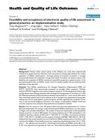
here. The genes chosen as normalizer genes were 18S,
GAPDH, GUSB, PGK, and ZNF592. (ZNF592 was identi-
fied from a large GeneChip database as expressed at very
consistent levels in human peripheral blood mononuclear
cells, A. Hill and M. O'Toole, unpublished observations).
The appropriateness of the 5 genes chosen as normalizers
is demonstrated by the consistent expression among sam-
ples of each of the 5, Figure 1. RQ values were calculated
using the delta delta C
T
method [12]. C
T
values of the
endogenous controls were averaged for each sample. This
average value was used to normalize RNA levels between
samples. Expression levels of test genes were calculated as
C
T
of gene - C
T
of the average of endogenous controls for
that sample (delta C
T
). Gene expression values were cal-
culated as: delta C
T
of gene minus delta C
T
of calibrator
(delta delta C
T
) Data were then expressed as RQ (fold
change over calibrator, 2E-delta delta C
T
or 2
-ΔΔC
T
).
Cynomolgus monkey PD biomarker assay development
animals and sample collection
Adult cynomolgus monkeys (Macaca fascicularis;
Charles River BRF, Inc, Houston, TX) weighing 6 to 9 kg
were singly or pair housed and cared for according to the
American Association for Accreditation of Laboratory
Animal Care guidelines. The Wyeth Institutional Animal
Arai et al. Journal of Translational Medicine 2010, 8:51
/>Page 4 of 13
Care and Use Committee approved all aspects of this
study. Under ketamine sedation (Ketaset, Fort Dodge
Laboratories Inc., Fort Dodge, IA, 10 mg/kg IM), the fem-
oral area was cleaned with povidone-iodine (Betadine;
Purdue Frederick Co, Norwalk, CT) preparation solution
followed by alcohol. Blood was drawn into Vacutainer
CPT mononuclear cell preparation tubes (Catalogue
#362761, BD, Franklin Lakes, NJ).
Ex vivo treatment of monkey blood
rhIL-21 was added to aliquoted blood on the same day
that the blood was drawn. When samples were treated
with both antibody and rhIL21, the antibody was added
and mixed thoroughly immediately prior to rhIL21 addi-
tion. Samples were then incubated at 37°C for 4 hours.
Aliquots (0.5 mL) were removed and added to 2.0 mL
microtubes (Axygen Scientific, #10011-744) containing
1.3 mLs of RNAlater
®
supplied with the Human
RiboPure™-Blood Kit and mixed thoroughly by 5 com-
plete inversions. Samples were stored at ambient temper-
ature overnight and then frozen at -80°C pending RNA
purification. This report and the report by Vugmeyster et
al. [13] document the ex vivo response to rhIL21 stimula-
tion in a combined total of 47 monkeys.
Measurement of Ab-01 PD activity in monkeys dosed with
Ab-01
Antibody was administered by means of bolus intrave-
nous (i.v.) infusion via saphenous vein catheter (22G 1"
Surflo, Terumo Co). Groups of animals were adminis-
tered IgG
1
TM control antibody (n = 3), or Ab-01 (n = 3)
at a dose of 10 mg/kg. Blood samples were drawn prior to
antibody administration and 5 minutes post dosing.
RNA isolation, description of custom TLDA and assay of
RNA concentration for monkey studies
RNA isolation was performed as described above for the
human blood assay. The pilot work for the assay was per-
formed on the Human Immune TLDA (ABI), which con-
tains assays measuring the levels of 96 different
Table 1: Assays used to measure human genes on custom TaqMan low density array for human studies
Gene Gene DescriptionAssay ID (ABI)
18S* Eukaryotic 18S rRNA Hs99999901_s1
CCL3 chemokine (C-C motif ligand Hs00234142_m1
CD19 CD19 Hs00174333_m
CXCL10 chemokine (C-X-C motif ligand 10) Hs00171042_m1
CXCL11 chemokine (C-X-C motif ligand 11) Hs00171138_m1
GNLY Granulysin Hs00246266_m1
GAPDH* glyceraldehyde 3 phosphate dehydrogenase Hs99999905_m1
GUSB* glucuronidase, beta Hs99999908_m1
GZMB Granzyme B (cytotoxic T lymphocyte-associated serine esterase 1) Hs00188051_m1
ICAM1 intercellular adhesion molecule 1 (CD54) Hs00164932_m1
IFNg interferon, gamma Hs00174143_m1
IL10 interleukin 10 Hs00174086_m1
IL12A interleukin 12A (natural killer cell stimulatory factor 1 Hs00168405_m1
IL1b interleukin 1, beta Hs00174097_m1
IL21R interleukin 21 receptor Hs00222310_m1
IL2RA interleukin 2 receptor, alpha, CD25 Hs00166229_m1
IL6 interleukin 6 Hs00174131_m1
IL8 interleukin 8 Hs00174103_m1
PGK1* phosphoglycerate kinase Hs99999906_m1
PRF1 perforin 1 (pore forming protein) Hs00169473_m1
STAT3 signal transducer and activator of transcription 3 Hs00234174_m1
TBX21 T box 21 Hs00203436_m1
TNF tumor necrosis factor (TNF superfamily, member 2) Hs00174128_m1
ZNF592* zinc finger protein 592 Hs00206029_m1
*Gene used as endogenous normalizer
Arai et al. Journal of Translational Medicine 2010, 8:51
/>Page 5 of 13
transcripts. Any assay that detected an IL21 response in
human and/or monkey blood was selected for inclusion
on a custom TLDA designed for the monkey studies. If
the assay for the human gene was capable of measuring
the monkey transcript, the human assay was retained on
the custom TLDA for monkey studies. For genes that
responded to IL21 in humans but were not detectable in
the monkey using primers and probes designed for the
human sequence, primers and probes designed to detect
rhesus genes were used for the custom TLDA because the
assay for cynomolgus monkeys were not, in general,
available as predesigned Gene Expression Assays from
ABI. All TaqMan assays included on the custom TLDA
for monkey studies were among the "inventoried" assays
available from ABI, and are described in Table 2. cDNA
synthesis, preparation of samples for TLDA assay and
measurements of RNA concentration were performed as
described for the human assay.
Results
Time course and dose response of ex vivo response of
human whole blood to rhIL21
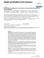
Whole blood samples from 5 healthy human donors were
incubated in the presence of 3.3, 10 or 30 ng/mL of
rhIL21 for 2, 4, 6 or 24 hours. Consistent with prior pilot
exploratory studies performed on 10 human blood
donors, the most significant and robust rhIL21-depen-
dent signals were obtained for six genes: IL6, IFN
γ
,
IL2RA, GZMB, PRF1, CD19 (Figure 2). These 6 genes
were therefore chosen as biomarkers of IL21 activity in
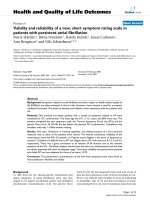
whole blood. The optimal signal for all but CD19 was
obtained at 2 hours (Figure 3). There was no difference in
the response obtained at 3.3, 10 or 30 ng/mL rhIL21.
Based on the results obtained with these 5 donors, the
assay conditions chosen for subsequent experiments on
ex vivo whole blood response to rhIL21 were 2 hour stim-
ulation with 10 ng/mL of rhIL21.
Figure 1 Expression levels of normalizer genes. The unadjusted C
T
values for 5 genes used as endogenous normalizers are shown and reveal very
similar levels of expression in all study samples.
Arai et al. Journal of Translational Medicine 2010, 8:51
/>Page 6 of 13
Titration of Ab-01 inhibition of ex vivo response to rhIL21
Samples from 4 individual healthy human donors were
pre-incubated for 2 hours at the indicated concentration
of Ab-01 or the control IgG
1
TM prior to addition of 10
ng/mL rhIL21 and 2 hr incubation, and the effect on the 6
biomarkers was then assessed (Figures 4 and 5). For the
first 2 donors tested, even the lowest concentration of
Ab-01 (0.1 μg/mL, 0.66 nM) resulted in complete inhibi-
tion of the rhIL21 response, therefore the two subsequent
donors were tested at increasing concentrations of Ab-01
starting at 0.003 μg/mL. Ab-01 inhibited the response of
all 6 genes in all 4 donors. IC50 values ranged between
0.003 and 0.015 μg/mL Ab-01(Figure 5). Control IgG
1
TM
had no significant effect on rhIL21 response (Figure4B).
Ab-01 blocks signal transduction through cynomolgus
rhIL21R
In order to determine if cynomolgus monkey was a suit-
able choice for safety studies, we tested whether the activ-
ity of rhIL21 on monkey cells was blocked by Ab-01. We
therefore first examined the activation effects of ex vivo
rhIL21 stimulation on cynomolgus blood cells, and
observed very similar results to those observed with
human blood. Results for 5 genes most significantly
increased in monkey blood stimulated ex vivo with
rhIL21 are shown in Table 3. The most robust (largest
magnitude change and most consistent change) rhIL21-
mediated change in cynomolgus monkeys was observed
for IL2RA. All animals tested (n = 48) for effects of rhIL21
on IL2RA expression levels gave a response of >1.5 fold
change [13], and therefore this gene was selected as the
biomarker for subsequent monkey studies. To determine
if Ab-01 had the desired blocking activity of rhIL21/
IL21R dependent activation of cynomolgus blood cells,
its ability to block rhIL21-dependent IL2RA activation
was tested. The IL2RA response was effectively blocked
by Ab-01 treatment (Table 4). There was no significant
difference between the response to rhIL21 in the pres-
ence and absence of control IgG
1
TM, while the response
in the presence of Ab-01 was blocked (P < 0.001). These
results show that signal induction through the interaction
of cynomolgus IL21R with rhIL21 is inhibited by Ab-01,
an antibody to human IL21R. Therefore Ab-01 has the
intended activity in cynomolgus monkeys.
Table 2: Assays used to measure monkey genes on custom TaqMan low density array for monkey studies
Gene ID Use Species Assay ID (ABI)
18S Manufacturing QC Human Hs99999901_s1
CD19 Effects of IL21/Ab-01 Human Hs00174333_m1
CSF1 Effects of IL21/Ab-01 Human Hs00174164_m1
GUSb Normalizer Human Hs99999908_m1
GZMB Effects of IL21/Ab-01 Rhesus Rh02621701_m
ICOS Effects of IL21/Ab-01 Rhesus Rh02621771_m1
IFN
γ
Effects of IL21/Ab-01 Rhesus Rh02621721_m1
IL10 Effects of IL21/Ab-01 Human Hs00174086_m1
IL12B Effects of IL21/Ab-01 Human Hs00233688_m1
IL21R Effects of IL21/Ab-01 Human Hs00174086_m
IL2RA Effects of IL21/Ab-01 Human Hs00166229_m1
IL6 Effects of IL21/Ab-01 Rhesus Rh02621719_u1
IL7 Effects of IL21/Ab-01 Human Hs00174202_m1
IL8 Effects of IL21/Ab-01 Human Hs00174103_m1
PGK1 Normalizer Human Hs99999906_m1
PRF1 Effects of IL21/Ab-01 Human Hs00169473_m1
STAT3 Effects of IL21/Ab-01 Human Hs00234174_m1
TBX2 Effects of IL21/Ab-01 Human Hs00203436_m1
TNF Effects of IL21/Ab-01 Human Hs00174128_m1
ZNF592 Normalizer Human Hs00206029_m1
In addition to the assays listed, assays for CCL19, CSF2, IL-17, and REN were run but found to be unreliable in these cynomolgus monkey samples
and are not shown.
Arai et al. Journal of Translational Medicine 2010, 8:51
/>Page 7 of 13
rhIL21-mediated activation is blocked in blood from
monkeys dosed with Ab-01
The utility of the ex vivo rhIL21 stimulation assay as a
read-out of Ab-01 PD activity in treated individuals was
tested by comparing the rhIL21 response in 3 monkeys
before and 5 minutes following administration of a 10
mg/kg i.v. dose of Ab-01. The control group consisted of 3
monkeys dosed with 10 mg/kg i.v. dose of IgG
1
TM. Con-
sistent with the results from the many other monkeys
tested, prior to dosing with Ab-01 an increase in IL2RA
expression was observed in the blood of all 6 monkeys
when stimulated ex vivo with rhIL21 (Table 5). No IL2RA
response was observed in blood drawn 5 minutes follow-
ing i.v. administration of Ab-01 (Table 5). Dosing with
control IgG
1
TM did not affect the subsequent ex vivo
response to rhIL21. Additional blood samples were tested
at later time points following the single dose of Ab-01.
Results showed that as the circulating levels of Ab-01 fell
over time, the ex vivo rhIL21-mediated response was
restored [13]. These results, together with the pre-dose
and 5 minute post-dose data in Table 5 establish that all
three monkeys dosed with Ab-01 were responsive to
rhIL21 before dosing, did not respond when Ab-01 was
present in the blood, and returned to responsiveness
when Ab-01 was cleared from circulation.
Discussion
We have developed a human blood biomarker assay that
detects the blocking activity of Ab-01, an antibody to
IL21R. In parallel we have developed an adaptation of this
assay and used it to demonstrate that the IL21-dependent
response in cynomolgus monkey blood is blocked both
by ex vivo addition of Ab-01 to blood, and by i.v. adminis-
tration prior to blood collection. These results support
the use of cynomolgus monkeys for safety studies by
establishing that Ab-01 hits its target in vivo and has the
Figure 2 Whole human blood response to ex vivo stimulation with 10 ng/mL rhIL21 for 2 hours. Data shown are for the 6 genes (of 19 tested)
with the most consistent response among the 9 individual donors, and are identified as 6 preferred biomarkers of rhIL21 activity in whole human
blood. These 6 genes were also identified as the best indicators of IL21 response in a series of pilot studies conducted with blood from a different
group of donors (data not shown).
Relative RNA Concentration
IL6
0
5
10
15
20
25
No IL21 10ng/ml IL-21
GZMB
0
1
2
3
4
5
6
No IL21 10ng/ml IL-21
CD19
0
0.5
1
1.5
2
2.5
3
No IL21 10ng/ml IL-21
IFNJ
J
0
4
8
12
16
20
No IL21 10ng/ml IL-21
IL2RA
0
2
4
6
8
No IL21 10ng/ml IL-21
PRF1
0
1
2
3
4
5
No IL21 10ng/ml IL-21
P=0.007P<0.001P=0.002
P=0.005 P=0.001 P<0.001
Arai et al. Journal of Translational Medicine 2010, 8:51
/>Page 8 of 13
desired biological activity in the species. We have shown
that this assay system is well suited to examining the rela-
tionship between pharmacokinetics and pharmacody-
namics (PK/PD), the intended clinical use of the assay
system [13]. A third critical contribution, demonstrating
lack of Ab-01 agonistic activity even under circumstances
designed to force an agonistic signal is described in Guo
et al [14]. The data reported here and in these related
reports on Ab-01 are unified by their focus on addressing
central challenges of translational medicine - dose selec-
tion, elucidation of PK/PD relationships, choice of safety
study species, and mitigation of risk of immunotoxicity.
The assay reported here relies on an ex vivo stimulation
procedure. It is difficult to develop a robust assay for
determining Ab-01 PD activity by relying upon the
endogenous levels of IL21 activity on biomarker gene
expression in samples upon collection, especially in
healthy donors. It is possible that such a strategy could
correlate PK parameters and biomarker movement dur-
ing the treatment period, but it would not show direct
linkage between the movement of the biomarker and the
engagement of the target. Following the biomarker assay
development strategy previously employed by others, we
have developed a whole blood ex vivo stimulation assay.
Our biomarker discovery and assay development strate-
gies have, from the start, proceeded with the realities of
the clinical setting in mind. The volume of blood required
is less than 5 mL, well below the limits of a routine blood
draw. The live blood sample is subjected to minimal
manipulation, consisting of separation into two aliquots,
addition of rhIL21 to one of the aliquots, and 2 hour incu-
bation at 37°C with rotation. RNA preserving solution is
then added, and the sample frozen. All subsequent proce-
dures can be carried out on batched samples in a vali-
dated lab. The assay is sensitive and robust, and has
shown consistent performance among healthy human
Figure 3 Determination of optimal time point for rhIL21-response for the 6 genes identified as preferred biomarkers. This time point exper-
iment was done using 5 of the donors shown in Figure 2. Earlier pilot studies had suggested that stimulation for 30 minutes was significantly sub-
optimal. There was no significant difference in response between all doses tested (30, 10, and 3 ng/mL). The lowest dose of rhIL21 that elicits a re-
sponse has therefore not been determined.
No IL21 3.3 ng/ml IL21
10 ng/ml IL21
30 ng/ml IL21
CD19
0
0.5
1
1.5
2
2.5
3
3.5
024624
0
0.5
1
1.5
2
2.5
3
3.5
024624
GZMB
0
1
2
3
4
5
6
7
8
9
024624
IL6
0
0.5
1
1.5
2
2.5
3
3.5
024624
PRF1
0
1
2
3
4
5
6
7
024624
IL2RA
0
1
2
3
4
5
6
7
8
024624
IFNJ
J
Incubation Time (hours)
Average Relative RNA Concentration + S.D.
Arai et al. Journal of Translational Medicine 2010, 8:51
/>Page 9 of 13
and primate subjects. We have shown that the assay
detects Ab-01 in the 100 pM range in humans, well
within the range for potential clinical utility.
We believe that the RNA assay system reported here
has significant advantages over a protein secretion assay.
First, the read-out of RNA is a more proximal event than
protein secretion, and we have found that, in this assay
system at least, the RNA signal is more easily and reliably
detected than the protein signal [14]. Detection of protein
secretion required a much longer incubation period,
necessitating a shift from an assay with whole blood to an
assay requiring purification and culturing of peripheral
blood mononuclear cells. Such sample manipulation
steps compromise the ease of adaptation for clinical use
and introduce additional sources of variability. Secondly,
measurements made on well purified RNA are not sub-
ject to the factors (such as for example, specific binding
factors, charged proteins, or rheumatoid factor), which
can confound ELISA assays performed in human serum
or plasma [15]. We have also found that the cost of RNA
assay systems compares favorably with that of ELISA sys-
tems, especially with considerations of standardization
methodology.
Concordance between humans and cynomolgus mon-
keys with respect to genes activated was observed for
IL2RA, GZMB, IL6 and PRF1. IFN
γ
also increases dra-
matically in both species, but detection of this increase in
cynomolgus monkeys required a protocol modification
(purification of PBMCs to increase IFN
γ
RNA yield) that
was not performed for the 18-monkey cohort shown in
Table 3. While CD19 is among the genes listed as most
significantly increased by rhIL21 in humans, its absence
from the list of genes in cynomolgus monkeys does not
reflect a difference between the species, but rather the
unavailability of a reliable TaqMan assay for CD19 in cyn-
Figure 4 Average percent inhibition of the expression level of 6 IL21-responsive genes. Percent inhibition values were calculated based on RQ
(relative quantification) values of untreated control and rhIL21-treated samples for each of the 4 donors, and subsequently the mean and standard
deviation were determined for each gene shown. A: Percent inhibition in presence of Ab-01. B: Percent inhibition in presence of control IgG
1
TM. Data
for the 0.1 μg/mL , 0.3 μg/mL and 1 μg/mL concentrations were generated using 4 donors. Data for the higher and the lower concentrations were
generated using 2 donors.
CD19 GZMB
IFNJ
IL2RA IL6 PRF1
A
0.003 0.01 0.03 0.1 0.3 1 3.3 10 30
P
g/ml Ab-01
-50
0
50
100
150
Average % Inhibition and St. Dev
B
-50
0
50
100
150
P
g/ml Control IgG
1
TM
0.003 0.01 0.03 0.1 0.3 1 3.3 10 30
Average % Inhibition and St. Dev
Arai et al. Journal of Translational Medicine 2010, 8:51
/>Page 10 of 13
Figure 5 Inhibition by Ab-01 at the indicated concentration is shown for 6 IL21-responsive genes. IC50 values of inhibition curves shown in
Figure 4A were calculated using curve fit (XLfit) program for each of the referred biomarker genes. Values for the 0.1 μg/mL, 0.3 μg/mL and 1 μg/mL
concentrations were generated using 4 donors. Data for the higher and the lower concentrations were generated using 2 donors each.
CD19
IC50 = 0.008 Pg/ml
R
2
= 0.86
0.01 1
10
50
100
150
GZMB
IC50 = 0.005 Pg/ml
R2 = 0.99
0.01 1
50
75
100
IFNJ
J
IC50 = 0.005 Pg/ml
R
2
= 0.99
0.01 1
50
100
50
75
100
0.01 1
IL2RA
IC50 = 0.003 Pg/ml
R
2
= 0.97
25
50
75
100
0.01 1
IL6
IC50 = 0.014 Pg/ml
R
2
= 0.99
25
50
75
100
0.01 1
PRF1
IC50 = 0.015 Pg/ml
R
2
= 0.99
% Inhibition % Inhibition
P
g/ml Ab-01
P
g/ml Ab-01
P
g/ml Ab-01
Table 3: rhIL21-responsive genes in whole cynomolgus monkey blood
Gene Average Fold Change (with IL21) SD P-value paired t-test
IL2RA 4.1 1.76 < 0.0001
PRF1 1.8 0.78 <0.0001
IL21R 2.3 0.63 <0.0001
GZMB 1.6 0.39 <0.0001
IL6 2.7 1.72 0.0007
rhIL21-mediated average fold increase in RNA expression in blood of 18 individual cynomolgus monkeys stimulated in vitro for 4 hours.
Control (no rhIL21) RNA expression values were normalized to 1, and p values were calculated using log
2
of the fold change paired with the
log2 of 1 (0).
Arai et al. Journal of Translational Medicine 2010, 8:51
/>Page 11 of 13
Table 4: Inhibition by Ab-01 of ex vivo rhIL21-dependent IL2RA expression
Animal rHuIL21 alone
rHuIL21 + Control IgG1TM
rHuIL21 + Ab-01
14.7 3.5 1.2
23.8 3.8 0.9
34.1 5.1 1.4
42.8 2.8 0.9
53.3 3.2 1.3
62.8 2.9 1
73.6 3.7 1.1
87.2 8.8 1.1
Each value represents the fold change over the no-treatment group in expression levels of IL2RA in monkey blood samples following 4 hour
ex vivo treatment as indicated.
Table 5: rhIL21 induced IL2RA expression in whole blood from cynomolgus monkeys dosed with Ab-01
Animal Treatment Group Time point Ex vivoIL2RA Fold Change Response to IL21 Stimulation
Cpeak
1 Ab-01 pre-dose 2.8 not applicable
1 Ab-01 5 minute post dose 0.8 200
2 Ab-01 pre-dose 4.8 not applicable
2 Ab-01 5 minute post dose 0.8 139
3 Ab-01 pre-dose 4.2 not applicable
3 Ab-01 5 minute post dose 0.9 153
4 Control IgG
1
TM pre-dose 4.2 not applicable
4 Control IgG
1
TM 5 minute post dose 4.3 not applicable
5 Control IgG
1
TM pre-dose 2.7 not applicable
5 Control IgG
1
TM 5 minute post dose 4.4 not applicable
6 Control IgG
1
TM pre-dose 9.1 not applicable
6 Control IgG
1
TM 5 minute post dose 7.7 not applicable
Relative RNA concentration of IL2RA induced by ex vivo addition of rhIL-21 to whole blood obtained from cynomolgus monkeys before and after
treatment with either Ab-01 or control IgG
1
TM is shown. Pre-dose whole blood sample was taken on day -13 for animal 1, and on day 0 (the day
of dosing) for all others. All monkeys were dosed with 10 mg/kg via intravenous (IV) route. The ex vivo response to IL21 was completely blocked
in monkeys treated with Ab-01 and was not blocked in control IgG
1
TM treated monkeys
Arai et al. Journal of Translational Medicine 2010, 8:51
/>Page 12 of 13
omolgus monkeys. A significant rhIL21-mediated IL21R
elevation was observed in both humans and monkeys.
The presence of IL21R on the top gene list for monkeys
and not for humans reflects a higher relative ranking in
monkeys due to the lack of IFN
γ
and CD19 data in cyno-
molgus monkeys (for the reason detailed above). We con-
clude that the whole blood rhIL21 responses of human
and monkey are remarkably similar, and our work does
not identify differences between them.
The goal of this work was to identify markers to be used
to assess the PD activity of Ab-01 in treated subjects. For
this reason, the most reliable read-outs detectable in
whole blood were selected as the most useful from a clin-
ical operations perspective. IL21 exerts many different
effects on a wide variety of lymphoid and non-lymphoid
cells including fibroblasts and intestinal epithelial cells
[16-18]. The activities of IL21 have been shown to be
highly dependent on the lineage of the target cell, the
activation state of the target cell and the presence of other
co-stimulators and immune modulators [19,20]. Previous
studies have shown that IL21 activates both IFN
γ
and
IL2RA in purified NK and purified T cells [21]. CD19 and
IL21R were also among the genes that changed signifi-
cantly upon ex vivo exposure of whole blood to rhIL21,
and IL21 has been shown to up regulate both CD19 and
IL21R expression in activatec B cells [22]. Therefore the
markers identified in this study include genes with previ-
ously demonstrated links to activation of the IL21 path-
way in a variety of cell types. In light of the well
established complexities of IL21 biological activity, it is
fortuitous from a clinical development perspective that
whole blood provides a useful analytical sample for
detecting inhibition by a candidate therapeutic of signal
transduction through IL21R.
Conclusions
The PD biomarker assay described here has been devel-
oped to facilitate safe and efficient clinical testing by posi-
tioning a clinical team to make dose selection decisions
based on reliable information on in vivo biological activ-
ity. The assay system monitors levels of activity in blood
and will indicate when levels are too low to block activity
and when levels are significantly higher than they need to
be. The basic approach here is also applicable to other
therapeutic candidates, especially in indications related
to inflammation. The demonstration of the desired bio-
logical activity in the safety study species is also a signifi-
cant contribution in preparing for transition to the clinic.
Additional material
Competing interests
All authors were employees of Pfizer (formerly Wyeth) at the time this work was
performed.
Authors' contributions
MA performed all the studies on human samples reported here, analyzed the
data and co-wrote the manuscript. SJ and AAW performed all the pilot studies
in humans and monkeys that identified the candidate biomarkers, and they
participated in the data analyses. AAH developed the customized Spotfire tool
used for data analyses and reviewed statistical analyses. YG participated in the
assessment and selection of TaqMan PCR assays for studies in monkeys and
participated in the biomarker discovery (pilot) phase of the project. AGB per-
formed all in vivo procedures on monkeys and contributed to the writing of
the manuscript. MFS was responsible for interface with the clinical team and
for securing and scheduling the resources that will be required upon hand-off
for assay validation. SJ, AW, SWA and KA performed the in vitro assays on mon-
keys treated with Ab-01 and control Ig and analyzed the data, and KA and SA
performed the experiments on monkeys dosed in vivo with Ab-01 or IgG
1
TM.
ERL performed data analysis and review and participated in the drafting of the
manuscript. DY co-led the Ab-01 team and was responsible for studies related
to the characterization of the biological properties of the antibody. LB co-led
the Ab-01 team and was responsible for lead antibody selection and develop-
ment. MO devised the PD assay strategy, directed the biomarker discovery pro-
cess, supervised assay development and co-wrote the manuscript. All authors
have read and approved the final manuscript.
Acknowledgements
We thank Holly Legault, Leslie Lowe and Vikki Spaulding for exploratory studies
related to assay development, Khetemenee Lam and Stephane Olland for pro-
viding rhIL21, and Mary Collins, Cheryl Nutter and Davinder Gill for critical
review of the manuscript.
Author Details
1
Global Biotherapeutic Technologies, Pfizer, 87 Cambridge Park Drive,
Cambridge, MA 02140, USA,
2
Massachusetts Research Business Technologies,
Pfizer, 35 Cambridge Park Drive, Cambridge, MA 02140, USA,
3
Inflammation
and Immunology, Pfizer, 200 Cambridge Park Drive,Cambridge, MA 02140, USA
,
4
Translational Medicine- Inflammation, Hoffmann-LaRoche, 340 Kingsland St.,
Nutley, NJ 07110, USA,
5
Investigative Toxicology, Pfizer, 1 Burtt Road, Andover,
MA 01810, USA,
6
Drug Safety Research and Development, Eastern Point Road,
MS8274-1222, Groton, CT 06340, USA and
7
Translational Medicine,
BioTherapeutic Research, Pfizer, 35 Cambridge Park Drive,Cambridge, MA
02140, USA
References
1. Kuhlmann J, Wensing G: The applications of biomarkers in early clinical
drug development to improve decision-making processes. Curr Clin
Pharmacol 2006, 1:185-191.
2. Guidance for Industry Dosage and Administration Section of Labeling
for Human Prescription Drug and Biological Products [http://
www.fda.gov/downloads/Drugs/GuidanceComplianceRegulatoryIn
formation/Guidances/UCM075066.pdf]
3. Danhof M, de Lange EC, Della Pasqua OE, Ploeger BA, Voskuyl RA:
Mechanism-based pharmacokinetic-pharmacodynamic (PK-PD)
modeling in translational drug research. Trends Pharmacol Sci 2008,
29:186-191.
4. Kola I, Landis J: Can the pharmaceutical industry reduce attrition rates?
Nat Rev Drug Discov 2004, 3:711-715.
5. Lavallie ER, Dorner AJ, Burczynski ME: Use of ex vivo systems for
biomarker discovery. Curr Opin Pharmacol 2008, 8:647-653.
6. Ray CA, Dumaual C, Willey M, Fill J, O'Brien PJ, Gourley I, Devanarayan V,
Konrad RJ: Optimization of analytical and pre-analytical variables
associated with an ex vivo cytokine secretion assay. J Pharm Biomed
Anal 2006, 41:189-195.
7. Parasrampuria DA, de Boer P, Desai-Krieger D, Chow AT, Jones CR: Single-
dose pharmacokinetics and pharmacodynamics of RWJ 67657, a
specific p38 mitogen-activated protein kinase inhibitor: a first-in-
human study. J Clin Pharmacol 2003, 43:406-413.
Additional file 1 Description of rhIL21. Sequence and information on
preparation and activity of the rhIL21 protein preparation used in these
studies is shown.
Received: 10 November 2009 Accepted: 28 May 2010
Published: 28 May 2010
This article is available from: 2010 Arai et al; licensee BioMed Central Ltd. This is an Open Access article distributed under the terms of the Creative Commons Attribution License ( which permits unrestricted use, distribution, and reproduction in any medium, provided the original work is properly cited.Journal of Tr anslational Medi cine 2010, 8:51
Arai et al. Journal of Translational Medicine 2010, 8:51
/>Page 13 of 13
8. Vugmeyster Y, Guay H, Szklut P, Qian MD, Jin M, Widom A, Spaulding V,
Bennett F, Lowe L, Andreyeva T, Lowe D, Lane S, Thom G, Valge-Archer V,
Gill D, Young D, Bloom L: In vitro potency, pharmacokinetic profiles, and
pharmacological activity of optimized anti-IL-21R antibodies in a
mouse model of lupus. MAbs 2010, 2:335-346.
9. Black R, Ekman L, Lieberburg I, Grundman M, Callaway J, Gregg KM,
Jacobsen JS, Gill D, Tchistiakova L, Windom A: Immunotherapy regimes
dependent on APOE status, patent application number 20090155256.
2009 [].
10. Kasaian MT, Tan XY, Jin M, Fitz L, Marquette K, Wood N, Cook TA, Lee J,
Widom A, Agostinelli R, Bree A, Schlerman FJ, Olland S, Wadanoli M, Sypek
J, Gill D, Goldman SJ, Tchistiakova L: Interleukin-13 neutralization by two
distinct receptor blocking mechanisms reduces immunoglobulin E
responses and lung inflammation in cynomolgus monkeys. J
Pharmacol Exp Ther 2008, 325:882-892.
11. Schroeder A, Mueller O, Stocker S, Salowsky R, Leiber M, Gassmann M,
Lightfoot S, Menzel W, Granzow M, Ragg T: The RIN: an RNA integrity
number for assigning integrity values to RNA measurements. BMC Mol
Biol 2006, 7:3.
12. Livak KJ, Schmittgen TD: Analysis of relative gene expression data using
real-time quantitative PCR and the 2(-Delta Delta C(T)) Method.
Methods 2001, 25:402-408.
13. Vugmeyster Y, Allen S, Szklut P, Bree A, Ryan M, Ma M, Spaulding V, Young
D, Guay H, Bloom L, Leach MW, O'Toole M, Adkins K: Correlation of
pharmacodynamic activity, pharmacokinetics, and anti-product
antibody responses to anti-IL-21R antibody therapeutics following IV
administration to cynomolgus monkeys. J Transl Med 2010, 8:41.
14. Guo Y, Hill A, Ramsey R, Immermann F, Corcoran C, Young D, LaVallie E,
Pfeifer R, Warner G , Bologna M, Bloom L, O'Toole M: Assessing agonistic
potential of a candidate therapeutic anti-IL21R antibody. J Transl Med
2010, 8:50.
15. O'Toole M, Legault H, Ramsey R, Wynn TA, Kasaian MT: A novel and
sensitive ELISA reveals that the soluble form of IL-13R-alpha2 is not
expressed in plasma of healthy or asthmatic subjects. Clin Exp Allergy
2008, 38:594-601.
16. Caruso R, Fina D, Peluso I, Stolfi C, Fantini MC, Gioia V, Caprioli F, Del
Vecchio Blanco G, Paoluzi OA, Macdonald TT, Pallone F, Monteleone G: A
functional role for interleukin-21 in promoting the synthesis of the T-
cell chemoattractant, MIP-3alpha, by gut epithelial cells.
Gastroenterology 2007, 132:166-175.
17. Monteleone G, Caruso R, Fina D, Peluso I, Gioia V, Stolfi C, Fantini MC,
Caprioli F, Tersigni R, Alessandroni L, MacDonald TT, Pallone F: Control of
matrix metalloproteinase production in human intestinal fibroblasts
by interleukin 21. Gut 2006, 55:1774-1780.
18. Jungel A, Distler JH, Kurowska-Stolarska M, Seemayer CA, Seibl R, Forster A,
Michel BA, Gay RE, Emmrich F, Gay S, Distler O: Expression of interleukin-
21 receptor, but not interleukin-21, in synovial fibroblasts and synovial
macrophages of patients with rheumatoid arthritis. Arthritis Rheum
2004, 50:1468-1476.
19. Leonard WJ, Spolski R: Interleukin-21: a modulator of lymphoid
proliferation, apoptosis and differentiation. Nat Rev Immunol 2005,
5:688-698.
20. Zeng R, Spolski R, Finkelstein SE, Oh S, Kovanen PE, Hinrichs CS, Pise-
Masison CA, Radonovich MF, Brady JN, Restifo NP, Berzofsky JA, Leonard
WJ: Synergy of IL-21 and IL-15 in regulating CD8 + T cell expansion and
function. J Exp Med 2005, 201:139-148.
21. Strengell M, Sareneva T, Foster D, Julkunen I, Matikainen S: IL-21 up-
regulates the expression of genes associated with innate immunity
and Th1 response. J Immunol 2002, 169:3600-3605.
22. Ettinger R, Kuchen S, Lipsky PE: The role of IL-21 in regulating B-cell
function in health and disease. Immunol Rev 2008, 223:60-86.
doi: 10.1186/1479-5876-8-51
Cite this article as: Arai et al., Development and application of a biomarker
assay for determining the pharmacodynamic activity of an antagonist candi-
date biotherapeutic antibody to IL21R in whole blood Journal of Translational
Medicine 2010, 8:51