Báo cáo hóa học: " Epigenetics of human cutaneous melanoma: setting the stage for new therapeutic strategies" pot
Bạn đang xem bản rút gọn của tài liệu. Xem và tải ngay bản đầy đủ của tài liệu tại đây (3.36 MB, 22 trang )
Sigalotti et al. Journal of Translational Medicine 2010, 8:56
/>Open Access
REVIEW
© 2010 Sigalotti et al; licensee BioMed Central Ltd. This is an Open Access article distributed under the terms of the Creative Commons
Attribution License ( which permits unrestricted use, distribution, and reproduction in
any medium, provided the original work is properly cited.
Review
Epigenetics of human cutaneous melanoma:
setting the stage for new therapeutic strategies
Luca Sigalotti*
1
, Alessia Covre
1,2
, Elisabetta Fratta
1
, Giulia Parisi
1,2
, Francesca Colizzi
1
, Aurora Rizzo
1
, Riccardo Danielli
2
,
Hugues JM Nicolay
2
, Sandra Coral
1
and Michele Maio
1,2
Abstract
Cutaneous melanoma is a very aggressive neoplasia of melanocytic origin with constantly growing incidence and
mortality rates world-wide. Epigenetic modifications (i.e., alterations of genomic DNA methylation patterns, of post-
translational modifications of histones, and of microRNA profiles) have been recently identified as playing an important
role in melanoma development and progression by affecting key cellular pathways such as cell cycle regulation, cell
signalling, differentiation, DNA repair, apoptosis, invasion and immune recognition. In this scenario, pharmacologic
inhibition of DNA methyltransferases and/or of histone deacetylases were demonstrated to efficiently restore the
expression of aberrantly-silenced genes, thus re-establishing pathway functions. In light of the pleiotropic activities of
epigenetic drugs, their use alone or in combination therapies is being strongly suggested, and a particular clinical
benefit might be expected from their synergistic activities with chemo-, radio-, and immuno-therapeutic approaches
in melanoma patients. On this path, an important improvement would possibly derive from the development of new
generation epigenetic drugs characterized by much reduced systemic toxicities, higher bioavailability, and more
specific epigenetic effects.
Introduction
Cutaneous melanoma (CM) is a highly aggressive malig-
nancy originating from melanocytes, which is character-
ized by constantly growing incidence and mortality rates
world-wide [1]. Unlike the majority of human cancers,
CM is frequently diagnosed in young and middle-aged
adults [2]. Despite representing only 3% of all skin malig-
nancies, CM is responsible for 65% of skin malignancy-
related deaths, and the 5-year survival of metastatic CM
patients is 7-19% [3,4].
The increasing incidence and the poor prognosis of
CM, along with the substantial unresponsiveness of
advanced disease to conventional therapies, have
prompted significant efforts in defining the molecular
alterations that accompany the malignant transformation
of melanocytes, identifying epigenetic modifications as
important players [5]. "Epigenetics" refers to heritable
alterations in gene expression that are not achieved
through changes in the primary sequence of genomic
DNA. In this respect, the most extensively characterized
mediators of epigenetic inheritance are the methylation
of genomic DNA in the context of CpG dinucleotides,
and the post-translational modifications of core histone
proteins involved in the packing of DNA into chromatin
[6]. Despite not yet having been extensively character-
ized, also microRNAs (miRNAs) are emerging as impor-
tant factors in epigenetic determination of gene
expression fate in CM [7].
DNA methylation occurs at the C5 position of cytosine
in the context of CpG dinucleotides and it is carried out
by different DNA methyltransferases (DNMT) that have
distinct substrate specificities: DNMT1 preferentially
methylates hemimethylated DNA and has been associ-
ated with the maintenance of DNA methylation patterns
[8]; DNMT3a and 3b do not show preference for hemim-
ethylated DNA and have been implicated in the genera-
tion of new methylation patterns [9,10]. Besides this
initial strict categorization, recent evidences are indicat-
ing that all three DNMTs may possess both de novo and
maintenance functions in vivo, and that they cooperate in
establishing and maintaining DNA methylation patterns
[11-14]. The methylation of promoter regions inhibits
gene expression either by directly blocking the binding of
* Correspondence:
1
Cancer Bioimmunotherapy Unit, Centro di Riferimento Oncologico, Istituto di
Ricovero e Cura a Carattere Scientifico, Via F. Gallini 2, 33081 Aviano, Italy
Full list of author information is available at the end of the article
Sigalotti et al. Journal of Translational Medicine 2010, 8:56
/>Page 2 of 22
transcriptional activators or by binding methyl-CpG-
binding domain (MBD) proteins that silence gene expres-
sion through the recruitment of chromatin remodeling
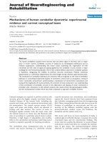
co-repressor complexes (Figure 1) [15,16].
Genomic DNA in the nucleus is packed into the chro-
matin, the base unit of which is the nucleosome: a histone
octamer core comprising two copies each of histones
H2A, H2B, H3 and H4, around which about 147 bp of
DNA are wrapped. Each histone contains flexible N-ter-
minal tails protruding from the nucleosomes, which are
extensively targeted by post-translational modifications,
including acetylation and methylation. These modifica-
tions determine how tightly the chromatin is compacted,
thus playing a central regulatory role in gene expression.
The acetylation status of histones is controlled by the bal-
anced action of histone acetyltransferases and histone
deacetylases (HDAC), and acetylated histones have been
associated with actively expressed genes. On the other
hand, methylation of histones, accomplished by histone
methyl transferases (HMT), may have both repressive
(H3 lysine (K) 9, H3K27) or promoting (H3K4) effects on
transcription, depending on the target residue (Figure 1)
[17]. Histone modifications comprehensively define the
so called "histone code" that is read by multi-protein
chromatin remodelling complexes to finally determine
the transcriptional status of the target gene by modulat-
ing chromatin compaction grade [18].
MiRNAs, the most recently discovered mediators of
epigenetic gene regulation, are endogenous non-coding
RNA about 22 nucleotide long. MiRNAs are transcribed
in the nucleus by RNA polymerase II into long primary
transcripts (pri-miRNAs), which are further processed by
a complex of the RNase III Drosha and its cofactor
DGCR8 into the about 60 nucleotides long precursor
miRNAs (pre-miRNAs). Pre-miRNAs are subsequently
exported to the cytoplasm where the RNase III Dicer cuts
off the loop portion of the stem-loop structure, thus
reducing pre-miRNAs to short double strands. Finally,
each pre-miRNA is unwound by a helicase into the func-
tional miRNA. Once incorporated into the RNA-induced
silencing complex, miRNAs recognize their target mRNA
through a perfect or nearly perfect sequence complemen-
tarity, and direct their endonucleolytic cleavage or inhibit
their translation (Figure 1). Each miRNA is predicted to
have many targets, and each mRNA may be regulated by
more than one miRNA [7].
Rather than acting separately, the above described epi-
genetic regulators just represent different facets of an
integrated apparatus of epigenetic gene regulation (Figure
1). Indeed, recent studies showed that DNA methylation
affects histone modifications and vice versa, to make up a
highly complex epigenetic control mechanism that coop-
erates and interacts in establishing and maintaining the
patterns of gene expression [19]. Along this line, miRNA
were demonstrated to be target of regulation by DNA
methylation, while concomitantly being able to regulate
the expression of different chromatin-modifying enzymes
[7].
Identifying epigenetic alterations in CM
The maintenance of epigenetic marks, either natural or
acquired through neoplastic transformation, requires the
function of specific enzymes, such as DNMT and HDAC.
The pharmacologic and/or genetic inactivation of DNMT
and/or HDAC erases these epigenetic marks, leading to
the reactivation of epigenetically-silenced genes [20].
This pharmacologic reversal has been widely exploited to
identify genes and cellular pathways that were potentially
inactivated by aberrant epigenetic alterations in CM
[21,22]: genes down-regulated in CM as compared to
melanocytes, and whose expression was induced/up-reg-
ulated by epigenetic drugs, were assumed to be epigeneti-
cally inactivated in CM. Gene expression microarrays
were recently used to assess the modulation of the whole
transcriptome by the DNMT inhibitor 5-aza-2'-deoxycy-
tidine (5-AZA-CdR) in different CM cell lines, allowing
to identify a large number of genes that were potentially
inactivated by promoter methylation in CM, as further
supported by preliminary methylation analyses per-
formed on 20 CM tissues [21]. A similar approach inves-
tigated genome-wide gene re-expression/up-regulation
following combined treatment with 5-AZA-CdR and the
HDAC inhibitor (HDACi) Trichostatin A (TSA), to iden-
tify genes suppressed in CM cells by aberrant promoter
hypermethylation and histone hypoacetylation [22].
Despite the power of these approaches, care must be
taken to correctly interpret these high-throughput results
[23]: an adequate statistical treatment of data is manda-
tory to obtain robust findings, which are finally required
to be validated through the direct evaluation of the corre-
lation between promoter methylation or histone post-
translational modifications and the expression of the
identified genes, in large cohorts of CM lesions. Along
this line, the specific functional role of each of these
genes in CM biology is being further examined either by
gene transfer or RNA interference approaches in CM cell
lines [21].
The direct evaluation of the DNA methylation status of
the genes of interest is performed through different tech-
nologies that usually rely on the modification of genomic
DNA with sodium bisulfite, which converts unmethy-
lated, but not methylated, cytosines to uracil, allowing
methylation data to be read as sequence data [24,25]. The
most widely used bisulfite-based methylation assays are:
i) bisulfite sequencing [25]; ii) bisulfite pyrosequencing
[26]; iii) Combined Bisulfite Restriction Analysis
(CoBRA) [27]; iv) Methylation-Specific PCR (MSP) [28];
v) MSP real-time PCR [29]. Global genomic DNA methy-
Sigalotti et al. Journal of Translational Medicine 2010, 8:56
/>Page 3 of 22
Figure 1 Epigenetic alterations in CM. Epigenetic regulation of gene expression involves the interplay of DNA methylation, histone modifications
and miRNAs. A. Transcriptionally inactive genes (crossed red arrow) are characterized by the presence of methylated cytosines within CpG dinucle-
otides (grey circles), which is carried out and sustained by DNA methyltransferases (DNMT). Transcriptional repression may directly derive from meth-
ylated recognition sequence preventing the binding of transcription factors, or may be a consequence of the binding of methyl-CpG-binding proteins
(MBP), which recruit chromatin remodelling co-repressor complexes. Transcriptionally active genes (green arrow) contain demethylated CpG dinu-
cleotides (green circles), which prevent the binding of MBP and co-repressor complexes, and are occupied by complexes including transcription fac-
tors and co-activators. B. Histones are subjected to a variety of post-translational modifications on their amino terminus (N), including methylation
and acetylation, which determine chromatin structure, resulting in the modulation of accessibility of DNA for the transcriptional machinery. The acety-
lation status of histones is controlled by the balanced action of histone acetyltransferases and histone deacetylases, and acetylated histones have
been associated with actively expressed genes. Histone methylation may have both repressive (H3K9, H3K27) or promoting (H3K4) effects on tran-
scription, depending on which residue is modified. C. MiRNAs are small non-coding RNAs that regulate the expression of complementary mRNAs.
Once incorporated into the RNA-induced silencing complex, miRNAs recognize their target mRNA through a perfect or nearly perfect sequence com-
plementarity, and direct their endonucleolytic cleavage or inhibit their translation. DICER, RNase III family endoribonuclease, ORF, open reading frame.
N
N
N
N
N
N
A. DNA methylation
B. Histone modifications
C. miRNA
Acetylation
DNMT
DNMT
DNMT
MBP
MBP
MBP
mRNA cleavage
Translational repression
ORF
ORF
DICER
Pre-miRNA
Mature-miRNA
Methylation
repressive
promoting
H3K9, H3K27
H3K4
Sigalotti et al. Journal of Translational Medicine 2010, 8:56
/>Page 4 of 22
lation assays may be used to directly assess the overall
role of aberrant DNA methylation in CM biology, and
include: i) methylation of the repetitive elements LINE-1
and Alu by CoBRA or pyrosequencing [30]; ii) 5-methyl-
cytosine content by HPLC or capillary electrophoresis
[31]; iii) whole genome evaluation of CpG island methyla-
tion by CpG island microarrays [32]. Along this line, a
genome-wide integrative analysis of promoter methyla-
tion and gene expression microarray data might assist in
the identification of methylation markers that are likely to
have a biologic relevance due to their association with
altered levels of expression of the respective gene [32].
The bias posed by the pre-definition of the sequences to
be investigated, which is inherently associated with CpG
island microarray analyses, will be most likely overcome
in the next few years by exploiting the next-generation
sequencing technologies [33]. The application of these
approaches on genomic DNA that has been enriched in
methylated sequences by affinity chromatography, with
either anti-5-methyl-cytosine antibodies or MBD pro-
teins, can be expected to provide a detailed and essen-
tially unbiased map of the whole methylome of CM.
On the other hand, global levels of histone modifica-
tions can be evaluated through either mass spectrometry
or Western blot analysis [34]. The direct evaluation of
gene-associated histone post-translational modifications
relies on immunoprecipitation of chromatin with anti-
bodies specifically recognizing histones with modified
tails, followed by PCR amplification of the gene of inter-
est. This immunoprecipitation approach might be even-
tually coupled to genomic microarray hybridization or
next-generation sequencing to examine at whole genome
level the aberrant genetic patterns of histone post-trans-
lational modifications [35].
DNA methylation
Neoplastic transformation is accompanied by a complex
deregulation of the cellular DNA methylation homeosta-
sis, resulting in both gene-specific hypermethylation and
genome-wide hypomethylation [6].
Aberrant DNA hypermethylation is a frequent event in
CM and represents an important mechanism utilized by
neoplastic cells to shut off different tumor suppressor
genes (TSG) (Figure 2, Table 1). Inactivation by DNA
hypermethylation was found to affect also genes that are
not typically targeted by gene deletion/mutation, provid-
ing complementary tools for melanocyte transformation.
Nevertheless, genetic and epigenetic alterations also co-
operate to shut off specific gene functions, as it was seen
for the CDKN2A locus [36,37]. CDKN2A can be regarded
as the major gene involved in CM pathogenesis and pre-
disposition, being inactivated in the majority of sporadic
CM and representing the most frequently mutated gene
inherited in familial CM [38]. CDKN2A locus encodes
two proteins, p16
INK4A
and p14
ARF
, which exert tumor
suppressor functions through the pRB and the p53 path-
ways, respectively [38]. Recent data have demonstrated
that aberrant promoter hypermethylation at CDKN2A
locus independently affects p16
INK4A
and p14
ARF
, which
are methylated in 27% and 57% of metastatic CM sam-
ples, respectively [37]. These epigenetic alterations had
an incidence comparable to gene deletions/mutations,
and frequently synergized with them to achieve a com-
plete loss of TSG expression: gene deletion eliminating
one allele, promoter hypermethylation silencing the
remaining one. This combined targeting of the CDKN2A
locus, through epigenetic and genetic alterations, led to
the concomitant inactivation of both p16
INK4A
and p14
ARF
in a significant proportion of metastatic CM examined,
likely allowing neoplastic cells to evade the growth arrest,
apoptosis and senescence programs triggered by the pRB
and p53 pathways. Besides specific examples, on the
whole, gene-specific hypermethylation has been demon-
strated to silence genes involved in all of the key pathways
of CM development and progression, including cell cycle
regulation, cell signalling, differentiation, DNA repair,
apoptosis, invasion and immune recognition (Figure 2,
Table 1). RAR-β2, which mediates growth arrest, differen-
tiation and apoptotic signals triggered by retinoic acids
(RA), together with RASSF1A, which promotes apoptosis
and growth arrest, and MGMT, which is involved in DNA
repair, are the most frequent and well-characterized
hypermethylated genes in CM, being methylated in 70%
[39], 55% [40,41] and 34% of CM lesions, respectively [39]
(Figure 2, Table 1). Notably, a very high incidence of pro-
moter methylation has been observed for genes involved
in the metabolic activation of chemotherapeutic drugs
(i.e., CYP1B1, methylated in 100% CM lesions [21], and
DNAJC15, methylated in 50% CM lesions [21]), which
might contribute, together with the impairment of the
apoptotic pathways, to the well-known resistance of CM
cells to conventional chemotherapy. The list of genes
hypermethylated in CM is continuously expanding, and it
is including new genes that are hypermethylated in virtu-
ally all CM lesions examined (e.g., QPCT, methylated in
100% CM [21]; LXN, methylated in 95% CM [21]), though
their function/role in CM progression has still to be
addressed. Interestingly, some genes, such as RAR-β2, are
found methylated with similar frequencies in primary
and metastatic CM, suggesting their methylation as being
an early event in CM, while others have higher frequen-
cies in advanced disease (e.g., MGMT, RASSF1A, DAPK),
suggesting the implication of their aberrant hypermethy-
lation in CM progression [39]. Along this line, a recent
paper by Tanemura et al reported the presence of a CpG
island methylator phenotype (i.e., high incidence of con-
comitant methylation of different CpG islands) in CM,
which was associated with advancing clinical tumor
Sigalotti et al. Journal of Translational Medicine 2010, 8:56
/>Page 5 of 22
Table 1: Genes with an altered DNA methylation status in human CM
PATHWAY GENE
METHYLATION
STATUS IN CMa
PERCENT FREQUENCY SOURCE MODULATED
BY 5-AZA-CdR
REF.
APOPTOSIS
DAPKb
methylated 19 16/86 tumor
ND
c
[39]
HSPB6 methylated 100 8/8 cell line YES [32]
HSPB8 methylated 69 11/16 tumor YES [128]
RASSF1A methylated NA NA cell line YES [41]
methylated 46 6/13 cell line YES [129]
methylated 69 11/16 cell line ND [44]
methylated 63 26/41 serum NA [130]
methylated 28 13/47 serum NA [124]
methylated 19 6/31 serum NA [39]
methylated 25 10/40 tumor ND [101]
methylated 36 9/24 tumor NA [129]
methylated 55 24/44 tumor YES [40]
methylated 57 49/86 tumor YES [39]
TMS1 methylated 8 3/40 tumor ND [101]
methylated 50 5/10 tumor YES [131]
TNFRSF10C methylated 57 23/40 tumor YES [101]
TNFRSF10D methylated 80 32/40 tumor YES [101]
TP53INP1 methylated 19 3/16 tumor YES [22]
TRAILR1 methylated 80 8/10 cell line YES [98]
methylated 13 5/40 tumor ND [101]
XAF1 methylated NA NA cell line YES [99]
ANCHORAGE-
INDEPENDENT GROWTH
TPM1 methylated 8 3/40 tumor ND [101]
CELL CYCLE CDKN1B methylated 0 0/13 cell line ND [129]
methylated 0 0/40 tumor ND [101]
methylated 9 4/45 tumor ND [132]
CDKN1C methylated 35 7/20 tumor YES [21]
CDKN2A methylated 76 31/41 serum NA [130]
methylated 10 3/30 tumor YES [36]
methylated 13 5/40 tumor ND [101]
methylated 19 11/59 tumor ND [133]
methylated 57 34/60 tumor ND [37]
TSPY methylated 100 5/5 male
patients
tumor and
cell line
YES [134]
CELL FATE
DETERMINATION
MIB2 methylated 19 6/31 tumor ND [135]
APC methylated 15 6/40 tumor ND [101]
methylated 17 9/54 tumor YES [136]
WIF1 methylated NA NA cell line YES [137]
Sigalotti et al. Journal of Translational Medicine 2010, 8:56
/>Page 6 of 22
CHROMATIN REMODELING NPM2 methylated 50 12/24 tumor YES [32]
DEGRADATION OF
MISFOLDED PROTEINS
DERL3 methylated 23 3/13 cell line NO [138]
DIFFERENTIATION ENC1 methylated 6 1/16 tumor YES [22]
GDF15 methylated 75 15/20 tumor YES [21]
HOXB13 methylated 20 4/20 tumor YES [21]
DNA REPAIR MGMT methylated 0 0/13 cell line ND [129]
methylated 50 8/16 cell line ND [44]
methylated 63 26/41 serum NA [130]
methylated 19 6/31 serum NA [39]
methylated 13 5/40 tumor ND [101]
methylated 31 26/84 tumor ND [139]
methylated 34 29/86 tumor YES [39]
DRUG METABOLISM CYP1B1 methylated 100 20/20 tumor YES [21]
DNAJC15 methylated 50 10/20 tumor YES [21]
EXTRACELLULAR MATRIX COL1A2 methylated 63 45/24 tumor YES [32]
methylated 80 16/20 tumor YES [21]
MFAP2 methylated 30 6/20 tumor YES [21]
IMMUNE RECOGNITION BAGE demethylated 83 10/12 cell line YES [140]
HLA class I methylated NA NA cell line YES [97]
HMW-MAA methylated NA NA tumor and
cell line
YES [93]
MAGE-A1 demethylated NA NA cell line YES [45]
MAGE-A2, -A3, -
A4
demethylated NA NA tumor YES [47]
INFLAMMATION PTGS2 methylated 20 4/20 tumor YES [21]
INVASION/METASTASIS CCR7 no CpG island NA NA cell line YES [141]
CDH1 methylated 88 14/16 cell line ND [44]
CDH8 methylated 10 2/20 tumor YES [21]
CDH13 methylated 44 7/16 cell line ND [44]
CXCR4 methylated NA NA cell line YES [141]
DPPIV methylated 80 8/10 cell line YES [142]
EPB41L3 methylated 5 1/20 tumor YES [21]
SERPINB5 methylated 100 7/7 cell line ND [143]
methylated 13 5/40 tumor YES [144]
LOX methylated 45 18/40 tumor YES [101]
SYK methylated 3 1/40 tumor ND [101]
Table 1: Genes with an altered DNA methylation status in human CM (Continued)
Sigalotti et al. Journal of Translational Medicine 2010, 8:56
/>Page 7 of 22
methylated 30 6/20 tumor YES [21]
TFPI-2 methylated 13 5/40 tumor ND [101]
methylated 29 5/17 tumor YES [145]
THBD methylated 20 8/40 tumor YES [101]
methylated 60 12/20 tumor and
cell line
YES [146]
TIMP3 methylated 13 5/40 tumor ND [101]
PROLIFERATION MT1G methylated 21 5/24 tumor YES [32]
WFDC1 methylated 20 4/20 tumor YES [21]
methylated 25 10/40 tumor ND [101]
SIGNALING DDIT4L methylated 29 7/24 tumor YES [32]
ERα methylated 17 2/12 cell line ND [129]
methylated 50 8/16 cell line ND [44]
methylated 24 26/109 serum NA [123]
methylated 51 55/107 tumor ND [123]
PGRβ methylated 56 9/16 cell line ND [44]
PRDX2 methylated 8 3/36 tumor YES [138]
PTEN methylated 23 3/13 cell line ND [129]
methylated 62 23/37 serum YES [147]
methylated 0 0/40 tumor NA [101]
3-OST-2 methylated 15 2/13 cell line ND [129]
methylated 56 14/25 tumor NA [129]
RARRES1 methylated 13 2/16 tumor YES [22]
RARβ2 methylated 44 7/16 cell line ND [44]
methylated 46 6/13 cell line YES [129]
methylated 13 4/31 serum NA [39]
methylated 22 5/23 tumor NA [129]
methylated 20 5/25 tumor YES [129]
methylated 60 24/40 tumor ND [101]
methylated 70 74/106 tumor YES [39]
RIL methylated 88 14/16 cell line ND [44]
SOCS1 methylated 75 30/40 tumor ND [101]
methylated 76 31/41 serum NA [130]
SOCS2 methylated 44 18/41 serum NA [130]
methylated 75 30/40 tumor ND [101]
SOCS3 methylated 60 3/5 tumor YES [148]
UNC5C methylated 23 3/13 cell line NO [138]
VESCICLE TRANSPORT Rab33A methylated 100 16/16 tumor and
cell line
YES [149]
TRANSCRIPTION HAND1 methylated 15 2/13 cell line ND [129]
HAND1 methylated 63 10/16 cell line ND [44]
OLIG2 methylated 63 10/16 cell line ND [44]
NKX2-3 methylated 63 10/16 cell line ND [44]
PAX2 methylated 38 6/16 cell line ND [44]
PAX7 methylated 31 5/16 cell line ND [44]
Table 1: Genes with an altered DNA methylation status in human CM (Continued)
Sigalotti et al. Journal of Translational Medicine 2010, 8:56
/>Page 8 of 22
RUNX3 methylated 23 3/13 cell line ND [129]
methylated 29 5/17 cell line ND [150]
methylated 4-17 2/52-5/30 tissues NA [150]
TBD BST2 methylated 50 10/20 tumor YES [21]
FAM78A methylated 8 1/13 cell line NO [138]
HS3ST2 methylated 56 14/25 tumor ND [129]
LRRC2 methylated 5 1/20 tumor YES [21]
LXN methylated 95 19/20 tumor YES [21]
PCSK1 methylated 60 12/20 tumor YES [21]
PPP1R3C methylated 25 4/16 tumor YES [22]
PTPRG methylated 8 1/13 cell line NO [138]
QPCT methylated 100 20/20 tumor YES [21]
SLC27A3 methylated 46 6/13 cell line NO [138]
a
, methylation status of the gene found in CM as compared to that found in normal tissue;
b
, gene symbol: APAF-1, Apoptotic Protease Activating Factor 1; APC, adenomatous polyposis coli; BAGE, B melanoma antigen; BST2, bone
marrow stromal cell antigen 2; CCR7, chemokine (C-C motif) receptor 7; CDH1, cadherin 1;CDH8, cadherin 8; CDH13, cadherin 13; CDKN1B, cyclin-
dependent kinase inhibitor 1B; CDKN1C, cyclin-dependent kinase inhibitor 1C; CDKN2A, cyclin-dependent kinase inhibitor 2A; COL1A2, alpha 2
type I collagen; CXCR4, chemokine (C-X-C motif) receptor 4; CYP1B1, cytochrome P450, family 1, subfamily B, polypeptide 1; DAPK, death-
associated protein kinase; DDIT4L, DNA-damage-inducible transcript 4-like; DERL3, Der1-like domain family, member 3; DNAJC15, DnaJ
homolog, subfamily C, member 15; DPPIV, dipeptidyl peptidase IV; ENC1, ectodermal-neural cortex-1; EPB41L3, erythrocyte membrane protein
band 4.1-like 3; ERα, Estrogen Receptor alpha; FAM78A, Family with sequence similarity 78, member A; GDF15, growth differentiation factor 15;
HAND1, heart and neural crest derivatives expressed 1; HLA class I, human leukocyte class I antigen; HMW-MAA, high molecular weight
melanoma associated antigen; HOXB13, homeobox B13; HS3ST2, heparan sulfate (glucosamine) 3-O-sulfotransferase 2; HSPB6, heat shock
protein, alpha-crystallin-related, B6; HSPB8 heat shock 22 kDa protein 8; LRRC2, leucine rich repeat containing 2; LOX, lysyl oxidase; LXN, latexin;
MAGE, melanoma-associated antigen, MFAP2, microfibrillar-associated protein 2; MGMT, O-6-methylguanine-DNA methyltransferase; MIB2,
mindbomb homolog 2; MT1G, metallothionein 1G; NKX2-3, NK2 transcription factor related, locus 3; NPM2, nucleophosmin/nucleoplasmin 2;
OLIG2, oligodendrocyte lineage transcription factor 2; PAX2, paired box 2; PAX7, paired box 7; PCSK1, proprotein convertase subtilisin/kexin type
1; PGRβ, progesterone receptor β; PPP1R3C, protein phosphatase 1, regulatory (inhibitor) subunit 3C; PRDX2, Peroxiredoxin; PTEN, Phosphatase
and tensin homologue; PTGS2, prostaglandin-endoperoxide synthase 2; PTPRG, Protein tyrosine phosphatase, receptor type, G; QPCT,
glutaminyl-peptide cyclotransferase; RARB, Retinoid Acid Receptor β2; RASSF1A, RAS associacion domain family 1; RIL, Reversion-induced LIM;
RUNX3, runt-related transcription factor 3; SERPINB5, serpin peptidase inhibitor, clade B, member 5; SLC27A3, Solute carrier family 27; SOCS,
suppressor of cytokine signaling; SYK, spleen tyrosine kinase; TFPI-2, Tissue factor pathway inhibitor-1; THBD, thrombomodulin; TIMP3, tissue
inhibitor of metalloproteinase 3; TMS1, Target Of Methylation Silencing 1; TNFRSF10C, tumor necrosis factor receptor superfamily, member 10C;
TNFRSF10D, tumor necrosis factor receptor superfamily, member 10D; TP53INP1, tumor protein p53 inducible nuclear protein 1; TPM1,
tropomyosin 1 (alpha); TRAILR1, TNF-related apoptosis inducing ligand receptor 1; TSPY, testis specific protein, Y-linked; UNC5C, Unc-5
homologue C; WFDC1, WAP four-disulfide core domain 1; WIF1, Wnt inhibitory factor 1; XAF1, XIAP associated factor 1.
c
, NA, not applicable; ND, not done; TBD, to be determined.
Table 1: Genes with an altered DNA methylation status in human CM (Continued)
stage. In particular, the TSG WIF1,TFPI2, RASSF1A, and
SOCS1, and the methylated in tumors (MINT) loci 17 and
31, showed a statistically significant higher frequency of
methylation from AJCC stage I to stage IV tumors [42].
Besides TSG hypermethylation, genome-wide hypom-
ethylation might contribute to tumorigenesis and cancer
progression by promoting genomic instability, reactivat-
ing endogenous parasitic sequences and inducing the
expression of oncogenes [43]. In this context, Tellez et al
measured the level of methylation of the LINE-1 and Alu
repetitive sequences to estimate the genome wide methy-
lation status of CM cell lines [44]. With this approach
they were able to demonstrate that CM cell lines do have
hypomethylated genomes as compared to melanocytes.
Moreover, the extent of repetitive elements hypomethyla-
tion inversely correlated with the number of TSG aber-
rantly inactivated by promoter hypermethylation. The
data obtained are particularly interesting since they shed
initial light on how the two apparently antithetical phe-
nomena of TSG hypermethylation and global loss of
genomic 5-methylcytosine content might be intercon-
nected. In fact, it could be speculated that, upon an initial
genome-wide demethylation wave, the cell attempts to
re-establish methylation patterns of repetitive elements.
This wave of re-methylation could find promoter CpG
islands more prone to de novo methylation, thus resulting
in a more frequent silencing of TSG [44]. On the other
hand, a direct association was found between genome-
wide demethylation and de novo expression of tumor
associated antigens belonging to the Cancer Testis Anti-
Sigalotti et al. Journal of Translational Medicine 2010, 8:56
/>Page 9 of 22
gens (CTA) family (e.g., MAGE-A and NY-ESO genes)
[45-47]. CTA are not expressed in normal tissues except
testis and placenta, while they are expressed with variable
frequencies in CM tissues [47]. This characteristic tissue
distribution, and their ability to generate both cellular
and humoral immune responses, identified CTA as ideal
targets for immunotherapy of CM patients, and led to the
development of several clinical trials that are providing
promising therapeutic results [48]. Recent data demon-
strated that the frequently observed intratumoral hetero-
geneity of CTA expression, which might impair the
clinical success of CTA-based immunotherapies, is itself
sustained by the intratumoral heterogeneous methylation
of their promoters [49]. This promoter methylation het-
erogeneity is further inherited at single cell level, propa-
gating the heterogeneous CTA expression profile to
daughter generations [50]. The reported association
between aberrant hypomethylation of CTA promoters
and CTA expression has been most recently confirmed
also on populations of putative CM stem cells [51], pro-
viding further support to the key role of deregulated
DNA methylation in CM development and progression,
and on the potential of CTA as therapeutic targets in CM
[52].
Histone post-translational modifications
In contrast to the massive information existing on the
altered DNA methylation patterns occurring in CM, the
data available on aberrant post-translational modifica-
tions of histones are comparatively limited and mostly
indirect, being frequently just inferred from the modula-
tion of gene expression observed following treatment
with pharmacologic inhibitors of histone-modifying
enzymes (i.e., HDACi). This essential lack of direct infor-
mation likely reflects the more challenging approaches
that are required for evaluating histone modifications
associated to the transcriptional status of specific genes.
In this respect, selected issues are: i) the myriad of combi-
nations of post-translational modifications that are possi-
ble for each histone; ii) the requirement of chromatin
immunoprecipitation approaches with antibodies spe-
cific for each histone modification; and, iii) the need of
huge amounts of starting DNA, which essentially pre-
cluded the evaluation of tumor tissues. These limitations,
however, are likely to be overcome soon thanks to the
availability of the new generation high-throughput tech-
nologies and whole genome amplification protocols.
Despite these restrictions, the available data suggest
that aberrant post-translational modifications of his-
tones, and in particular their hypoacetylation, profoundly
influence CM cell biology by affecting cell cycle regula-
tion, cell signaling, differentiation, DNA repair, apoptosis,
invasion and immune response (Table 2). Among these,
the alterations of cell cycle regulation and apoptosis are
the better characterized, and mainly involve histone
hypoacetylation-mediated down-regulation of CDKN1A/
P21, and of the pro-apoptotic proteins APAF-1, BAX,
BAK, BID, BIM, caspase 3 and caspase 8 [53-56]. These
findings might, to some extent, provide a molecular back-
ground for a peculiar characteristic of CM. In fact, CM
cells usually express high levels of wild type p53, which
represents the master regulator of DNA repair that
directs cells to apoptosis in case of DNA repair failure
[57]. Despite this, CM cells are extremely resistant to
undergoing apoptosis following conventional cytotoxic
therapies. In light of the information above, it could be
speculated that this behaviour of CM cells could depend,
at least in part, on the epigenetic impairment of apoptotic
pathways.
Besides histone acetylation status, initial studies have
addressed a possible role of aberrant histone methylation
in CM. Along this line, CM cells were found to express
up-regulated levels of the H3K27 HMT EZH2 [58]. Even
though no direct evidence has been provided, over-
expression of EZH2 could help CM cells to evade senes-
cence, by suppressing p16
INK4A
expression, and to invade
surrounding tissues, by repressing E-cadherin [59].
Moreover, a reduced expression of the histone demethy-
lase KDM5B, which targets trimethylated H3K4, was
found in advanced CM [60]. In A375 CM cells, ectopic
expression of KDM5B resulted in the block of the cell
cycle in G1/S, accompanied by a significant decrease of
DNA replication and cellular proliferation, suggesting
this histone demethylase might function as a TSG in CM
[60]. These are clearly very preliminary data, which need
confirmation in large series of CM tissues and the direct
identification of the target genes to define the role of his-
tone methylation in CM biology.
MicroRNAs
Up to now only limited data is available on miRNA dereg-
ulation in CM and on its potential involvement in driving
CM tumorigenesis and progression (Table 3). Most of the
information were derived from general studies on
miRNA expression in tumors of different histotype,
among which CM represented a variable proportion
(reviewed in [61]). Yet, a CM-specific miRNA profiling
study has been recently published, reporting extensive
modifications of miRNA patterns in CM as compared to
normal melanocytes, as well as identifying modifications
of miRNA expression that are potentially associated to
the different phases of CM pathogenetic process [62].
Accordingly, Levati et al showed that miR-17-5p, miR-
18a, miR-20a and miR-92a were over-expressed, while
miR-146a, miR-146b, and miR155 were down-regulated
in the majority of examined CM cell lines as compared to
normal melanocytes. Furthermore, the ectopic expres-
sion of miR-155 in CM cells significantly inhibited prolif-
Sigalotti et al. Journal of Translational Medicine 2010, 8:56
/>Page 10 of 22
eration and induced apoptosis, though the miRNA target
mRNA(s) responsible for this activity have not been iden-
tified yet [63]. These upcoming evidences, together with
initial studies that have identified the target genes regu-
lated by specific miRNA and their functional effect on
tumor biology, strongly suggest that miRNA deregulation
might play an important role in CM. Along this line, the
transcription factor MITF, a master regulator of melano-
cytes biology, was found to be regulated by at least 2 dif-
ferent miRNAs, miR-137 and miR-182, which showed
opposite alterations. MiR-137 was shown to be down-
regulated in selected CM cell lines through the amplifica-
tion of a Variable Number of Tandem Repeats sequence
in its 5' untranslated region, which altered the secondary
structure of pri-miR-137, preventing the production of
the mature miRNA. This lack of inhibition by miR-137
resulted in the over-expression of MITF in CM cells [64].
On the other hand, miR-182 has been identified as being
frequently over-expressed through gene amplification in
different CM cell lines and tissues, where it contributed
to an increased survival and metastatic potential of neo-
plastic cells by repressing MITF and FOXO3. Of note,
miR-182 appeared to be particularly involved in CM pro-
gression, being increasingly over-expressed with evolu-
tion from primary to metastatic disease [65]. The
interplay between the reported opposing alterations
involving miR-137 and miR-182 might represent a molec-
ular mechanism able to orchestrate the complex modula-
tion of MITF expression that appears to be required
during CM "lifespan", including its up-regulation in the
initial phases of CM tumorigenesis and its down-regula-
tion necessary for CM cells to acquire invasive and meta-
Figure 2 Selected pathways altered by DNA hypermethyation in CM. Aberrant promoter hypermethylation in CM may suppress the expression
of APC, PTEN, RASSF1A, TMS1, TRAIL-R1, XAF1, and WIF1, leading to deregulation of different pathways, including apoptosis, cell cycle, cell-fate deter-
mination, cell growth, and inflammation. Gene symbol: APAF1, apoptotic peptidase activating factor 1; APC, adenomatous polyposis coli; BAX, BCL2-
associated X protein; CYT C, cytochrome C; DIABLO, direct IAP-binding protein with low pI; DVL, dishevelled; FADD, Fas-associating protein with death
domain; GF, Growth Factor; GSK3β, glycogen synthase kinase 3 beta; IL, interleukin; LRP, LDL receptor family; MOAP1, modulator of apoptosis 1; mTOR,
mammalian target of rapamycin; PI3K, phosphoinositide-3-kinase; PIP3, phosphatidylinositol (3,4,5)-trisphosphate; PTEN, phosphatase and tensin ho-
molog; RAR, retinoic acid receptor; RASSF1A, Ras association domain family 1; RTK, Receptor Tyrosine Kinase; TCF/LEF, T-cell factor/lymphoid enhancer
factor; TMS1, Target Of Methylation Silencing 1; TRAIL, TNF-related apoptosis inducing ligand; TRAIL-R1, TRAIL receptor 1; WIF1, Wnt inhibitory factor
1; XAF1, XIAP associated factor 1; XIAP, X-linked inhibitor of apoptosis.
TRAIL-R1
TRAIL
FADD
CASPASE-8
CASPASE-3
XIAP
5m
C
XAF1
TMS1
CASPASE-1
IL1β
IL18
RTK
GF
RASSF1A
MOAP1
BAX
5m
C
OMI
DIABLO
CYT
C
APAF1
APOPTOSIS
PRO-INFLAMMATORY
CYTOKINES
FRIZZLED
LRP
RAS
PIP3
PI3K
PTEN
AKT
mTOR
TRANSLATION
GROWTH
5m
C
WNT
WIF1
APC
β-CATENIN
GSK3β
DVL
CELL-FATE
DETERMINATION
β-CATENIN
TCF/LEF
CELL-CYCLE ARREST
DIFFERENTIATION
RAR
RA
RA
5m
C
Sigalotti et al. Journal of Translational Medicine 2010, 8:56
/>Page 11 of 22
Table 2: Genes potentially regulated by modifications of histone acetylation in human CM
PATHWAY GENE SOURCE HDACi MODULATION
BY HDACi
FUNCTION REFERENCE
APOPTOSIS
BAK
a
cell line
SBHA
b
up-regulation pro-apoptotic [54,106,151]
BAX cell line SBHA, NaB up-regulation pro-apoptotic [54,56,106,151]
BCL-X cell line SAHA, SBHA, TSA down-regulation anti-apoptotic [54,106,151,152]
BID cell line SBHA up-regulation pro-apoptotic [151]
BIM cell line SBHA up-regulation pro-apoptotic [54,106,151]
CASP3 cell line SBHA up-regulation pro-apoptotic [151]
CASP8 cell line SBHA up-regulation pro-apoptotic [151]
MCL-1 cell line SBHA down-regulation anti-apoptotic [54,106,151]
TRAILR1 cell line SAHA, TSA up-regulation pro-apoptotic [152]
TRAILR2 cell line TSA, SAHA up-regulation pro-apoptotic [152]
XIAP cell line SBHA down-regulation anti-apoptotic [54,106,151]
CELL CYCLE CCNA cell line TSA down-regulation promotes cell
cycle progression
[53]
CCND1 cell line TSA, VPA down-regulation promotes cell
cycle progression
[53,114]
CCND3 cell line TSA up-regulation promotes cell
cycle progression
[53]
CCNE cell line TSA up-regulation promotes cell
cycle progression
[53,153]
CDKN1A cell line LAQ824, VPA, MS-
275, NaB, TSA
up-regulation inhibits cell cycle
progression
[53,110,114,154-
157]
CDKN2A cell line VPA up-regulation inhibits cell cycle
progression
[114]
TP53 cell line TSA down-regulation inhibits cell cycle
progression
[53]
DNA REPAIR KU70 cell line NaB, SAHA, TSA down-regulation repairing
radiation-induced
DNA damages
[119,120]
KU80 cell line SAHA down-regulation repairing
radiation-induced
DNA damages
[120]
KU86 cell line NaB, TSA down-regulation repairing
radiation-induced
DNA damages
[119]
RAD50 cell line SAHA down-regulation repairing
radiation-induced
DNA damages
[120]
INVASION/
METASTASIS
CCR7 cell line TSA up-regulation promotes cell
migration
[141]
CXCR4 cell line TSA up-regulation promotes cell
migration
[141]
MMP10 cell line Apicidin down-regulation promotes
invasion
[158]
MMP2 cell line Apicidin up-regulation promotes
invasion
[158]
Sigalotti et al. Journal of Translational Medicine 2010, 8:56
/>Page 12 of 22
static potential. Recent data have suggested that also the
expression of the oncogene MET, which is involved in
triggering an "invasive growth" program characterized by
enhanced cell motility, invasion and resistance to apopto-
sis, might be regulated by miRNA in CM. Indeed, miR-
34b, miR-34c, and miR-199a* were found to negatively
regulate MET in cancer cell lines of different histotype,
and their exogenous expression in primary CM cell cul-
tures led to a reduced expression of MET and to an
impaired MET-mediated motility [66]. Another gene that
is crucial for CM progression is integrin β3. Its over-
expression is frequently observed in CM and leads to
enhanced migratory and invasive potential of neoplastic
cells. In this context, Muller et al have recently demon-
strated that the miRNA let-7a directly regulates integrin
β3 by targeting its 3' untranslated region, and that the fre-
quent loss of let-7a in CM is the major cause of integrin
β3 over-expression [67]. Another member of the let-7
family, let7-b, was shown to be down-regulated in CM.
Let-7b was able to suppress, both directly and indirectly,
different cell cycle promoting proteins, including cyclins
A, D1, D3 and Cyclin-dependent kinase 4. Thus, it
appears that Let-7b is an important negative regulator of
CM cell growth and proliferation, and its loss likely plays
a crucial role in providing neoplastic cells of the melano-
cytic lineage with oncogenic properties [68].
As suggested by the case of let-7b, a peculiar behaviour
of miRNA deregulation is that the specific alteration of a
single miRNA species may impact the biology of CM cells
by concurrently affecting multiple proteins/pathways.
Along with this notion, the increased expression of miR-
221/222, occurring during CM progression from primary
to metastatic disease, was described to down-regulate
both p27 and c-KIT, leading to a concomitant increase in
cell proliferation and differentiation blockade of CM cells
[69].
Lastly, besides mediating epigenetic regulation of gene
expression, miRNA can be themselves targets of epige-
netic regulation. This is the case, for instance, of miR-34a,
which is silenced by aberrant CpG island methylation at
its promoter in 43.2% of CM cell lines and 62.5% of pri-
mary CM tissues analyzed [70]. However, despite its fre-
quent inactivation in CM, further studies are required to
define its role in CM biology.
Epigenetic drugs
Epigenetic deregulation leads to the concomitant impair-
ment of multiple cellular pathways in CM, and the preser-
vation of this aberrant status is dependent on the retained
activity of DNMT and/or HDAC. Thus, both enzymes
clearly represent the designated targets for epigenetic
intervention in CM, and different inhibitors of their activ-
ity have been so far described and utilized in the clinical
setting.
DNMT inhibitors (for review see [71])
Nucleoside inhibitors are represented by different cyto-
sine analogues that function as substrate for DNMT,
including 5-azacytidine (Vidaza), 5-AZA-CdR (Dacogen),
S110 [72] and zebularine. To exert their activity, nucleo-
side inhibitors must be incorporated into the genomic
DNA of the target cell during the S-phase of the cell
cycle. Their methylation by DNMT results in a stable
covalent bond between the modified DNA and the
enzyme, which is irreversibly inactivated and trapped
into the DNA [73,74]. The resulting cellular depletion of
DNMT activity leads to the passive demethylation of the
neosynthesized DNA [73,74]. These cytidine analogs are
the most potent DNA hypomethylating agents available
so far, and 5-aza-cytidine and 5-AZA-CdR have been
positively used in hematologic malignancies, being also
able to induce in vivo the expression of specific genes
(P16, several CTA) in both hemopoietic [acute myeloid
leukemia (AML), myelodysplastic syndrome (MDS)] [75]
and solid tumors (lung cancer, esophageal cancer, malig-
nant pleural mesothelioma) [76]. Their use, however, is
associated with a significant cytotoxicity that may be
mediated, at least in part, by the triggering of additional
cellular events (e.g., genotoxic stress responses), which
are not related to hypomethylation but strictly inherent
with the mode of action of these drugs.
SIGNALING OSMR cell line TSA up-regulation anti-proliferative
signals
[159]
RAP 1 cell line FK228 up-regulation inhibits RAS
signaling
[160]
RARB cell line LAQ824 up-regulation transduces RA
signals
[110]
a
, gene symbol: BAK, BCL2-antagonist/killer; Bax, BCL2-associated X protein; Bid, BH3 interacting domain death agonist; BIM, bcl-2 interacting
mediator of cell death; CASP3,caspase-3; CASP8, Caspase 8; CCNA, cyclin A; CCND1, cyclin D1; CCND3, cyclin D3; CCNE, cyclin E; CCNE, cyclin E;
CDKN1A, cyclin-dependent kinase inhibitor 1A; LEF-1, lymphoid enhancer factor-1; MCL-1, myeloid cell leukemia sequence 1; MMP2, matrix
metallopeptidase 2; MMP10, matrix metallopeptidase 10; OSMR, oncostatin M receptor beta; TP53, tumor protein p53; TRAILR2, TNF-related
apoptosis inducing ligand receptor 2; XIAP, X-linked inhibitor of apoptosis.
b
, HDACi: NaB, sodium butyrate; SAHA, suberoylanilide hydroxamic acid; SBHA, suberic bishydroxamic acid; TSA, tricostatin A; VPA, valproic acid.
Table 2: Genes potentially regulated by modifications of histone acetylation in human CM (Continued)
Sigalotti et al. Journal of Translational Medicine 2010, 8:56
/>Page 13 of 22
Non nucleoside inhibitors directly block the DNMT
activity without needing to be incorporated into the
DNA, thus are not expected to give toxicity related to the
covalent trapping of the enzyme. Within this class, differ-
ent compounds have been associated with different
modalities of action: i) procaine and procainamide inter-
fere with the binding of DNMT to the substrate DNA; ii)
(-)-epigallocatechin-3-gallate and RG108 bind and block
the DNMT catalytic site; iii) the MG98 antisense oligonu-
cleotide triggers degradation of DNMT mRNA. Of these,
MG98 has undergone clinical evaluation in Phase I and II
trials conducted in patients with solid (colorectal, cervix,
esophagus, lung, ovary, renal) or hematopoietic (AML,
MDS) malignancies, but failed to demonstrate any signif-
icant clinical activity [77-79].
HDAC inhibitors
HDACi (for review see [80]) can be classified into differ-
ent classes based on their chemical structure: short chain
fatty acids (e.g., butyrate, valproic acid), hydroxamic acids
[e.g., TSA, suberoylanilide hydroxamic acid (SAHA, vor-
inostat, ZOLINZA), suberic bishydroxamic acid (SBHA),
PXD101 (Belinostat), LAQ824], cyclic tetrapeptides [e.g.,
trapoxin A, apicidin, depsipeptide (romidepsin)], benz-
amides [e.g., MS-275 (SNDX-275), CI-994]. Most of them
are suggested to act by blocking the Zn
2+
containing cata-
lytic site of HDAC. HDACi cause accumulation of acety-
lated histones into the nucleosomes, resulting in a more
accessible and transcriptionally active chromatin struc-
ture. This activity has been linked to their ability to revert
aberrant epigenetic marks in human neoplasia. Histones,
however, are not the only targets for HDAC, and the
comprehensive effects of HDACi may result, at least in
part, from mechanisms that are unrelated to direct chro-
matin remodelling.
Clinical translation
Epigenetic therapies
Treatment of CM cells with epigenetic drugs has clearly
demonstrated to have pleiotropic effects sustained by the
reactivation of different pathways that became aberrantly
inactivated during neoplastic transformation of melano-
cytes [81]. From a therapeutic perspective, it would thus
be tempting to combine epigenetic intervention with
conventional and/or innovative therapeutic approaches
that would take specific advantages from the epigeneti-
cally-restored functionality of deregulated pathways. To
this end, despite different epigenetic drugs have already
been used extensively in the clinic (Table 4) [82-84], and
recent in vitro and in vivo evidences show that these
drugs preferentially target neoplastic cells [85-88], addi-
tional pre-clinical studies are likely required to more pre-
cisely define their effects on normal cells and to predict
their safety for patients. Along this line, validation of
recent investigations, reporting potential molecular
markers of in vitro sensitivity/resistance to epigenetic
drugs [89], is required prior to their clinical application
for selecting patients who will benefit most from epige-
netic treatment.
A growing body of experimental evidences identifies a
potent immunomodulatory activity of epigenetic drugs.
In fact, 5-AZA-CdR was able to induce or to up-regulate
the expression of CTA in CM cells both in vitro and in
vivo, allowing their recognition by CTA-specific cyto-
toxic T lymphocytes (CTL), and generating high titre
anti-CTA antibodies in vivo [47,85,90-92]. Moreover, 5-
AZA-CdR was able to revert the constitutively heteroge-
neous intratumoral expression of CTA, allowing an
homogeneous intratumoral targeting of CM cells by
CTA-specific CTL [49]. CTA do not appear to be the sole
immunotherapeutic targets modulated by hypomethylat-
ing treatment, since the High Molecular Weight-Mela-
noma Associated Antigen was recently reported to be re-
activated by 5-AZA-CdR in CM cells [93], and the tyrosi-
nase-related protein 2 was reactivated by the hypomethy-
lating treatment in B16 murine CM cells [94]. Besides
tumor antigens, 5-AZA-CdR has a broader immunomod-
ulatory activity, being able to concomitantly up-regulate
molecules that are essential for the presentation of immu-
nogenic peptides to immune cells, and for the recognition
and cytotoxicity of CM cells by effector T-cells: 5-AZA-
CdR up-regulated HLA class I antigens and accessory/co-
stimulatory molecules (e.g., CD54, CD58), resulting per
se in an increased recognition of CM cells by antigen-spe-
cific CTL [90,95-97]. The ability of 5-AZA-CdR to re-
establish the expression of different molecules required
by CM cells to undergo immune-triggered apoptosis
(TRAILR1, XAF1, RASSF1A, caspase 8), represents a fur-
ther important effect that may ensure an efficient
immune eradication of neoplastic cells [41,98-100]. Nev-
ertheless, this effect might not be taken as granted, since
Liu et al have recently reported that demethylating agents
may also up-regulate TRAIL decoy receptors that antago-
nize TRAIL-induced apoptosis [101]. In this epigenetic
immunomodulatory scenario, HDACi may contribute
with their demonstrated ability to up-regulate different
molecules, including: FAS, the melanoma antigen gp100,
molecules involved in antigen processing and presenta-
tion (MHC class I and II antigens, TAP1, TAP2, LMP2,
LMP7 and Tapasin), and the co-stimulatory molecules
CD40 and B7-1/2 in B16 murine CM cells [102-105].
These modulations of the antigenic profile of CM cells
associated with a significant increase in direct presenta-
tion of MHC class I- and II-restricted peptides by
HDACi-treated B16 cells, and to their increased apopto-
sis following FASL treatment [103-105]. Similarly, human
CM cells underwent increased apoptosis upon the syner-
gistic action of TRAIL and the HDACi SBHA [106]. The
above reported immunologic modulations, which also
Sigalotti et al. Journal of Translational Medicine 2010, 8:56
/>Page 14 of 22
Table 3: miRNAs altered in human CM
PATHWAY miRNA TARGETED GENE EXPRESSION
a
SOURCE REFERENCE
APOPTOSIS miR-15b up-regulated tumors and cell lines [122]
miR-155
NIK
b
(?)
c
, SKI (?)
down-regulated cell lines [63]
CELL CYCLE miR-193b cyclin D1 down-regulated tumors [161]
miR 17-92 cluster c-MYC up-regulated cell lines [62,63]
miR 106-363 cluster Rbp1-like (?) up-regulated cell lines [62]
miR-137 MITF down-regulated cell lines [61,64]
miR-182 MITF, FOXO3 up-regulated tumors and cell lines [61,65]
miR-221/-222 c-KIT, p27 up-regulated cell lines [61,69]
let-7b cyclins A, D1, D3, CDK4 down-regulated tumors [61,68]
INVASION/
METASTASIS
miR-373 up-regulated cell lines [62]
miR-137 MITF down-regulated cell lines [61,64]
miR-182 MITF, FOXO3 up-regulated tumors and cell lines [61,65]
let-7a ITGB3 down-regulated cell lines [61]
miR-34b MET down-regulated cell lines [66]
miR-34c MET down-regulated cell lines [66]
miR-199a* MET down-regulated cell lines [66]
TBD
d
miR-17-5p up-regulated tumors and cell lines [62,63,161]
miR-146a down-regulated cell lines [63]
miR-146b down-regulated cell lines [63]
miR-16 up-regulated tumors [161]
miR-21 up-regulated tumors [161]
miR-22 up-regulated tumors [161]
miR-106b up-regulated tumors [161]
miR-125b down-regulated tumors [161]
miR-200c down-regulated tumors [161]
miR-203 down-regulated tumors [161]
miR-204 down-regulated tumors [161]
miR-205 down-regulated tumors [161]
miR-211 down-regulated tumors [161]
miR-214 down-regulated tumors [161]
miR-768-3p down-regulated tumors [161]
a
, level of expression of miRNAs in CM as compared to that found in normal melanocytes;
b
, gene symbol: CDK4, cycline dependent kinase 4; FOXO3, forkhead box O3; ITGB3, integrin beta 3; MITF, microphthalmia-associated
transcription factor; NIK, nuclear factor-inducing kinase; Rbp1-like, retinoblastoma binding protein 1 like; SKI, v-ski sarcoma viral oncogene
homolog;
c
, predicted target genes that have not been confirmed are indicated with question marks (?);
d
, TBD: to be determined.
Sigalotti et al. Journal of Translational Medicine 2010, 8:56
/>Page 15 of 22
include an increased antigen cross presentation in vivo,
likely explain the observation that vaccination of mice
with HDACi-treated B16 cells induced specific anti-
tumor immunity that was able to control the growth of
established B16 tumors (therapeutic vaccination) and to
prevent tumor take by subsequent challenge with B16
CM cells (prophylactic vaccination) [104]. Altogether, the
information above provide a strong scientific background
to translate treatments combining epigenetic drugs and
immunotherapies into clinical development. Along this
line, Kozar et al demonstrated that IL12 immunotherapy
improves the antitumor effectiveness of 5-AZA-CdR in
B16 CM model in mice, and that this synergism requires
the presence of CD4+ and CD8+ T lymphocytes [107].
Moreover, in vivo administration of HDACi has proven
particularly effective in enhancing the antitumor activity
of adoptively transferred antigen- or tumor-specific T
cells in mice, through a coordinate action on both tumor
and T cells [105,108]. Indeed, besides the phenotypic
modulations induced on CM cells, the immunotherapeu-
tic activity of HDACi appeared to also rely on their ability
to: i) provide a proliferative advantage to adoptively
transferred cells, mediated by a preferential depletion of
naïve endogenous lymphocytes in the recipient mice; ii)
improve the functionality of the adoptively transferred
lymphocytes, which showed a higher cytotoxic potential
in vivo [108]. In this context, Gollob et al have recently
performed a phase I trial of 5-AZA-CdR plus high-dose
IL2 in CM and renal carcinoma patients, demonstrating
that the combination is well-tolerated and that 5-AZA-
CdR may enhance the activity of IL2 [109]. In light of
these promising data, additional epigenetic-based immu-
notherapy studies are likely to be expected in the next
future (Table 4).
Keeping on in the field of biologic therapies, 5-AZA-
CdR or HDACi in association with RA demonstrated to
be able to re-express RAR-β2. Combined treatment
resulted in a reduced clonogenicity and in an impaired
growth of CM cells in vivo, suggesting for the potential
clinical effectiveness of this therapeutic association
[39,110]. Recent data, however, showed that the expres-
sion of PRAME may prevent the re-activation of RAR-β2
by epigenetic drugs. This observation led to the patenting
of a therapeutic strategy that foresees treatment with an
inhibitor of PRAME in conjunction, or prior, to HDACi
and RA therapy (Table 5, Pat. n: CA2553886). Tumor
angiogenesis has become an attractive therapeutic target
in different malignancies, though it is now clear that the
most efficient clinical use of anti-angiogenic drugs is
through combination therapies. In this respect, epige-
netic drugs might represent appealing combination part-
ners in light of the recent demonstration that 5-AZA-
CdR, zebularine and TSA counteract the pro-angiogenic
stimuli mediated by tumor conditioned medium, finally
resulting in a reduced vessel formation in different tumor
models [111].
In addition to biologic therapies, epigenetic drugs are
expected to be successful also in combination with stan-
dard cancer chemo- and radio-therapeutic approaches
(Table 4). In fact, re-expression/up-regulation of caspase
8 and/or of APAF-1 by 5-AZA-CdR may sensitize CM
cells to apoptosis induced by adriamycin, cisplatinum,
doxorubicin, and etoposide [55,100]. Furthermore, resis-
tance of tumor cells to alkylating drugs is associated to an
increased expression of MGMT, which repairs the DNA
alterations induced by these drugs. Although surprising,
recent reports indicate an association between MGMT
re-expression in CM cells and intragenic hypermethyla-
tion around exon 3 [112,113]. Consistently, 5-AZA-CdR
treatment down-regulated MGMT activity in CM cells,
partly reverting their sensitivity to alkylating drugs
[112,113]. As far as HDACi, these agents were demon-
strated to be able to sensitize CM cells to apoptosis
induced by cisplatinum and topoisomerase inhibitors
[114,115]. These data led to the development of different
clinical trials with HDACi alone or combined with chemo
or chemoimmunotherapeutic regimens in CM (Table
4)[116-118]. Results were promising, being the combina-
tion generally well-tolerated and frequently associated
with stabilization of the disease [116,118]. Nevertheless,
Rocca et al reported that combination of valproic acid
and dacarbazine plus interferon-α resulted in an
increased toxicity and no superior clinical efficacy as
compared to the standard therapy in patients with
advanced CM [117]. Thus, it appears that the clinical effi-
cacy of HDACi combinations strictly depends on the set-
ting in which they are utilized. Besides chemotherapeutic
drugs, HDACi were demonstrated to synergize also with
radiation, reducing the clonogenic survival of CM cells.
The radiosensitizing activity of HDACi seems to be
related to their ability to sensitize CM cells to radiation-
induced apoptosis and to impair the ability of CM cells to
repair radiation-induced DNA damages (down-regula-
tion of the repair proteins Ku70, Ku80, Ku86 and Rad50)
[119,120]. Altogether, the above reported data strongly
support the future development of combined epigenetic
chemo/radiotherapies that might overcome the currently
limited efficacy of conventional therapies in CM.
Prognostic and predictive epigenetic markers
The epigenetic alterations found in CM may be exploited
also to define new markers for diagnosis or prediction of
disease outcome and/or response to therapy. Along this
line, different patents exist promoting the analysis of the
methylation status of selected genes (e.g., TSPY1, CYBA,
MX1, MT2A, RPL37A, HSPB1, FABP5, BAGE) as addi-
tional diagnostic tool for CM, which might also predict
the likelihood of metastatic spreading (Table 5). Upcom-
ing literature data have provided some initial validation
Sigalotti et al. Journal of Translational Medicine 2010, 8:56
/>Page 16 of 22
Table 4: Ongoing clinical trials with epigenetic drugs in CM patients
Molecule (commercial name) Tumor Phase Combination
Identifier
a
inhibitors of DNMT 5-azacytidine
(Vidaza)
Melanoma (Skin) I Recombinant
Interferon α-2b
NCT00398450
Kidney Cancer, Melanoma
(Skin)
I Recombinant
Interferon α-2b
NCT00217542
5-Aza-2'-deoxycytidine (Dacogen,
Decitabine)
Melanoma I, II Pegylated
Interferon α-2b
NCT00791271
Metastatic Melanoma I, II Temozolomide,
Panobinostat
NCT00925132
Melanoma I, II Pegylated
Interferon α-2b
NCT00791271
Melanoma I, II Temozolomide NCT00715793
inhibitors of HDAC Valproic acid
(Depakote, Depakote ER, Depakene,
Depacon, Stavzor)
Melanoma I, II Karenitecin NCT00358319
FR901228
(Romidepsin)
Intraocular Melanoma,
Unresectable stage III or stage
IV Melanoma
II NCT00104884
MS-275
(Entinostat, SNDX-275, BAY86-5274)
Melanoma II NCT00185302
Suberoylanilide hydroxamic acid,
SAHA (Vorinostat, Zolinza)
NSCLC, Pancreatic Cancer,
Melanoma, Lymphoma
I Protesome inhibitor
NPI-0052
NCT00667082
Intraocular Melanoma,
Metastatic or Unresectable
Melanoma
II NCT00121225
a
, identifier of the trial as retrieved from: .
on the potential prognostic role of epigenetic alterations
in CM. By analyzing 230 primary CM, Lahtz et al demon-
strated that PTEN methylation in CM tissues is an inde-
pendent predictor of impaired patient survival, though its
prognostic relevance was not superior to tumor thickness
and ulceration [121]. On the other hand, aberrant hyper-
methylation of MINT31 locus was recently found to be a
significant predictor of improved overall survival in stage
III CM patients. Nevertheless, the small number of
patients examined in this study requires further valida-
tions to draw general conclusions [42]. Besides DNA
methylation markers, initial data are suggesting that also
alterations in miRNAs expression might have a prognos-
tic role in CM. Indeed, a recent paper described a signifi-
cant association between an up-regulated expression of
miRNA miR-15b in primary CM lesions and a poor
recurrence-free and overall survival of patients [122]. In
line with these data, different studies have investigated
the methylation status of several genes in sera of CM
patients, with the aim to provide reliable soluble prognos-
tic epigenetic markers that could be easily assayable in
the routine laboratory. Albeit conducted on a small num-
ber of patients, the results of these initial studies are
encouraging: i) serum ER-α methylation in stage IV CM
patients was a negative predictor of overall and progres-
sion-free survival in patients treated with biochemother-
Sigalotti et al. Journal of Translational Medicine 2010, 8:56
/>Page 17 of 22
apy (dacarbazine or temozolomide, cisplatinum,
vinblastine, interferon-α2b, IL2, and tamoxifen) [123]; ii)
serum RASSF1A methylation inversely correlated with
overall survival and biochemotherapy response in CM
patients [124]. Most recently, methylation of the p73 gene
was found to be associated to an increased sensitivity of
CM cells to alkylating agents in vitro [125], suggesting it
as a potential marker to be assayed in patients to predict
response to therapy. Along this line, MGMT promoter
methylation has been evaluated in CM patients undergo-
ing therapy with the alkylating agent temozolomide. A
trend towards a positive correlation was found between
MGMT promoter methylation level ≥ 25% and the
achievement of partial clinical responses to the drug, sug-
gesting further evaluations in clinical trials [126]. The
development of new diagnostic or prognostic epigenetic
tools is clearly an exploding field in the translational
research of CM, and it might also take advantage of the
recent identification of genes that are hypermethylated in
virtually all CM lesions (e.g., QPCT, LXN) [21] (Table 5).
Conclusion
Epigenetic alterations clearly play a major role in CM
biology, and epigenetics of CM is a rapidly growing field
that promises appealing therapeutic and diagnostic
developments. The upcoming availability of next-genera-
tion sequencing technologies, at increasingly affordable
costs, is expected to allow defining the complete epige-
nome of CM in the near future. This in-depth knowledge
will provide a full understanding of the biological aspects
altered by epigenetic modifications during CM tumori-
genesis and progression, granting new therapeutic tar-
gets, as well as more effective prognostic and/or
predictive markers to be implemented in the daily clinical
management of CM patients. Concomitantly, new gener-
ation epigenetic drugs can be expected to be developed to
achieve reduced systemic toxicities, higher bioavailability,
and a more specific epigenetic effect. Concerning the lat-
ter aspect, it should be kept in mind that: i) the highly
effective nucleoside inhibitors of DNMT may trigger a
DNA hypomethylation-unrelated cytotoxic response
caused by covalent trapping of DNMT into DNA; ii)
HDACi induce hyperacetylation of many non-histone
proteins, resulting in cellular effects that may not depend
on epigenetic regulation of gene expression. One clear
future direction is therefore to find more specific epige-
netic remodelling agents. Along this path, the recent defi-
nition of a three-dimensional model for the catalytic site
of the human DNMT1 allowed to select in silico the small
molecule RG108 as a specific inhibitor of DNMT1.
RG108 was then demonstrated to inhibit the activity of
purified DNMT in vitro and to hypomethylate tumor
Table 5: Published patents on CM epigenetics
a
TITLE PATENT NUMBER PUBLICATION DATE
ADMINISTRATION OF AN INHIBITOR OF HDAC AND AN HMT INHIBITOR WO2009126537 15/10/2009
USE OF METHYLATION STATUS OF MINT LOCI AND TUMOR-RELATED GENES
AS A MARKER FOR MELANOMA AND BREAST CANCER
WO2009086472 09/07/2009
GENE METHYLATION IN DIAGNOSIS OF MELANOMA US2009170083 02/07/2009
ADMINISTRATION OF AN INHIBITOR OF HDAC WO2009067500 28/05/2009
MARKERS FOR MELANOMA US2009093424 09/04/2009
USE OF HDAC INHIBITORS FOR THE TREATMENT OF MELANOMA CA2684114 20/11/2008
UTILITY OF HIGH MOLECULAR WEIGHT MELANOMA ASSOCIATED ANTIGEN
IN DIAGNOSIS AND TREATMENT OF CANCER
WO2008121125 09/10/2008
INHIBITORS OF DNA METHYLATION IN TUMOR CELLS US2008138329 12/06/2008
METHODS AND PRODUCTS FOR DIAGNOSING CANCER WO2008066878 05/06/2008
UTILITY OF HIGH MOLECULAR WEIGHT MELANOMA ASSOCIATED ANTIGEN
IN DIAGNOSIS AND TREATMENT OF CANCER
WO2007123697 01/11/2007
MARKERS FOR MELANOMA EP1840227 03/10/2007
MARKERS FOR MELANOMA WO2006092610 08/09/2006
COMBINED USE OF PRAME INHIBITORS AND HDAC INHIBITORS CA2553886 18/08/2005
USE OF BAGE (B MELANOMA ANTIGENS) LOCI AS TUMOUR MARKERS WO2004101822 25/11/2004
a
, as retrieved from database search on:
Sigalotti et al. Journal of Translational Medicine 2010, 8:56
/>Page 18 of 22
suppressor genes in human neoplastic cell lines, yet hav-
ing a negligible toxicity as compared to nucleoside ana-
logs [127]. This encouraging result prompts further
efforts in designing new drugs with specific epigenetic
remodelling properties, which could represent even more
suitable agents to be implemented in epigenetic therapies
in CM patients.
Competing interests
The authors declare that they have no competing interests.
Authors' contributions
LS and MM both conceived of the manuscript, and participated in its design
and coordination. All authors made intellectual contributions and participated
in the acquisition, analysis and interpretation of literature data, have been
involved in drafting the manuscript and approved the final manuscript.
Acknowledgements
This work was supported in part by grants from the Associazione Italiana per la
Ricerca sul Cancro (grants number: IG 6038 and MFAG 9195), Progetto 3 Istituto
Superiore di Sanità-Alleanza Contro il Cancro, Istituto Superiore di Sanità-Malat-
tie Rare, Istituto Toscano Tumori, Fondazione Monte dei Paschi di Siena, and
The Harry Lloyd Charitable Trust.
Author Details
1
Cancer Bioimmunotherapy Unit, Centro di Riferimento Oncologico, Istituto di
Ricovero e Cura a Carattere Scientifico, Via F. Gallini 2, 33081 Aviano, Italy and
2
Division of Medical Oncology and Immunotherapy, Department of Oncology,
University Hospital of Siena, Istituto Toscano Tumori, Strada delle Scotte 14,
53100 Siena, Italy
References
1. Markovic SN, Erickson LA, Rao RD, Weenig RH, Pockaj BA, Bardia A, Vachon
CM, Schild SE, McWilliams RR, Hand JL, Laman SD, Kottschade LA, Maples
WJ, Pittelkow MR, Pulido JS, Cameron JD, Creagan ET: Malignant
melanoma in the 21st century, part 1: epidemiology, risk factors,
screening, prevention, and diagnosis. Mayo Clin Proc 2007, 82:364-380.
2. Houghton AN, Polsky D: Focus on melanoma. Cancer Cell 2002,
2:275-278.
3. Balch CM, Buzaid AC, Soong SJ, Atkins MB, Cascinelli N, Coit DG, Fleming
ID, Gershenwald JE, Houghton A Jr, Kirkwood JM, McMasters KM, Mihm
MF, Morton DL, Reintgen DS, Ross MI, Sober A, Thompson JA, Thompson
JF: Final version of the American Joint Committee on Cancer staging
system for cutaneous melanoma. J Clin Oncol 2001, 19:3635-3648.
4. Cummins DL, Cummins JM, Pantle H, Silverman MA, Leonard AL,
Chanmugam A: Cutaneous malignant melanoma. Mayo Clin Proc 2006,
81:500-507.
5. Schwabe M, Lubbert M: Epigenetic lesions in malignant melanoma.
Curr Pharm Biotechnol 2007, 8:382-387.
6. Jones PA, Baylin SB: The epigenomics of cancer. Cell 2007, 128:683-692.
7. Guil S, Esteller M: DNA methylomes, histone codes and miRNAs: tying it
all together. Int J Biochem Cell Biol 2009, 41:87-95.
8. Pradhan S, Bacolla A, Wells RD, Roberts RJ: Recombinant human DNA
(cytosine-5) methyltransferase. I. Expression, purification, and
comparison of de novo and maintenance methylation. J Biol Chem
1999, 274:33002-33010.
9. Okano M, Xie S, Li E: Cloning and characterization of a family of novel
mammalian DNA (cytosine-5) methyltransferases. Nat Genet 1998,
19:219-220.
10. Okano M, Bell DW, Haber DA, Li E: DNA methyltransferases Dnmt3a and
Dnmt3b are essential for de novo methylation and mammalian
development. Cell 1999, 99:247-257.
11. Rhee I, Jair KW, Yen RW, Lengauer C, Herman JG, Kinzler KW, Vogelstein B,
Baylin SB, Schuebel KE: CpG methylation is maintained in human cancer
cells lacking DNMT1. Nature 2000, 404:1003-1007.
12. Rhee I, Bachman KE, Park BH, Jair KW, Yen RW, Schuebel KE, Cui H,
Feinberg AP, Lengauer C, Kinzler KW, Baylin SB, Vogelstein B: DNMT1 and
DNMT3b cooperate to silence genes in human cancer cells. Nature
2002, 416:552-556.
13. Kim GD, Ni J, Kelesoglu N, Roberts RJ, Pradhan S: Co-operation and
communication between the human maintenance and de novo DNA
(cytosine-5) methyltransferases. EMBO J 2002, 21:4183-4195.
14. Paz MF, Wei S, Cigudosa JC, Rodriguez-Perales S, Peinado MA, Huang TH,
Esteller M: Genetic unmasking of epigenetically silenced tumor
suppressor genes in colon cancer cells deficient in DNA
methyltransferases. Hum Mol Genet 2003, 12:2209-2219.
15. Klose RJ, Bird AP: Genomic DNA methylation: the mark and its
mediators. Trends Biochem Sci 2006, 31:89-97.
16. Strathdee G, Brown R: Aberrant DNA methylation in cancer: potential
clinical interventions. Expert Rev Mol Med 2002, 4:1-17.
17. Cheung P, Lau P: Epigenetic regulation by histone methylation and
histone variants. Mol Endocrinol 2005, 19:563-573.
18. Taverna SD, Li H, Ruthenburg AJ, Allis CD, Patel DJ: How chromatin-
binding modules interpret histone modifications: lessons from
professional pocket pickers. Nat Struct Mol Biol 2007, 14:1025-1040.
19. Hashimshony T, Zhang J, Keshet I, Bustin M, Cedar H: The role of DNA
methylation in setting up chromatin structure during development.
Nat Genet 2003, 34:187-192.
20. Yoo CB, Jones PA: Epigenetic therapy of cancer: past, present and
future. Nat Rev Drug Discov 2006, 5:37-50.
21. Muthusamy V, Duraisamy S, Bradbury CM, Hobbs C, Curley DP, Nelson B,
Bosenberg M: Epigenetic silencing of novel tumor suppressors in
malignant melanoma. Cancer Res 2006, 66:11187-11193.
22. Bonazzi VF, Irwin D, Hayward NK: Identification of candidate tumor
suppressor genes inactivated by promoter methylation in melanoma.
Genes Chromosomes Cancer 2009, 48:10-21.
23. Hoek KS: DNA microarray analyses of melanoma gene expression: a
decade in the mines. Pigment Cell Res 2007, 20:466-484.
24. Wang RY, Gehrke CW, Ehrlich M: Comparison of bisulfite modification of
5-methyldeoxycytidine and deoxycytidine residues. Nucleic Acids Res
1980, 8:4777-4790.
25. Frommer M, McDonald LE, Millar DS, Collis CM, Watt F, Grigg GW, Molloy
PL, Paul CL: A genomic sequencing protocol that yields a positive
display of 5-methylcytosine residues in individual DNA strands. Proc
Natl Acad Sci USA 1992, 89:1827-1831.
26. Colella S, Shen L, Baggerly KA, Issa JP, Krahe R: Sensitive and quantitative
universal Pyrosequencing methylation analysis of CpG sites.
Biotechniques 2003, 35:146-150.
27. Brena RM, Auer H, Kornacker K, Hackanson B, Raval A, Byrd JC, Plass C:
Accurate quantification of DNA methylation using combined bisulfite
restriction analysis coupled with the Agilent 2100 Bioanalyzer
platform. Nucleic Acids Res 2006, 34:e17.
28. Herman JG, Graff JR, Myohanen S, Nelkin BD, Baylin SB: Methylation-
specific PCR: a novel PCR assay for methylation status of CpG islands.
Proc Natl Acad Sci USA 1996, 93:9821-9826.
29. Eads CA, Danenberg KD, Kawakami K, Saltz LB, Blake C, Shibata D,
Danenberg PV, Laird PW: MethyLight: a high-throughput assay to
measure DNA methylation. Nucleic Acids Res 2000, 28:E32.
30. Yang AS, Estecio MR, Doshi K, Kondo Y, Tajara EH, Issa JP: A simple
method for estimating global DNA methylation using bisulfite PCR of
repetitive DNA elements. Nucleic Acids Res 2004, 32:e38.
31. Stach D, Schmitz OJ, Stilgenbauer S, Benner A, Dohner H, Wiessler M, Lyko
F: Capillary electrophoretic analysis of genomic DNA methylation
levels. Nucleic Acids Res 2003, 31:E2.
32. Koga Y, Pelizzola M, Cheng E, Krauthammer M, Sznol M, Ariyan S, Narayan
D, Molinaro AM, Halaban R, Weissman SM: Genome-wide screen of
promoter methylation identifies novel markers in melanoma. Genome
Res 2009, 19:1462-1470.
33. Ansorge WJ: Next-generation DNA sequencing techniques. N
Biotechnol 2009, 25:195-203.
34. Esteller M: Cancer epigenomics: DNA methylomes and histone-
modification maps. Nat Rev Genet 2007, 8:286-298.
35. Dasgupta P, Chellappan SP: Chromatin immunoprecipitation assays:
molecular analysis of chromatin modification and gene regulation.
Methods Mol Biol 2007, 383:135-152.
36. Gonzalgo ML, Bender CM, You EH, Glendening JM, Flores JF, Walker GJ,
Hayward NK, Jones PA, Fountain JW: Low frequency of p16/CDKN2A
Received: 16 February 2010 Accepted: 11 June 2010
Published: 11 June 2010
This article is available from: 2010 Sigalotti et al; licensee BioMed Central Ltd. This is an Open Access article distributed under the terms of the Creative Commons Attribution License ( ), which permits unrestricted use, distribution, and reproduction in any medium, provided the original work is properly cited.Journal of Translational Medicine 2010, 8:56
Sigalotti et al. Journal of Translational Medicine 2010, 8:56
/>Page 19 of 22
methylation in sporadic melanoma: comparative approaches for
methylation analysis of primary tumors. Cancer Res 1997, 57:5336-5347.
37. Freedberg DE, Rigas SH, Russak J, Gai W, Kaplow M, Osman I, Turner F,
Randerson-Moor JA, Houghton A, Busam K, Timothy BD, Bastian BC,
Newton-Bishop JA, Polsky D: Frequent p16-independent inactivation of
p14ARF in human melanoma. J Natl Cancer Inst 2008, 100:784-795.
38. Palmieri G, Capone M, Ascierto ML, Gentilcore G, Stroncek DF, Casula M,
Sini MC, Palla M, Mozzillo N, Ascierto PA: Main roads to melanoma. J
Transl Med 2009, 7:86.
39. Hoon DS, Spugnardi M, Kuo C, Huang SK, Morton DL, Taback B: Profiling
epigenetic inactivation of tumor suppressor genes in tumors and
plasma from cutaneous melanoma patients. Oncogene 2004,
23:4014-4022.
40. Spugnardi M, Tommasi S, Dammann R, Pfeifer GP, Hoon DS: Epigenetic
inactivation of RAS association domain family protein 1 (RASSF1A) in
malignant cutaneous melanoma. Cancer Res 2003, 63:1639-1643.
41. Reu FJ, Leaman DW, Maitra RR, Bae SI, Cherkassky L, Fox MW, Rempinski
DR, Beaulieu N, MacLeod AR, Borden EC: Expression of RASSF1A, an
epigenetically silenced tumor suppressor, overcomes resistance to
apoptosis induction by interferons. Cancer Res 2006, 66:2785-2793.
42. Tanemura A, Terando AM, Sim MS, van Hoesel AQ, de Maat MF, Morton
DL, Hoon DS: CpG island methylator phenotype predicts progression of
malignant melanoma. Clin Cancer Res 2009, 15:1801-1807.
43. Esteller M: Epigenetics in cancer. N Engl J Med 2008, 358:1148-1159.
44. Tellez CS, Shen L, Estecio MR, Jelinek J, Gershenwald JE, Issa JP: CpG island
methylation profiling in human melanoma cell lines. Melanoma Res
2009, 19:146-155.
45. De Smet C, De Backer O, Faraoni I, Lurquin C, Brasseur F, Boon T: The
activation of human gene MAGE-1 in tumor cells is correlated with
genome-wide demethylation. Proc Natl Acad Sci USA 1996,
93:7149-7153.
46. De Smet C, Lurquin C, Lethe B, Martelange V, Boon T: DNA methylation is
the primary silencing mechanism for a set of germ line- and tumor-
specific genes with a CpG-rich promoter. Mol Cell Biol 1999,
19:7327-7335.
47. Sigalotti L, Coral S, Nardi G, Spessotto A, Cortini E, Cattarossi I, Colizzi F,
Altomonte M, Maio M: Promoter methylation controls the expression of
MAGE2, 3 and 4 genes in human cutaneous melanoma. J Immunother
(1997) 2002, 25:16-26.
48. Caballero OL, Chen YT: Cancer/testis (CT) antigens: potential targets for
immunotherapy. Cancer Sci 2009, 100:2014-2021.
49. Sigalotti L, Fratta E, Coral S, Tanzarella S, Danielli R, Colizzi F, Fonsatti E,
Traversari C, Altomonte M, Maio M: Intratumor heterogeneity of cancer/
testis antigens expression in human cutaneous melanoma is
methylation-regulated and functionally reverted by 5-aza-2'-
deoxycytidine. Cancer Res 2004, 64:9167-9171.
50. Fratta E, Sigalotti L, Colizzi F, Covre A, Nicolay HJ, Danielli R, Fonsatti E,
Altomonte M, Calabro L, Coral S, Maio M: Epigenetically regulated clonal
heritability of CTA expression profiles in human melanoma. J Cell
Physiol 2010, 223:352-358.
51. Sigalotti L, Covre A, Zabierowski S, Himes B, Colizzi F, Natali PG, Herlyn M,
Maio M: Cancer testis antigens in human melanoma stem cells:
expression, distribution, and methylation status. J Cell Physiol 2008,
215:287-291.
52. Sigalotti L, Covre A, Nicolay HJ, Coral S, Maio M: Cancer testis antigens
and melanoma stem cells: new promises for therapeutic intervention.
Cancer Immunol Immunother 2010, 59:487-488.
53. Florenes VA, Skrede M, Jorgensen K, Nesland JM: Deacetylase inhibition
in malignant melanomas: impact on cell cycle regulation and survival.
Melanoma Res 2004, 14:173-181.
54. Zhang XD, Gillespie SK, Borrow JM, Hersey P: The histone deacetylase
inhibitor suberic bishydroxamate regulates the expression of multiple
apoptotic mediators and induces mitochondria-dependent apoptosis
of melanoma cells. Mol Cancer Ther 2004, 3:425-435.
55. Soengas MS, Capodieci P, Polsky D, Mora J, Esteller M, Opitz-Araya X,
McCombie R, Herman JG, Gerald WL, Lazebnik YA, Cordon-Cardo C, Lowe
SW: Inactivation of the apoptosis effector Apaf-1 in malignant
melanoma. Nature 2001, 409:207-211.
56. Bandyopadhyay D, Mishra A, Medrano EE: Overexpression of histone
deacetylase 1 confers resistance to sodium butyrate-mediated
apoptosis in melanoma cells through a p53-mediated pathway. Cancer
Res 2004, 64:7706-7710.
57. Box NF, Terzian T: The role of p53 in pigmentation, tanning and
melanoma. Pigment Cell Melanoma Res 2008, 21:525-533.
58. McHugh JB, Fullen DR, Ma L, Kleer CG, Su LD: Expression of polycomb
group protein EZH2 in nevi and melanoma. J Cutan Pathol 2007,
34:597-600.
59. Richards HW, Medrano EE: Epigenetic marks in melanoma. Pigment Cell
Melanoma Res 2009, 22:14-29.
60. Roesch A, Becker B, Schneider-Brachert W, Hagen I, Landthaler M, Vogt T:
Re-expression of the retinoblastoma-binding protein 2-homolog 1
reveals tumor-suppressive functions in highly metastatic melanoma
cells. J Invest Dermatol 2006, 126:1850-1859.
61. Mueller DW, Bosserhoff AK: Role of miRNAs in the progression of
malignant melanoma. Br J Cancer 2009, 101:551-556.
62. Mueller DW, Rehli M, Bosserhoff AK: miRNA expression profiling in
melanocytes and melanoma cell lines reveals miRNAs associated with
formation and progression of malignant melanoma. J Invest Dermatol
2009, 129:1740-1751.
63. Levati L, Alvino E, Pagani E, Arcelli D, Caporaso P, Bondanza S, Di LG,
Ferracin M, Volinia S, Bonmassar E, Croce CM, D'Atri S: Altered expression
of selected microRNAs in melanoma: antiproliferative and
proapoptotic activity of miRNA-155. Int J Oncol 2009, 35:393-400.
64. Bemis LT, Chen R, Amato CM, Classen EH, Robinson SE, Coffey DG, Erickson
PF, Shellman YG, Robinson WA: MicroRNA-137 targets microphthalmia-
associated transcription factor in melanoma cell lines. Cancer Res 2008,
68:1362-1368.
65. Segura MF, Hanniford D, Menendez S, Reavie L, Zou X, varez-Diaz S,
Zakrzewski J, Blochin E, Rose A, Bogunovic D, Polsky D, Wei J, Lee P,
Belitskaya-Levy I, Bhardwaj N, Osman I, Hernando E: Aberrant miR-182
expression promotes melanoma metastasis by repressing FOXO3 and
microphthalmia-associated transcription factor. Proc Natl Acad Sci USA
2009, 106:1814-1819.
66. Migliore C, Petrelli A, Ghiso E, Corso S, Capparuccia L, Eramo A, Comoglio
PM, Giordano S: MicroRNAs impair MET-mediated invasive growth.
Cancer Res 2008, 68:10128-10136.
67. Muller DW, Bosserhoff AK: Integrin beta 3 expression is regulated by let-
7a miRNA in malignant melanoma. Oncogene 2008, 27:6698-6706.
68. Schultz J, Lorenz P, Gross G, Ibrahim S, Kunz M: MicroRNA let-7b targets
important cell cycle molecules in malignant melanoma cells and
interferes with anchorage-independent growth. Cell Res 2008,
18:549-557.
69. Felicetti F, Errico MC, Bottero L, Segnalini P, Stoppacciaro A, Biffoni M, Felli
N, Mattia G, Petrini M, Colombo MP, Peschle C, Care A: The promyelocytic
leukemia zinc finger-microRNA-221/-222 pathway controls melanoma
progression through multiple oncogenic mechanisms. Cancer Res
2008, 68:2745-2754.
70. Lodygin D, Tarasov V, Epanchintsev A, Berking C, Knyazeva T, Korner H,
Knyazev P, Diebold J, Hermeking H: Inactivation of miR-34a by aberrant
CpG methylation in multiple types of cancer. Cell Cycle 2008,
7:2591-2600.
71. Lyko F, Brown R: DNA methyltransferase inhibitors and the
development of epigenetic cancer therapies. J Natl Cancer Inst 2005,
97:1498-1506.
72. Yoo CB, Jeong S, Egger G, Liang G, Phiasivongsa P, Tang C, Redkar S, Jones
PA : Delivery of 5-aza-2'-deoxycytidine to cells using
oligodeoxynucleotides. Cancer Res 2007, 67:6400-6408.
73. Santi DV, Norment A, Garrett CE: Covalent bond formation between a
DNA-cytosine methyltransferase and DNA containing 5-azacytosine.
Proc Natl Acad Sci USA 1984, 81:6993-6997.
74. Schermelleh L, Spada F, Easwaran HP, Zolghadr K, Margot JB, Cardoso MC,
Leonhardt H: Trapped in action: direct visualization of DNA
methyltransferase activity in living cells. Nat Methods 2005, 2:751-756.
75. Sigalotti L, Altomonte M, Colizzi F, Degan M, Rupolo M, Zagonel V, Pinto A,
Gattei V, Maio M: 5-Aza-2'-deoxycytidine (decitabine) treatment of
hematopoietic malignancies: a multimechanism therapeutic
approach? Blood 2003, 101:4644-4646.
76. Schrump DS, Fischette MR, Nguyen DM, Zhao M, Li X, Kunst TF, Hancox A,
Hong JA, Chen GA, Pishchik V, Figg WD, Murgo AJ, Steinberg SM: Phase I
study of decitabine-mediated gene expression in patients with
cancers involving the lungs, esophagus, or pleura. Clin Cancer Res 2006,
12:5777-5785.
77. Klisovic RB, Stock W, Cataland S, Klisovic MI, Liu S, Blum W, Green M,
Odenike O, Godley L, Burgt JV, Van LE, Cullen M, MacLeod AR, Besterman
Sigalotti et al. Journal of Translational Medicine 2010, 8:56
/>Page 20 of 22
JM, Reid GK, Byrd JC, Marcucci G: A phase I biological study of MG98, an
oligodeoxynucleotide antisense to DNA methyltransferase 1, in
patients with high-risk myelodysplasia and acute myeloid leukemia.
Clin Cancer Res 2008, 14:2444-2449.
78. Winquist E, Knox J, Ayoub JP, Wood L, Wainman N, Reid GK, Pearce L, Shah
A, Eisenhauer E: Phase II trial of DNA methyltransferase 1 inhibition with
the antisense oligonucleotide MG98 in patients with metastatic renal
carcinoma: a National Cancer Institute of Canada Clinical Trials Group
investigational new drug study. Invest New Drugs 2006, 24:159-167.
79. Davis AJ, Gelmon KA, Siu LL, Moore MJ, Britten CD, Mistry N, Klamut H,
D'Aloisio S, MacLean M, Wainman N, Ayers D, Firby P, Besterman JM, Reid
GK, Eisenhauer EA: Phase I and pharmacologic study of the human DNA
methyltransferase antisense oligodeoxynucleotide MG98 given as a
21-day continuous infusion every 4 weeks. Invest New Drugs 2003,
21:85-97.
80. Bolden JE, Peart MJ, Johnstone RW: Anticancer activities of histone
deacetylase inhibitors. Nat Rev Drug Discov 2006, 5:769-784.
81. Sigalotti L, Fratta E, Coral S, Cortini E, Covre A, Nicolay HJ, Anzalone L,
Pezzani L, Di Giacomo AM, Fonsatti E, Colizzi F, Altomonte M, Calabro L,
Maio M: Epigenetic drugs as pleiotropic agents in cancer treatment:
biomolecular aspects and clinical applications. J Cell Physiol 2007,
212:330-344.
82. Lane AA, Chabner BA: Histone deacetylase inhibitors in cancer therapy.
J Clin Oncol 2009, 27:5459-5468.
83. Graham JS, Kaye SB, Brown R: The promises and pitfalls of epigenetic
therapies in solid tumours. Eur J Cancer 2009, 45:1129-1136.
84. Garcia-Manero G: Demethylating agents in myeloid malignancies. Curr
Opin Oncol 2008, 20:705-710.
85. Coral S, Sigalotti L, Colizzi F, Spessotto A, Nardi G, Cortini E, Pezzani L,
Fratta E, Fonsatti E, Di Giacomo AM, Nicotra MR, Natali PG, Altomonte M,
Maio M: Phenotypic and functional changes of human melanoma
xenografts induced by DNA hypomethylation: immunotherapeutic
implications. J Cell Physiol 2006, 207:58-66.
86. Weiser TS, Guo ZS, Ohnmacht GA, Parkhurst ML, Tong-On P, Marincola FM,
Fischette MR, Yu X, Chen GA, Hong JA, Stewart JH, Nguyen DM,
Rosenberg SA, Schrump DS: Sequential 5-Aza-2 deoxycytidine-
depsipeptide FR901228 treatment induces apoptosis preferentially in
cancer cells and facilitates their recognition by cytolytic T lymphocytes
specific for NY-ESO-1. J Immunother (1997) 2001, 24:151-161.
87. Guo ZS, Hong JA, Irvine KR, Chen GA, Spiess PJ, Liu Y, Zeng G, Wunderlich
JR, Nguyen DM, Restifo NP, Schrump DS: De novo induction of a cancer/
testis antigen by 5-aza-2'-deoxycytidine augments adoptive
immunotherapy in a murine tumor model. Cancer Res 2006,
66:1105-1113.
88. Schmelz K, Sattler N, Wagner M, Lubbert M, Dorken B, Tamm I: Induction
of gene expression by 5-Aza-2'-deoxycytidine in acute myeloid
leukemia (AML) and myelodysplastic syndrome (MDS) but not
epithelial cells by DNA-methylation-dependent and -independent
mechanisms. Leukemia 2005, 19:103-111.
89. Halaban R, Krauthammer M, Pelizzola M, Cheng E, Kovacs D, Sznol M,
Ariyan S, Narayan D, Bacchiocchi A, Molinaro A, Kluger Y, Deng M, Tran N,
Zhang W, Picardo M, Enghild JJ: Integrative analysis of epigenetic
modulation in melanoma cell response to decitabine: clinical
implications. PLoS One 2009, 4:e4563.
90. Coral S, Sigalotti L, Gasparollo A, Cattarossi I, Visintin A, Cattelan A,
Altomonte M, Maio M: Prolonged upregulation of the expression of HLA
class I antigens and costimulatory molecules on melanoma cells
treated with 5-aza-2'-deoxycytidine (5-AZA-CdR). J Immunother 1999,
22:16-24.
91. dos Santos NR, Torensma R, de Vries TJ, Schreurs MW, de Bruijn DR, Kater-
Baats E, Ruiter DJ, Adema GJ, Van Muijen GN, van Kessel AG:
Heterogeneous expression of the SSX cancer/testis antigens in human
melanoma lesions and cell lines. Cancer Res 2000, 60:1654-1662.
92. Weber J, Salgaller M, Samid D, Johnson B, Herlyn M, Lassam N, Treisman J,
Rosenberg SA: Expression of the MAGE-1 tumor antigen is up-
regulated by the demethylating agent 5-aza-2'-deoxycytidine. Cancer
Res 1994, 54:1766-1771.
93. Luo W, Wang X, Kageshita T, Wakasugi S, Karpf AR, Ferrone S: Regulation
of high molecular weight-melanoma associated antigen (HMW-MAA)
gene expression by promoter DNA methylation in human melanoma
cells. Oncogene 2006, 25:2873-2884.
94. Sanchez-Perez L, Kottke T, Diaz RM, Ahmed A, Thompson J, Chong H,
Melcher A, Holmen S, Daniels G, Vile RG: Potent selection of antigen loss
variants of B16 melanoma following inflammatory killing of
melanocytes in vivo. Cancer Res 2005, 65:2009-2017.
95. Fonsatti E, Sigalotti L, Coral S, Colizzi F, Altomonte M, Maio M:
Methylation-regulated expression of HLA class I antigens in
melanoma. Int J Cancer 2003, 105:430-431.
96. Fonsatti E, Nicolay HJ, Sigalotti L, Calabro L, Pezzani L, Colizzi F, Altomonte
M, Guidoboni M, Marincola FM, Maio M: Functional up-regulation of
human leukocyte antigen class I antigens expression by 5-aza-2'-
deoxycytidine in cutaneous melanoma: immunotherapeutic
implications. Clin Cancer Res 2007, 13:3333-3338.
97. Serrano A, Tanzarella S, Lionello I, Mendez R, Traversari C, Ruiz-Cabello F,
Garrido F: Rexpression of HLA class I antigens and restoration of
antigen-specific CTL response in melanoma cells following 5-aza-2'-
deoxycytidine treatment. Int J Cancer 2001, 94:243-251.
98. Bae SI, Cheriyath V, Jacobs BS, Reu FJ, Borden EC: Reversal of methylation
silencing of Apo2L/TRAIL receptor 1 (DR4) expression overcomes
resistance of SK-MEL-3 and SK-MEL-28 melanoma cells to interferons
(IFNs) or Apo2L/TRAIL. Oncogene 2008, 27:490-498.
99. Reu FJ, Bae SI, Cherkassky L, Leaman DW, Lindner D, Beaulieu N, MacLeod
AR, Borden EC: Overcoming resistance to interferon-induced apoptosis
of renal carcinoma and melanoma cells by DNA demethylation. J Clin
Oncol 2006, 24:3771-3779.
100. Fulda S, Kufer MU, Meyer E, van VF, Dockhorn-Dworniczak B, Debatin KM:
Sensitization for death receptor- or drug-induced apoptosis by re-
expression of caspase-8 through demethylation or gene transfer.
Oncogene 2001, 20:5865-5877.
101. Liu S, Ren S, Howell P, Fodstad O, Riker AI: Identification of novel
epigenetically modified genes in human melanoma via promoter
methylation gene profiling. Pigment Cell Melanoma Res 2008,
21:545-558.
102. Khan AN, Magner WJ, Tomasi TB: An epigenetically altered tumor cell
vaccine. Cancer Immunol Immunother 2004, 53:748-754.
103. Khan AN, Gregorie CJ, Tomasi TB: Histone deacetylase inhibitors induce
TAP, LMP, Tapasin genes and MHC class I antigen presentation by
melanoma cells. Cancer Immunol Immunother 2008, 57:647-654.
104. Khan AN, Magner WJ, Tomasi TB: An epigenetic vaccine model active in
the prevention and treatment of melanoma. J Transl Med 2007, 5:64.
105. Murakami T, Sato A, Chun NA, Hara M, Naito Y, Kobayashi Y, Kano Y,
Ohtsuki M, Furukawa Y, Kobayashi E: Transcriptional Modulation Using
HDACi Depsipeptide Promotes Immune Cell-Mediated Tumor
Destruction of Murine B16 Melanoma. J Invest Dermatol 2008,
128:1506-1516.
106. Gillespie S, Borrow J, Zhang XD, Hersey P: Bim plays a crucial role in
synergistic induction of apoptosis by the histone deacetylase inhibitor
SBHA and TRAIL in melanoma cells. Apoptosis 2006, 11:2251-2265.
107. Kozar K, Kaminski R, Switaj T, Oldak T, Machaj E, Wysocki PJ, Mackiewicz A,
Lasek W, Jakobisiak M, Golab J: Interleukin 12-based immunotherapy
improves the antitumor effectiveness of a low-dose 5-Aza-2'-
deoxycitidine treatment in L1210 leukemia and B16F10 melanoma
models in mice. Clin Cancer Res 2003, 9:3124-3133.
108. Vo DD, Prins RM, Begley JL, Donahue TR, Morris LF, Bruhn KW, de la RP,
Yang MY, Mok S, Garban HJ, Craft N, Economou JS, Marincola FM, Wang E,
Ribas A: Enhanced antitumor activity induced by adoptive T-cell
transfer and adjunctive use of the histone deacetylase inhibitor
LAQ824. Cancer Res 2009, 69:8693-8699.
109. Gollob JA, Sciambi CJ, Peterson BL, Richmond T, Thoreson M, Moran K,
Dressman HK, Jelinek J, Issa JP: Phase I trial of sequential low-dose 5-aza-
2'-deoxycytidine plus high-dose intravenous bolus interleukin-2 in
patients with melanoma or renal cell carcinoma. Clin Cancer Res 2006,
12:4619-4627.
110. Kato Y, Salumbides BC, Wang XF, Qian DZ, Williams S, Wei Y, Sanni TB,
Atadja P, Pili R: Antitumor effect of the histone deacetylase inhibitor
LAQ824 in combination with 13-cis-retinoic acid in human malignant
melanoma. Mol Cancer Ther 2007, 6:70-81.
111. Hellebrekers DM, Jair KW, Vire E, Eguchi S, Hoebers NT, Fraga MF, Esteller
M, Fuks F, Baylin SB, van EM, Griffioen AW: Angiostatic activity of DNA
methyltransferase inhibitors. Mol Cancer Ther 2006, 5:467-475.
112. Alvino E, Castiglia D, Caporali S, Pepponi R, Caporaso P, Lacal PM, Marra G,
Fischer F, Zambruno G, Bonmassar E, Jiricny J, D'Atri S: A single cycle of
treatment with temozolomide, alone or combined with O(6)-
Sigalotti et al. Journal of Translational Medicine 2010, 8:56
/>Page 21 of 22
benzylguanine, induces strong chemoresistance in melanoma cell
clones in vitro: role of O(6)-methylguanine-DNA methyltransferase and
the mismatch repair system. Int J Oncol 2006, 29:785-797.
113. Christmann M, Pick M, Lage H, Schadendorf D, Kaina B: Acquired
resistance of melanoma cells to the antineoplastic agent fotemustine
is caused by reactivation of the DNA repair gene MGMT. Int J Cancer
2001, 92:123-129.
114. Valentini A, Gravina P, Federici G, Bernardini S: Valproic acid induces
apoptosis, p16INK4A upregulation and sensitization to chemotherapy
in human melanoma cells. Cancer Biol Ther 2007, 6:185-191.
115. Daud AI, Dawson J, DeConti RC, Bicaku E, Marchion D, Bastien S, Hausheer
FA III, Lush R, Neuger A, Sullivan DM, Munster PN: Potentiation of a
topoisomerase I inhibitor, karenitecin, by the histone deacetylase
inhibitor valproic acid in melanoma: translational and phase I/II clinical
trial. Clin Cancer Res 2009, 15:2479-2487.
116. Munster PN, Marchion D, Thomas S, Egorin M, Minton S, Springett G, Lee
JH, Simon G, Chiappori A, Sullivan D, Daud A: Phase I trial of vorinostat
and doxorubicin in solid tumours: histone deacetylase 2 expression as
a predictive marker. Br J Cancer 2009, 101:1044-1050.
117. Rocca A, Minucci S, Tosti G, Croci D, Contegno F, Ballarini M, Nole F,
Munzone E, Salmaggi A, Goldhirsch A, Pelicci PG, Testori A: A phase I-II
study of the histone deacetylase inhibitor valproic acid plus
chemoimmunotherapy in patients with advanced melanoma. Br J
Cancer 2009, 100:28-36.
118. Hauschild A, Trefzer U, Garbe C, Kaehler KC, Ugurel S, Kiecker F, Eigentler T,
Krissel H, Schott A, Schadendorf D: Multicenter phase II trial of the
histone deacetylase inhibitor pyridylmethyl-N-{4-[(2-aminophenyl)-
carbamoyl]-benzyl}-carbamate in pretreated metastatic melanoma.
Melanoma Res 2008, 18:274-278.
119. Munshi A, Kurland JF, Nishikawa T, Tanaka T, Hobbs ML, Tucker SL, Ismail S,
Stevens C, Meyn RE: Histone deacetylase inhibitors radiosensitize
human melanoma cells by suppressing DNA repair activity. Clin Cancer
Res 2005, 11:4912-4922.
120. Munshi A, Tanaka T, Hobbs ML, Tucker SL, Richon VM, Meyn RE:
Vorinostat, a histone deacetylase inhibitor, enhances the response of
human tumor cells to ionizing radiation through prolongation of
gamma-H2AX foci. Mol Cancer Ther 2006, 5:1967-1974.
121. Lahtz C, Stranzenbach R, Fiedler E, Helmbold P, Dammann RH:
Methylation of PTEN as a Prognostic Factor in Malignant Melanoma of
the Skin. J Invest Dermatol 2009, 130:620-622.
122. Satzger I, Mattern A, Kuettler U, Weinspach D, Voelker B, Kapp A, Gutzmer
R: MicroRNA-15b represents an independent prognostic parameter
and is correlated with tumor cell proliferation and apoptosis in
malignant melanoma. Int J Cancer 2009, 126:2553-2562.
123. Mori T, Martinez SR, O'Day SJ, Morton DL, Umetani N, Kitago M, Tanemura
A, Nguyen SL, Tran AN, Wang HJ, Hoon DS: Estrogen receptor-alpha
methylation predicts melanoma progression. Cancer Res 2006,
66:6692-6698.
124. Mori T, O'Day SJ, Umetani N, Martinez SR, Kitago M, Koyanagi K, Kuo C,
Takeshima TL, Milford R, Wang HJ, Vu VD, Nguyen SL, Hoon DS: Predictive
utility of circulating methylated DNA in serum of melanoma patients
receiving biochemotherapy. J Clin Oncol 2005, 23:9351-9358.
125. Shen L, Kondo Y, Ahmed S, Boumber Y, Konishi K, Guo Y, Chen X,
Vilaythong JN, Issa JP: Drug sensitivity prediction by CpG island
methylation profile in the NCI-60 cancer cell line panel. Cancer Res
2007, 67:11335-11343.
126. Rietschel P, Wolchok JD, Krown S, Gerst S, Jungbluth AA, Busam K, Smith K,
Orlow I, Panageas K, Chapman PB: Phase II study of extended-dose
temozolomide in patients with melanoma. J Clin Oncol 2008,
26:2299-2304.
127. Brueckner B, Boy RG, Siedlecki P, Musch T, Kliem HC, Zielenkiewicz P, Suhai
S, Wiessler M, Lyko F: Epigenetic reactivation of tumor suppressor genes
by a novel small-molecule inhibitor of human DNA methyltransferases.
Cancer Res 2005, 65:6305-6311.
128. Sharma BK, Smith CC, Laing JM, Rucker DA, Burnett JW, Aurelian L:
Aberrant DNA methylation silences the novel heat shock protein H11
in melanoma but not benign melanocytic lesions. Dermatology 2006,
213:192-199.
129. Furuta J, Umebayashi Y, Miyamoto K, Kikuchi K, Otsuka F, Sugimura T,
Ushijima T: Promoter methylation profiling of 30 genes in human
malignant melanoma. Cancer Sci 2004, 95:962-968.
130. Marini A, Mirmohammadsadegh A, Nambiar S, Gustrau A, Ruzicka T,
Hengge UR: Epigenetic inactivation of tumor suppressor genes in
serum of patients with cutaneous melanoma. J Invest Dermatol 2006,
126:422-431.
131. Guan X, Sagara J, Yokoyama T, Koganehira Y, Oguchi M, Saida T, Taniguchi
S: ASC/TMS1, a caspase-1 activating adaptor, is downregulated by
aberrant methylation in human melanoma. Int J Cancer 2003,
107:202-208.
132. Worm J, Bartkova J, Kirkin AF, Straten P, Zeuthen J, Bartek J, Guldberg P:
Aberrant p27Kip1 promoter methylation in malignant melanoma.
Oncogene 2000, 19:5111-5115.
133. Straume O, Smeds J, Kumar R, Hemminki K, Akslen LA: Significant impact
of promoter hypermethylation and the 540 C > T polymorphism of
CDKN2A in cutaneous melanoma of the vertical growth phase. Am J
Pathol 2002, 161:229-237.
134. Gallagher WM, Bergin OE, Rafferty M, Kelly ZD, Nolan IM, Fox EJ, Culhane
AC, McArdle L, Fraga MF, Hughes L, Currid CA, O'Mahony F, Byrne A,
Murphy AA, Moss C, McDonnell S, Stallings RL, Plumb JA, Esteller M, Brown
R, Dervan PA, Easty DJ: Multiple markers for melanoma progression
regulated by DNA methylation: insights from transcriptomic studies.
Carcinogenesis 2005, 26:1856-1867.
135. Takeuchi T, Adachi Y, Sonobe H, Furihata M, Ohtsuki Y: A ubiquitin ligase,
skeletrophin, is a negative regulator of melanoma invasion. Oncogene
2006, 25:7059-7069.
136. Worm J, Christensen C, Gronbaek K, Tulchinsky E, Guldberg P: Genetic and
epigenetic alterations of the APC gene in malignant melanoma.
Oncogene 2004, 23:5215-5226.
137. Lin YC, You L, Xu Z, He B, Yang CT, Chen JK, Mikami I, Clement G, Shi Y,
Kuchenbecker K, Okamoto J, Kashani-Sabet M, Jablons DM: Wnt
inhibitory factor-1 gene transfer inhibits melanoma cell growth. Hum
Gene Ther 2007, 18:379-386.
138. Furuta J, Nobeyama Y, Umebayashi Y, Otsuka F, Kikuchi K, Ushijima T:
Silencing of Peroxiredoxin 2 and aberrant methylation of 33 CpG
islands in putative promoter regions in human malignant melanomas.
Cancer Res 2006, 66:6080-6086.
139. Kohonen-Corish MR, Cooper WA, Saab J, Thompson JF, Trent RJ, Millward
MJ: Promoter hypermethylation of the O(6)-methylguanine DNA
methyltransferase gene and microsatellite instability in metastatic
melanoma. J Invest Dermatol 2006, 126:167-171.
140. Grunau C, Sanchez C, Ehrlich M, van der BP, Hindermann W, Rodriguez C,
Krieger S, Dubeau L, Fiala E, De SA: Frequent DNA hypomethylation of
human juxtacentromeric BAGE loci in cancer. Genes Chromosomes
Cancer 2005, 43:11-24.
141. Mori T, Kim J, Yamano T, Takeuchi H, Huang S, Umetani N, Koyanagi K,
Hoon DS: Epigenetic up-regulation of C-C chemokine receptor 7 and C-
X-C chemokine receptor 4 expression in melanoma cells. Cancer Res
2005, 65:1800-1807.
142. McGuinness C, Wesley UV: Dipeptidyl peptidase IV (DPPIV), a candidate
tumor suppressor gene in melanomas is silenced by promoter
methylation. Front Biosci 2008, 13:2435-2443.
143. Denk AE, Bettstetter M, Wild PJ, Hoek K, Bataille F, Dietmaier W, Bosserhoff
AK: Loss of maspin expression contributes to a more invasive potential
in malignant melanoma. Pigment Cell Res 2007, 20:112-119.
144. Wada K, Maesawa C, Akasaka T, Masuda T: Aberrant expression of the
maspin gene associated with epigenetic modification in melanoma
cells. J Invest Dermatol 2004, 122:805-811.
145. Nobeyama Y, Okochi-Takada E, Furuta J, Miyagi Y, Kikuchi K, Yamamoto A,
Nakanishi Y, Nakagawa H, Ushijima T: Silencing of tissue factor pathway
inhibitor-2 gene in malignant melanomas. Int J Cancer 2007,
121:301-307.
146. Furuta J, Kaneda A, Umebayashi Y, Otsuka F, Sugimura T, Ushijima T:
Silencing of the thrombomodulin gene in human malignant
melanoma. Melanoma Res 2005, 15:15-20.
147. Mirmohammadsadegh A, Marini A, Nambiar S, Hassan M, Tannapfel A,
Ruzicka T, Hengge UR: Epigenetic silencing of the PTEN gene in
melanoma. Cancer Res 2006, 66:6546-6552.
148. Tokita T, Maesawa C, Kimura T, Kotani K, Takahashi K, Akasaka T, Masuda T:
Methylation status of the SOCS3 gene in human malignant
melanomas. Int J Oncol 2007, 30:689-694.
149. Cheng E, Trombetta SE, Kovacs D, Beech RD, Ariyan S, Reyes-Mugica M,
McNiff JM, Narayan D, Kluger HM, Picardo M, Halaban R: Rab33A:
Sigalotti et al. Journal of Translational Medicine 2010, 8:56
/>Page 22 of 22
characterization, expression, and suppression by epigenetic
modification. J Invest Dermatol 2006, 126:2257-2271.
150. Kitago M, Martinez SR, Nakamura T, Sim MS, Hoon DS: Regulation of
RUNX3 tumor suppressor gene expression in cutaneous melanoma.
Clin Cancer Res 2009, 15:2988-2994.
151. Zhang XD, Gillespie SK, Borrow JM, Hersey P: The histone deacetylase
inhibitor suberic bishydroxamate: a potential sensitizer of melanoma
to TNF-related apoptosis-inducing ligand (TRAIL) induced apoptosis.
Biochem Pharmacol 2003, 66:1537-1545.
152. Facchetti F, Previdi S, Ballarini M, Minucci S, Perego P, La Porta CA:
Modulation of pro- and anti-apoptotic factors in human melanoma
cells exposed to histone deacetylase inhibitors. Apoptosis 2004,
9:573-582.
153. Bandyopadhyay D, Okan NA, Bales E, Nascimento L, Cole PA, Medrano EE:
Down-regulation of p300/CBP histone acetyltransferase activates a
senescence checkpoint in human melanocytes. Cancer Res 2002,
62:6231-6239.
154. Kahnberg P, Lucke AJ, Glenn MP, Boyle GM, Tyndall JD, Parsons PG, Fairlie
DP: Design, synthesis, potency, and cytoselectivity of anticancer
agents derived by parallel synthesis from alpha-aminosuberic acid. J
Med Chem 2006, 49:7611-7622.
155. Holsken A, Eyupoglu IY, Lueders M, Trankle C, Dieckmann D, Buslei R,
Hahnen E, Blumcke I, Siebzehnrubl FA: Ex vivo therapy of malignant
melanomas transplanted into organotypic brain slice cultures using
inhibitors of histone deacetylases. Acta Neuropathol 2006, 112:205-215.
156. Vozza A, Borriello A, Criniti V, Vozza G, Della RF: New established
melanoma cell lines: genetic and biochemical characterization of cell
division cycle. J Eur Acad Dermatol Venereol 2003, 17:37-41.
157. Demary K, Wong L, Spanjaard RA: Effects of retinoic acid and sodium
butyrate on gene expression, histone acetylation and inhibition of
proliferation of melanoma cells. Cancer Lett 2001, 163:103-107.
158. Kim SH, Ahn S, Han JW, Lee HW, Lee HY, Lee YW, Kim MR, Kim KW, Kim WB,
Hong S: Apicidin is a histone deacetylase inhibitor with anti-invasive
and anti-angiogenic potentials. Biochem Biophys Res Commun 2004,
315:964-970.
159. Lacreusette A, Nguyen JM, Pandolfino MC, Khammari A, Dreno B, Jacques
Y, Godard A, Blanchard F: Loss of oncostatin M receptor beta in
metastatic melanoma cells. Oncogene 2007, 26:881-892.
160. Kobayashi Y, Ohtsuki M, Murakami T, Kobayashi T, Sutheesophon K,
Kitayama H, Kano Y, Kusano E, Nakagawa H, Furukawa Y: Histone
deacetylase inhibitor FK228 suppresses the Ras-MAP kinase signaling
pathway by upregulating Rap1 and induces apoptosis in malignant
melanoma. Oncogene 2006, 25:512-524.
161. Chen J, Feilotter HE, Pare GC, Zhang X, Pemberton JG, Garady C, Lai D,
Yang X, Tron VA: MicroRNA-193b represses cell proliferation and
regulates cyclin D1 in melanoma. Am J Pathol 2010, 176:2520-2529.
doi: 10.1186/1479-5876-8-56
Cite this article as: Sigalotti et al., Epigenetics of human cutaneous mela-
noma: setting the stage for new therapeutic strategies Journal of Transla-
tional Medicine 2010, 8:56