Báo cáo hóa học: "Early combined treatment with sildenafil and adipose-derived mesenchymal stem cells preserves heart function in rat dilated cardiomyopathy" doc
Bạn đang xem bản rút gọn của tài liệu. Xem và tải ngay bản đầy đủ của tài liệu tại đây (4.15 MB, 16 trang )
RESEARC H Open Access
Early combined treatment with sildenafil and
adipose-derived mesenchymal stem cells
preserves heart function in rat dilated
cardiomyopathy
Yu-Chun Lin
1,2†
, Steve Leu
1,2
, Cheuk-Kwan Sun
3†
, Chia-Hung Yen
4
, Ying-Hsien Kao
5
, Li-Teh Chang
6
, Tzu-Hsien Tsai
1
,
Sarah Chua
1
, Morgan Fu
1
, Sheung-Fat Ko
7
, Chiung-Jen Wu
1
, Fan-Yen Lee
8†
, Hon-Kan Yip
1,2*
Abstract
Background: We investigated whether early combined autologous adipose-derived mesenchymal stem cell
(ADMSC) and sildenafil therapy offers an additive benefit in preserving heart function in rat dilated cardiomyopathy
(DCM).
Methods: Adult Lewis rats (n = 8 per group) were divided into group 1 (normal control), group 2 (saline-treated
DCM rats), group 3 [2.0 × 10
6
ADMSC implanted into left ventricular (LV) myocardium of DCM rats], group 4 (DCM
rats with sildenafil 30 mg/kg/day, orally), and group 5 (DCM rats with combined ADMSC-sildenafil). Treatment was
started 1 week after DCM induction and the rats were sacrificed on day 90.
Results: The results showed that mitochondrial protein expressions of connexin43 and cytochrome-C were lowest
in group 2, and lower in groups 3 and 4 than in group 5 (p < 0.002). Conversely, oxidative index was highest in
group 2, and also higher in groups 3 and 4 than in group 5 (p < 0.0003). The mRNA expressions of interleukin (IL)-
10, Gro/IL-8, endothelial nitric oxide synthase, and Bcl-2 were lowest in group 2, and lower in groups 3 and 4
compared with group 5 (p < 0.0001). The mRNA expressions of matrix metalloproteinase-9, Bax, caspase 3, and
stromal-cell derived factor-1a were highest in group 2, and higher in groups 3 and 4 than in group 5 (p < 0.0004).
Apoptosis and fibrosis in LV myocardium were most prominent in group 2 and higher in groups 3 and 4 than in
group 5, whereas angiogenesis and LV ejection fraction were lowest in group 2 and lower in groups 3 and 4 than
in group 5 (p < 0.003).
Conclusion: Early combined ADMSC/sildenafil is superior to either treatment alone in preserving LV function.
Background
Different treatment st rategies for patients with sympto-
matic dilated cardiomyopathy (DCM) have been exten-
sively investigated [1-5]. Although medications including
angiotensin converting enzyme inhibitors/angiotensin II
type I block ers, and beta-blockers have been recognized
as some of the most effective therapeutic regimes i n
improving left ventricular (LV) function, congest ive
heart failure (CHF), and long-term outcome for patients
with DCM [1-3,6,7], the mortality rate of this patient
population remains high. A safe and more effective ther-
apeu tic option for impr oving LV function and the long-
term outcome of DCM patients is urgently needed.
Growing data demonstrate that cell therapy can
improve cardiac function both in the rat model of acute
myocardial infarction (AMI) and in patients with
ischemic cardiomyopathy or following AMI [8-12]. Cell
therapy, therefore, has been suggested to be a pro mising
novel therapeutic strategy for restoration of heart func-
tion in the settings of ischemic cardiomyopathy or AMI
[8-13]. However, the potential impact of cell ther apy on
* Correspondence:
† Contributed equally
1
Division of cardiology, Department of Internal Medicine, Chang Gung
Memorial Hospital-Kaohsiung Medical Center, Chang Gung University
College of Medicine, Kaohsiung, Taiwan
Full list of author information is available at the end of the article
Lin et al. Journal of Translational Medicine 2010, 8:88
/>© 201 0 Lin et al; licensee BioMed Central Ltd. This is an Open Access article distributed under the terms of the Creative Commons
Attribution L icense (http://creativecommons .org/licenses/by/2.0), which permits u nrestricted use, distribution, and reproduction in
any medium, provided the original work is properly cited.
DCM in attenuating LV remodeling and preserving LV
function has not been fully inve sti ga ted [13]. Addit ion-
ally, before envisaging cell-based therapy for improving
ischemia-related myocardial dysfunction, some unre-
solved issues still need to be clarified: 1) the ideal cell
source for transplantation, 2) the most appropriate
route of cell administration, and, 3) the best approach
to achieve an optimal cellular uptake by the recipient
organ, thereby attaining a functional integration of the
transplanted cells and the host tissue.
As compared with embryonic stems cells and bone
marrow-derived mesenchymal stem cells, adipose-
derived mesenchymal stem cells (ADMSCs) ha ve the
distinct advantages of being abundant, easy to obtain
with minimal invasiveness, and readily cultured to a suf-
ficient number for autologous transplantation without
ethical issue. Previous study has also demonstrated a
therapeutic superiority of ADMSCs over bone marrow-
derived mesenchymal stem cells in an animal model of
liver injury [14]. It is, therefore, conceivable that
ADMSC s would be of tremendous momentum in trans-
lational medicine for potential clinical application in
patients with cardiovascular ischemic syndrome in the
near future.
Sildenafil, a phosphodiesterase type-5 (PDE-5) inhibi-
tor, has been widely utilized in the management of erec-
tile dysfunction in men [15,16]. Consistently,
experimental studies have identified abundant distribu-
tion of PDE-5 in vascular smooth muscle cells [17] that
has been demonstrated to cause vasodilatation through
an increase of cyclic guanosine 3’,5’-monophospahte
(cGMP) concentration [18,19]. In addition, a small clini-
cal trial has recently reported an improvement in the
symptoms of CHF in patients with DCM [20].
Although our recent study has shown that implanta-
tion of bone marrow-derived MSCs can effectively pre-
serve cardiac function in an animal model of DCM, LV
dysfunction and remodeling were actually partially
rather than completely reversed by this treatment strat-
egy [13]. Importantly, based on t he experience from our
clinical practice, early management is always better than
a delayed treatment at all stages of development of the
disease. Accordingly, the experimental protocol was
designed to focus on the treatment of early stages of
DCM during disea se initiation rather than treatment of
the established condition. The purposes of this study
were to test the hypothesis that early combined treat-
ment with autologous ADMSC implantation into LV
myocardium and oral sildenafil is superior to either
autologous ADMSC transplantation or sildenafil alone
in the preservation of LV function in early DCM as well
as to elucidate the underlying mechanisms of biologic
signaling. The model used in this study is based on the
development of cardiomyopathy from autoimmune
myositis elicited through the administration of porcine
heart myosin plus Freund complete adjuvant which is
known to induce selective DCM in male Lewis rats [21].
Methods
Ethics
All experimental animal procedures were approved by
the Institute of Animal Care and Use Committee at our
hospital and performed in accordance with the Guide
for the Care and Use of Laboratory Animals (NIH publi-
cation No. 85-23, National Academy Press, Washington,
DC, USA, revised 1996).
Animal Model of DCM
Experimental procedures were performed in pathogen-
free, adult male Lewis rats weighing 275-300 g (Charles
River Technology, BioLASCO Taiwan Co., Ltd., Tai-
wan). The rats were initially randomized into five groups
before isolation of ADMSCs. DCM was induced via
experimental myocarditis based on previous studies [21]
and our recent reports [ 13]. Briefly, 1 mg (0.1 mL) of
porcine heart myosin (Sigma) was mixed with an equal
volume of Freund complete adjuvant (Sigma) and
injected into the footpad of each animal on day 1 and
day 7. Five weeks after immunization, these rats served
as models for heart failure due to DCM [13,21].
Isolation of Adipose-Derived Mesenchymal Stem Cells
from Rat
The 20 rats in groups 3 and 5 were anesthetized with
inhalational isoflurane on day 7 prior to DCM induc-
tion. Adipose tissue surrounding the epididymis was
carefully dissected and excised. Then 200-300 μLof
sterile saline was added to every 0.5 g of tissue to pre-
vent dehydration. The tissue was cut into < 1 mm
3
size
pieces using a pair of sharp, sterile surgical scissors.
Sterile saline (37°C) was added to the homogenized adi-
pose tissue in a ratio of 3:1 (saline: adipose tissue), fol-
lowed by the addition of stock collagenase solution to a
final concentration of 0.5 units/mL. The centrifuge
tubes with the contents were placed and secured on a
Thermaline shaker and incubated with constant agita-
tion for 60 ± 15 min at 37°C. After 40 minutes of incu-
bation, the content was triturated w ith a 25 mL pipette
for 2-3 min. The cells obtained were plac ed back to the
rocker for incubation. The contents of the flask were
transferred to 50 mL tubes after digestion, followed by
cent rifugation at 600 g, for 5 minutes at room tempera-
ture. The fat layer and saline supernatant from the tube
were poured out gently in one smooth motion or
removed using vacuum suction. The cell pellet thus
obtained was resuspended in 40 mL saline and then
centrifuged again at 600 g for 5 minutes at room tem-
perature. After being resuspended again in 5 mL saline,
Lin et al. Journal of Translational Medicine 2010, 8:88
/>Page 2 of 16
the cell suspension was filtered through a 100 mm filter
into a 50 mL conical tube to which 2 mL of saline was
added to rinse the remaining cells through the filter.
The flow-through was pipetted into a new 50 mL coni-
cal tube through a 40 mm filter. The tubes were centri-
fuged for a third time at 600 g for 5 minutes at room
temperature. The cells were resuspended in saline. An
aliquot of cell suspension was then removed for cell cul-
ture in DMEM-low glucose medium containing 10%
FBS for two weeks. Approximately 2.0 × 10
6
ADMSCs
were obtained from each rat. Flow cytometric analysis
was performed for identification of cellular characteris-
tics after cell-labeling with appropriate antibodies 30
minutes before transplantation (Table 1).
Randomization
Eight he althy Lewis rats served as sham controls (group
1) in this stud y. DCM was induce d in 32 Lewis rats,
including those 20 rats of ADMSC isolation which were
then randomized into group 2 (saline-treated DCM),
group 3 (2.0 × 10
6
ADMSC implanted into LV anterior
wall), group 4 (sildenafil 30 mg/kg/day, orally), and
group 5 (combined sildenafil and ADMSC). ADMSC
transplantation and oral sildenafil were given on day 7
after DCM induction, while all the animals were sacri-
ficed on day 90.
Rationale of Sildenafil Dosage and Early Combined
Therapy
The dosage of sildenafil in this study was according to
our rec ent report [22]. In addition, the choice of a rela-
tively early timing of treatment was based on our aim of
evaluating the therapeutic effect of the combined regi-
men on early DCM.
ADMSC Labeling and Implantation
On day 14, CM-Dil (Vybrant™ Dil cell-labeling solution,
Molecular Probes, Inc.) (50 μg/ml) was added to the
culture medium 30 minutes before implantation of
ADMSCs. After completion of ADMSC labeling, all ani-
mals were anesthetized by chloral hydrate (35 m g/kg i.
p.) and placed in a supine position on a warming pad at
37°C, followed by endotracheal intubation with positive-
pressure ventilation (180 mL/min) with room air using a
Small Animal Ventilator (SAR-830/A, CWE, Inc., USA).
Under sterile conditions, the heart was exposed via a
left thoracotomy. Using a 30 -gauge needle, approxi-
mately 2 × 10
6
ADMSCs in 100 μl culture medium
IMDM were implanted in myocardium of LV anterior
wall over six different sites in groups 3 and 5, while
group 2 rats received 100 μl saline over the same
regionsofLV.Groups1and4animalsreceivedthora-
cotomy only without cardiac injection. After the proce-
dures, all animals were allowed to remain on the
warming pad and recover under care.
Functional Assessment by Echocardiography
Transthoracic echocardiography was performed in each
group prior to and on day 35 and day 90 after DCM
induction with the anesthetized rats in a supine position
by an animal cardiologist blinded to the design of the
experiment using a commercially available echocardio-
graphic system (UF-750XT) equipped with a 8-MHz lin-
ear-array transducer for animals (FUKUDA Denshi Co.
Hongo, Bunkyo-Ku, Tokyo, Japan). M-mode tracings of
LV were obtained with the heart being imaged in 2-
dimensional mode in short-axis at the level of th e papil-
lary muscle. Left ventricular internal dimensions [end-
systolic diameter (ESD) and end-diastolic diameter
(EDD)] were measured according to the American
Society of Echocardiography leading-edge method using
at least three consecutives cardiac cycles. The LV ejec-
tion fraction (LVEF) was calculated as follows:
LVEF LVEDD LVEDS LVEDD 1
33 3
%/
()
=−
()
⎡
⎣
⎤
⎦
× 00
Histological and Immunohistochemical Studies
Engraftment of troponin I-positive, CD31-positive, and
a-smooth muscle actin (a -SMA)-positive ADMSCs was
assessed by examining the implanted areas after immu-
nohistochemical labeling using respective primary anti-
bodies based on our recent study [13]. Irrelevant
antibodies were used as controls.
TUNEL Assay for Apoptotic Nuclei
For each rat, 6 sections (3 longitudinal and 3 transverse
sections of LV myocardium) were analyzed by an in situ
Cell Death Detection Kit, AP (Roche) according to the
Table 1 Flow Cytometric Results of ADMSC Surface
Markers on Days 0 and 14 Cell Culture
ADMSC surface markers Day 0 (n = 6) Day 14 (n = 6) p value*
CD31+ 23.1 ± 6.3 43.1 ± 15.3 0.067
KDR+ 18.0 ± 9.8 44.6 ± 14.7 0.040
CD45+ 20.4 ± 10.5 44.6 ± 14.5 0.034
CD27+ 13.5 ± 2.8 42.5 ± 16.5 0.009
VEGF+ 15.3 ± 8.3 41.6 ± 17.8 0.045
vWF+ 14.7 ± 8.3 43.7 ± 18.1 0.021
c-Kit+ 8.8 ± 5.1 11.8 ± 7.9 0.443
Sca-1+ 1.4 ± 1.1 0.7 ± 0.5 0.283
CD29+ 33.8 ± 22.7 64.6 ± 19.1 0.013
CD34+ 18.6 ± 7.3 4.5 ± 3.7 0.006
CD90+ 42.3 ± 12.2 54.8 ± 22.0 0.257
Troponin-I+ 15.4 ± 5.6 20.6 ± 15.4 0.551
*By paired-T test.
ADMSC = adipose-derived mesenchymal stem cell; VEGF = vascular
endothelial cell growth factor; vWF = von Willebrand factor.
Lin et al. Journal of Translational Medicine 2010, 8:88
/>Page 3 of 16
manufacturer’ s guidelines. The TUNEL-positive cells
were examined in 3 randomly chosen high-power fields
(HPFs) (×400). The mean number per HPF for each ani-
mal was then determined by summation of all numbers
divided by 18.
Integrated Area of CD31-Positively stained cells
The integrated area (μm
2
) of CD31+ spot area in the tis-
sue sections was calculated using Image Tool 3 (IT3)
image analysis software (University of Texas, Health
Science Center, San Antonio, UTHSCSA; Image Tool
for Windows, Version 3.0, USA) as described previously
[13]. Three selected sections were quantified for each
animal. Three randomly selected HPFs (400 ×) were
analyzed in each section. After determining the number
of pixels in each CD31+ spot area per HPF, the num-
bers of pixels obtained from the three HPFs were sum-
mated. The procedure was repeated in two other
sections for each animal. The mean pixel number per
HPF for each animal was then determined by summat-
ing all pixel numbers and dividing by 9. The mean area
of CD31+ sp ot area per HPF was obtained using a con-
version factor of 19.24 (1 μm
2
represented 19.24 pixels).
Histological Study of Fibrosis Area
Masson’s trichrome staining was used for studying fibro-
sis of LV myocardium. The method of calculating the
integrated area (μm
2
) of fibrosis in LV myocardium i n
the tissue sections was identical to that f or the inte-
grated area (μm
2
) of CD31+ spot area using Image Tool
3 (IT3) image analysis software.
Western Blot Analysis for Connexin (Cx)43, Cytochrome-C
in Mitochondria and Oxidative Stress Reaction in LV
Myocardium
Equal am ounts (10-30 mg) of protein extracts from
remote viable LV myocardium were loaded and sepa-
rated by SDS-PAGE using 8-10% acrylamide gradients.
Following electrophoresis, the separated proteins were
transferred electrophoretically to a polyvinylidene
difluoride (PVDF) membrane (Amersham Biosciences).
Nonspecific proteins were blocked by incubating the
membrane in blocking buffer (5% nonfat dry milk in T-
TBS containing 0.05% Tween 20) overnight. The mem-
branes were incubated with the indicated primary anti-
bodies (Cx43, 1:1000, Chemicon; Cytochrome C, 1:1000,
BD Biosciences; Actin, 1:10000, Chemicon) for 1 h at
room temperature. Horseradish peroxidase-conjugated
anti-mouse immunoglobulin IgG (1:2000, Amersham
Bio sciences) was applied as the second antibody for 1 h
at room temperature. The washing procedure was
repeated eight times within 1 h. The Oxyblot Oxidized
Protein Detection Kit was purchased from Chemicon
(S7150). The oxyblot procedure was performed
accordi ng to a previo us study [13,22]. The procedure of
2,4-dinitrophenylhydrazine (DNPH) derivatization was
carried out on 6 μg of protein for 15 minutes ac cording
to manufacturer’s instructions. One-dimensional electro-
phoresis was carried out on 12% SDS/polyacrylamide gel
after DNPH derivatization. Proteins were transferred to
nitrocellulose membranes which were then incubat ed in
the primary antibody solution (anti-DNP 1: 150) for 2 h,
followed by incubation with second antibody solution
(1:300) for 1 h at room temperature. The washing pro-
cedure was repeated eight times within 40 minutes.
Immunoreactive bands were visualized by enhanced che-
miluminescence (ECL; Amersham Biosciences) which
was then exposed to Biomax L film (Kodak). For quanti-
fication, ECL signals were digitized using Labwork soft-
ware (UVP). For oxyblot protein analysis, a standard
control was loaded on each gel.
Vessel Density in LV Myocardium
Immunohistochemical staining of blood vessels was per-
formed with a-SMA (1:400) a s primary antibody at
room temperature for 1 h, followed by washing with
PBS thrice. Ten minutes after the addition of the anti-
mouse-HRP conjugated secondary antibody, the tissue
sections were washed with PBS thrice. The 3,3’ diamino-
benzidine (DAB) ( 0.7 gm/tablet) (Sigma) was then
added, followed by washing with PBS thrice after one
minute. Finally, hematoxylin was added as a counter-
stain for nuclei, followed by washing twice with PBS
after one minute. Three sections of LV myocardium
were analyzed in each rat. For quantifi cation, three ran-
domly selected HPFs (×100) were analyzed in each sec-
tion. The mean number per HPF for each animal was
then determined by summation of all numbers divided
by 9.
Real-Time Quantitative PCR Analysis
Real-time polymerase chain reaction (RT-PCR) was con-
ducted using LightCycler TaqMan Master (Roche, Ger-
many) in a single capillary tube according to the
manufacturer’s guidelines for individual component con-
centrations. Forward and reverse primers were each
designed based on individual exon of the target gene
sequence to avoid amplifying genomic DNA.
During PCR, the probe was hybridized to its comple-
mentary single-strand DNA sequence within the PCR
target. As amplification occurred, the probe was
degraded due to the exonuclease activity of Taq DNA
polymerase, thereby separating the quencher from
reporter dye during extension. During the entire amplifi-
cation cycle, light emission increased exponentially. A
positive result was determined by identifying the thresh-
old cycle value at which report er dye emissi on appeared
above background.
Lin et al. Journal of Translational Medicine 2010, 8:88
/>Page 4 of 16
Statistical Analysis
Data were expressed as mean values (mean ± SD). Sta-
tistical analysis was adequately performed by unpaired
Student t test or analysis of variance, followed by T ukey
multiple comparison procedure. SAS statistical software
for Windows version 8.2 was utilized (SAS institute,
Cary, NC). A probability value <0.05 was considered sta-
tistically significant.
Results
Group Mortality Rates
No mortality was noted in group 1 (sham control) within
the study period. However, two rats died in groups 2 to 5
during the procedure or less than 3 days after the proce-
dure. Fischer exact test revealed no significant difference
in mortality rates among the five groups (p = 0.666).
Flow Cytometry Findings of Cultured ADMSC Surface
Markers
Flow cytometric analysis demonstrated that cellular
expressions of the surface makers for endothelial pro-
genitor cells (EPC) (C-kit, Sca-1) were relatively low
prior to cell culture and did not significantly change
after 14-day culture (Table 1). Additionally, the percen-
tage of cells positively stained for troponin, an index of
myogenic-like cell marker, was also relatively low prior
to cell culture and did not significantly change after 14-
day culture. However, surface makers for EPCs (CD31,
CD34, KDR,) and endothelial cell (VEGF, vWF) were
remarkab ly increased after 14-day culture. Furthermo re,
the expressions of su rface markers of mesenchymal
stem cell (CD27, CD29, CD45 and CD90) were remark-
ably higher following 14 days of culturing.
Body Weight, Heart Weight, Lung Weight, and Serial
Echocardiographic Findings
The initial and final body weight did not differ among
the five groups, whereas t he final heart weight and left
lung weight were significantly higher in group 2 (i.e.
DCM only) than in other groups (Table 2). There was
also no significant difference in initial LVEF among all
groups. Additionally, the femoral arterial blood pressure
did not differ among five groups on day 90 following
DCM induction. However, on day 35 and day 90 follow-
ing DCM induction, the LVEF was significantly reduced
in group 2 compared with that in other groups, and
notably lower in groups 3 (ADMSC therapy) a nd 4 (sil-
denafil therapy) than in groups 1 (nor mal control) and 5
(combined ADMSC and sildenafil therapy) but it did
not differ between groups 1 and 5 (Table 2).
Identification of Implanted ADMSCs and CD31+ Cells in
LV Myocardium
By day 90 following DCM induction, the rats were sacri-
ficed for identifying implanted ADM SCs in LV myoca r-
dium. Numerous CM-Dil-stained undifferentiated
ADMSCs were found to have engrafted (Figure 1B and
1E). However, only some implanted CM-Dil-stained cells
presenting as myogenic-like cells were stained positively
for troponin I (Figure 1C and 1F). In contrast, numerous
CD31+ stained spots were identified in group 3 (Figure
1I), and significantly higher spot a rea (Figure 1L) was
noted in group 5 as compared with group 3 in LV myo-
cardium on day 90 after DCM induction (Figure 1M).
Apoptosis in LV Myocardium
The number of apoptotic nuclei was similar between
groups 3 and 4 (Figure 2A-F). However, the number of
apoptotic nuclei was substantially higher in group 2
than in other groups, remarkably higher in groups 3 and
4 than in groups 1 and 5, and it was also notably higher
in group 5 than in group 1.
Fibrosis of LV Myocardium
Mean area of fibrotic tissue did not differ between groups
3and4onMasson’s trichrome staining (Figure 2G-L).
However, the mean area of fibrotic tissue was substantially
higher in group 2 than in other groups, remarkably higher
Table 2 Summarized Body Weight, Heart Weight, Left Lung Weight and Heart Function in Studied Animals
Variables Group 1† (n = 8) Group 2† (n = 8) Group 3† (n = 8) Group 4† (n = 8) Group 5† (n = 8) p value*
Initial body weight (g) 328 ± 15.9 323 ± 11.8 323 ± 12.6 317 ± 24.3 325 ± 25.8 0.792
Final body weight (g) 450.0 ± 35.7 468.9 ± 26.4 476.5 ± 25.9 446.0 ± 29.9 465.7 ± 59.0 0.498
Final left lung weight (g) 1.70
a
± 0.09 2.01
b
± 0.15 1.76
a
± 0.16 1.78
a
± 0 21 1.78
a
± 0.08 0.0009
Final heart weight (g) 1.46
a
± 0.11 1.80
b
± 0.24 1.55
a,b
± 0.24 1.51
a,b
± 0.28 1.42
a
± 0.26 0.023
Day 0 LVEF (%) 79.3 ± 2.0 79.3 ± 3.5 80.9 ± 2.3 79.3 ± 3.2 80.9 ± 4.7 0.744
Day-30 LVEF (%) 79.8
a
± 2.5 72.1
b
± 1.7 74.8
a,b
± 3.2 75.9
a,b
± 5.0 78.7
a
± 4.0 0.0005
Day-90 LVEF (%) 79.1
a
± 3.3 70.2
b
± 2.3 74.3
a,b
± 0.6 74.5
a,b
± 3.8 78.6
a,b
± 3.0 < 0.0001
FASBP on day 90, mmHg 122 ± 26 116 ± 21 109 ± 19 103 ± 22 108 ± 21 0.213
*: by one-way ANOVA. Different superscript letters between the groups indicate sign ificant difference (at 0.05 level) by Tukey’s multiple comparison procedure.
†: Group 1 = normal control; Group 2 = DCM only; Group 3 = DCM plus ADMSC; Group 4 = DCM plus sildenafil; Group 5 = DCM plus combined sildenafil and
ADMSC.
FABP = femoral arterial blood pressure; LVEF = left ventricular ejection fraction.
Lin et al. Journal of Translational Medicine 2010, 8:88
/>Page 5 of 16
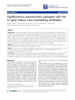
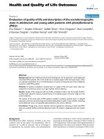
Figure 1 Identification of Implanted ADMSCs and CD31+ Cells in LV Myocardium. Confocal imaging study on day 90 following dilated
cardiomyopathy (DCM induction). Merged image (C) of double staining [troponin-I (A) plus Dil (B) (yellow arrows)] in Group 3 (ADMSC-treated)
showing few troponin I-positive myogenic-like cells (pink arrows) and undifferentiated adipose-derived mesenchymal stem cells (ADMSCs)
(yellow arrows) in LV myocardium. Merged image (F) of double staining [troponin-I (D) plus Dil (E) (yellow arrows)] in Group 5 (combined
ADMSCs and sildenafil) showing some troponin I-positive myogenic-like cells (pink arrows) and undifferentiated ADMSCs (yellow arrows) in LV
myocardium. CD31-positively stained cells in Group 3 (G) and Group 5 (J) indicating endothelial phenotype. Confocal image study
demonstrating rich engrafting of Dil-positively stained ADMSCs (yellow arrows) in LV myocardium of Group 3 (H) and Group 5 (K). The mean
CD-31 positively stain areas (pink arrows) were significantly higher (M) in Group 5 (L) than in Group 3 (I).
Lin et al. Journal of Translational Medicine 2010, 8:88
/>Page 6 of 16
in groups 3 and 4 than group 5, and notably higher in
group 5 than in group 1 (i.e. negative staining).
CD40+ cell Expression in LV Myocardium and Intensity of
Oxidative Stress
To determine whether inflammatory cells were up-regu-
lated in LV myocardium on day 90 following DCM
induction, immunohistochemical staining for detection
of CD40-positively stained cells was performed (Figure
3A-F). Density of CD40-positively stained cells in LV
myocardium were significantly higher in group 2 than in
other groups, significantly higher in groups 3 and 4 than
in groups 1 and 5, and also notably higher in group 5
than in group 1.
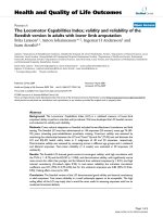
Figure 2 Apoptosis and Fibrosis in LV Myocardium. TUNEL assay (400×) of apoptotic nuclei (A-E) (red arrows) of LV myocardium on day 90
following DCM induction (n = 8). F) * p < 0.0001 between the indicated groups. Symbols (*, †, ‡) indicate significant difference (at 0.05 level) by
Tukey multiple comparison procedure. Scale bars in right lower corner represent 20 μm. Mean fibrotic area (μm
2
)/high-power field (HPF) (200×)
in each group (n = 8) of rats on day 90 following DCM induction. Masson’s trichrome stain (G-K) demonstrating markedly increased fibrosis area
(yellow arrows) in DCM group compared to other groups. L) * p < 0.001 between the indicated groups. Symbols (*, †, ‡, §, ¶) indicate significant
difference (at 0.05 level) by Tukey multiple comparison procedure. Scale bars in right lower corner represent 50 μm.
Lin et al. Journal of Translational Medicine 2010, 8:88
/>Page 7 of 16
The oxidative stress in mitochondria did not differ
between groups 1 and 5, groups 3 and 4, and groups 4
and 5 on day 90 following DCM induction (Figure 3G).
However,asignificantlyhigher mitochondrial oxidative
stress was noted in group 2 than in other groups, and in
groups3and4thaningroup1.Theoxidativestress
was also notably higher in group 3 than in group 5.
Protein Expressions of Cytochrome C and Cx43 in LV
Western blotting for Cx43 in LV demonstrated that
Cx43proteinexpressionwassimilar between groups 1
and 5 and between groups 3 and 4 (Figure 4A). How-
ever, this protein expression was substantially lower in
group 2 than in other groups and notably lower in
groups 3 and 4 than in groups 1 and 5 (Figure 4A).
The total amount of cytochrome C protein expression
in mitochondria was similar among groups 3 and 4, and
was also similar between group 1 and group 5 (Figure
4B). However, this protein expression in mitochondria
was sig nificantly lower in group 2 than in other gr oups,
was also notably lower in groups 3 and 4 than in groups
1 and 5. The total cytochrome C protein expression in
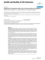
Figure 3 CD40+ Cell Expression in LV Myocardium and Intensity of Oxidative Stress. Immunohistochemical staining (400×) (A-E) for
identifying CD40-positive cells (red arrows) in LV myocardium on day 90 following DCM induction (n = 8 in each group). F) * p < 0.0001
between the indicated groups. Symbols (*, †, ‡, ¶) indicate significant difference (at 0.05 level) by Tukey multiple comparison procedure. Scale
bars in right lower corner represent 20 μm. Western blotting results (G) of oxidative index, protein carbonyls, in LV myocardium on day 90
following DCM induction (upper panel), with quantification results of each group (n = 8) (lower panel). * p < 0.0003 between the indicated
groups. Symbols (*, †, ‡, ¶) indicate significant difference (at 0.05 level) by Tukey multiple comparison procedure. Note: Right lane and left lane
shown on upper panel represent control oxidized molecular protein standard and protein molecular weight marker, respectively.
Lin et al. Journal of Translational Medicine 2010, 8:88
/>Page 8 of 16
cytosol did not differ between groups 1 and 5, as well as
between groups 3 and 4 (Figure 4C). However, this cyto-
solic protein expression was significantly higher in group
2 than i n other groups, and notably higher in groups 3
and 4 than in groups 1 and 5. These findings indicate
that the expression of cytochrome C, an index of energy
supply and storage in mitochondria, was notably lower
in group 2 than in groups 1 and 5. The increase in cyto-
solic cytochrome C content also suggested significant
mitochondrial damage with cytochrome C release into
the cytosol in the myocardium of group 2 animals.
RT-PCR of LV Myocardium on Day 90 Following DCM
Induction
The mRNA expression of matrix metalloproteinase-9
mRNA, an indicator of inflammat ion, was markedly
higher in group 2 than in other groups, notably higher
in groups 3 and 4 than in groups 1 and 5 (Figure 5A).
Conversely, interleukin (IL)-10 mRNA expression, an
index of anti-inflammation, was significantly lower in
group 2 than in other groups, notably lower in groups 3
and 4 than in groups 1 and 5, and significantly lower in
group 5 than in group 1 (Figure 5B). Additionally,
eNOS mRNA expression, an index of anti-inflammation
and endothelial function, was notably lower in group 2
than in other groups, significantly lower groups 3 and 4
than in group 1 (Figure 5C). On the other hand, this
mRNA expression was simil ar between group 1 and
group 5, and was also similar among groups 3, 4, and 5.
The mRNA expressions of caspase 3 (Figure 5D) and
Bax (Figure 5E), indexes of apoptosis, were remarkably
higher in group 2 than in other groups, markedly higher
in groups 3, 4, and 5 than in group 1, and also notably
lower in groups 3 and 4 than in group 5. In contrast,
mRNA expression of Bcl-2, an index of anti-apoptosis,
was sig nificantly lower in group 2 than in other gr oups,
significantlyloweringroups3and4thaninthanin
groups 1 and 5, and also significantly lower in group 5
than in group 1 (Figure 5F). The IL-8/Gro mRNA
expression, an essential chemokine guiding stem cell
homing from bone marrow to damaged myocardium
[23], was notably lower in groups 1 and 2 t han in other
groups, significantly lower in groups 3 and 4 than in
group 5 (Figure 5G). Conversely, the stromal cell-
derived factor (SDF) -1a mRNA expression, an index of
endothelial proge nitor cell chemo kine attractant, wa s
markedlyincreasedingroup2thaninothergroups,
notably increased in groups 3 and 4 than in groups 1
and 5 (Figure 5H).
The peroxisome proliferator activated receptor-g coac-
tivator (PGC)-1a mRNA expression, an energy tran-
scription marker, did not differ between gro ups 1 and 5,
or among groups 3, 4, and 5. On the other hand, the
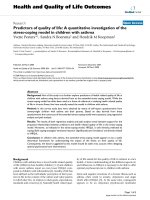
Figure 4 Protein Expressions of Cytochrome C and Cx43 in LV
Myocardium. (A) Connexin43 protein expression of LV myocardium
on day 90 after DCM induction. * p < 0.0007 between the indicated
groups. B) Cytochrome C protein expression in mitochondria of LV
myocardium on day 90 after DCM induction. * p < 0.009 between
the indicated groups. C) Cytochrome C protein expression in
cytosol of LV myocardium on day 90 after DCM induction. * p <
0.002 between the indicated groups. All symbols (*, †, ‡,¶)inA), B)
and C) indicate significant difference (at 0.05 level) by Tukey
multiple comparison procedure (n = 8 in each group)
Lin et al. Journal of Translational Medicine 2010, 8:88
/>Page 9 of 16
mRNA expression was notably lower in group 2 than in
other groups, and significantly lower in groups 3 a nd 4
than in group 1 (Figure 5I).
Protein Level of eNOS, SDF-1a, Caspase 3 and Bcl-2 of LV
Myocardium on Day 90 Following DCM Induction
Western blot was performed to determine whether the
initially elicited mRNA expressions of eNOS, SDF-1a,
caspase 3 and Bcl-2 participated in translation (Figure
6). The finings showed consistent changes in protein
production compared with mRNA expressions.
Small Arteriolar Density Analysis and Cardiac Hypotrophic
Gene Expression
The number of small arterioles (Figure 7A-F) (≤ 25 μmin
diameter) in LV myocardium was remarkably lower in
group 2 than in other groups. Moreover, the number of
small arterioles was notably lower in group 1 than in
groups 3, 4, and 5, sig nificantly lower in group 4 than in
groups 3 and 5, and also notably lower in group 3 than in
group 5. This finding indicates that early combined treat-
ment with sildenafil and ADMSCs is better than ADMSCs
or sildenafil alone in inducing angiogenesis/vasculogenesis.
Cardiac hypertrophy is characterized by a switch of
mRNA expression from a-tob-myosin heavy chain
(MHC) (i.e. reactivation of fetal gene program) [24]. In
the present study, the mRNA expression of b-MCH was
significantly higher in group 2 than in other groups,
notablyhigheringroups3,4,and5thaningroup1,
and also significantly higher in groups 3 and 4 than in
group 5 (Figure 7H). No significant difference was
noted, however, between group 3 and group 4. On the
other hand, a-MHC in LV was expressed in a reversed
manner in these groups (Figure 7G).
Discussion
Effect of Combined Therapy with ADMSCs and Sildenafil
on Early DCM
Our recent study demonstrated an increase in h eart
weight and LV remodeling i n the rat D CM model [13].
Figure 5 RT-PCR of LV Myocardium on Day 90 Following DCM Induction. The mRNA expressions of A) matrix metalloproteinase (MMP)-9, *
p < 0.0001 between the indicated groups; B) interleukin (IL)-10, * p < 0.0001 between the indicated groups; C) endothelial nitric oxide synthase
(eNOS), * < 0.0008 between the indicated groups; D) caspase 3, * p < 0.0004 between the indicated groups; E) Bax, * p < 0.0002 between the
indicated groups; F) Bcl-2, * p < 0.0001 between the indicated groups; G) IL-8/Gro, * p < 0.0001 between the indicated groups; H) stromal cell-
derived factor(SDF)-1a, * p < 0.0001 between the indicated groups; I) peroxisome proliferator activated receptor-g coactivator(PGC)-1a,*p<
0.002 between the indicated groups. All symbols (*, †, ‡,¶)inA) to I) indicate significant difference (at 0.05 level) by Tukey multiple comparison
procedure (n = 8 in each group).
Lin et al. Journal of Translational Medicine 2010, 8:88
/>Page 10 of 16
One interesting finding in the present study is that the
left lung weight was notably higher in group 2 than in
other groups on day 90 after DCM induction. This
finding may implicate that an increase in left lung
weight in DCM rat resulted in a sequestration of trans-
udate due to CHF. Additionally, the heart weight was
notably increased in group 2 than in groups 3 and 4,
and it was remarkably increased as compared with
group 5. Furthermore, RT-PCR showed substantially
higher expression of the b-MHC gene in LV in group
2 than in other groups, whereas an opposite trend was
noted in a-MHC gene expression in group 2 compared
to other groups. Moreover, the arterial blood pressure
did not differ among the five groups. These findings,
in addition to supporting the reproducibility of results
using our DCM model [13], further indicate that either
sildenafil or ADMSC therapy offered similar effect on
attenuating the progression of the hypertrophic
changes in DCM that were not due to the alternation
in the blood pressure. Of importance is the fact that
combined therapy with ADMSCs and sildenafil is
superior to either therapy alone in abrogating the pro-
gression of DCM.
Lack of Evidence Supporting Differentiation of ADMSCs
into the Myogenic-Like Cells for Preserving Heart
Function
Serial echocardiographic measurements in the current
study showed that LV function was significantly pre-
served in the DCM animals with ADMSC therapy com-
pared with that in the DCM group without treatment
on day 90 after DCM induction. Recently, we have
demonstrated that bone marrow-derived mononuclear
cell therapy alleviated left ventricular remodeling and
improved heart f unction in rat DCM [13]. Accordingly,
the results of our present study reinforce the findings of
our recent report [13]. Interestingly, flow cytome tric
analysis in the present study showed only a few cells
that were positively stained for troponin-I prior to or on
day 14 of cell culturing. Additionally, confocal image
study identified that only a few implanted ADMSCs
actually differentiated into troponin-I positively stained
cells in LV myocardium, a phenotype o f myogenic-like
cells. In contrast, a fairly la rge number of engrafted
ADMSCs were found to exhibit the undifferentiated
phenotype in LV myocardium on confocal microscopic
examination on day 90 following DCM induction. These
findings raise the suspicion that the number of myo-
genic-like cells in the LV myocardium differentiated
from ADMSCs is insufficient for sustaining cardiac
function. Other confounding fa ctors, therefore, may
contribute to the preservatio n of heart function in DCM
after cellular therapy.
Synergic Action of Early Combined Therapy with
Sildenafil and ADMSCs in Preservation of LV Function in
Rat DCM
Interestingly, while the effect of si ldenafil on improving
outcome of pulmonary arterial hypertension through
enhanced vasodilatatory effect of cGMP [18,19] has
been extensively investigated in b oth clinical trials [25]
and experimental studies [23,26,27], data regarding the
impact of sildenafil on improving clinical outcome of
patients with DCM has been seldom reported [20].
Thus, the role of sildenafil in the DCM setting is cur-
rently unclear. An important finding in the current
study was that sildenafil therapy offered similar effect
compared with ADMSC therapy on preservation of
heart function in DCM rats. Accordingly, our finding
strengthens the finding o f previous study [20]. Anot her
importan t finding in the current study is that combined
therapy with ADMSCs and sildenafil more significantly
preserved rat LV function than either ADMSCs or silde-
nafil alone on days 30 and 90 after DCM induction.
These findings, therefore, highlight a potential role of
this combined therapy in translational clinical applica-
tion in patients with DCM.
Possible Mechanisms Underlining Improvements of Heart
Function in Setting of DCM Following Cellular and
Sildenafil Therapy
Recently, studies have demonstrated that angiogenesis/
vasculogenesis play an essential role in improving ische-
mia-related LV dysfunction [10-13,25,28]. In the present
study, we found that the number of small vessels and
CD31-positively stained cells, an surface marker of
endothelial cells, in LV myocardium were remarkably
higher in DCM rats treated with ADMSCs or sildenafil
than in those DCM animals without treatment, whereas
it was signi ficantly higher in the combined therapy
group than in other groups. Moreover, eNOS gene and
protein expressio ns, an index of endothelial function
and angiogenesis [22], were found to show a similar
increase comparable to the number of small vessels and
CD31-positively stained cells in LV myocardium of each
group. Interestingly, previous experimental study has
demonstrated that sildenafil enhanced eNOS mRNA
expression [22]. Our findings, in addition to co rroborat-
ing the results of recent studies [10-13,23,25,28], may at
least in part explain the mechanisms underlying sildena-
fil and cell therapy in preserving LV function in the
rodent DCM model. Besides, our results also revealed
the difference in therapeutic benefits offered b y the
three regimens in our experiment setting.
Growing data suggest that cytokine effect [12,13,28] is
another important mechanism underlying the restora-
tion of ischemia-related LV dysfunction. Both SDF-1a
Lin et al. Journal of Translational Medicine 2010, 8:88
/>Page 11 of 16
and IL-8/Gro CXC chemokines have been found to be
crucial in the mobilization, incorporation, homing, sur-
vival, proliferation, and differentiation of stem cells
[29,30]. In the current study, IL-8/Gro mRNA expres-
sion was found to be notably higher in animals receiving
either ADMSC or sildenafil therapy and remarkably
increased in the combined treatment group as compared
with DCM alone. Our finding was comparable to those
of previous studies [29,30]. Interestingly, mRNA and
protein expressions of SDF-1a were found to show a
negative correlation with that of IL-8/Gro in the
animals. This finding may suggest that higher level of
SDF-1 may be secr eted by theischemictissuein
response to the severity of ischemia to attract the
endothelial progenitor cells for tissue repair.
The link between an increase of inflammation/reactive
oxygen species (ROS) and apoptosis/cellular death in
ischemic myocardium has been established [12,13,31-33].
Accordingly, our results demonstrated remarkably
increased gene and protein expressions of MMP-9, the
number of CD40-positively stained cells, and oxidative
stress in group 2 compared with other groups. These
Figure 6 Protein Expressions of eNOS, SDF-1a, Caspase 3 and Bcl-2 in LV Myocardium on Day 90 after DCM Induction. A) eNOS protein
expression. * p < 0.01 between the indicated groups. B) SDF-1a protein expression. * p < 0.045 between the indicated groups C) Caspase 3
protein expression. * p < 0.05 between the indicated groups. D) Bcl-2 protein expression. * p < 0.05 between the indicated groups. All symbols
(*, †, ‡,¶)inA) to D) indicate significant difference (at 0.05 level) by Tukey multiple comparison procedure (n = 8 in each group).
Lin et al. Journal of Translational Medicine 2010, 8:88
/>Page 12 of 16
parameters were also notably elevated in groups 3 and
4 than in group 5. In contrast, mRNA expressions of
IL-10 and eNOS, the anti-inflammatory indicators,
were lowest in group 2 and significantly decreased in
groups 3 and 4 compared to that in group 5. Further-
more, the apoptotic biomarkers of apoptotic nuclei and
BaxaswellasmRNAandproteinexpressionsofcas-
pase 3 were notably higher, whereas both mRNA and
protein expressions of Bcl-2, an ant i-apoptosis biomar-
ker, was remarkably lowest in group 2. Stem cell ther-
apy has been proposed to be immune-modulatory and
anti-inflammatory through down-regulating both
innate and adaptive immunity [13,34]. Additionally, sil-
denafil has been found to possess anti-inflammatory,
anti-fibroproliferative, and an ti-apoptotic properties
[23]. Our findings not only strengthen the hypothesis
[13,23] and the findings of previous report [34], but
also provide insight into the mechanisms underlying
the reduction in fibrosis and cellular apoptosis of LV
myocardium in rodent DCM after ADMSC treatment.
Besides, our findings further supports that combined
therapy with ADMSCs and sildenafil provide an addi-
tional benefit compared to either ADMSC or sildenafil
therapy alone in significantly limiting DCM-related
cardiac dysfunction.
The principal finding in the current s tudy is that RT-
PCR showed a markedly lower mRNA expressions of
PGC-1a which is a transcriptional coactivator of oxidative
Figure 7 Small Arteriolar Densit y Analysis and Cardiac Hypotrophic Gene Expression. a-SMA immunohistochemical staining (A-E) (200×)
for the number of small arterioles (≤ 25 μm in diameter) in LV myocardium on day 90 after DCM induction. The results showing notably lower
small vessel (red arrows) number in DCM group than in other groups. F) * p < 0.0001 between the indicated groups. Scale bars in right lower
corner represent 50 μm (n = 8 in each group). The mRNA expressions of a-myosin heavy chain (MHC) (G) and b-MHC of LV myocardium on day
90 after DCM induction. G) * p < 0.003 between the indicated groups. H) * p < 0.0003 between the indicated groups. All symbols (*, †, ‡,§,¶)
in F), G) and H) indicate significant difference (at 0.05 level) by Tukey multiple comparison procedure (n = 8 in each group).
Lin et al. Journal of Translational Medicine 2010, 8:88
/>Page 13 of 16
metabolism, mitochondrial metabolism and biogenesis
[13,35,36] in group 2 than in other groups, and lower in
groups 3 and 4 than in group 5. Moreover, Western blot
analysis identified a significantly lower mitochondrial cyto-
chrome C content in group 2 compared with that in other
groups. It was also notably lower in groups 3 and 4 than
in group 5, whereas its cytosolic counterpart showed
respectively o pposi te changes in the study groups. These
findings suggest a significant preservation of mitochondrial
integrity and functions from combined treatment. Further-
more, changes in Cx43 expression pattern have been
reported to be associated with various cardiac pathologies
and contribute to the development of cardiac arrhythmia
[37]. The reduction in Cx43 protein expression in a DCM
setting implies a perturbation in cell-to-cell interconnec-
tions [13] and hence electrical coupling and cellular signal
transductions [37]. Of impo rtance in the present study is
that combined therapy with ADMSCs and sildenafil was
better than either therapeutic option alone in preventing
the down- regulation of Cx43 expressio n in the rodent
DCM model. Therefore, not only may the current study
provide explanations for the improved cardiac function
after combined therapy with ADMSCs and sildenafil in
the DCM animals, it also further strengthens the findings
from previous studies [13,35-37].
Study Limitation
This study has limitations. First, although sildenafil has
been clearly shown to cause vasodilatation through an
increase of cGMP concentration in smooth muscle
[17,18], the mechanisms through which sildenafil
enhanced ADMSCs’ participation in the process of myo-
cardial regeneration has not been investigated in thi s
study. The precise role of cGMP-dependent signal ing in
the setting of DCM, therefor e, remains unclear. Second,
except for LVEF and arterial blood pressure, other phy-
siological parameters for monitoring LV remodeling
including pressure-volume loop, left vent ricular end-dia-
stolic pressure, and pulmonary vascular resistance were
not provided in the current study.
In con clusion, our results demonstrated that early
combined treatment with ADMSCs and sildenafil for
DCM rats not only is superior to either ADMSC or sil-
denafil alone through eliciting serial molecular-cellular
biological effects in the preservation of LV function.
These findings may raise the need for further
Figure 8 The Proposed Mechanisms. ADMSCs: Adipose-derived mesenchymal stem cells; ROS: Reactive oxygen species; PGC-1a:Peroxisome
proliferator activated receptor-g coactivator-1a; MMP-9: Matrix metalloproteinase-9; eNOS: Endothelial nitric oxide synthase; SDF-1a: Stromal cell-
derived factor-1a; LVEF: Left ventricular ejection fraction; Cx43: Connexin 43
Lin et al. Journal of Translational Medicine 2010, 8:88
/>Page 14 of 16
prospective studies on assessing the therapeutic poten-
tial of combined ADMESC-sildenafil regimen in human
subjects with DCM. The proposed mechanisms underly-
ing the potential impacts of combined ADMSC-sildena-
fil therapy against DCM rats have been summarized in
Figure 8.
Acknowledgements
This study is supported by a program grant from Chang Gung Memorial
Hospital, Chang Gung University (grant no. CMRPG 881241).
Author details
1
Division of cardiology, Department of Internal Medicine, Chang Gung
Memorial Hospital-Kaohsiung Medical Center, Chang Gung University
College of Medicine, Kaohsiung, Taiwan.
2
Center for Translational Research in
Biomedical Sciences, Chang Gung Memorial Hospital-Kaohsiung Medical
Center, Chang Gung University College of Medicine, Kaohsiung, Taiwan.
3
Division of General Surgery, Department of Surgery, Chang Gung Memorial
Hospital-Kaohsiung Medical Center, Chang Gung University College of
Medicine, Kaohsiung, Taiwan.
4
Department of Life Science, National
Pingtung University of Science and Technology, Pingtung, Taiwan.
5
Department of Medical Research, E-DA Hospital, I-Shou University,
Kaohsiung, Taiwan.
6
Basic Science, Nursing Department, Meiho University,
Pingtung, Taiwan.
7
Department of Radiology, Chang Gung Memorial
Hospital-Kaohsiung Medical Center, Chang Gung University College of
Medicine, Kaohsiung, Taiwan.
8
Division of Cardiovascular Surgery,
Department of Surgery, Chang Gung Memorial Hospital-Kaohsiung Medical
Center, Chang Gung University College of Medicine, Kaohsiung, Taiwan.
Authors’ contributions
All authors have read and approved the final manuscript. Dr. Fan-Yen Lee
contributed equally to this work compared with the corresponding author.
YCL, CKS, and SL designed the experiment, drafted the manuscript, and
performed animal experiments. LTC, CHY, THT, SC, MF, SFK, CJW, and YHK
were responsible for the laboratory assay and troubleshooting. FYL and HKY
participated in refinement of experiment protocol and coordination and
helped in drafting the manuscript.
Competing interests
The authors declare that the y have no competing interests.
Received: 21 June 2010 Accepted: 26 September 2010
Published: 26 September 2010
References
1. Garg R, Yusuf S: Overview of randomized trials of angiotensin-converting
enzyme inhibitors on mortality and morbidity in patients with heart
failure. Collaborative Group on ACE Inhibitor Trials. Jama 1995,
273:1450-1456.
2. Pitt B, Poole-Wilson PA, Segal R, Martinez FA, Dickstein K, Camm AJ,
Konstam MA, Riegger G, Klinger GH, Neaton J, et al: Effect of losartan
compared with captopril on mortality in patients with symptomatic
heart failure: randomised trial–the Losartan Heart Failure Survival Study
ELITE II. Lancet 2000, 355:1582-1587.
3. Solomon SD, Wang D, Finn P, Skali H, Zornoff L, McMurray JJ, Swedberg K,
Yusuf S, Granger CB, Michelson EL, et al: Effect of candesartan on cause-
specific mortality in heart failure patients: the Candesartan in Heart
failure Assessment of Reduction in Mortality and morbidity (CHARM)
program. Circulation 2004, 110:2180-2183.
4. Frazier OH, Benedict CR, Radovancevic B, Bick RJ, Capek P, Springer WE,
Macris MP, Delgado R, Buja LM: Improved left ventricular function after
chronic left ventricular unloading. Ann Thorac Surg 1996, 62:675-681,
discussion 681-672.
5. Dandel M, Weng Y, Siniawski H, Potapov E, Lehmkuhl HB, Hetzer R: Long-
term results in patients with idiopathic dilated cardiomyopathy after
weaning from left ventricular assist devices. Circulation 2005, 112:I37-45.
6. Poole-Wilson PA, Swedberg K, Cleland JG, Di Lenarda A, Hanrath P,
Komajda M, Lubsen J, Lutiger B, Metra M, Remme WJ, et al: Comparison of
carvedilol and metoprolol on clinical outcomes in patients with chronic
heart failure in the Carvedilol Or Metoprolol European Trial (COMET):
randomised controlled trial. Lancet 2003, 362:7-13.
7. Di Lenarda A, Remme WJ, Charlesworth A, Cleland JG, Lutiger B, Metra M,
Komajda M, Torp-Pedersen C, Scherhag A, Swedberg K, Poole-Wilson PA:
Exchange of beta-blockers in heart failure patients. Experiences from the
poststudy phase of COMET (the Carvedilol or Metoprolol European
Trial). Eur J Heart Fail 2005, 7:640-649.
8. Tomita S, Li RK, Weisel RD, Mickle DA, Kim EJ, Sakai T, Jia ZQ: Autologous
transplantation of bone marrow cells improves damaged heart function.
Circulation 1999, 100:II247-256.
9. Strauer BE, Brehm M, Zeus T, Kostering M, Hernandez A, Sorg RV, Kogler G,
Wernet P: Repair of infarcted myocardium by autologous intracoronary
mononuclear bone marrow cell transplantation in humans. Circulation
2002, 106:1913-1918.
10. Davani S, Marandin A, Mersin N, Royer B, Kantelip B, Herve P, Etievent JP,
Kantelip JP: Mesenchymal progenitor cells differentiate into an
endothelial phenotype, enhance vascular density, and improve heart
function in a rat cellular cardiomyoplasty model. Circulation 2003,
108(Suppl 1):II253-258.
11. Dai W, Hale SL, Martin BJ, Kuang JQ, Dow JS, Wold LE, Kloner RA:
Allogeneic mesenchymal stem cell transplantation in postinfarcted rat
myocardium: short-and long-term effects. Circulation 2005, 112:214-223.
12. Yip HK, Chang LT, Wu CJ, Sheu JJ, Youssef AA, Pei SN, Lee FY, Sun CK:
Autologous bone marrow-derived mononuclear cell therapy prevents
the damage of viable myocardium and improves rat heart function
following acute anterior myocardial infarction. Circ J 2008,
72:1336-1345.
13. Sun CK, Chang LT, Sheu JJ, Chiang CH, Lee FY, Wu CJ, Chua S, Fu M,
Yip HK: Bone marrow-derived mononuclear cell therapy alleviates left
ventricular remodeling and improves heart function in rat-dilated
cardiomyopathy. Crit Care Med 2009, 37:1197-1205.
14. Boolell M, Allen MJ, Ballard SA, Gepi-Attee S, Muirhead GJ, Naylor AM,
Osterloh IH, Gingell C: Sildenafil: an orally active type 5 cyclic GMP-
specific phosphodiesterase inhibitor for the treatment of penile erectile
dysfunction. Int J Impot Res 1996, 8:47-52.
15. Goldenberg MM: Safety and efficacy of sildenafil citrate in the treatment
of male erectile dysfunction. Clin Ther 1998, 20:1033-1048.
16. Ahn HS, Foster M, Cable M, Pitts BJ, Sybertz EJ: Ca/CaM-stimulated and
cGMP-specific phosphodiesterases in vascular and non-vascular tissues.
Adv Exp Med Biol 1991, 308:191-197.
17. Zhao L, Mason NA, Morrell NW, Kojonazarov B, Sadykov A, Maripov A,
Mirrakhimov MM, Aldashev A, Wilkins MR: Sildenafil inhibits hypoxia-
induced pulmonary hypertension. Circulation 2001, 104:424-428.
18. Shekerdemian LS, Ravn HB, Penny DJ: Intravenous sildenafil lowers
pulmonary vascular resistance in a model of neonatal pulmonary
hypertension. Am J Respir Crit Care Med 2002, 165:1098-1102.
19. Banas A, Teratani T, Yamamoto Y, Tokuhara M, Takeshita F, Osaki M,
Kawamata M, Kato T, Okochi H, Ochiya T: IFATS collection: in vivo
therapeutic potential of human adipose tissue mesenchymal stem cells
after transplantation into mice with liver injury. Stem Cells 2008,
26:2705-2712.
20. Behling A, Rohde LE, Colombo FC, Goldraich LA, Stein R, Clausell N: Effects
of 5’-phosphodiesterase four-week long inhibition with sildenafil in
patients with chronic heart failure: a double-blind, placebo-controlled
clinical trial. J Card Fail 2008, 14:189-197.
21. Muranaka H, Marui A, Tsukashita M, Wang J, Nakano J, Ikeda T, Sakata R:
Prolonged mechanical unloading preserves myocardial contractility but
impairs relaxation in rat heart of dilated cardiomyopathy accompanied
by myocardial stiffness and apoptosis. J Thorac Cardiovasc Surg 2010.
22. Yen CH, Leu S, Lin YC, Kao YH, Chang LT, Chua S, Fu M, Wu CJ, Sun CK,
Yip HK: Sildenafil Limits Monocrotaline-Induced Pulmonary Hypertension
in Rats through Suppression of Pulmonary Vascular Remodeling. J
Cardiovasc Pharmacol 2010, 55:574-584.
23. Kocher AA, Schuster MD, Bonaros N, Lietz K, Xiang G, Martens TP,
Kurlansky PA, Sondermeijer H, Witkowski P, Boyle A, et al: Myocardial
homing and neovascularization by human bone marrow angioblasts is
regulated by IL-8/Gro CXC chemokines. J Mol Cell Cardiol 2006,
40:455-464.
24. Nagaya N, Kangawa K, Itoh T, Iwase T, Murakami S, Miyahara Y, Fujii T,
Uematsu M, Ohgushi H, Yamagishi M, et al: Transplantation of
Lin et al. Journal of Translational Medicine 2010, 8:88
/>Page 15 of 16
mesenchymal stem cells improves cardiac function in a rat model of
dilated cardiomyopathy. Circulation 2005, 112:1128-1135.
25. Blanco I, Gimeno E, Munoz PA, Pizarro S, Gistau C, Rodriguez-Roisin R,
Roca J, Barbera JA: Hemodynamic and gas exchange effects of sildenafil
in patients with chronic obstructive pulmonary disease and pulmonary
hypertension. Am J Respir Crit Care Med 181:270-278.
26. Schermuly RT, Kreisselmeier KP, Ghofrani HA, Yilmaz H, Butrous G, Ermert L,
Ermert M, Weissmann N, Rose F, Guenther A, et al: Chronic sildenafil
treatment inhibits monocrotaline-induced pulmonary hypertension in
rats. Am J Respir Crit Care Med 2004, 169:39-45.
27. Tse HF, Kwong YL, Chan JK, Lo G, Ho CL, Lau CP: Angiogenesis in
ischaemic myocardium by intramyocardial autologous bone marrow
mononuclear cell implantation. Lancet 2003, 361:47-49.
28. Kucia M, Reca R, Miekus K, Wanzeck J, Wojakowski W, Janowska-
Wieczorek A, Ratajczak J, Ratajczak MZ: Trafficking of normal stem cells
and metastasis of cancer stem cells involve similar mechanisms: pivotal
role of the SDF-1-CXCR4 axis. Stem Cells 2005, 23:879-894.
29. Kee HJ, Sohn IS, Nam KI, Park JE, Qian YR, Yin Z, Ahn Y, Jeong MH, Bang YJ,
Kim N, et al: Inhibition of histone deacetylation blocks cardiac
hypertrophy induced by angiotensin II infusion and aortic banding.
Circulation 2006, 113:51-59.
30. Frangogiannis NG, Smith CW, Entman ML: The inflammatory response in
myocardial infarction. Cardiovasc Res 2002, 53:31-47.
31. Frangogiannis NG: The immune system and cardiac repair. Pharmacol Res
2008, 58:88-111.
32. Garrido AM, Griendling KK: NADPH oxidases and angiotensin II receptor
signaling. Mol Cell Endocrinol 2009, 302:148-158.
33. Thum T, Bauersachs J, Poole-Wilson PA, Volk HD, Anker SD: The dying stem
cell hypothesis: immune modulation as a novel mechanism for
progenitor cell therapy in cardiac muscle. J Am Coll Cardiol 2005,
46:1799-1802.
34. Garnier A, Fortin D, Delomenie C, Momken I, Veksler V, Ventura-Clapier R:
Depressed mitochondrial transcription factors and oxidative capacity in
rat failing cardiac and skeletal muscles. J Physiol 2003, 551:491-501.
35. Suwa M, Nakano H, Kumagai S: Effects of chronic AICAR treatment on
fiber composition, enzyme activity, UCP3, and PGC-1 in rat muscles. J
Appl Physiol 2003, 95:960-968.
36. Kirchhoff S, Nelles E, Hagendorff A, Kruger O, Traub O, Willecke K: Reduced
cardiac conduction velocity and predisposition to arrhythmias in
connexin40-deficient mice. Curr Biol 1998, 8:299-302.
37. Peters NS, Green CR, Poole-Wilson PA, Severs NJ: Cardiac
arrhythmogenesis and the gap junction. J Mol Cell Cardiol
1995, 27:37-44.
doi:10.1186/1479-5876-8-88
Cite this article as: Lin et al.: Early combined treatment with sildenafil
and adipose-derived mesenchymal stem cells preserves heart function
in rat dilated cardiomyopathy. Journal of Translational Medicine 2010 8:88.
Submit your next manuscript to BioMed Central
and take full advantage of:
• Convenient online submission
• Thorough peer review
• No space constraints or color figure charges
• Immediate publication on acceptance
• Inclusion in PubMed, CAS, Scopus and Google Scholar
• Research which is freely available for redistribution
Submit your manuscript at
www.biomedcentral.com/submit
Lin et al. Journal of Translational Medicine 2010, 8:88
/>Page 16 of 16