Báo cáo hóa học: " S110, a novel decitabine dinucleotide, increases fetal hemoglobin levels in baboons (P. anubis)" potx
Bạn đang xem bản rút gọn của tài liệu. Xem và tải ngay bản đầy đủ của tài liệu tại đây (1.09 MB, 8 trang )
RESEARC H Open Access
S110, a novel decitabine dinucleotide, increases
fetal hemoglobin levels in baboons (P. anubis)
Donald Lavelle
1,2*
, Yogen Saunthararajah
1,3
, Kestis Vaitkus
1,2
, Mahipal Singh
1,2,5
, Virryan Banzon
1,2
,
Pasit Phiasivongsva
4
, Sanjeev Redkar
4
, Sarath Kanekal
4
, David Bearss
4
, Chongtie Shi
4
, Roger Inloes
4
,
Joseph DeSimone
1,2
Abstract
Background: S110 is a novel dinucleoside analog that could have advantages over existing DNA methyltransferase
(DNMT) inhibitors such as decitabine. A potential therapeutic role for S110 is to increase fetal hemoglobin (HbF)
levels to treat b-hemoglobinopathies. In these experiments the effect of S110 on HbF levels in baboons and its
ability to reduce DNA methylation of the g-globin gene promoter in vivo were evaluated.
Methods: The effect of S110 on HbF and g-globin promoter DNA methylation was examined in cultured human
erythroid progenitors and in vivo in the baboon pre-clinical model. S110 pharmacokinetics was also examined in the
baboon model.
Results: S110 increased HbF and reduced DNA methylation of the g-globin promoter in human erythroid
progenitors and in baboons when administered subcutaneously. Pharmaco kinetic analysis was consistent with
rapid conversion of S110 into the deoxycytosine analog decitabine that binds and depletes DNA.
Conclusion: S110 is rapidly converted into decitabine, hypomethylates DNA, and induce s HbF in cultured human
erythroid progenitors and the baboon pre-clinical model.
Background
Increased fetal hemoglobin levels are beneficial to patients
with sickle cell disease and b-thalassemia. Patients with
sickle cell disease with increased fetal h emoglobin levels
have less pain crises [1] and longer life spans [2]. Th ere-
fore pharmacolog ical agents that can elevate fetal hemo-
globin have great potential as therapeutic agents. The
DNA methyltransferase (DNMT) inhibitors 5-azacytidine
and 5-aza-2’deoxycyidine (decitabine) have been shown to
increase fetal hemoglobin levels in clinical trials in patients
with sickle cell diseas e [3 -6]. Although the clinical effec-
tiveness of decitabine in alleviating the symptoms asso-
ciated with the d isease remains to be demonstrated in
multi-center clinical trials, recent results in patients with
severe sickle cell disease strongly suggest that this agent
may have a major impact on the treatment of this disease
[7]. Although decitabine and 5-azacytidine have a potential
role as HbF inducers to treat b-hemoglobinopathies, these
agents have pharmacological limitations including rapid
destruction by the enzyme cytidine deaminase that is the
principal barrier to oral administration [8,9]. The novel
dinu cleotide S110 (Figure 1) can also inhibit DNMT and
is resistant to cytidine deaminase [10]. Hence, S110 could
have advantages as a potential HbF inducer.
In this investigation our goal was to determine
whether S110 increa sed fetal hemoglobin level s and
reduced DNA methylation in cultured human erythroid
progenitor cells and in baboons. Our results indicate
that S110 administered by subcutaneous injection is
rapidly converted to decitabine, hypomethylates the
g-globin gene promoter, and induces HbF. These results
are the first demonstration t hat S110, a novel decitabine
dinucleotide compound, can increase fetal hemoglobin
and cause DNA hypomethylation in vivo and represent
an import ant step towards understanding if S110 has a
potential role in the treatment of b-hemoglobinopathies.
* Correspondence:
1
Department of Medicine, University of Illinois at Chicago, 840 S. Wood St.
Chicago, Illinois 60612-7323, USA
Full list of author information is available at the end of the article
Lavelle et al. Journal of Translational Medicine 2010, 8:92
/>© 2010 Lavelle et al; licensee BioMed Central Ltd. T his is an Open Access article distributed under the terms of the Creative Commons
Attribution License ( which permits unrestricted use, distribution, and reproduction in
any medium, provided the original work is properly cited.
Methods
Drugs
Decitabine and S110 were obtained from SuperGen, Inc,
Dublin, Ca.
Cell Culture
Frozen CD34+ human cells purified from the peripheral
blood of mobilized donors were purchased from Allcells,
Inc. These cells were cultured in Iscove’s media contain-
ing 20% fetal bovine serum, stem cell factor (SCF), ery-
thropoietin (epo), estradiol, and dexamethasone [11]. On
day 8, S110 or decitabine were added to the culture.
After 24 hours, cells were transferred to f resh Iscove’s
media supplemented with 20% fetal bovine serum, epo,
and insulin. One day 10, RNA was purified for analysis
of globin mRNA expression. On day 11, lysates were
prepared for high performance liquid chromatograp hy
(HPLC) analysis of globin chain expression and DNA
was isolated for bisulfite sequence analysis.
Baboon Treatments
Two baboons (P. anubis), PA 7256 and 7470, were used
in these experiments. Prior to drug treatment, animals
were phlebotomized to attain a hematocrit (Hct) of 20
by daily removal of 16-18% of the packed cell volume.
Each animal was treated initially with S110 (1 mg/kg /d)
for ten days, followed by a washout period prior to
initiation of the second cycle of phlebotomy and subse-
quent administration of decitabine (0.5 mg/kg/d). The
fir st do se of drug was administered IV followed by pro-
curement of samples for pharmacokinetic analysis, with
the remaining nine injections administered by subc uta-
neous injection on the subsequent days. Bone marrow
(BM) aspirations from the hips were performed follow-
ing the last day of drug administration. HbF levels were
determined by alkali denaturation [12] and confirmed
by H PLC [13]. All procedures were approved by Institu-
tional Animal Care and Use Committee ( IACUC) of the
University of Illinois at Chicago.
Figure 1 Comparison of structures of cytidine, 5-aza-2-deoxycytidine, 5-azacytidine, and S110.
Lavelle et al. Journal of Translational Medicine 2010, 8:92
/>Page 2 of 8
Real Time PCR Analysis of Globin mRNA
RNA was purified f rom cultured erythroid progenitors
using the RNeasy Mini Kit ( QIAGEN) according to
manufacturer’ s instructions. RNA was treated with
DNaseI(Ambion)andusedtopreparecDNAusing
kits (Ferment as). Levels of a-, g-andb-globin tran-
scripts were determined by rea l time PCR analysis using
Taqman probe and primer sets (Applied Biosystems).
Absolute numbers of a-, g-andb-globin transcripts
were determined by ext rapolation from standard curves
prepared from the cloned amplicons. Results were
expressed as g/ g + b mRNA ratio. Statistical significance
was assessed using a two-tailed T test.
HPLC analysis of Globin Chain Expression
For analysis of globin chain expression in cultured
human erythroid progenitor cells, cells (5-10 × 10
6
)
were harvested and washed three times in PBS. Lysates
were prepared by addition of H
2
O to the packed cell
pellet followed by three cycles of f reezing and thawing
in a dry-ice methanol bath. Analysis of globin chains
was performed on a TSP Spectra HPLC system using a
LiChristopher 100 RP-8 5 mM column and a gradient
of acetonitrile-methanol-NaCl as described [13]. Absor-
bance was monitored at 215 nm. Quantitation of globin
chains was performed by integra tion of peaks represent-
ing the separated a-, b-, and g-globin chains using
ChromQuest 4.1 software.
Bisulfite Sequence Analysis
The DNA methylation status of 5 CpG sites (-54, -51, +5,
+16, +48) within the 5’ g-globin promoter region was ana-
lyzed by bisulfite sequencing according to previously pub-
lished methods [14,15]. Nucleated erythroid cells were
purified from baboon bone marrow aspirates by Perco ll
density gradient sedimentation followed by immunomag-
netic column (Miltenyi) purification using an anti-baboon
red blood cell mouse monoclonal antibody (Clone E34-
731, #551299, BD Bioscience) as the primary reagent and
magnetically labeled rat anti-mouse IgG1 microbeads
(Miltenyi) as the secondary reagent. DNA was isolated
from purified bab oon nucleated erythroid bone marro w
cells and from cultured human e rythroid progenitors
using Qiagen blood mini kits. Bisulfite modification was
performed as described following digestion with Hind III.
The g-globin gene promoter region was amplified by two
rounds of PCR using semi-nested primers. The primer set
BG1 (TATGGTGGGAGAAGAAATTAGTAAAGG) and
BG2 (AATAACCTTATCCTCCTCTATAAAATAACC)
were used in the first round and BG2 and BG5
(GGTTGGTTAGTTTTGTTTTGATTAATAG) in the
second round. Amplicons were cloned in the PCR4 vector
in the TOP10 E. coli strain. At least ten independent
clones were sequenced from each sample.
Pharmacokinetic Studies
Blood samples were collected from the femoral vein
prior to drug administration (pre-dose) and 15, 30, 60,
120, 150, 180, and 240 minutes following intravenous
administration of either decitabine or S110 in 3 mL K
2
EDTA tubes pre-loaded with 8 μL of tetrahydrouridine
(THU-500 μg/mL solution) and maintained on ice.
Blood samples were centrifuged at 1,8 00 × g for 10 min
at 4°C. The resulting plasma was decanted into a screw
top tube and stored at -70°C until analyzed. S amples
were shipped to SuperGen, Inc. on dry ice for analy sis
of decitabine and S110 levels. Levels of decitabine a nd
S110 were determined using a liquid chromatography-
tandem mass spectrometry method [16]. Values for HL
LAMBDA (half life), T max (time of maximum concen-
tration), Cmax (concentration at Tmax), AUCall (area
under the curve from time of dosing to last observa-
tion), and AUCinf Obs (area under the curve from time
of dosing to infinity) were calculated using WinNonLin
version 5.0 (Pharsight).
Results
Effect of S110 in Human Erythroid Progenitor Cell
Cultures
Globin Transcripts
Initial experiments were performed in human erythroid
progenitor cell cultures to determine whether S110
increased g-globin expression. Human CD34+ cells,
purified from the peripheral blood of mobilized donors
(AllCells), were cultured as described [11]. Because
globin synthesis occurs between days 8 and 13 in these
cultures [11], drugs, either S110 (1 or 5 μM) or decita-
bine (1 μM), were added on day 8. Analysis of levels of
g-andb-globin mRNA 48 hours post-decitabine addi-
tion showed that the g/g+b mRNA ratio in drug-trea-
ted cells was increased approximately twofold (p < .05)
compared to untreated control cultures. (Table 1;
Figure 2A). No significant difference in the a/g+b
mRNA ratio was observed between untreated co ntrols
and drug-treated cultures.
Globin Chain Ratio
HPLC analysis of globin chain expression was also per-
formed in human er ythr oid progenitor cultures treated
with S 110 or decitabine. Analysis of lysates prepared 72
hours following drug addition showed that the g/g+b
chain ratio was increased 1.6 fold (p < .05) in cultures
treated with decitabine and S110 compared to untreated
controls. (Table 1; Figure 2B).
DNA Methylation of the g-globin Gene Promoter
Bisulfite sequence analysis was performed to determine
the effect of S110 on the level of DNA methylation of
the g-globin gene promoter. Marked DNA hypomethyla-
tion of the g-globin promoter was apparent following
Lavelle et al. Journal of Translational Medicine 2010, 8:92
/>Page 3 of 8
treatment with either decitabine or S110 compared to
untreated controls (Figure 3). The 1 × 10
-6
M decitabine
dose and the 5 × 10
-6
M S110 dose induced similar
levels of DNA hypomethylation
Effect of S110 in the Baboon
Fetal Hemoglobin S110 was administered to baboons to
evaluate its in vivo activity. Two phlebotomized
baboons, PA 7256 and 7470, were treated with S110
(1.0 mg/kg/d) for t en days. The first injection was given
IV and blood samples were obtained pharmacokinetic
studies. The remaining nine drug treatments were admi-
nistered by subcutaneous injection which avoids the
Table 1 Effect of S110 on g-globin expression in human
erythroid progenitor cell cultures
Treatment Dose
(μM)
g/g + b
mRNA
g/g + b
polypeptide chain
ratio
Control 0 0.162 ± .091 (n = 4) 18.3 ± 3.3 (n = 3)
Decitabine 1 0.337 ± .135 (n = 4) 29.8 ± 3.2 (n = 3)
S110 1 0.355 ± .038 (n = 4) 27.8 ± 1.9 (n = 3)
S110 5 0.310 ± .136 (n = 3) 29.2 ± 2.9 (n = 3)
The effect of decitabine and S110 on globin mRNA (n = 4) and globin chain
expression (n = 3) was measured in cultured human erythroid progenitor
cells. Difference in g/g+b mRNA and g/g+b chain ratios between untreated
controls and drug-treated cultures was significant (p < .05).
Figure 2 Comparison of the effects of S110 and decityabine on globin gene expression in cultured human erythroid progenitor cells.
A. Effect of decitabine and S110 on expression of g-globin mRNA in cultured human erythroid progenitor cells. Results are expressed as fold
change (± SD) relative to untreated controls. The difference in g/g+b mRNA between the untreated controls and drug-treated cultures was
significant (p < .05). B. Effect of decitabine and S110 on the g/g + b chain ratio in cultured human erythroid progenitor cells. The difference in
g/g + b chain ratio between the untreated controls and drug-treated cultures was significant (p < .05).
Lavelle et al. Journal of Translational Medicine 2010, 8:92
/>Page 4 of 8
need to anesthetize the baboons. An ident ical of cour se
of decitabine using an equivalent molar dose (0.5 mg/
kg/d), was given following a 60 day wash out period.
Induction of HbF occurred following admi nistration of
both S110 and decitabine. Individual differences in max-
imal HbF attained were observed between the two
baboons, and decitabine induced a slightly higher HbF
response in each. The kinetics of response to S110 and
decitabine were similar, with peak HbF attained approxi-
mately 10 days following the last day of drug adminis-
tration (Figure 4).
DNA Methylation of the g-globin Gene Promoter DNA
was isolated from purified BM erythroid precursor cells
obtained from baboons following the c ourse of S110
administration to evaluate the effect of the drug on
DNA methylation levels of the g-globin gene promoter.
The level of DNA methylation of 5 CpG sites within the
g-globin promoter was determined by bisulfite sequence
analysis. S110 induced DNA hypomethylation of these
CpG residues in both PA 7256 and 7470 compared to
bled controls (Figure 5). The level of DNA hypomethy-
lation of the g-globin promoter induced by S110 was
equivalent to that observed in three other baboons pre-
viously treated with decitabine [15].
Platelet and Neutrophils Both S110 and decitabine
induced similar effects on neut rophil and platelet counts.
Platelets counts rose approximately 2 weeks post-drug
administration. The rise in platelet counts was m irrored
by a decrease in neutrophils at this time following admin-
istration of both S110 and decitabine (Figure 6). This
effect was pr eviously observed in patie nts with sickl e cell
disease treated with decitabine [5].
Pharmacokinetic analysis A summary of the pharma-
cokinetic data obtained is presented in Table 2. In
baboons treated with S110, both S110 and decitabine
were detected following administration of the drug. Peak
levels of decitabine (17 ng/ml) were approximately 3
fold higher than peak levels of S110 (6 ng/ml) consistent
Figure 3 Comparison of the effects of S110 and decitabine on DNA methylation of the g-globin gene promoter region in cultured
human erythroid progenitor cells. The effects of decitabine and S110 on the DNA methylation of 5 CpG sites located within the 5’ g-globin
promoter region are shown. Red rectangles = methylated CpG; green rectangles = unmethylated CpG. Results are expressed as the %
deoxymethylcytosine (dmC) of cytosines located within CpG dinucleotides at positions -54, -51, +5, +16, and +48 with respect to the
transcriptional start site of the human g-globin gene promoter. Each row corresponds to the sequence analysis of an individual cloned PCR
product derived from bisulfite-treated DNA. Results for each CpG site (-54, -51, +5, +16, +48) are in each corresponding column.
Lavelle et al. Journal of Translational Medicine 2010, 8:92
/>Page 5 of 8
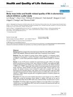
Figure 4 Comparison of the effects of S110 and dec itabine on fetal hemoglobin levels in baboons. Kinetics of change in fetal hemoglobin
levels during treatment with decitabine and S110 in PA 7256 and 7470. animals were treated with either S110 or decitabine between days 1-10.
Figure 5 Com parison of the effects of S110 and decitabine on DNA methylation of the g-globin gene promoter region in baboons.Red
rectangles = methylated CpG; green rectangles = unmethylated CpG, yellow rectangles = polymorphic sites where no CpG dinucleotides are present.
Results are expressed as the % deoxymethylcytosine (dmC) of cytosines located within CpG dinucleotides at positions -54, -51, +5, +16, and +48 with
respect to the transcriptional start site of the baboon g-globin gene promoter. Each row corresponds to the sequence analysis of an individual cloned
PCR product derived from bisulfite-treated DNA. Results at each CpG site (-54, -51, +5, +16, +48) are within each corresponding column.
Lavelle et al. Journal of Translational Medicine 2010, 8:92
/>Page 6 of 8
with a rapid conversion of S110 into decitabine.
Increased in vivo half life or AUC was not observed for
S110 compared to decitabine when these drugs were
administered intravenously.
Conclusion
Our results clearly demonstrate that subcutaneous admin-
istration of S110, a new decitabine dinucleotide, increases
expression of g -globin and reduces DNA methylation of
the g-globin promoter in cultured human erythroid pro-
genitor cells, and also in baboons. The ability of S110 to
induce HbF in vivo appears to be comparable to that of
decitabine. Both decitabine and S110 are inhibitors of
DNMT. The mechanism responsible for increased HbF by
DNMT inhibitors is a matter of current controversy, how-
ever [17,18]. Decitabine has been observed to activate p38
MAP kinase and increase the rate of terminal erythroid
different iation in cultured erythroid progenitor cells [19],
effects that have been associated with increased HbF
[20,21]. Both S110 and decitabine decrease the level of
DNA methylation of the g-globin promo ter, but the role
of DNA hypomethylation in the mechanism of action of
these drugs was not addressed in these experiments.
A previous report documented that S110 could
demethylate and reactivate the expression of a silenced
methylated p16INK4A tumor s uppressor gene in cancer
cell lines [10]. Results from these experiments strongly
sugge sted that S110 dinucleo tide was cleaved into indivi-
dual nucleotides and nucleosides that were incorporated
into DNA as the active form of the drug. It was specu-
lated that S110 entered the cell as a dinucleotide where it
was cleaved into its active form by phosphodiesterases.
Our results demonstrate that S110 is rapidly clea ved
in vivo into decitabine following intravenous administra-
tion. Pharmacokinetic analysis showed that levels of deci-
tabine were approximately 3 fold higher than those of
S110 following administration of S110. These results are
consistent with rapid conversion of S110 into decitabine
Figure 6 Comparison of the effects of decitabine and S110 on platelets and Absolute Neutrophil Count (ANC) in baboons.Plateletand
absolute neutrophil count during the course of treatment of baboons with S110 and decitabine are shown. Animals were treated with either S110 or
decitabine between days 1-10.
Table 2 Pharmacokinetic data
Parameter Units Decitabine Injection
(0.5 mg/kg)
S110 injection
(1.0 mg/kg)
Compound Decitabine S110 Decitabine
HL_LAMBDA_z min 93 39 58
Tmax min 30 16 15
Cmax ng/ml 16 6 17
AUCall min*ng/ml 1149 397 494
AUCINF_OBS min*ng/ml 1463 516 593
Pharmacokinetic data calculated for baboons treated with decitabine and S110.
HLLambda z- half life, Tmax- time of maximal drug concentration, Cmax-
concentration at Tmax, AUCall-area under the curve from time of dosing to last
observation, AUCINF_OBS-area under the curve from time of dosing to infinity.
Lavelle et al. Journal of Translational Medicine 2010, 8:92
/>Page 7 of 8
suggesting that S110 acts as a pro-drug. Similar molar
doses of S110 and decitabine induce comparable levels of
fetal hemoglobin, therefore most of the S110 must be
bioavailable as the active decitabine. S110 i s therefore an
effective drug in vivo that produces effects comparable to
decitabine when administered subcutaneously.
Effective oral administration of DNMT inhibitors
requires either high doses of drug or co-administration of
the cytidine deaminase inhibitor tetrahydouridine (THU;
8, 9). Even though S110 is resistant to cytidine deaminase,
the rapid conversion of S110 into decitabine in serum sug-
gests that S110 would not likely offer a significant advan-
tage over decitabine for oral administration. To exploit the
property of cytidine deaminase resistance to achieve effec-
tive oral delivery will require further modification of S110
to control its rapid conversion to decitabine.
Abbreviations
HBF: (fetal hemoglobin); THU: (tetrahydrouridine); PBS: (phosphate buffered
saline); HPLC: (high performance liquid chromatography); SCF: (stem cell
factor); EPO: (erythropoietin); HCT: (hematocrit); IACUC: (Institutional Animal
Care and Use Committee); DMC: (deoxymethylcytosine); ANC: (absolute
neutrophil count); HLLAMBDA Z: (half life); TMAX: (time of maximal drug
concentration); CMAX: (concentration at Tmax); AUCALL: (area under the
curve from time of dosing to last observation); AUCINF_OBS: (area under the
curve from time of dosing to infinity); BM: (bone marrow); DNMT: (DNA
methyltransferase)
Acknowledgements
This work was supported by NIH
Author details
1
Department of Medicine, University of Illinois at Chicago, 840 S. Wood St.
Chicago, Illinois 60612-7323, USA.
2
Jesse Brown VA Medical Center, 820 S.
Damen Ave., Chicago, Illinois 60612, USA.
3
Department of Hematologic and
Blood Disorders, Cleveland Clinic, 9500 Euclid St., Cleveland, Ohio 44195,
USA.
4
SuperGen, Inc., 4140 Dublin Blvd., Dublin, California 94568, USA.
5
Department of Animal Science/Molecular Biology, Agricultural Research
Station, Fort Valley State University, Fort Valley, Georgia 31030-4313, USA.
Authors’ contributions
DL, KV, MS, and VB performed the experiments in human erythroid
progenitor cells and baboons. PP, SR, SK, and DB developed the S110
reagent.
CS, and RI performed the pharmacokinetic analysis. DL, YS, and JD
interpreted the data and wrote the manuscript. All authors read and
approved the final manuscript.
Competing interests
DL, YS, KV, MS, and VB, and JDS have no competing interests. These
investigators were not employed by SuperGen and received no funds from
SuperGen for this work. SuperGen supplied S110 and conducted
pharmacokinetic studies but supplied no additional funds to the University
of Illinois at Chicago, Jesse Brown VA Medical Center, or its employees to
conduct these studies. PP, SR, SK, DB, CS, and RI were employees of
SuperGen, Inc.
Received: 11 January 2010 Accepted: 8 October 2010
Published: 8 October 2010
References
1. Platt OS, Thorington BD, Brambilla DJ, Milner PF, Rosse WF, Vichinsky E,
Kinney TR: Pain in sickle cell disease. Rates and risk factors. N Engl J Med
1991, 325:11-16.
2. Platt OS, Brambilla DJ, Rosse WF, Milner PF, Castro O, Steinberg MH,
Klug PP: Mortality in sickle cell disease. Life expectancy and risk factors
for early death. N Engl J Med 1994, 330:1639-1644.
3. Koshy M, Dorn L, Bressler L, Molokie R, Lavelle D, Talischy N, Hoffman R, van
Overveld W, DeSimone J: 2-deoxy 5-azacytidine and fetal hemoglobin
induction in sickle cell anemia. Blood 2000, 96:2379-384.
4. DeSimone J, Koshy M, Dorn L, Lavelle D, Bressler L, Molokie R, Talischy N:
Maintenance of elevated fetal hemoglobin levels by decitabine during
dose interval treatment of sickle cell anemia. Blood 2000, 99:3905-3908.
5. Saunthararajah Y, Hillery CA, Lavelle D, Molokie R, Dorn L, Bressler L,
Gavasova S, Chen YH, Hoffman R, DeSimone J: Effects of 5-aza-2’-
deoxycytidine on fetal hemoglobin levels, red cell adhesion, and
hematopoietic differentiation in patients with sickle cell disease. Blood
2000, 102:3865-3870.
6. Saunthararajah Y, Lavelle D, DeSimone J: DNA hypomethylating agents
and sickle cell disease. Br J Hematol 2004, 126:629-636.
7. Saunthararajah Y, Molokie R, Saraf S, Sidhwani S, Gowhari M, Vara S,
Lavelle D, DeSimone J: Clinical effectiveness of decitabine in severe sickle
cell disease. Br J Hematol 2008, 141:126-129.
8. DeSimone J, Heller P, Molokie R, Hall L, Zwiers D: Tetrahydrouridine,
cytidine analogues, and hemoglobin F. Am J Hematol 1985, 18:283-288.
9. Lavelle D, Chin J, Vaitkus K, Redkar S, Phiasivongsva P, Tang C, Will R,
Hankewych M, Roxas B, Singh M, Saunthararajah Y, DeSimone J: Oral
decitabine reactivates expression of the methylated gamma-globin gene
in Papio anubis . Am J Hematol 2007, 82:981-985.
10. Yoo CB, Jeong S, Egger G, Liang G, Phiasivongsva P, Tang C, Redkar S,
Jones PA: Delivery of 5-aza-2’-deoxycytidine to cells using
oligodeoxynucleotides. Cancer Res 2007, 67:6400-6408.
11. Migliaccio G, Di Pietro R, di Giacomo V, Di Baldassarre A, Migliaccio AR,
Maccioni L, Galanello R, Papayannopoulou Th: In vitro mass production of
human erythroid cells from the blood of normal donors and of
thalassemic patients. Blood Cells Mol Dis 2002, 28:169-180.
12. Singer K, Chernoff AL, Singer L: Studies on abnormal hemoglobins. I. Their
demonstration in sickle cell anemia and other hematologic disorders by
means of alkali denaturation. Blood 1951, 6:413-428.
13. Leone L, Monteleone M: Reversed-phase high-performance liquid
chromatography of human hemoglobin chains. J Chromatog 1985,
321:407-419.
14. Raizis AM, Schmitt F, Jost J-P: A bisulfite method of 5-methylcytosine
mapping that minimizes template degradation. Anal Biochem 1995,
226:161-166.
15. Lavelle D, Vaitkus K, Hankewych M, Singh M, DeSimone J: The effect of 5-
aza-2’-deoxycytidine (Decitabine) on covalent histone modifications of
chromatin associated with the ε-, γ-, and β-globin genes in baboon (P.
anubis). Exp Hematol 2006, 34:339-347.
16. Cashen AF, Shah AK, Todt L, Fisher N, DiPersio J: Pharmacokinetics of
decitabine administered as a 3-h infusion to patients with acute
myeloid leukemia (AML) or myelodysplastic syndrome. Cancer Chemother
Pharmacol 2008, 61:759-766.
17. Mabraera R, Greene MR, Richardson CA, Conine SJ, Kozul CD, Lowrey CH:
Neither DNA hypomethylation nor changes in the kinetics of erythroid
differentiation explain 5-azacytidine’s ability to induce human fetal
hemoglobin. Blood 2007, 111:411-420.
18. Lavelle D, Saunthararajah Y, DeSimone J: DNA methylation and the
mechanism of action of 5-azacytidine. Blood 2008, 111:2485.
19. Ibanez V, Banzon V, Kousnetzova T, Vaitkus K, Peterson K, DeSimone J,
Lavelle D: The role of DNA damage/stress response pathways in the
mechanism of action of decitabine. Blood 2008, 112:490A.
20. Sangerman J, Lee MS, Yao X, Oteng E, Hsiao CH, Li W, Zein S, Ofori-
Acquah SF, Pace BS: Mechanism for fetal hemoglobin induction by
histone deacetylase inhibitors involves gamma-globin activation by
CREB1 and ATF-2. Blood 2006, 108:3590-3599.
21. Papayannopoulou T, Brice M, Stamatoyannopouolos G: Hemoglobin F
synthesis in vitro: evidence for control at the level of proimitive
erythroid stem cells. Proc Natl Acad Sci USA 1977, 74:2923-2927.
doi:10.1186/1479-5876-8-92
Cite this article as: Lavelle et al.: S110, a novel decitabine dinucleotide,
increases fetal hemoglobin levels in baboons (P. anubis). Journal of
Translational Medicine 2010 8:92.
Lavelle et al. Journal of Translational Medicine 2010, 8:92
/>Page 8 of 8