Báo cáo hóa học: "Expression of Msx-1 is suppressed in bisphosphonate associated osteonecrosis related jaw tissue-etiopathology considerations respecting jaw developmental biology-related unique features" docx
Bạn đang xem bản rút gọn của tài liệu. Xem và tải ngay bản đầy đủ của tài liệu tại đây (779.01 KB, 9 trang )
RESEA R C H Open Access
Expression of Msx-1 is suppressed in
bisphosphonate associated osteonecrosis related
jaw tissue-etiopathology considerations
respecting jaw developmental biology-related
unique features
Falk Wehrhan
1*
, Peter Hyckel
2
, Jutta Ries
1
, Phillip Stockmann
1
, Emeka Nkenke
1
, Karl A Schlegel
1
,
Friedrich W Neukam
1
, Kerstin Amann
3
Abstract
Background: Bone-destructive disease treatments include bisphosphonates and antibodies against the osteoc last
differentiator, RANKL (aRANKL); however, osteonecrosis of the jaw (ONJ) is a frequent side-effect. Current models
fail to explain the restriction of bisphosphonate (BP)-related and denosumab (anti-RANKL antibody)-related ONJ to
jaws. Msx-1 is exclusively expressed in craniofacial structures and pivotal to cranial neural crest (CNC)-derived
periodontal tissue remodeling. We hypothesised that Msx-1 expression might be impaired in bisphosphonate-
related ONJ. The study aim was to elucidate Msx-1 and RANKL-associated signal transduction (BMP-2/4, RANKL) in
ONJ-altered and healthy periodontal tissue.
Methods: Twenty ONJ and twenty non-BP exposed periodontal samples were processed for RT-PCR and
immunohistochemistry. An automated staining-based alkaline phosphatase-anti-alkaline phosphatase method was
used to measure the stained cells:total cell-number ratio (labelling index, Bonferroni adjustment). Real-time RT-PCR
was performed on ONJ-affected and healthy jaw periodontal samples (n = 20 each) to quantitatively compare Msx-
1, BMP-2, RANKL, and GAPDH mRNA levels.
Results: Semi-quantitative assessment of the ratio of stained cells showed decreased Msx-1 and RANKL and
increased BMP-2/4 (all p < 0.05) expression in ONJ-adjacent periodontal tissue. ONJ tissue also exhibited decreased
relative gene expression for Msx-1 (p < 0.03) and RANKL (p < 0.03) and increased BMP-2/4 expression (p < 0.02)
compared to control.
Conclusions: These results explain the sclerotic and osteopetrotic changes of periodontal tissue following BP
application and substantiate clinical findings of BP-related impaired remodeling specific to periodontal tissue.
RANKL suppression substantiated the clinical finding of impaired bone remodelling in BP- and aRANKL-induced
ONJ-affected bone structures. Msx-1 suppression in ONJ-adjacent periodontal tissue suggested a bisphosphonate-
related impairment in cellular differentiation that occurred exclusively jaw remodelling. Further research on
developmental biology-related unique features of jaw bone structures will help to elucidate pathologies restricted
to maxillofacial tissue.
* Correspondence:
1
Department of Oral and Maxillofacial Surgery University of Erlangen-
Nuremberg Glueckstrasse 11, 91054 Erlangen, Germany
Full list of author information is available at the end of the article
Wehrhan et al. Journal of Translational Medicine 2010, 8:96
/>© 2010 Wehrhan et al; licensee BioMed Central Ltd. This is an Open Access article distributed under the terms of the Creative
Commons Attribution License ( g/licenses/by/2.0), which permits unrestricted use, distribution, and
reproductio n in any medium, provi ded the original work is properly cit ed.
Introduction
Numerous attempts have targeted explaining the etiol-
ogy of the restriction of amino-bisphosphonate (BP)-
associated osteonecrosis of the jaw (BONJ) to the jaws,
but an accepted model of formal pathology has been
lacking [1,2]. Existing hypotheses have focused on accu-
mulation of BP in the jaw or BP-specific tissue toxicity
as a factor [3]. Howe ver, denusomab (humanized anti-
RANKL antibody, Prolia, Amgen, USA) also has been
demonstrated to cause osteonecrosis specifically of the
jaw (ONJ) [4-6]. Thus, any hypothesized etiology of
BONJ requires incorporation of these findings [1].
Potential factors to consider include the unique
biological features of the alveolar bone of the jaw. Impair-
ment of cranial neural crest (CNC)-specific RANKL-
associated cell signaling as an underlying mechanism of
ONJ is an attractive hypothesis because CNC-derived
periodontal progenitor cells are involved in remodeling
of both hard and soft jaw tissues [7-9]. Impairment of
CNC cell plasticity affects remodeling of jaw bone and
periodontal structures [7-9]. In addition, the transcrip-
tion factor Msx-1 mediates the innate cellular plasticity
of CNC and is expressed exclusively in CNC-derived
bone and bone progenitor structures including oral peri-
ost and periodontal ligamentum (PDL) throughout ado-
lescence [10,11]. Within the jaw, Msx-1 is expressed with
the highest co ncentration in the PDL [9,11-13] and is co-
expressed with RANKL on CNC-derived osteoblast and
chondro blast pro genitors [14-16]. Because of the restric-
tion of Msx-1 to the adult jaw and its co-exp ression with
RANKL, a BP- and denusomab-related loss of RANKL
and Msx-1 expression might explain the BP- and denosu-
mab-related impairment of hard and soft tissue remodel-
ing that is restricted to the jaw bone in ONJ [4,14]. Thus,
the aim of this study was to compare Msx-1, BMP-2/4,
and RANKL expression at the protein and mRNA levels
in samples of BONJ-related oral mucoperiosteal tissue
compared to healthy oral periodonta l tissue to test the
hypothesized impairment of jaw-specific Msx-1-RANKL-
associated cell signaling in periodontal progenitor cells.
Materials and methods
Patients and Material Harvesting
This study included oral mucoperiosteal specimens from
40 patients. Of these, 20 were from periodontal soft tis-
sue adjacent to clinically and histologically confirmed
BONJ of 20 consecutively treated patients undergoing
radical sequestrotomy, taken as part of the tissue samples
provided for routine histopathological diagnostics. The
study was approved by the ethical committee of the Uni-
versity of Erlangen-Nuremberg. All patients gave their
info rmed consent to participation. Additional criteria for
specimen inclusion were intravenous application of either
pamidronate or z oledronate for at least 12 months and
clinical evidence of an exposed jaw bone for at least 8
weeks. Any former radiotherapy was excluded. Details
about patient data, surgical treatment, and the follow-up
period were previously documented [17]. Controls were
20 alveolar mucoperiosteal specimens, harvested during
intraoral surgery in patients negative fo r BP history and
presenting no clinical signs of intraoral inflammatory
processes or periodontitis. The 40 specimens measured
on average 5 × 3 × 3 mm and were immediately sepa-
rated into two equal parts. One part was immediately
flash frozen at -80°C in liquid nitrogen. Mature bone
pieces were detached from the other part, and the period-
ontal soft tissue was immersed in RNA-preserving
reagent (RNALater, Qiagen, Hilden, Germany) for 24 h
at 4°C and then frozen and stored at -80°C.
Immunohistochemical Staining
Tissue samples were processed for immunohistochemis-
try as previously described[18]. Antibodies and dilutio ns
were as follows: Msx-1, polyclonal rabbit-IgG anti-
human Msx-1 antibody ( anti-Msx-1; M0944-100G,
Sigma-Aldrich, Taufkirchen, Germany; dilution 1:10 0);
BMP-2/4, polyclonal rabbit-IgG (anti-human BMP-2/4,
sc-9003, Santa Cruz Biotechnology, Santa Cruz, CA,
USA; dilution: 1:100); and RANKL, polyclonal goat-anti-
human RANKL antibody (sc-7628, Santa Cruz, dilution
1:100). Secondary antibody was used according to the
staining kit [biotinylated polyclonal, goat-anti-rabbit IgG
(Msx-1, BMP-2/4) and rabbit-anti-goat (RANKL) (E
0466, DAKO, dilution 1:100)]. Visualization was per-
formed using Fast Red solution, and localized by biotin-
ass ociated activation of the staining kit (ChemMate-Kit,
Dako) followed by incubation in hematoxylin for nuclear
counterstaining. Two consecutive tissue samples were
processed per immunohistochemical staining, one for
experimental staining and the other as a negative con-
trol (replacement of primary antibody incubation with
incubation with istotype-IgG of the primary antibody).
A known positive staining sample was also included in
each series as a positive control.
Semiquantitative Immunohistochemical Analysis
Sections were examined qualitatively under a bright-field
microscope (Axioskop, Zeiss, Jena, Ger many) at 100-
400× magnification for number and localization of
stained osteoblast progenitors and fibroblasts. In healthy
periodontal samples, subepithelial tissue was observed,
including c onnective, submucous, and periosteal struc-
tures. Mature bone tissue, including osteocytes, was
excluded from any analysis. In BONJ samples, soft tissue
adjacent to the necrotic zone was identified, an d three
visual fields per section for each sample were digitized
Wehrhan et al. Journal of Translational Medicine 2010, 8:96
/>Page 2 of 9
at 200× magnificat ion using a CCD camera (Axiocam 5,
Zeiss, Jena, Germany) and the program AxioVision
(AxioVison, Zeiss, Jena, Germany). For this purpose,
randomized systematic subsampling was performed as
previously described [18]. Semiquantitative analysis of
cytoplasmic expression of Msx-1, BMP-2/4, and RANKL
was perfo rmed by determining the labeling index as the
ratio of positively stained cells to the total number of
cells per visual field.
Quantitative mRNA Analysis and Real-time Reverse
Transcriptase Polymerase Chain Reaction (RTqPCR)
Frozen tissues were agitated (Mixer Mill, Qiagen,
Hilden, Germany) in lysis buffer (RNeasy Kit, Qiagen,
Hilden, Germany), and whole RNA from tissues was
extracted using the RNeasy Kit according to the manu-
facturer’ s protocol. Quantitative measurement of
mRNA in each probe was performed using a c ommer-
cial microfluid Lab-on-a-Chip technology (Agilent
RNA 6000 Pico Kit and the Agilent 2100 Bioanalyzer,
Agilent, Waldbronn, Ge rmany). The cDNAs from total
RNA were synthesized using the High Capacity cDNA
Archive Kit (Cat. 4322171; Applied Biosystems, Foster
City, CA, USA) according to the manufacturer’sproto-
col. Real-time RT qPCR analyses were done using
QuantiTect Primer Assay (200) [Hs_BMP2_1_SGQuan-
tiTect Primer Assay (200) (Cat. GT00012544) for
BMP-2; Hs_MSX1_SG QuantiTect Primer Assay (200)
(Cat. GT00224350) for Msx-1; and Hs_TNFS
F11_va.1_SG QuantiTect Primer Assay (200) (Cat.
QT01011381) for RANKL]. For normalization, GAPDH
was used [Hs_GAPDH_1_SG QuantiTect Primer Assay
(200) (Cat. QT00079247), Qiagen)]. The QuantiTect
TM SYBR Green PCR kit (Cat. 204143; Qiagen) was
used for PCR amplification. The relativ e quantification
of mRNA was performed with the ABI Prism 7300
Sequence Detection System (Applied Biosystems). In
total, 40 ng of cDNA was used for e ach PCR reaction
in a total volume of 25 μl. Each PCR run included a
15-min activation time at 95°C,followedbyathree-
step cycle: denaturing at 94°C for 15 s, annealing at
55°C for 30 s, and extension at 72°C for 34 s. Forma-
tion during PCR of undesired side products that con-
tribute to fluorescence was assessed by melting curve
analysis after PCR. Msx-1, BMP-2, and RANKL mRNA
quantities were analyzed in duplicate, normalized
against GAPDH as an internal control, and expressed
in relation to mRNA isolated from healthy periodontal
tissue as a calibrator. Relative gene expression was
determined using the ΔΔCt method. RNA isolated
from healthy oral periodontal tissue (pool of 20
patients) was used as controls.
Statistical Analysis
To analyze the immunohistochemical cytoplasmic stain-
ing and the spatial pattern of expression, the labeling
index of positively stained cells per visual field was
assessed. Comparing the relative gene expression,
addressedbythereal-timeRT-PCR,themediangene
expression for Msx-1, BMP-2, and RANKL in the pool
of healthy oral mucoperiosteum was set as 1. Gene
expression in both grou ps was stated as relative expres-
sion compared to healthy mucoperiosteal expression.
Multiple measurements per group of investigation were
aggregated prior to analysis. Descriptive analysis of
labeling index and relative gene expression data were
performed u sing the median (ME) a nd the interquartile
range (IQR). Graphical representations use diagrams
representing ME, IQR, minimum, and maximum. Con-
firmatory comparisons were made between treatment
and control groups using generalized estimating equa-
tions with “treatment modality” and “subject id” as inde-
pendent factors for a ppropriate analysis of repea ted
measurements per individual. Multiple p values were
adjusted according to Bonferroni by multiplying each
p value obt ained by the number of confirmatory tests
performed (n = 10). Two-sided adjusted p v alues of p ≤
0.05 were considered to be significant. All calculations
were made using SPSS 18.0 for Windows ( SPSS Inc,
Chicago, IL, USA).
Results
Immunohistochemistry
All examined BONJ sa mples had multinucleated cells
and a thickened epithelial l ayer above necrotic tissuear-
eas between vital zones (Figures 1b, 2b, 3b). Observation
consistently showed necrotic lesions of partial con-
fluency. Empty o steocyte lacunae were detected. The
mucoperiosteal soft tissue presented variable thickness
including inflammatory infiltrate s within the connective
tissue layers. Capillaries were seen in BONJ-related
mucoperiosteal specimens and healthy jaw connective
tissue.
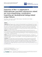
In control jaw periodontal tissue, Msx-1 expression
was localized in the nucleus and cytoplasm of osteo-
blasts, fibroblasts, and progenitors within the co nnective
tissue layer (Figure 1a). In the BONJ-related tissue, a
reduced cellular density of Msx-1 expressing osteoblasts,
fibroblasts, and progenitor cells was noted (Figure 1b).
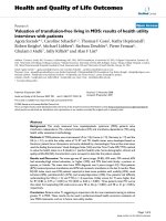
BMP-2/4 expression was found in osteoblast progenitors
of adjacent periosteal tissue in both healthy jaw bone
(Figure 2a) and the BONJ samples (Figure 2b).
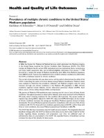
RANKL expression was present throughout the soft
tissue in normal jaw samples (Figure 3a), including peri-
osteal and subepithelial tissue; however, in BONJ sam-
ples, RANKL expression was present sparsely in the
Wehrhan et al. Journal of Translational Medicine 2010, 8:96
/>Page 3 of 9
Figure 1 Msx-1 expression was reduced in ONJ-related periodontal tissue. (a) The Msx-1 staining was a ccentuated in periost eal cells,
attached to the mineralized bone matrix. The bone trabeculae interconnecting fibrous tissue presented nuclear and cytoplasmic Msx-1 staining.
(b) In the BONJ group, staining of periosteal cells was rare, and cytoplasmic staining was decreased, as was the cellular density of Msx-1-
expressing fibroblasts in the fibrous and inflammatory tissue surrounding the bone matrix. (c) Relative cellular expression (labeling index) for Msx-
1 was significantly reduced (Controls-ME: 34.29, IQR 24.0 vs. BONJ-ME: 14.03, IQR: 6.0; p < 0.05) in ONJ-related oral mucoperiosteum. (d) Relative
gene expression for Msx-1 was suppressed 6.8-fold at the mRNA level in ONJ-related periosteum samples (Controls-ME: 1.00, IQR 0.25 vs. BONJ-
ME: 0.15, IQR: 0.31; p < 0.03). Horizontal bars indicate median (ME), and error bars indicate interquartile range (IQR).
Wehrhan et al. Journal of Translational Medicine 2010, 8:96
/>Page 4 of 9
Figure 2 BMP-2/4 expression was incr eased at the protein and mRNA level s in BP-altered oral mucoperiosteum. (a) Rarely, there was
pronounced BMP-2/4 staining in healthy jaw periosteum. (b) BMP-2/4-expressing osteocytes showed higher cellular density in the BONJ group.
(c) The labeling index of BMP-2/4-expressing osteoblasts and osteocytes was significantly increased compared to control (Controls-ME: 22.06, IQR
25.0 vs. BONJ-ME: 53.97, IQR: 25.0; p < 0.05). (d) Relative BMP-2 gene expression at the mRNA level was elevated 8.9-fold in ONJ samples
(Controls-ME: 1.14, IQR 1.07 vs. BONJ-ME: 8.9, IQR: 6.1; p < 0.02) related to healthy samples. Horizontal bars indicate median (ME), and error bars
indicate interquartile range (IQR).
Wehrhan et al. Journal of Translational Medicine 2010, 8:96
/>Page 5 of 9
Figure 3 RANKL was suppre ssed in ONJ-adjacent soft tissue. (a, b) Spatial distribution of RANKL-expressing cells in the soft tissue areas of
BONJ samples (b) was non-homogeneous compared to normal jaw periodontal samples (a). A local high concentration of RANKL-expressing
multinucleated cells was detected only at zones of tissue resorption in BONJ samples. (c) The relative cellular expression (labeling index) of
RANKL-positive cells was significantly lower in ONJ samples (Controls-ME: 59.38, IQR 21.0 vs. BONJ-ME: 23.25, IQR: 12.0; p < 0.05). (d) A 2.94-fold
suppression of RANKL mRNA was detected in ONJ-related bone samples (Controls-ME: 1.00, IQR 0.13 vs. BONJ-ME: 0.34, IQR: 0.44; p < 0.03).
Horizontal bars indicate median (ME), and error bars indicate interquartile range (IQR).
Wehrhan et al. Journal of Translational Medicine 2010, 8:96
/>Page 6 of 9
endosteal and periosteal tissue at the site of bone
resorption (Figure 3b).
The labeling index of Msx-1-expressing (Figure 1c)
and RANKL-expressing (Figure 3c) cells was signifi-
cantly diminished compared to normal bone. The label-
ing index of BMP- 2/4-expressing osteoblasts and
osteocytes (Figure 2c) was significantly increased com-
pared to control.
PCR
The patterns for mRNA expression reflected those for
protein expression. Msx-1 mRNA levels were signifi-
cantly suppressed 6.8-fold in BONJ samples compared
to control periodontal tissue (Figure 1d). BMP-2/4
mRNA expression was signifi cantly higher by about 8.9-
fold in BONJ tissue than in normal jaw mucoperiosteal
tis sue (Figure 2d), whil e RANKL mRNA expression was
significantly suppressed 2.9-fold in BONJ samples rela-
tive to control (Figure 3d).
Discussion
This study identified a significantly diminished expres-
sion of Msx-1, a cellular plasticity and proliferation-
mediating transcripti on factor, in BONJ-affected jaw
periodontal tissue at the protein and mRNA levels. Sig-
nificantly elevated expression of BMP-2/4 in the BONJ -
related periodontal and periosteal tissue revealed an
increased osseous differentiation stimulation in progeni-
tors of osteoblastic lineage in BP-compromised jaw
mucoperiosteal tissue. As wit h Msx-1 expressio n,
RANKL expression in the jaw bone overlying mucoper-
iosteal t issue was significantly reduced, suggesting sup-
pressed osteoclast activation by osteoblasts [19].
BP-related Msx-1 loss in the PDL can explain the
sclerotic, periapical hypermineralized thin lines around
dental roots of BP-altered PDL tissue, which is known
for having the highest endogenous Msx-1 expression in
the jaw [9,12,13,20]. In addition, Msx-1 is critically
involved in cellular plasticity and differentiation. Within
the PDL, a balanced progenitor cell differentiatio n
towards fibrous soft tissue takes place between dental
and bone hard tissue. The clinical observation of sclero-
tic remodeling of the PDL is substantiated by the
experimental finding of BP-induced osteogenetic cell
recruitment and trans-differentiation of progenitor cells
within the PDL [21]. Because Msx-1 has been reported
to prevent terminal differentiation and to stim ulate pro-
liferation of progenitors, loss of Msx-1 in the presence
of BMP-2 is likel y to be associated with poor cell prolif-
eration and als o with overwhelming mineralization in
periodontal tissue [22,23].
The significantly increased expression of BMP-2/4
identified here at the cellular and mRNA levels in
BONJ-affected jaw periosteum is consistent with the
clinical and radiologic observation of the osteopetrotic
aspect of ONJ-related jaw bone: BMP-2/4 is an essential
osteoinductive factor and induces terminal osseous dif-
ferentiation through DLX5 signaling in the absence of
Msx-1 [24]; [25]. Increased terminal osseus differentia-
tion and reduced proliferation of progenitor cells within
the periodontal tissue might explain sclerosis and osteo-
petrosis of the alveolar bone and the reduced periodon-
tal soft tissue proliferation. The immunohistochemical
and molecular results in this study are consistent with
those found in osteopetrotic bone [26], and BONJ has
been described as local osteopetrosis [24,27].
The finding of BP-related RANKL suppression in peri-
odontal progenitor ce lls in vivo is described here for the
first time a nd indicates the relevance of BP effects on
cellular differentiation in explaining the etiology of
BONJ. The significantly reduced expression of RANKL
in ONJ-adjacent periodontal tissue at the protein and
mRNA levels d emonstrates the effect of BP action on
soft-tissue remodeling. Suppression of RANKL has been
described as the main action of BP, preventing osteo-
clast activation and bone resorption in malignancies and
osteoporosis [28-31]. This suggestion finds strong sup-
port from clinical findings of ONJ onset following appli-
cation of the anti-RANKL denosumab without any BP
involvement [4,6]. The concerted regulation of RANKL
and Msx-1 identified here connects jaw-specific and
common bone remodeling mechanisms, but the details
remain to be elucidated at the cellular and subcellular
levels.
Conclusion
These findings help to explain some of the molecular
underpinnings of the restriction of BONJ to the jaw
bone. Jaw restricted osteopetrosis implicated in BONJ
can be explained by loss of Msx-1. Msx-1, kn own to
be a key regulator of cellular plasticity and constitu-
tively expressed in CNC-derived jaw hard and soft
tissue progenitor cells, could be of relevance in jaw-
restricted diseases associated with impaired bone and
soft tissue remodeling [32-34]. Addressing the Msx-1-
RANKL-associated signaling could help to elucidate
mechanisms of CNC-related jaw bone and periodontal-
tissue-specific homeostasis [7-9]. In a greement with
leading international experts in the field of ONJ, we
found that t argeting the unique features o f the jaw
bone is a promising approach to elucidating the under-
lying pathologic mechanisms of ONJ [35]. Of note, BP
and aRANKL h ad differential impacts on proliferation,
vascularisation, and surface marker expression [36,37].
This suggests that BP and aRANKL effects on Msx-
and RANKL-related interactions in CNC- and MsC-
derived osteoblasts, osteoclasts, and bone structures
should be investigated in more detail in the future.
Wehrhan et al. Journal of Translational Medicine 2010, 8:96
/>Page 7 of 9
Acknowledgements
The authors thank Heidemarie Heider, Andrea Kosel, and Miriam Ramming
for technical assistance with the immunohistochemistry autostainer. In
addition, we thank Andrea Krautheim-Zenk for help with mRNA processing
and RT-PCR.
This study was funded by the ELAN-Fonds of the University of Erlangen-
Nuremberg.
Author details
1
Department of Oral and Maxillofacial Surgery University of Erlangen-
Nuremberg Glueckstrasse 11, 91054 Erlangen, Germany.
2
Department of
Plastic Surgery/St. Georg-hospital Eisenach University of Jena Erlanger Allee
101, 07747 Jena, Germany.
3
Institute of Pathology University of Erlangen-
Nuremberg Universitaetsstrasse 22, 91054 Erlangen, Germany.
Authors’ contributions
FW was responsible for the application of grant support (ELAN-Fonds,
university of Erlangen), the conduction of study, built the hypothesis,
established and conducted the methods and analytic procedures and wrote
the manuscript. PH built the hypothesis and did the interpretation of the
data. JR established the m-RNA analysis and RT-PCR and wrote the
manuscript, section RT-PCR. PS and KS did the immunohistochemistry
analysis.
FN interpreted the data and wrote the manuscript, section discussion. EN
interpreted the data and conducted the study by harvesting samples. KA
established immunohistochemistry, analysed the tissue samples, interpreted
the data and was responsible for the histopatholgical analysis of ONJ- and
control tissue samples. All authors read and approved the final manuscript.
Competing interests
There are no competing interests of the authors to be declared.
This study was funded by the ELAN-Fonds of the University of Erlangen-
Nuremberg, Germany.
Received: 20 June 2010 Accepted: 13 October 2010
Published: 13 October 2010
References
1. Reid IR: Osteonecrosis of the jaw: who gets it, and why? Bone 2009,
44:4-10.
2. Ruggiero SL, Drew SJ: Osteonecrosis of the jaws and bisphosphonate
therapy. J Dent Res 2007, 86:1013-1021.
3. Agis H, Blei J, Watzek G, Gruber R: Is zoledronate toxic to human
periodontal fibroblasts? J Dent Res 89:40-45.
4. Taylor KH, Middlefell LS, Mizen KD: Osteonecrosis of the jaws induced by
anti-RANK ligand therapy. Br J Oral Maxillofac Surg 2010, 48(3):221-3.
5. Henry D, vonMoos R, Vadhan-Raj S, et al: A double-blind, randomized
study of denosumab versus zoledronic acid for the treatment of bone
metastases in patients with advanced cancer (excluding breast and
prostate cancer) or multiple myeloma. Eur J Can Suppl 2009, 7(3):12.
6. Stopeck A, Body J, Fujiwara Y, et al: Denosumab versus zoledronic acid for
the treatment of breast cancer patients with bone metastases: rusults of
a randomized phase 3 study. Eur J Can Suppl 2007, 5(3):31.
7. Trainor PA: Specification and patterning of neural crest cells during
craniofacial development. Brain Behav Evol 2005, 66:266-280.
8. Luan X, Dangaria S, Ito Y, Walker CG, Jin T, Schmidt MK, Galang MT,
Druzinsky R: Neural crest lineage segregation: a blueprint for periodontal
regeneration. J Dent Res 2009, 88:781-791.
9. Chung IH, Yamaza T, Zhao H, Choung PH, Shi S, Chai Y: Stem cell property
of postmigratory cranial neural crest cells and their utility in alveolar
bone regeneration and tooth development. Stem Cells 2009, 27:866-877.
10. Blin-Wakkach C, Lezot F, Ghoul-Mazgar S, Hotton D, Monteiro S, Teillaud C,
Pibouin L, Orestes-Cardoso S, Papagerakis P, Macdougall M, et al:
Endogenous Msx1 antisense transcript: in vivo and in vitro evidences,
structure, and potential involvement in skeleton development in
mammals. Proc Natl Acad Sci USA 2001, 98:7336-7341.
11. Orestes-Cardoso S, Nefussi JR, Lezot F, Oboeuf M, P ereira M,
Mesbah M, Robert B, Berdal A: Msx1 is a regulator of bone
formation during development and postnatal growth: in vivo
investigations in a transgenic mouse model. Connect Tissue Res
2002, 43:153-160.
12. Babajko S, Petit S, Fernandes I, Meary F, LeBihan J, Pibouin L, Berdal A:
Msx1 expression regulation by its own antisense RNA: consequence on
tooth development and bone regeneration. Cells Tissues Organs 2009,
189:115-121.
13. Ruhin-Poncet B, Ghoul-Mazgar S, Hotton D, Capron F, Jaafoura MH,
Goubin G, Berdal A: Msx and dlx homeogene expression in epithelial
odontogenic tumors. J Histochem Cytochem 2009, 57:69-78.
14. Houpis CH, Tosios KI, Papavasileiou D, Christopoulos PG, Koutlas IG,
Sklavounou A, Alexandridis C: Parathyroid hormone-related peptide
(PTHrP), parathyroid hormone/parathyroid hormone-related peptide
receptor 1 (PTHR1), and MSX1 protein are expressed in central and
peripheral giant cell granulomas of the jaws. Oral Surg Oral Med Oral
Pathol Oral Radiol Endod 2010, 109(3):415-24.
15. Idowu BD, Thomas G, Frow R, Diss TC, Flanagan AM: Mutations in SH3BP2,
the cherubism gene, were not detected in central or peripheral giant
cell tumours of the jaw. Br J Oral Maxillofac Surg 2008, 46:229-230.
16. Miah SM, Hatani T, Qu X, Yamamura H, Sada K: Point mutations of 3BP2
identified in human-inherited disease cherubism result in the loss of
function. Genes Cells 2004, 9:993-1004.
17. Stockmann P, Vairaktaris E, Wehrhan F, Seiss M, Schwarz S, Spriewald B,
Neukam FW, Nkenke E: Osteotomy and primary wound closure in
bisphosphonate-associated osteonecrosis of the jaw: a prospective
clinical study with 12 months follow-up. Support Care Cancer 2010,
18(4):449-60.
18. Wehrhan F, Rodel F, Grabenbauer GG, Amann K, Bruckl W, Schultze-
Mosgau S: Transforming growth factor beta 1 dependent regulation of
Tenascin-C in radiation impaired wound healing. RadiotherOncol 2004,
72:297-303.
19. Boyce BF, Xing L: Functions of RANKL/RANK/OPG in bone modeling and
remodeling. Arch Biochem Biophys 2008, 473(2):139-46.
20. Groetz KA, Al-Nawas B: Persisting alveolar sockets-a radiologic symptom
of BP-ONJ? J Oral Maxillofac Surg 2006, 64:1571-1572.
21. Lekic P, Rubbino I, Krasnoshtein F, Cheifetz S, McCulloch CA, Tenenbaum H:
Bisphosphonate modulates proliferation and differentiation of rat
periodontal ligament cells during wound healing. Anat Rec 1997,
247:329-340.
22. Newberry EP, Boudreaux JM, Towler DA: Stimulus-selective inhibition of
rat osteocalcin promoter induction and protein-DNA interactions by the
homeodomain repressor Msx2. JBiolChem 1997, 272:29607-29613.
23. Dodig M, Kronenberg MS, Bedalov A, Kream BE, Gronowicz G, Clark SH,
Mack K, Liu YH, Maxon R, Pan ZZ, et al: Identification of a TAAT-containing
motif required for high level expression of the COL1A1 promoter in
differentiated osteoblasts of transgenic mice. JBiolChem 1996,
271:16422-16429.
24. Marx RE, Sawatari Y, Fortin M, Broumand V: Bisphosphonate-induced
exposed bone (osteonecrosis/osteopetrosis) of the jaws: risk factors,
recognition, prevention, and treatment. J Oral Maxillofac Surg 2005,
63:1567-1575.
25. Ryoo HM, Lee MH, Kim YJ: Critical molecular switches involved in BMP-2-
induced osteogenic differentiation of mesenchymal cells. Gene 2006,
366:51-57.
26. Cohen MM Jr: The new bone biology: pathologic, molecular, and clinical
correlates. Am J Med Genet A 2006, 140:2646-2706.
27. Favia G, Pilolli GP, Maiorano E:
Histologic and histomorphometric features
of bisphosphonate-related osteonecrosis of the jaws: An analysis of 31
cases with confocal laser scanning microscopy. Bone 2009, 45(3):406-13.
28. Nishida S, Tsubaki M, Hoshino M, Namimatsu A, Uji H, Yoshioka S,
Tanimori Y, Yanae M, Iwaki M, Irimajiri K: Nitrogen-containing
bisphosphonate, YM529/ONO-5920 (a novel minodronic acid), inhibits
RANKL expression in a cultured bone marrow stromal cell line ST2.
Biochem Biophys Res Commun 2005, 328:91-97.
29. Viereck V, Emons G, Lauck V, Frosch KH, Blaschke S, Grundker C,
Hofbauer LC: Bisphosphonates pamidronate and zoledronic acid
stimulate osteoprotegerin production by primary human osteoblasts.
BiochemBiophysResCommun 2002, 291:680-686.
30. McGonigle JS, Giachelli CM, Scatena M: Osteoprotegerin and RANKL
differentially regulate angiogenesis and endothelial cell function.
Angiogenesis 2009, 12:35-46.
31. Buckle CH, Neville-Webbe HL, Croucher PI, Lawson MA: Targeting RANK/
RANKL in the treatment of solid tumours and myeloma. Curr Pharm Des
16:1272-1283.
Wehrhan et al. Journal of Translational Medicine 2010, 8:96
/>Page 8 of 9
32. Lezot F, Coudert A, Petit S, Vi-Fane B, Hotton D, Davideau JL, Kato S,
Descroix V, Pibouin L, Berdal A: Does Vitamin D play a role on Msx1
homeoprotein expression involving an endogenous antisense mRNA? J
Steroid Biochem Mol Biol 2004, 89-90:413-417.
33. Galle S, Yanze N, Seipel K: The homeobox gene Msx in development and
transdifferentiation of jellyfish striated muscle. Int J Dev Biol 2005,
49:961-967.
34. Gersch RP, Lombardo F, McGovern SC, Hadjiargyrou M: Reactivation of Hox
gene expression during bone regeneration. J Orthop Res 2005,
23:882-890.
35. Khosla S, Burr D, Cauley J, Dempster DW, Ebeling PR, Felsenberg D,
Gagel RF, Gilsanz V, Guise T, Koka S, et al: Bisphosphonate-associated
osteonecrosis of the jaw: report of a task force of the American Society
for Bone and Mineral Research. J Bone Miner Res 2007, 22:1479-1491.
36. Stefanik D, Sarin J, Lam T, Levin L, Leboy PS, Akintoye SO: Disparate
osteogenic response of mandible and iliac crest bone marrow stromal
cells to pamidronate. Oral Dis 2008, 14:465-471.
37. Matsubara T, Suardita K, Ishii M, Sugiyama M, Igarashi A, Oda R,
Nishimura M, Saito M, Nakagawa K, Yamanaka K, et al: Alveolar bone
marrow as a cell source for regenerative medicine: differences between
alveolar and iliac bone marrow stromal cells. J Bone Miner Res 2005,
20:399-409.
doi:10.1186/1479-5876-8-96
Cite this article as: Wehrhan et al.: Expression of Msx-1 is suppressed in
bisphosphonate associated osteonecrosis related jaw tissue-
etiopathology considerations respecting jaw developmental biology-
related unique features. Journal of Translational Medicine 2010 8:96.
Submit your next manuscript to BioMed Central
and take full advantage of:
• Convenient online submission
• Thorough peer review
• No space constraints or color figure charges
• Immediate publication on acceptance
• Inclusion in PubMed, CAS, Scopus and Google Scholar
• Research which is freely available for redistribution
Submit your manuscript at
www.biomedcentral.com/submit
Wehrhan et al. Journal of Translational Medicine 2010, 8:96
/>Page 9 of 9