Báo cáo hóa học: " Rapid induction of autoantibodies during ARDS and septic shock" pptx
Bạn đang xem bản rút gọn của tài liệu. Xem và tải ngay bản đầy đủ của tài liệu tại đây (483.82 KB, 9 trang )
RESEARC H Open Access
Rapid induction of autoantibodies during ARDS
and septic shock
Peter D Burbelo
1*
, Nitin Seam
2,3
, Sandra Groot
1
, Kathryn H Ching
1
, Brian L Han
1
, G Umberto Meduri
4
,
Michael J Iadarola
1
, Anthony F Suffredini
2
Abstract
Background: Little is known about the induction of humoral responses directed against human autoantigens
during acute inflammation. We utilized a highly sensitive antibody profiling technology to study autoantibodies in
patients with acute respiratory distress syndrome (ARDS) and severe sepsis, conditions characterized by intensive
immune activation leading to multiple organ dysfunction.
Methods: Using Luciferase Immunoprecipitation Systems (LIPS), a cohort of control, ARDS and sepsis patients were
tested for antibodies to a panel of autoantigens. Autoantibody titers greater than the mean plus 3 SD of the 24
control samples were used to identify seropositive samples. Available longitudinal samples from different
seropositive ARDS and sepsis patient samples, starting from within the first two days after admission to the
intensive care, were then analyzed for changes in autoantibody over time.
Results: From screening patient plasma, 57% of ARDS and 46% of septic patients without ARDS demonstrated at
least one statistically significant elevated autoantibody compared to the controls. Frequent high titer antibodies
were detected against a spectrum of autoantigens in cluding potassium channel regulator, gastric ATPase, glutamic
decarboxylase-65 and several cytokines. Analysis of serial samples revealed that several seropositive patients had
low autoantibodies at early time points that often rose precipitously and peaked between days 7-14. Further, the
use of therapeutic doses of corticosteroids did not diminish the rise in autoantibody titers. In some cases, the
patient autoantibody titers remained elevated through the last serum sample collected.
Conclusion: The rapid induction of autoantibodies in ARDS and severe sepsis suggests that ongoing systemi c
inflammation and associated tissue destruction mediate the break in tolerance against these self proteins.
Introduction
Serum antibodies are essential components of adaptive
immunity, but are also i nvolved in the pathogenesis of
many autoimmune diseases. While much is known about
the control o f host an tibody production following patho-
gen exposure or vaccination [1], the induction of autoan-
tibodies in human autoimmune and other diseases
remains poorly defined. In genetically susceptible indivi-
duals, infection and other environmental insults h ave
been speculated to trigger immune responses by different
mechanisms including induction of cytokines, stimula-
tion of toll-like receptors and other pattern recognition
receptors, the release of self antigens by damaged cells
and tissues and/or molecular mimicry [2]. However to
date, little is known about the spectrum of autoantibody
responses and the kinetics of autoantibody induction
during acute infection and systemic inflammation.
Acute respiratory distress syndrome (ARDS) and severe
sepsis are acute inflammatory conditi ons associated with
high morb idity a nd mor tality, often inv olving multiple
organ failure [3,4]. ARDS is caused b y a w ide variety of
infectious or inflammatory insults to the lung that may
occur by direct (e.g. pneumonia) or indirect injury (e.g.
peritonitis). The pathologic hallmarks of ARDS are dif-
fuse alveolar damage manifested by disruption of the
alveolar-capillary interface, as well as the accumulation of
inflammatory cells and protein-rich exudates in the
alveolar spaces [4]. Patients with ARDS have elevated
levels of inflammatory mediators such as TNF-a,IL-1b,
* Correspondence:
1
Neurobiology and Pain Therapeutics Section, Laboratory of Sensory Biology,
National Institute of Dental and Craniofacial Research, National Institutes of
Health, Bethesda, Maryland 20892, USA
Full list of author information is available at the end of the article
Burbelo et al. Journal of Translational Medicine 2010, 8:97
/>© 2010 Burbelo et al; licensee Bio Med Central Ltd. This is an Open Access article distributed under the terms of the Creative Commons
Attribution License ( which permits unrestricted use, distribution, and reproduction in
any medium, provided the original work is properly cited.
IL-6 and IL-8 in lung lining fluid as well as in the circula-
tion [5]. In sepsis a nidus of infe ction causes a l ocal and
systemic inflammatory response [3]. However as sepsis
persists, there is a rapid shift towards an anti-inflamma-
tory immunosuppressive state that likely involves T-cell
anergy [6,7], increased anti-inflammatory c ytokines [8]
and the loss of dendritic cells, B ly mphocytes and CD4+
T lymphocytes [9,10].
Luciferase immunop recipitation systems (LIPS), offers
ahighlyquantitativeandsensitivemethodtomeasure
antibody responses against large numbers of foreign
antigens and autoantigens [11-16]. In thi s study, LIPS
was used to profile plasma from patients with ARDS or
sepsis against a panel of known autoantigens. Within 10
to 14 days after the onset of illness, nearly 50% of the
patients show high antibody titers to at least one auto-
antigen. Remarkably, analysis of serial sa mples revealed
that the induction of these autoant ibodies occurred
rapidly, often within 1-7 days after intensive care unit
admission and in some cases remained elevated for sev-
eral weeks. The mechanisms and time course for the
rapid induction of autoantibodies seen in ARDS and
sepsis may occur i n other conditions including autoim-
mune diseases.
Methods
Patient Samples
Plasma samples were obtained fr om patients and healthy
control subjects under institutional review board-
approved protocols at the NIH Clinical Center and from
the University of Tennessee Health Science Center [17].
Plasma samples were obtained from heparinized venous
whole blood b y centrifugation and stored in aliquot s at
-80°C. Plasma samples from the 24 normal volunteers
were collected at the NIH Clinical Center, while the 35
ARDS patients were selected from a randomized trial
investigating prolonged methylprednisolone treatment in
early severe ARDS conducted at the University of Ten-
nessee [17]. The 13 patients with sepsis were selected
from a randomized trial investigating prolonged hydro-
cortisone therapy in severe sepsis not complicated by
ARDS, also conducted at the University of Tennessee
Health Science Center. These ARDS and sepsis plasma
samples were used in this retrospective, exploratory
study to examine w hether autoantibodies are generated
in periods of acute inflammation. Samples were selected
from patients whom time points were available at least 7
days into acute inflammatory conditio ns. We attempted
to use samples from patients with positive culture results:
all patients in the sepsis cohort had positive culture
results, as did 80% of the patients in the ARDS cohort.
As part of a clinical protocol, all ARDS and sepsis
patients were in the intensive care unit (ICU) and evalu-
ated with a battery of clinical tests including lung injury
scores (LIS) and multiple organ dysfunction syndrome
(MODS) scores. For the 35 ARDS patients, 22 patients
were treated with methylprednisolone and 13 were not
treated with steroids. For severe sepsis patients, ten
received stress-doses of hydrocortisone, and three did
not. The characteristics of the ARDS and sepsis pa tients
are summarized in Table 1 and include age, gender,
APACHE 3 scoring, methylprednisolone/hydrocortisone
treatment frequency, infection st atus and in-hospital sur-
vival rate. None of the patients had a known history of
autoimmune disorders or were receiving outpatient treat-
ment with corticosteroids or other immunosuppressants
prior to clinical presentation.
Ruc-antigen fusions and LIPS analysis
Many of the autoantigens used in these LIPS studies
including those fo r glutamic decarboxylas e-65 (GAD65),
AQP-4,gastricATPaseandafragmentofRo52(Ro52-
Δ2) have been previously described [14-16]. Four cyto-
kines (Interf eron-g, I nterferon-ω, Interleukin-6 and
interleukin-1a) corresponding to the processed cytoki ne
missing the signal peptide sequences were also gener-
ated as C-terminal Ruc-antigen fusions [18]. In addition
a new lun g autoantigen, KCNRG and four other cyto-
kines were constructed as C-terminal antigen fusions
downstream of Renilla luciferase (Ruc) using the pREN2
vector [13]. DNA sequencing was used to ensure the
integrity of this new construct.
LIPS assay was performed at room temperature as
described [19]. In these assays, sera were processed in a
96-well format. A “master plate” was first constructed
by diluting patient sera 1:10 in assay buffer A (50 mM
Tris,pH7.5,100mMNaCl,5mMMgCl
2
,1%Triton
X-100) in a 96-well polypropylene microtiter plate. For
evaluating antibody titers by LIPS, 40 μl of buffer A,
10 μl of diluted human sera (1 μl equivalent), and 1 ×
10
7
light units (LU) of Ruc-antigen Cos1 cell extract,
diluted i n buffer A to a volume of 50 μl, were added to
each well of a polypropylene plate and incubated for
60 minutes at room temperature on a rotary shaker.
Next, 5 μl of a 30% suspen sion of Ultralink protein A/G
beads (Pierce Biotechnology, Rockford, IL) in PBS were
added to the bottom of each well of a 96-well filter HTS
plate (Millipore, Bedford, MA). To this filter plate, the
100 μl antigen-antibody reaction mixture was trans-
ferred and incubated for 60 minutes at room tempera-
ture on a rotary shaker. The washing steps of the
retained protein A/G beads were performed on a Tecan
plate washer with a vacuum manifold. After the final
wash, LU w ere measured in a Berthold LB 960 Centro
microplate luminometer (Berthold Technologies, Bad
Wilbad, Germany) using coelentera zine substrate mix
(Promega, Madison, WI). All light unit (LU) data were
obtained from the average of two separate experiments
Burbelo et al. Journal of Translational Medicine 2010, 8:97
/>Page 2 of 9
and not corrected for negligible background protein
A/G bead binding. Patient samples positive at day 10 for
ARDS or day 14 for sepsis were reexamined for changes
in antibody titers using all available serial samples.
Statistical analysis
GraphPad Prism s oftware (San Diego, CA) was used for
statistical analysis. Due to the overdispersed nature of
the autoantibody titers, the healthy control subjects
(CTRL) are reported as the geometric mean titer (GMT)
± 95% confidence interval. For determining the cut-off
limits for each of the LIPS tests, the mean value of the
24 control samples plus 3 s tandard deviations (SD) in
the first cohort was used and is indicated in the figures.
The non-parametric Mann-Whitney U test was used for
compari son of antibody titers in different groups. Using
contingency tables, the Fischer’s exact test was used to
determine the statistical significance between autoanti-
body seropositivity and in-hospital survival.
Data transformation and a heatmap were used to visua-
lize the autoantibody profiles of the partic ipants as a sin-
gle graphic. In order to create this heatmap, the mean
and stan dard deviation of the antibody titers for each
antigen in the 24 control samp les was first generated as a
reference scale. Next, antibody titer values for each anti-
gen-antibody measurement greater than the control
mean plus 3 SD were color-coded to signify the relative
number of standard deviations above these cut-off values.
Lastly, the samples were rank ordered with respect to
anti-KCNRG autoantibodies, the most informative auto-
antigen in the ARDS and sepsis patients.
Results
Detection of high titer autoantibodies to proinflammatory
cytokines in selected ARDS and sepsis patients
Based on the hypothesis that anti-cytokine autoantibodies
might predispose a patient to infection or inflammation,
24 controls, 35 ARDS and 13 sepsis patients were screened
for autoantibodies to a panel of cytoki nes using LIPS. To
increase the likelihood of detecting anti-cytokine antibo-
dies at peak levels, patient samples were analyzed from
ARDS at day 14 and sepsis at day 10. Since the normal
range of anti-cytokine autoantibody titers is not known,
and in order to facilitate the identification of elevated anti-
cytokine autoantibodies, a cut-off threshold based on auto-
antibody titers greater than the mean plus 3 SD of the 24
control samples was used to identify potential seropositive
samples. Based on this criterion, a selected number of
ARDS and sepsis patie nts showed autoantibodies against
several cytokines that were often 10 to 1,000-fold higher
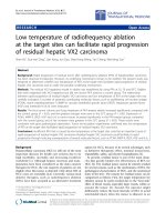
than the GMT of the c ontrols (Figure 1). For example,
three ARDS serum samples had high anti-interleukin-6
Table 1 Clinical Characteristics Based on Autoantibody Status
Autoantibody Positive
a
(n = 20) Autoantibody Negative
a
(n = 15)
ARDS
Age yrs (mean ± SD) 45 ± 14 52 ± 16
Gender 8 male (40%) 10 male (67%)
APACHE 3 score (mean ± SD) 58 ± 17 62 ± 16
Methylprednisolone treatment
b
13/20 (65%) 9/15 (60%)
Infections Gram positive bacteria: 12
Gram negative bacteria: 3
Fungal: 1
Culture negative: 5
Gram positive bacteria: 4
Gram negative Bacteria: 7
Fungal: 1
Viral: 1
Culture negative: 2
In-hospital survival 18/20 (90%) 9/15 (60%)
Severe Sepsis
Autoantibody Positive
a
(n = 6) Autoantibody Negative
a
(n = 7)
Age yrs (mean ± SD) 54 ± 21 63 ± 18
Gender 5 male (83%) 7 male (100%)
APACHE 3 score (mean ± SD) 75 ± 26 68 ± 23
Hydrocortisone treatment
c
5/6 (83%) 5/7 (71%)
Infections Gram positive bacteria: 6
Gram negative bacteria: 0
Gram positive bacteria: 3
Gram negative bacteria: 4
In-hospital survival 4/6 (67%) 5/7 (71%)
a
As determined by LIPS.
b
Methylprednisolone dose - 1 mg/kg/day for 14 days then tapered.
c
Hydrocortisone dosage - 300 mg initially then 10 mg per hour for seven days.
Burbelo et al. Journal of Translational Medicine 2010, 8:97
/>Page 3 of 9
(IL-6) autoantibody titers with values of 34,213, 60,719,
and 255,074 LU, which were all markedly higher than the
GMT of anti-IL-6 antibodies in the controls with a value
of 2,347 LU [95% confidence interval (CI); 2,080-2,649]
(Figure 1A). As shown in Figure 1B, two ARDS samples
were positive for anti-interferon-ω (INF-ω) autoantibodies
with values of 34 ,348 and 70,77 9 LU, while the GMT of
the control group was only 8,658 LU (95% CI; 7,993-
9,379). Significantly elevated anti-interferon-g (INF-g) anti-
bodies were also detected in one ARDS and one septic
patient (Figure 1C). Finally, one ARDS samp le showed a
positive anti-int erleukin-1a (IL1-a ) antibody titer of
1,136,872 LU, which was above the cut-off derived from
the controls (Figure 1D). Testing a number of other cyto-
kines, including interferon-a, BAFF (TNF family member),
April (a proliferation-inducing ligand) and IL-12, did not
reveal autoantibody positivity in any of the ARDS or sepsis
patients (data not shown). Together these results suggest
that some ARDS and sepsis patients generate high levels
of serum autoantibodies to certain cytokines which might
reflect autoimmunization against these parti cular cyto-
kines seen in these patients.
Detection of immunoreactivity to diverse autoantigen
targets in ARDS and sepsis
In light of detecting anti-cytokine autoantibodies in both
ARDS and sepsis patients, other potential autoantigens
were also evaluated. Since we hypothesized that ARDS
and septic patients might show immunoreactivity with
antigens derived f rom damaged tissue and organs, we
tested a panel of known autoantigens associated with
several autoimmune diseases. The autoantigens Jo-1,
MuSK, and La failed to show any statistically significant
responses in b oth patients with ARDS a nd those with
severe sepsis (data not shown). From screening several
other autoantigens, we detected autoantibodies against
the lung-specif ic autoantig en potassium channel regula-
tor (KCNRG). Although the anti-KCNRG autoantibody
titers were modestly elevated compared to the anti-cyto-
kine autoantibodies, 23% (8/35) of the ARDS and 25%
(3/12) of the sepsis patients had statistically significant
autoantibody titers that were higher than the control
cut-off (Figure 1E). Mann Whitney U test analysis
revealed significantly higher detectable anti-KCNRG
autoantibody titers in both the ARDS (P <0.006)and
sepsis patient g roups (P < 0.03) compared to the con-
trols (Figure 1E). These results suggest that the KCNRG
protein is a target of autoantibodies in patients with
ARDS and sepsis.
Screening of several other autoantigens, including gas-
tric ATPase, GAD65, AQP-4 and Ro52 also revealed
high titer autoantibodies in several patients from the
ARDS and severe sepsis cohorts. For example, elevated
anti-gastric ATPase autoantibodies, higher than th e cut-
off derived from the controls, were found in 14% of the
ARDS patients (5/35) as well as one patient with severe
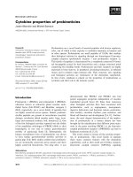
Figure 1 Autoantibodies in patients with ARDS or severe sepsis. Shown are results from 24 controls, 35 ARDS and 13 sepsis patients. Each
symbol represents a sample from one individual patient. The autoantibody titers for (A) IL-6 (B) IFN-ω, (C) IFN-g, (D) IL1-a, (E) KCNRG and (F)
gastric ATPase, (G) AQP-4 and (H) Ro52 antibody titers are plotted on the Y-axis using a log
10
scale. The geometric mean antibody titer for the
ARDS, sepsis and controls are shown by the short solid lines. The dashed line represents the cut-off level for determining seropositivity and is
derived from the mean plus 3 SD of the antibody titer of the 24 controls. P values were calculated using the Mann Whitney U test and were
only significant for anti-KCNRG autoantibodies (control vs. ARDS; P = 0.006 and control vs. sepsis; P = 0.03).
Burbelo et al. Journal of Translational Medicine 2010, 8:97
/>Page 4 of 9
sepsis (Figure 1F). Testing for anti-AQP-4 antibodies
revealed that 9% (3/35) of the ARDS and 15% (2/13) of
sepsis samples had antibody titers above the cut-off
value of the mean plus 3 SD of the 24 control samples
(Figure 1G). High titer autoantibodies above the control
cut-off were also detected to GAD65 in three ARDS and
two sepsis patients (data not shown). Lastly, one ARDS
and o ne sepsis patient had statistically significant levels
of autoantibodies to Ro52 ( Figure 1H). Together, these
results suggest that ARDS and sepsis patients have a
high frequency o f autoantibodies against a number of
diverse autoantigen targets that are classic ally associated
with several different autoimmune conditions.
Autoantibody profiles in ARDS and sepsis
To more easily understand patient immunoreactivity to
the different antigens and relative titers, a colored heat-
map was employed. For this heatmap, antibody titer
values for each antigen-anti body measurem ent greater
than the cut-off of the control mean plus 3 SD were
color-coded to signify the relative number of standard
deviations above these cut-off values. Analysis of con-
trols revealed that 5 of the normal volunteers s howed
positive single autoantibody responses in the range of
3-4SD(datanotshownandFigure1).Incontrast,
some but not all ARDS and seps is patients showed het-
erogeneous immunoreactivity to the autoantigen panel
with antibody titers ranging from 3 to 394 SD above the
mean of controls (Figure 2). The most frequently posi-
tive autoantigen was the KCNRG lung protein, followed
by the gastric ATPase, AQP-4, GAD65 neural autoanti-
gens and finally the Ro52 protein (Figure 2). As evident
from the heatmap, several of the ARDS and septic
shock patients showed positive autoantibody responses
to multiple autoantigens. In general, patients showing
autoantibodies to multiple targets were patients with the
highest autoantibody titers. The most dramatic example
of this was a patient with b acterial meningitis (S3) who
showed high titer autoantib odies to four different auto-
antigens including KCNRG, AQP-4, GAD65 and INF-g
(Figure 2). Some of the other patients with high titer
anti-cytokine autoantibodies also showed interesting
co-profiles: two ARDS patients (A14 and A30) were
co-positive for only IL-6 and interferon-ω autoantibo-
dies, one ARDS patient (A35) was co-positive for IL-1a
and ATPase autoantibodies and one sepsis patient (S13)
with interferon-g autoantibodies was also positive for
anti-KCNRG and anti-GAD65 autoantibodies (Figure 2).
Inspection of the heatmap also shows that the re was no
difference in the prevalence or relative autoantibody in
patients treated with and without steroids (Figure 2 and
Table 1). Taken together these results highlight the he t-
erogeneity of targets and autoantibody titers seen
acutely in ARDS and sepsis patients and s uggest that
steroid treatment has little or no effect on the produc-
tion of autoantibodies in these conditions.
Since only 57% of ARDS and 46% of septic patients
demonstrated at least one statistically significant ele-
vated autoantibody compared to the contro ls, at present
it is difficult to make any general conclusions about the
predictive value of these autoantibodies for determining
severity. However, the relationship between short-term
survival and autoantibodies was examine d. As shown in
Table 1, the ARDS autoantibody positive patients
showed a 90% (18/20) in-hospital survival rate, while the
autoantibody negative samples showed a 60% survival
rate (9/15). Similarly, the autoantibody positive sepsi s
patients showed a 67% (4/6) survival rate and the auto-
antibody negative sepsis patients had a 71% (5/7) survi-
val rate. Statistical analysis using Fischer’s exact tests did
not reveal any significant differences between the differ-
ent groups. Lastly, this study with short-term samples
from ARDS and sepsis patients was not designed to ana-
lyze the significance of these autoantibodies as they
relate to long-term morbidity and mortality.
Kinetics of autoantibody induction in ARDS and sepsis
Since 57% of the ARDS and 46% of the septic shock
patients showed high antibody titers against at least one
autoantigen, we analyzed serial samples to determine
whether these were pre-existing antibodies or were gen-
era ted during the acute inflammatory process. Available
longitudinal samples, typically 3-5 different samples
starting within the first two days after admission to th e
ICU were analyzed. Analysis of the ARDS autoantibody
positive patients revealed dynamic changes in antibody
titers over time. In some cases, the induced autoanti-
body titers sh owed a marked increase of 50 to 100-fold
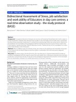
over the co urse of a few days (Figure 3). For example,
the anti-Ro52 auto antibodies in ARDS patient A31
increased from 1,000 LU at day 10 to over 1 million LU
by da y 14 (Figure 3). A similar rapid rise in anti-ATPase
and anti- KCNRG autoantibod ies was also seen in
patient A31 (F igure 3). Another patient (A5) showed a
rapid rise in anti-Ro52 and to lesser extent anti-GAD65
autoantibodies, between days 1 and 10 after ICU admis-
sion (Figure 3). F or patient A23, ther e was a dramati c
rise in anti-IL-6 autoantibodies between day 0 and day 8
(Figure 3). A number of other patients including A14,
A22, A10, and A17 showed autoantibody titer increases
over time (Figure 3). Other patients, however, displayed
high antibody titers from the beginning of their ICU
admission, but showed an upward increase in antibody
titers that peaked at day 8 (see the IL-6 serial titers
inpatient A30; Figure 3). Lastly, some of these elevated
autoantibodies remained high at the last serum sample
collected at later time points such as at days 20 and 28
(Figure 3). Similar autoantibody titer increases and
Burbelo et al. Journal of Translational Medicine 2010, 8:97
/>Page 5 of 9
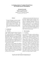
Figure 2 Heatmap analysis of autoantibody profiles in ARDS and sepsis patients. Autoantibody titers to the informative autoantigens are
shown for each of the 35 ARDS patient and 13 sepsis patients. The titer values greater than the mean of the 24 normal volunteers plus 3 SD
were color-coded from green to dark purple to signify the relative number of SD above these reference values. Shaded codes denote patients
who received corticosteroids as part of their treatment.
Burbelo et al. Journal of Translational Medicine 2010, 8:97
/>Page 6 of 9
fluctuations were also seen in many of the sepsis
patients (Figure 3). These results strongly suggest that
autoantibodies can be ra pidly induced and can markedly
fluctuate during conditions of severe inflammation and/
or infection such as ARDS and sepsis.
Discussion
Our finding s document the relatively high prevalence of
autoantibodies in acute, inflammatory, high mortality
conditions of ARDS and severe sepsis. The high detec-
tion rate of autoantibodies, 57% in ARDS and 46% in
severe sepsis patients using a relatively small panel of
autoantigens, suggests that the observed immunoreactiv-
ity to self proteins is a relatively common phenomenon
in these two conditions. The most frequent autoantigen
target in ARDS and sepsis was KCNRG, a protein highly
expressed in the lung [20]. While autoantibodies to
KCNRG have only been previously reported in a subset
of autoimmune polyendocrine syndrome patients with
lung complications [20], our finding of anti-KCNRG
autoantibodies in ARDS and sepsis patie nts is consistent
with the pulmonary injury and tissue destruction asso-
ciated with these conditions. The detection of autoanti-
bodies to the gastric ATPase autoantigen, a frequent
targ et in a number of autoimmune conditions including
autoimmune gastritis [21], type I diabetes [22] and Sjög-
ren’s syndrome [16], suggests that the stomach may be a
highly promiscuous target of autoantibody attack in
diverse inflammatory and autoimmune conditions. It
should also be noted that many of the patients were
concurrently on corticosteroids, but did not appear to
block autoantibody production. The finding of the rapid
induction of autoantibodies against the Ro52 autoanti-
gen, one of the major rheumatological antigens compris-
ing the SSA test, may coincide with the massive increase
in antibodies directed at potential pathogens and human
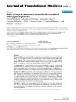
Figure 3 Rapid and dynamic changes in autoantibody titer in ARDS and Sepsis patients. Representative patient samples positive at day 10
for ARDS or day 14 for sepsis were reexamined for changes in antibody titers using all available serial samples. The antibody titers in LU plus
standard error bars are plotted on the Y-axis using a log
10
scale. The X-axis represents time in days following admission to the ICU.
Burbelo et al. Journal of Translational Medicine 2010, 8:97
/>Page 7 of 9
autoantigens that occur during ARDS and sepsis. Recent
studies suggest that Ro52 autoantigen plays an impor-
tant role in quality control of misfolded immunoglobu-
lins produced by B-lymphocytes [23] and may be
released from dying lymphocytes and other cells.
Consistent with the intense host inflammato ry
response found in ARDS (5) and sepsis [3], high titer
autoantibodies were detected to a number of cytokines
including IL-6, interferon-ω, interferon-g and interleu-
kin-1-a. In contrast to a previous report [24], we were
unable to detect autoantibodies to IL-8 in any of the sam-
ples. Nevertheless, the finding that some patients sho w
autoantibodies to a number of cytokines suggests that
these antibodies may be biomarkers for the high levels of
cytokines which may ca use autoimmunizati on and possi-
bly contribut e to immune dysfunction seen in ARDS and
sepsis. Alternativ ely these au toantibodies may play a role
patient susceptibility to opportunistic infection. For
example, anti-INF-g au toantibodies are found in pati ents
with susceptibility to non-tuberculosis mycobacterium
infection [25-27], anti-IL-6 has been reported in a patient
with chronic skin infection [28], and a variety of anti-
cytokine autoantibodies are detected in a subset of thy-
moma patients with o pportunistic i nfections [18,29, 30].
Inter estingly, 1 sepsis and 3 ARDS patients had relatively
high titer autoantibodies against IL-6 and/or INF-ω sug-
gesting that these autoantibodies might have a role in
dampening the activi ty of these cytokines. Future investi-
gations using other bioassays, such as looking for cyto-
kine neutralizing activity, are necessary to further
understand the functional significance of anti-cytokine
autoantibodies in ARDS and sepsis.
Many of the autoantibody re sponses detected in ARDS
and severe sepsis patients showed dynamic responses and
marked changes in titer over a short period of time.
Overall the findings of the rapid induction of autoantibo-
dies against one or several autoantigen targets in the
same patients do not support a role of molecular mimicry
in inducing these antibodies. The mechanism for the
rapid production of auto antibodies is int riguing. Long-
term memory B-cells which are responsible for the extra-
ordinary longevity of human serological memory [31]
may also be involved in the rapid synthesis of autoantibo-
dies described here. Rather than the long-term memory
B-cells directed against pathogen proteins, small numbers
of memory B-cells directed against self proteins may be
present in all humans, but in most cases remain dormant.
Following re-exposure to these self-antigens from tissue
destruction and/or othe r antigen-independent mechan-
ism including activation of cytokines and toll receptors,
these memory B-cells may expand and differentiate into
autoantibody producing plasma cells. Consistent with
this notion is the finding that many of the autoantibody
titers peaked at days 7-14 which may correlate with the
time frame needed to induce these autoantibodies after
the start of the inflammatory host response. Lastly, the
time course for the rapid induction of autoantibodies
seen in ARDS and sepsis may occur in other conditions
including autoimmune diseases.
Although this study focused on short-term outcomes, it
is intriguing that at these early time points autoantibodies
associated with neurological targets are detected. There is
evidence suggesting that ARDS patients su ffer long-term
adverse neuromuscular sequelae [32], and it is possible
that autoantibodies and T-cell-mediated autoimmunity
might contribute to these problems at later time points.
For example, the presence of autoantibodies against AQP-
4 and GAD65 in some ARDS and sepsis patients may be
related to long-term neurological deficits seen in these
patients. Anti-AQP-4 autoantibodies are found in patients
with neurological complications including autoimmune
attack on the optic nerve, spinal cord and peripheral
nerves [16,33,34]. Anti-GAD65 autoantibodies have also
been reported in a number of d ifferent neurological dis-
eases including Stiff person syndrome, encephalitis and
epilepsy , as well as being the major autoantigen in type I
diabetes [35]. It is possible that the anti-AQP-4 and anti-
GAD65 autoantibodies reflect autoimmune attack on the
nervous system t riggered by these condi tions. Consistent
with this possibi lity, it is interesting to note that some of
autoantibodies detected in ARDS including to KCNRG,
AQP-4 and GAD65, show sustained elevation past the last
collected plasma samples at day 20 to 28. Since we were
unable to analyze long-term outcome of these patients, it
is unclear whether the presence of these autoantibodies
are associated with long-term sequelae of critical illness. It
is also unclear whether subsequent mild infections, inflam-
mation and other trauma might reactivate autoantibody
production at a later time in certain seropositive patients.
Future studies expanding the autoantigen panel, profiling
later time points and attempting to correlate autoantibody
elevation with relevant clinical outcomes are needed to
understand whether these autoantibodie s have pathophy-
siological consequences.
Acknowledgements
The authors thank the patients who volunteered for these studies. This work
was supported by in part by the Intramural Research Program of the NIH,
the National Institute of Dental and Craniofacial Research, the NIH Clinical
Center and in part a grant from the Biomarker subsection of the Center for
Neuroscience and Regenerative Medicine.
Author details
1
Neurobiology and Pain Therapeutics Section, Laboratory of Sensory Biology,
National Institute of Dental and Craniofacial Research, National Institutes of
Health, Bethesda, Maryland 20892, USA.
2
Critical Care Medicine Department,
Clinical Center, National Institutes of Health, Bethesda, Maryland 20892, USA.
3
Pulmonary and Critical Care Medicine Department, Veterans Affairs Medical
Center, Washington, District of Columbia 20422, USA.
4
Division of Pulmonary,
Critical Care, and Sleep Medicine, Veterans Affairs Medical Center, Memphis,
Tennessee 38163, USA.
Burbelo et al. Journal of Translational Medicine 2010, 8:97
/>Page 8 of 9
Authors’ contributions
PDB, NS, SG and AFS conceived of the study. GUM collected the patient
plasma samples and provided the clinical characteristics. PDB, SG, KHC and
BH analyzed the sera by LIPS. PDB, NS and AFS analyzed the data. PDB
drafted the manuscript. PDB, NS, MJI, GUM and AFS were involved in critical
revision and final approval. All authors read and approved the manuscript.
Competing interests
The authors declare that they have no competing interests.
Received: 10 September 2010 Accepted: 14 October 2010
Published: 14 October 2010
References
1. Manz RA, Hauser AE, Hiepe F, Radbruch A: Maintenance of serum
antibody levels. Annu Rev Immunol 2005, 23:367-386.
2. Munz C, Lunemann JD, Getts MT, Miller SD: Antiviral immune responses:
triggers of or triggered by autoimmunity? Nat Rev Immunol 2009,
9(4):246-258.
3. Hotchkiss RS, Karl IE: The pathophysiology and treatment of sepsis. N Engl
JMed2003, 348(2):138-150.
4. Ware LB, Matthay MA: The acute respiratory distress syndrome. N Engl J
Med 2000, 342(18):1334-1349.
5. Meduri GU, Annane D, Chrousos GP, Marik PE, Sinclair SE: Activation and
regulation of systemic inflammation in ARDS: rationale for prolonged
glucocorticoid therapy. Chest 2009, 136(6):1631-1643.
6. Heidecke CD, Hensler T, Weighardt H, Zantl N, Wagner H, Siewert JR,
Holzmann B: Selective defects of T lymphocyte function in patients with
lethal intraabdominal infection. Am J Surg 1999, 178(4):288-292.
7. Hensler T, Heidecke CD, Hecker H, Heeg K, Bartels H, Zantl N, Wagner H,
Siewert JR, Holzmann B: Increased susceptibility to postoperative sepsis
in patients with impaired monocyte IL-12 production. J Immunol 1998,
161(5):2655-2659.
8. Gogos CA, Drosou E, Bassaris HP, Skoutelis A: Pro- versus anti-
inflammatory cytokine profile in patients with severe sepsis: a marker
for prognosis and future therapeutic options. J Infect Dis 2000,
181(1):176-180.
9. Hotchkiss RS, Tinsley KW, Swanson PE, Grayson MH, Osborne DF,
Wagner TH, Cobb JP, Coopersmith C, Karl IE: Depletion of dendritic cells,
but not macrophages, in patients with sepsis. J Immunol 2002,
168(5):2493-2500.
10. Hotchkiss RS, Tinsley KW, Swanson PE, Schmieg RE Jr, Hui JJ, Chang KC,
Osborne DF, Freeman BD, Cobb JP, Buchman TG, et al: Sepsis-induced
apoptosis causes progressive profound depletion of B and CD4+ T
lymphocytes in humans. J Immunol 2001, 166(11):6952-6963.
11. Burbelo PD, Ching KH, Bush ER, Han BL, Iadarola MJ: Antibody-profiling
technologies for studying humoral responses to infectious agents. Expert
review of vaccines 2010, 9(6):567-578.
12. Burbelo PD, Ching KH, Han BL, Bush ER, Reeves WH, Iadarola MJ:
Extraordinary antigenicity of the human Ro52 autoantigen. Am J Transl
Res 2010, 2(2):145-155.
13. Burbelo PD, Goldman R, Mattson TL: A simplified immunoprecipitation
method for quantitatively measuring antibody responses in clinical sera
samples by using mammalian-produced Renilla luciferase-antigen fusion
proteins. BMC Biotechnol 2005, 5:22.
14. Burbelo PD, Groot S, Dalakas MC, Iadarola MJ:
High definition profiling of
autoantibodies to glutamic acid decarboxylases GAD65/GAD67 in stiff-
person syndrome. Biochem Biophys Res Commun 2008, 366(1) :1-7.
15. Burbelo PD, Hirai H, Issa AT, Kingman A, Lernmark A, Ivarsson SA,
Notkins AL, Iadarola MJ: Comparison of radioimmunoprecipitation with
luciferase immunoprecipitation for autoantibodies to GAD65 and IA-
2beta. Diabetes Care 2010, 33(4):754-756.
16. Burbelo PD, Leahy HP, Issa AT, Groot S, Baraniuk JN, Nikolov NP, Illei GG,
Iadarola MJ: Sensitive and robust luminescent profiling of anti-La and
other autoantibodies in Sjogren’s syndrome. Autoimmunity 2009,
42(6):515-524.
17. Meduri GU, Golden E, Freire AX, Taylor E, Zaman M, Carson SJ, Gibson M,
Umberger R: Methylprednisolone infusion in early severe ARDS: results of
a randomized controlled trial. Chest 2007, 131(4):954-963.
18. Burbelo PD, Browne SK, Sampaio EP, Giaccone G, Zaman R, Kristosturyan E,
Rajan A, Ding L, Ching KH, Berman A, et al: Anti-cytokine autoantibodies
are associated with opportunistic infection in patients with thymic
neoplasia. Blood 2010.
19. Burbelo PD, Ching KH, Klimavicz CM, Iadarola MJ: Antibody profiling by
Luciferase Immunoprecipitation Systems (LIPS). J Vis Exp 2009, 32.
20. Alimohammadi M, Dubois N, Skoldberg F, Hallgren A, Tardivel I,
Hedstrand H, Haavik J, Husebye ES, Gustafsson J, Rorsman F, et al:
Pulmonary autoimmunity as a feature of autoimmune polyendocrine
syndrome type 1 and identification of KCNRG as a bronchial
autoantigen. Proc Natl Acad Sci USA 2009, 106(11):4396-4401.
21. Toh BH, Gleeson PA, Simpson RJ, Moritz RL, Callaghan JM, Goldkorn I,
Jones CM, Martinelli TM, Mu FT, Humphris DC, et al: The 60- to 90-kDa
parietal cell autoantigen associated with autoimmune gastritis is a beta
subunit of the gastric H+/K(+)-ATPase (proton pump). Proc Natl Acad Sci
USA 1990, 87(16):6418-6422.
22. De Block CE, De Leeuw IH, Bogers JJ, Pelckmans PA, Ieven MM, Van
Marck EA, Van Acker KL, Van Gaal LF: Autoimmune gastropathy in type 1
diabetic patients with parietal cell antibodies: histological and clinical
findings. Diabetes Care 2003, 26(1):82-88.
23. Takahata M, Bohgaki M, Tsukiyama T, Kondo T, Asaka M, Hatakeyama S:
Ro52 functionally interacts with IgG1 and regulates its quality control
via the ERAD system. Molecular immunology 2008, 45(7):2045-2054.
24. Kurdowska A, Miller EJ, Noble JM, Baughman RP, Matthay MA, Brelsford WG,
Cohen AB: Anti-IL-8 autoantibodies in alveolar fluid from patients with
the adult respiratory distress syndrome. J Immunol 1996,
157(6):2699-2706.
25. Hoflich C, Sabat R, Rosseau S, Temmesfeld B, Slevogt H, Docke WD, Grutz G,
Meisel C, Halle E, Gobel UB, et al: Naturally occurring anti-IFN-gamma
autoantibody and severe infections with Mycobacterium cheloneae and
Burkholderia cocovenenans. Blood 2004, 103(2)
:673-675.
26. Kampmann B, Hemingway C, Stephens A, Davidson R, Goodsall A,
Anderson S, Nicol M, Scholvinck E, Relman D , Waddell S, et al: Acquired
predisposition to mycobacterial disease due to autoantibodies to IFN-
gamma. J Clin Invest 2005, 115(9):2480-2488.
27. Patel SY, Ding L, Brown MR, Lantz L, Gay T, Cohen S, Martyak LA, Kubak B,
Holland SM: Anti-IFN-gamma autoantibodies in disseminated
nontuberculous mycobacterial infections. J Immunol 2005,
175(7):4769-4776.
28. Puel A, Picard C, Lorrot M, Pons C, Chrabieh M, Lorenzo L, Mamani-
Matsuda M, Jouanguy E, Gendrel D, Casanova JL: Recurrent staphylococcal
cellulitis and subcutaneous abscesses in a child with autoantibodies
against IL-6. J Immunol 2008, 180(1):647-654.
29. Kisand K, Boe Wolff AS, Podkrajsek KT, Tserel L, Link M, Kisand KV, Ersvaer E,
Perheentupa J, Erichsen MM, Bratanic N, et al: Chronic mucocutaneous
candidiasis in APECED or thymoma patients correlates with
autoimmunity to Th17-associated cytokines. The Journal of experimental
medicine 2010, 207(2):299-308.
30. Meager A, Vincent A, Newsom-Davis J, Willcox N: Spontaneous
neutralising antibodies to interferon–alpha and interleukin-12 in
thymoma-associated autoimmune disease. Lancet 1997,
350(9091):1596-1597.
31. Lanzavecchia A, Bernasconi N, Traggiai E, Ruprecht CR, Corti D, Sallusto F:
Understanding and making use of human memory B cells. Immunol Rev
2006, 211:303-309.
32. Wilcox ME, Herridge MS: Long-term outcomes in patients surviving acute
respiratory distress syndrome. Semin Respir Crit Care Med 2010,
31(1):55-65.
33. Lennon VA, Kryzer TJ, Pittock SJ, Verkman AS, Hinson SR: IgG marker of
optic-spinal multiple sclerosis binds to the aquaporin-4 water channel.
The Journal of experimental medicine 2005, 202(4):473-477.
34. Paul F, Jarius S, Aktas O, Bluthner M, Bauer O, Appelhans H, Franciotta D,
Bergamaschi R, Littleton E, Palace J, et al: Antibody to aquaporin 4 in the
diagnosis of neuromyelitis optica. PLoS Med 2007, 4(4):e133.
35. Vianello M, Tavolato B, Giometto B: Glutamic acid decarboxylase
autoantibodies and neurological disorders. Neurol Sci 2002, 23(4):145-151.
doi:10.1186/1479-5876-8-97
Cite this article as: Burbelo et al.: Rapid induction of autoantibodies
during ARDS and septic shock. Journal of Translational Medicine 2010 8:97.
Burbelo et al. Journal of Translational Medicine 2010, 8:97
/>Page 9 of 9