báo cáo hóa học:" Rapid serological detection of autoantibodies associated with Sjögren''''s syndrome" docx
Bạn đang xem bản rút gọn của tài liệu. Xem và tải ngay bản đầy đủ của tài liệu tại đây (430.28 KB, 8 trang )
BioMed Central
Open Access
Page 1 of 8
(page number not for citation purposes)
Journal of Translational Medicine
Research
Rapid serological detection of autoantibodies associated
with Sjögren's syndrome
Peter D Burbelo*
1,2
, Kathryn H Ching
1,2
, Alexandra T Issa
1,2
,
Caroline M Loftus
1,2
, Yi Li
1,2
, Minoru Satoh
1,2
, Westley H Reeves
1,2
and
Michael J Iadarola
1,2
Address:
1
Neurobiology and Pain Therapeutics Section, Laboratory of Sensory Biology, National Institute of Craniofacial Research, National
Institutes of Health, Bethesda, Maryland, USA and
2
Division of Rheumatology and Clinical Immunology and Center for Autoimmune Diseases,
University of Florida, Gainesville, Florida, USA
Email: Peter D Burbelo* - ; Kathryn H Ching - ; Alexandra T Issa - ;
Caroline M Loftus - ; Yi Li - ; Minoru Satoh - ;
Westley H Reeves - ; Michael J Iadarola -
* Corresponding author
Abstract
Background: Sjögren's syndrome (SjS) is a relatively common autoimmune disease characterized
by oral and ocular dryness. There is an increasing need for simple, sensitive and rapid technologies
for the diagnosis of SjS and other autoimmune diseases. Here we investigated whether a quick
version of luciferase immunoprecipitation systems (QLIPS) could be used to produce a rapid,
specific and quantitative test to detect autoantibodies associated with SjS.
Methods: Using QLIPS, which requires only ten minutes of incubation, a cohort of control and SjS
sera were tested for antibodies to three SjS autoantigens (La, Ro60 and Ro52). Sensitivity and
specificity of the QLIPS tests were compared with LIPS and existing ELISA data. The QLIPS test for
Ro52 was then evaluated with a new validation cohort and its diagnostic performance determined.
Results: Using QLIPS, autoantibodies to three SjS antigens, La, Ro60, and Ro52 were detected in
49%, 56% and 70%, respectively, of the SjS patients and none of the controls (100% specificity).
With antibody titers in the Ro52-seropositive SjS samples approximately 1,000 times higher than
the healthy controls, not only was Ro52 the most informative, but detection of anti-Ro52
antibodies under these non-equilibrium conditions was improved compared to the standard 2 hour
LIPS format. Validation of the anti-Ro52 QLIPS test in a new, independent cohort of SjS and control
serum samples showed 66% sensitivity and 100% specificity.
Conclusion: Together these results suggest that the QLIPS format for Ro52 yields both a more
rapid and more discriminating test for detecting Ro52 autoantibodies than existing immunoassays
and has the potential to be adapted for point-of-care evaluation of patients with SjS and other
rheumatologic diseases.
Published: 24 September 2009
Journal of Translational Medicine 2009, 7:83 doi:10.1186/1479-5876-7-83
Received: 23 July 2009
Accepted: 24 September 2009
This article is available from: />© 2009 Burbelo et al; licensee BioMed Central Ltd.
This is an Open Access article distributed under the terms of the Creative Commons Attribution License ( />),
which permits unrestricted use, distribution, and reproduction in any medium, provided the original work is properly cited.
Journal of Translational Medicine 2009, 7:83 />Page 2 of 8
(page number not for citation purposes)
Introduction
There is an increasing desire in the medical community to
develop rapid and personalized serum-based diagnostic
tests to detect autoimmune [1], neoplastic [2] and infec-
tious diseases [3]. One major approach involves using
antibody-based tests to diagnose and even predict the
onset of various diseases [1,4]. However, most current
quantitative immunoassays used to measure antibodies
are impractical for rapid point-of-care testing because they
are complex, time consuming, and difficult to standardize
[3]. As an alternative, rapid tests such as lateral flow
immunoassays, which can more easily be integrated in
point of care settings, are used for the diagnosis of several
infectious agents such as HIV and HCV. However, one
limitation of these assays is that they produce a qualitative
(i.e. positive or negative) rather than a quantitative result.
Currently there are no serological tests for rapidly detect-
ing autoantibodies associated with autoimmune diseases
that also satisfy the growing demand for high analytical
sensitivity and reproducibility.
Sjögren's syndrome (SjS) is a common autoimmune dis-
order associated with epithelial inflammation and exo-
crine gland dysfunction [5]. SjS is often associated with
polyclonal B cell activation resulting in the presence of
multiple autoantibodies including the well-known SSA
and SSB antibodies. While positive SSA (Ro52 and Ro60)
and SSB (La) autoantibodies are part of the diagnostic cri-
teria, five other clinical signs including ocular and oral
dryness and evidence of inflammation from minor sali-
vary gland biopsy are required for the diagnosis of pri-
mary SjS [6]. This is because antibodies to SSA and SSB are
not specific to SjS, but are also found in other rheumato-
logical diseases including systemic lupus erythematosus
(SLE), and myositis [7]. Nevertheless, in the 2002 classifi-
cation standards for SjS diagnosis, positive SSA and SSB
autoantibody tests were the only mandatory criteria if the
salivary gland biopsy was negative [6]. Current SSA and
SSB ELISAs, which employ native antigen complexes iso-
lated from calf thymus, show positive SSA and SSB serol-
ogy in 50-70% and 40-45% of SjS, respectively [8,9].
Previously, luciferase immunoprecipitation assay systems
(LIPS), which employs Renilla luciferase (Ruc)-antigen
fusions produced in mammalian Cos1 cells, was used to
detect patient antibodies to a variety of pathogen antigens
[10-17] and also to detect human autoantibodies associ-
ated with several autoimmune diseases including Type 1
diabetes [18], Stiff-person syndrome [19] and Sjögren's
syndrome [20]. In the SjS studies, detection of anti-La/SSB
antibodies by LIPS showed improved performance com-
pared to existing ELISA and offered a highly sensitive,
robust and high-throughput testing format [20]. LIPS pro-
filing of additional autoantigens revealed that certain SjS
patients also showed positive immunoreactivity with
Ro52, Ro60 and other extraglandular autoantigens
including thyroid peroxidase, the aquaporin-4 water
channel and the gastric H
+
/K
+
ATPase.
A quicker version of LIPS (called QLIPS) has also been
used to detect antibodies to several pathogen antigens
associated with human infection [14,16], in which the
two incubation steps of 1 hour were each reduced to 5
minutes. In the present study, we describe QLIPS tests for
evaluating antibodies to the 3 major SjS recombinant
autoantigens. Results from this study demonstrate that
detection of anti-Ro52 antibodies by QLIPS was rapid,
robust and has the potential to be used in the diagnosis of
SjS and other rheumatologic diseases in point-of-care set-
tings.
Methods
Patients
The SjS patients from both cohorts used in this study ful-
filled the revised European consensus criteria [6]. The ini-
tial cohort of sera was from patients with primary SjS
participating in a longitudinal natural history study and
was analyzed by the standard LIPS format in a previous
study [20]. These sera included 57 well-characterized
patients diagnosed with primary SjS and 25 healthy vol-
unteers evaluated under Institutional Review Board-
approved protocols at the SjS clinic of the National Insti-
tute of Dental and Craniofacial Research, National Insti-
tutes of Health, Bethesda, MD. As described, SSA (anti-
Ro52 and anti-Ro60 antibodies) and SSB (anti-La anti-
body) tests on these samples were measured in the Labo-
ratory of Clinical Medicine, NIH using a commercial
ELISA obtained from BioRad (Hercules, CA) that employs
native, extractable bovine nuclear antigens [20]. The cut-
off value used for the ELISA was determined from the
internal standards according to the manufacturer.
A second, independent validation cohort collected at the
University of Florida under Institutional Review Board-
approved protocols consisted of 105 SjS and 30 control
sera. For comparison, anti-Ro60 and anti-La (SSB) serop-
ositive status was evaluated in a subset of these samples
using a radiobinding immunoprecipitation assay (RBA)
[21] in the clinical laboratory of the Division of Rheuma-
tology and Clinical Immunology and Center for Autoim-
mune Diseases, University of Florida. In these tests,
35
S-
labeled whole K562 cell protein extract was used in
immunoprecipitation and following autoradiography
was scored positive or negative based on the presence or
absence of the La or Ro60 immunoprecipitated protein.
Renilla luciferase antigen constructs and extracts
A mammalian Renilla luciferase (Ruc) expression vector,
pREN2 [22], expressing Ruc-antigen fusion constructs for
La, Ro52, Ro52-Δ2 (spanning amino acid residues 278-
Journal of Translational Medicine 2009, 7:83 />Page 3 of 8
(page number not for citation purposes)
475) and Ro60, has been previously described [20]. In the
case of Ro60, a new pREN2 construct (Ro60-Δ2) express-
ing a C-terminal protein fragment spanning amino acid
residues 336-576, was generated. DNA sequencing con-
firmed the integrity of this Ro60-Δ2 plasmid construct.
Cos1 cells in 100 mm
2
dishes were transfected with Ruc-
antigen plasmids and lysates prepared as described [22].
Briefly, Cos1 cells in 100 mm
2
dishes were transfected
using FuGENE 6 (Roche) with 1-2 μg of pREN2 plasmid
constructs. Forty-eight hours after transfection, tissue cul-
ture media was removed and the plates were washed with
PBS. The cells were then scrapped in 1.4 ml of cold lysis
buffer composed of 50 mM Tris, pH 7.5, 100 mM NaCl, 5
mM MgCl
2
, 1% Triton X-100, 50% glycerol and protease
inhibitors (Mini protease inhibitor cocktail, Roche). The
cell lysate was sonicated, centrifuged and the cleared
supernatants were collected and used immediately or
stored at -80°C. Total luciferase activity in 1 μl of each
crude extract was determined by adding it to 9 μl of PBS
in a 1.5 ml clear microfuge tube, followed by the addition
of 100 μl of substrate mixture (Renilla Luciferase Reagent
Kit, Promega), vortexing, and immediately measuring
light-forming units with a luminometer (20/20
n
Turner
Scientific) for 5 sec.
QLIPS
A shortened version of LIPS designated QLIPS (for quick
LIPS) was employed [14,16]. In these assays, sera were
processed in a 96-well format. A "master plate" was first
constructed by diluting patient sera 1:10 in assay buffer A
(50 mM Tris, pH 7.5, 100 mM NaCl, 5 mM MgCl
2
, 1% Tri-
ton X-100) in a 96-well polypropylene microtiter plate.
For evaluating antibody titers by LIPS, 40 μl of buffer A, 10
μl of diluted human sera (1 μl equivalent), and 1 × 10
7
light units (LU) of Ruc-antigen Cos1 cell extract, diluted
in buffer A to a volume of 50 μl, were added to each well
of a polypropylene plate and incubated for 5 minutes at
room temperature with shaking. Next, 5 μl of a 30% sus-
pension of Ultralink protein A/G beads (Pierce Biotech-
nology, Rockford, IL) in PBS were added to the bottom of
each well of a 96-well filter HTS plate (Millipore, Bedford,
MA). To this filter plate, the 100-μl antigen-antibody reac-
tion mixture was transferred and incubated for 5 minutes
at room temperature on a rotary shaker. The washing steps
of the retained protein A/G beads were performed on a
BioMek FX work station (Beckman Coulter, Fullerton,
CA) using an integrated vacuum manifold. For these
washes, each well is washed 8 times with 100 μl of buffer
A, followed by two times with 100 μl of PBS. After the
final wash, the filter plate is blotted dry and LU were
measured in a Berthold LB 960 Centro microplate lumi-
nometer (Berthold Technologies, Bad Wilbad, Germany)
using coelenterazine substrate mix (Promega, Madison,
WI). For these measurements, 50 μl of coelenterazine sub-
strate is injected, the plate is shaken for 2 sec, followed by
a 5 sec read of luminescence. All LU data were obtained
from the average of at least two independent experiments,
and the resulting LU values were used without subtracting
the buffer blank.
Statistical analysis
The GraphPad Prism software (San Diego, CA) was used
for statistical analyses. Results for quantitative antibody
levels of the controls and SjS serum samples are reported
as the geometric mean titer (GMT) ± 95% confidence
interval (due to the typically overdispersed nature of these
data). Correlations among antibody responses to the anti-
gens tested were assessed by the Spearman rank test (r
S
).
The level of statistical significance for all tests was set at P
< 0.05. For determining the cut-off limits for each of the
QLIPS tests, the mean value of the 25 control samples plus
5 SD in the first cohort was used and is indicated in the fig-
ures. Additional analysis using a cut-off derived from the
mean plus 3 SD is also included in the text. Test perform-
ance was evaluated using area under the curve (AUC)
from receiver operator characteristic (ROC) analysis.
Results
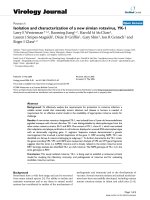
Detection of anti-La autoantibodies in SjS by QLIPS
The diagnostic performance of a previously described
QLIPS format was evaluated for measuring autoantibod-
ies to the three major SjS antigens. From testing a cohort
of 57 SjS and 25 healthy control sera with a full-length La
recombinant fusion protein, the geometric mean titer
(GMT) of anti-La antibodies was 44,692 LU (95% CI,
28,604-69,827) for SjS sera, which was 4 times higher
than the GMT of the control sera of 9,156 LU (95% CI,
8,097-10,355) (Figure 1). Compared to our previously
published study [20], the results using the QLIPS format
showed anti-La antibody titers that were ~10-fold lower
than the anti-La antibody titers reported by the standard 2
hour incubation format (data not shown). Nevertheless,
evaluation by the Mann Whitney U test still showed a sig-
nificant difference between the anti-La antibody titers in
the SjS samples and the controls (P < 0.0001). To examine
the diagnostic utility of the anti-La QLIPS test, the sensi-
tivity and specificity were determined. For this calcula-
tion, a cut-off value of 26,869 LU derived from the mean
plus 5 SD of the 25 control samples was used. Based on
this cut-off, the anti-La antibody test showed 49% sensi-
tivity and 100% specificity in distinguishing the 57 SjS
sera from the 25 control sera. An even lower cut-off
derived from the mean plus 3 SD yielded 56% sensitivity
and 96% specificity. While these QLIPS results for detect-
ing anti-La antibodies were similar to an established
ELISA test for anti-SSB/La antibodies (46% sensitivity),
this shortened assay was not as useful as the our previous
LIPS results on these same samples with 75% sensitivity
and 100% specificity [20]. Finally, the results from the
Journal of Translational Medicine 2009, 7:83 />Page 4 of 8
(page number not for citation purposes)
duplicate interassay QLIPS tests for anti-La antibodies
showed that they were reproducible and had a coefficient
of variation of 18.9%.
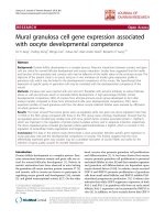
Rapid detection of anti-Ro60 and anti-Ro52
autoantibodies in SjS by QLIPS
We have previously reported using a full-length Ro60-Ruc
antigen fusion in the LIPS format, which required a cum-
bersome 1:200 dilution of human sera to place detection
of anti-Ro60 autoantibodies in the linear range [20]. In
order to simplify testing, a C-terminal Ro60 deletion frag-
ment (Ro60-Δ2) was generated and found to yield values
in the linear range without the need to dilute the sera. As
shown in Figure 2A, testing of this Ruc-Ro60-Δ2 fusion by
QLIPS revealed that the GMT of the anti-Ro60 antibody in
the 57 SjS samples was 18,967 LU (95% CI, 12,659-
27,613), which was over 4-fold higher than the GMT of
3,917 LU (95% CI, 3,574-4293) of the controls (Mann
Whitney U test, P < 0.0005). The anti-Ro60-Δ2 antibody
titers detected by QLIPS were ~10-fold lower compared to
when the same sera were tested in the standard 2 hour
incubation LIPS format (data not shown). Calculations of
the diagnostic performance of the Ro60-Δ2 QLIPS test
based on the mean plus 5 SD of the 25 control samples
(i.e. 8,466 LU) showed 56% sensitivity and 100% specifi-
city in distinguishing the 57 SjS sera from the 25 control
sera. An even lower cut-off derived from the mean plus 3
SD yielded 60% sensitivity and 100% specificity.
QLIPS testing for anti-Ro52 autoantibodies using a C-ter-
minal fragment (Ro52-Δ2) showed that the GMT of the
SjS sera was 198,110 LU (95% CI, 107,237-365,988),
which was 30-times higher than the GMT of the control
sera of 6,351 LU (95% CI, 5,068-7,957) (Figure 2B). The
results from the duplicate interassay QLIPS tests for anti-
Ro52-Δ2 antibodies were reproducible and had a coeffi-
cient of variation of 19%. To examine the diagnostic util-
ity of Ro52-Δ2, the sensitivity and specificity of the QLIPS
test were calculated using a cut-off value (i.e. 37,806 LU)
derived from the mean plus 5 SD of the 25 control sam-
ples. Using this cut-off, the Ro52-Δ2 antibody test showed
70% sensitivity and 100% specificity in distinguishing the
57 SjS patient from controls. An even lower cut-off
derived from the mean plus 3 SD still showed 70% sensi-
tivity and 96% specificity. Among the 40 Ro52-Δ2 serop-
ositive SjS samples, the Ro52-Δ2 antibody titers were
approximately 1,000 times higher than the controls or
Ro52-Δ2 seronegative SjS samples.
Interestingly, the Ro52-Δ2 QLIPS test showed higher sen-
sitivity and specificity than the standard 2 hour LIPS for-
mat on the same samples[20], which was only 65%
sensitive and 96% specific. Additional analysis revealed
that the antibody values in the 57 SjS samples by QLIPS
and LIPS were similar and correlated well (r
s
= 0.72). For
example, the mean and standard deviation in the 57 SJS
samples by QLIPS and LIPS were similar with values of
650,273 ± 485, 490 and 548,504 ± 398,083 LU, respec-
tively. In contrast, the antibody titer values for the 25 con-
trol samples showed a mean and standard deviation by
QLIPS and LIPS of 7,576 ± 6,046 and 29,770 ± 78,495 LU,
respectively. Of note, the markedly higher average and
standard deviation of the control group measured by LIPS
were due to several high titer outliers among the control
samples, which were likely due to low affinity antibodies
since they disappeared in the QLIPS format. While all the
LIPS Ro52 positives were also positive by QLIPS, 2 sam-
ples that were negative by LIPS were now positive by
QLIPS. The net result of these high titer controls in the
LIPS format was a much higher cut-off with an AUC value
of 0.83 for test performance. The AUC value for QLIPS test
was 0.85 and was slightly higher than LIPS reflecting the
higher sensitivity and specificity.
Based on these findings, the most informative autoanti-
gen in the QLIPS test for SjS was Ro52-Δ2 with 70% sen-
sitivity and 100% specificity. Including the results from
the anti-La and anti-Ro60 QLIPS tests did not add any
new positives to the existing Ro52-Δ2 QLIPS test. Per-
QLIPS detection of anti-La autoantibodiesFigure 1
QLIPS detection of anti-La autoantibodies. QLIPS
detection of autoantibodies against La in 25 normal controls
and 57 primary SjS patients. Each circle or square symbol
represents the anti-La antibody titer of a normal control or
SjS patient, respectively. The solid lines represent the GMT
for each group. For determining sensitivity and specificity for
this anti-La antibody test, the dashed line represents the cut-
off level derived from mean plus 5 SD of the antibody titers
of the 25 normal volunteers.
C
on
tr
ols
S
jS
10
3
10
4
10
5
10
6
La
LU
Journal of Translational Medicine 2009, 7:83 />Page 5 of 8
(page number not for citation purposes)
formance of the SSA ELISA on the same samples, which
measures antibodies to both Ro52 and Ro60 proteins,
had 72% sensitivity but included 3 ELISA borderline pos-
itive cases. These results suggest that QLIPS Ro52-Δ2 test
shows similar sensitivity to an ELISA, but produces
quicker and more robust results.
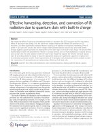
The Ro52-
Δ
2 QLIPS test in a new SjS cohort shows 66%
sensitivity and 100% specificity
A new, independent validation cohort of 105 SjS and 30
control sera was tested to verify the diagnostic utility of
the Ro52-Δ2 QLIPS test. Robust signals were detected in
the SjS samples from this validation cohort, in which the
GMT in the SjS samples was 119,092 LU (95% CI, 72,924-
194,489), and the GMT of the controls was 3,735 LU
(95% CI, 3,400-4,102). Comparison of the anti-Ro52-Δ2
antibody data plots between the initial and validation
cohorts show that they are remarkably similar (Figure 2B
vs. Figure 3), in which the mean LU values for Ro52 anti-
body titers in the initial and validation cohort are almost
identical with values of 650,273 and 529,711 LU, respec-
tively. Using the previous cut-off of 37,806 LU, the QLIPS
test distinguished 69 of the 104 SjS positive samples (66%
sensitivity) from the 30 controls with 100% specificity
(Figure 3). If a cut-off derived from the controls of the val-
idation cohort (i.e. 9,000 LU) is used, an even higher sen-
sitivity of 70% is achieved, while still maintaining 100%
specificity. Furthermore, compared to RBA for anti-Ro60
and anti-La antibodies performed on the same validation
samples, the QLIPS test for Ro52 had a significantly
higher sensitivity (66% versus 56%).
Discussion
Rapid and comprehensive serum-based diagnostic tests
that can be used in point-of-care settings for diagnosis and
even pre-symptom screening of autoimmunity are
urgently needed. A significant challenge in the develop-
ment of such assays is that, unlike antibodies associated
with infectious agents, the detection of autoantibodies
associated with autoimmunity requires more sensitive
tests than ELISAs or other solid phase immunoassays such
as protein arrays, which miss many conformational
epitopes [23]. Typically, liquid phase immunoprecipita-
tion assays such as the radiobinding assay (RBA), which
QLIPS detection of anti-Ro60 and anti-Ro52 autoantibodies in SjSFigure 2
QLIPS detection of anti-Ro60 and anti-Ro52 autoantibodies in SjS. QLIPS detection of autoantibodies against Ro60-
Δ2 and a C-terminal fragment of Ro52 (Ro52-Δ2) in 25 normal volunteers and 57 primary SjS patients. (A) The anti-Ro60-Δ2
antibody test. (B) The anti-Ro52-Δ2 antibody test. Each circle or square symbol represents individual normal controls or SjS
patient samples, respectively. The solid lines represent the GMT for each group. For determining sensitivity and specificity, the
dashed line represents the cut-off level derived from the control mean plus 5 SD.
Co
ntro
l
s
Sj
S
10
3
10
4
10
5
10
6
Ro60-'2
LU
Controls
Sj
S
10
3
10
4
10
5
10
6
10
7
Ro52-'2
A.
B.
Journal of Translational Medicine 2009, 7:83 />Page 6 of 8
(page number not for citation purposes)
show much higher sensitivity, specificity and signal to
noise ratios than ELISAs are needed for detecting autoan-
tibodies in most autoimmune diseases [23,24]. However,
a significant drawback of RBAs is the requirement for radi-
oactively-tagged antigens. In this study we demonstrate
that QLIPS, which utilizes a non-radioactive, luciferase
enzyme-based tracer in a liquid phase assay, can rapidly
and sensitively detect autoantibodies associated with SjS.
The QLIPS format could easily be integrated into a point-
of-care test because it requires only about 25 minutes of
total processing time per 94 sera samples, which includes
a 5 minute set-up, two five minute incubations steps, 10
minutes of washing and reading of the plate with a lumi-
nometer.
Due to the high signal to noise and large dynamic range
of the LIPS assay, the coefficient of variation (CV) of
approximately 20% for LIPS still provides remarkable
diagnostic accuracy. One likely cause of the near 20% CV
is due to the fact that the QLIPS sample processing (i.e.
pipetting and washing) was preformed rapidly in less than
15 minutes with 84 or greater samples. With some of
these high signals in the SjS positive samples, small pipet-
ting errors can resulting in large changes in antibody titer
(e.g. 10% pipetting error can result in over 100,000 LU
differences). Despite the 20% CV, the SjS positive samples
show 1000-fold higher anti-Ro52 antibody titers than the
negative samples and evaluation of each of the two runs
independently shows that show that the same samples are
positive.
Previously, we found that all individuals have detectable
anti-Ro52 antibodies by LIPS [20]. However, unlike the
normal range of anti-Ro52 antibody titers in healthy indi-
viduals, some patients with SjS or other rheumatological
diseases have markedly higher anti-Ro52 antibody titers
that can be detected by LIPS and other immunoassays. In
this study, the QLIPS test for anti-Ro52-Δ2 antibodies had
a higher diagnostic performance than the standard LIPS
format. The reason for this increased performance is due
to the loss of anomalously high signals in some of the
control samples observed in the standard LIPS format.
These anomalously high Ro52 antibody signals in several
of the controls were no longer positive under the rapid,
non-equilibrium conditions of QLIPS. Taken together
these results also suggest that performing QLIPS and LIPS
in parallel may allow a simple method of more accurately
assessing antibody avidity in some situations. An analo-
gous increase in specificity of QLIPS compared to LIPS has
also been observed for distinguishing antibodies to Loa
loa and Onchocerca volvulus antigenic proteins from anti-
bodies to antigens from related filarial infections [14,16].
In contrast, the QLIPS tests for anti-La and anti-Ro60 anti-
bodies showed a marked drop in test performance in the
QLIPS format. For example, the LIPS test for detecting
anti-La antibodies was 75% sensitive, versus the 49% sen-
sitivity of QLIPS. The decreased detection of anti-La and
anti-Ro60 seropositive antibodies under these rapid con-
ditions compared to LIPS is likely due in part to the ina-
bility to detect the low affinity/low titer autoantibodies
present in some of the SjS samples. However, we show
that using QLIPS, the SjS patients can be distinguished
from controls using the Ro52-Δ2 fragment alone.
While the standard LIPS format yielded 76% sensitivity
and required two independent assays (anti-La and anti-
Ro52 autoantibodies) to be performed [20], QLIPS, with
a single antigenic fragment of Ro52, showed approxi-
mately 70% sensitivity. Ironically, this C-terminal frag-
ment of Ro52 used in QLIPS is the same antigenic
fragment that shows no useful diagnostic immunoreactiv-
ity in ELISA and Western blotting [25,26]. The detection
of diagnostically useful antibodies to the C-terminus of
Ro52 by LIPS is supportive of the improved conforma-
tional epitopes using mammalian recombinant proteins
in this liquid phase QLIPS/LIPS compared to ELISA. Both
the LIPS and QLIPS formats are also as good as a conven-
tional ELISA for measuring SSA and SSB. However, an
ELISA requires significantly more time to complete (e.g. 5-
24 hours). Furthermore, the QLIPS Ro52 test showed
higher sensitivity in the validation cohort than an estab-
lished RBA for SSA and SSB (66% vs. 56%).
The short incubation time, high performance and relative
simplicity of the Ro52-Δ2 QLIPS test has practical impli-
cations for developing even simpler assay formats. The
finding that the Ro52-Δ2 QLIPS test showed antibody tit-
ers that were 1000 times higher in the SjS positive samples
compared to the control samples also provides a large
diagnostic window for detecting seropositive samples.
Furthermore, no other immunoassay format such as
ELISA or RBA shows such a large signal-to-noise ratio. It is
likely that additional assay modification, including reduc-
ing the volume of the reaction, may yield even more
robust signals. We speculate that a microfluidic device
configured for the QLIPS format might be suitable for
point-of-care testing. Using such a microfluidics device,
the addition of sera, Ruc-antigen mixture to immobilized
protein A/G, washing, and the addition of coelenterazine
luciferase substrate could all be automated and performed
rapidly. The ability to stably freeze the Ruc-antigen rea-
gents also has practical implications for point-of-care test-
ing. Due to the highly scalable format of QLIPS,
additional reagents for detecting anti-pathogen antibod-
ies (e.g. HIV, HCV, and HSV-2) could also be employed
for side-by-side diagnosis of these infections.
Conclusion
Ro52 autoantibodies are not only found in SjS, but are
found in SLE, myositis and several other autoimmune dis-
Journal of Translational Medicine 2009, 7:83 />Page 7 of 8
(page number not for citation purposes)
orders. These results suggest that the rapid and robust
Ro52-Δ2 QLIPS test has the potential to aid in point-of-
care evaluation of patients with SjS and other rheumato-
logic diseases. However, since not all SjS patients show
anti-Ro52 positive antibodies, the addition of other
autoantigens might improve the diagnostic performance
of the QLIPS test. In particular, antigens that produce
robust signals in Ro52-negative sera might be part of an
antigen mixture used in the QLIPS format to further
increase the sensitivity of this test. The ability of using
QLIPS for screening for anti-Ro52 and other autoantibod-
ies in early phases of the disease might make it possible to
diagnose and even treat autoimmune diseases before
more severe disease and/or substantial organ damage has
occurred.
Competing interests
Two of the authors (P.D.B., and M.J.I.) have a patent
application submitted using LIPS for detecting autoanti-
bodies associated with Sjögren's syndrome.
Authors' contributions
PDB conceived of the study, developed the needed con-
structs, analyzed the sera by LIPS, analyzed the data,
drafted the manuscript and made critical revisions; ATI,
KHC and CL generated the needed constructs and/or
lysates; YL and MS analyzed the sera by conventional
immunoprecipitation assays; WHR, provided patient sera
from cohort 2 with clinical information and was involved
in critical revision; MJI helped develop the high-through-
put assay and was involved in critical revision and final
approval and all authors commented on and approved
the manuscript.
Acknowledgements
This work was supported by the Division of Intramural Research, National
Institute of Dental and Craniofacial Research and, in part, by a Bench to
Bedside award from the NIH Clinical Research Center and by NIH grant
(AR44731). We greatly thank Dr. Gabor Illei and Dr. Nikolay Nikolov for
providing the initial cohort of SjS and control sera samples.
References
1. Sharp V, Utz PJ: Technology insight: can autoantibody profiling
improve clinical practice? Nat Clin Pract Rheumatol 2007,
3:96-103.
2. Kijanka G, Murphy D: Protein arrays as tools for serum autoan-
tibody marker discovery in cancer. J Proteomics 2009,
72:936-944.
3. Yager P, Domingo GJ, Gerdes J: Point-of-care diagnostics for glo-
bal health. Annu Rev Biomed Eng 2008, 10:107-144.
4. Bizzaro N: Autoantibodies as predictors of disease: the clinical
and experimental evidence. Autoimmun Rev 2007, 6:325-333.
5. Fox RI: Sjogren's syndrome. Lancet 2005, 366:321-331.
6. Vitali C, Bombardieri S, Jonsson R, Moutsopoulos HM, Alexander EL,
Carsons SE, Daniels TE, Fox PC, Fox RI, Kassan SS, et al.: Classifica-
tion criteria for Sjogren's syndrome: a revised version of the
European criteria proposed by the American-European
Consensus Group. Ann Rheum Dis 2002, 61:554-558.
7. Franceschini F, Cavazzana I: Anti-Ro/SSA and La/SSB antibodies.
Autoimmunity 2005, 38:55-63.
8. Kapogiannis B, Gussin HA, Teodorescu MR, Teodorescu M: Differ-
ences in clinical sensitivity of ELISA tests for autoantibodies
with human and bovine extractable nuclear antigens. Lupus
2000, 9:343-352.
9. Lyons R, Narain S, Nichols C, Satoh M, Reeves WH: Effective use
of autoantibody tests in the diagnosis of systemic autoim-
mune disease. Ann N Y Acad Sci 2005, 1050:217-228.
10. Burbelo PD, Ching KH, Mattson TL, Light JS, Bishop LR, Kovacs JA:
Rapid antibody quantification and generation of whole pro-
teome antibody response profiles using LIPS (luciferase
immunoprecipitation systems). Biochem Biophys Res Commun
2007, 352:889-895.
11. Burbelo PD, Hoshino Y, Leahy H, Krogmann T, Hornung RL, Iadarola
MJ, Cohen JI: Serological diagnosis of human herpes simplex
virus type 1 and 2 infections by luciferase immunoprecipita-
tion system assay.
Clin Vaccine Immunol 2009, 16:366-371.
12. Burbelo PD, Issa AT, Ching KH, Exner M, Drew WL, Alter HJ, Iad-
arola MJ: Highly quantitative serological detection of anti-
cytomegalovirus (CMV) antibodies. Virol J 2009, 6:45.
13. Burbelo PD, Leahy HP, Groot S, Bishop LR, Miley W, Iadarola MJ,
Whitby D, Kovacs JA: Four-antigen mixture containing v-cyclin
for serological screening of human herpesvirus 8 infection.
Clin Vaccine Immunol 2009, 16:621-627.
14. Burbelo PD, Leahy HP, Iadarola MJ, Nutman TB: A Four-Antigen
Mixture for Rapid Assessment of Onchocerca volvulus Infec-
tion. PLoS Negl Trop Dis 2009, 3:e438.
15. Burbelo PD, Meoli E, Leahy HP, Graham J, Yao K, Oh U, Janik JE,
Mahieux R, Kashanchi F, Iadarola MJ, Jacobson S: Anti-HTLV anti-
body profiling reveals an antibody signature for HTLV-I-
associated myelopathy/tropical spastic paraparesis (HAM/
TSP). Retrovirology 2008, 5:96.
16. Burbelo PD, Ramanathan R, Klion AD, Iadarola MJ, Nutman TB:
Rapid, novel, specific, high-throughput assay for diagnosis of
Loa loa infection. J Clin Microbiol 2008, 46:2298-2304.
17. Ramanathan R, Burbelo PD, Groot S, Iadarola MJ, Neva FA, Nutman
TB: A luciferase immunoprecipitation systems assay
Ro52-Δ2 QLIPS testing with an independent validation cohortFigure 3
Ro52-Δ2 QLIPS testing with an independent valida-
tion cohort. Antibodies were evaluated by QLIPS with 107
SjS and 30 control sera. Each circle or square symbol repre-
sents individual normal controls or SjS patient samples,
respectively. The solid lines represent the GMT for each
group. The cutoff of 37,806 LU, previously determined from
the initial cohort in Figure 2B, is shown by the dotted line
and results in 66% sensitivity and 100% specificity.
Controls
Sj
S
10
3
10
4
10
5
10
6
10
7
LU
Ro52-'2
Publish with Bio Med Central and every
scientist can read your work free of charge
"BioMed Central will be the most significant development for
disseminating the results of biomedical research in our lifetime."
Sir Paul Nurse, Cancer Research UK
Your research papers will be:
available free of charge to the entire biomedical community
peer reviewed and published immediately upon acceptance
cited in PubMed and archived on PubMed Central
yours — you keep the copyright
Submit your manuscript here:
/>BioMedcentral
Journal of Translational Medicine 2009, 7:83 />Page 8 of 8
(page number not for citation purposes)
enhances the sensitivity and specificity of diagnosis of
Strongyloides stercoralis infection. J Infect Dis 2008,
198:444-451.
18. Burbelo PD, Hirai H, Leahy H, Lernmark A, Ivarsson SA, Iadarola MJ,
Notkins AL: A new luminescence assay for autoantibodies to
mammalian cell-prepared insulinoma-associated protein 2.
Diabetes Care 2008, 31:1824-1826.
19. Burbelo PD, Groot S, Dalakas MC, Iadarola MJ: High definition
profiling of autoantibodies to glutamic acid decarboxylases
GAD65/GAD67 in stiff-person syndrome. Biochem Biophys Res
Commun 2008, 366:1-7.
20. Burbelo PD, Leahy HP, Issa AT, Groot S, Baraniuk JN, Nikolov NP,
Illei GG, Iadarola MJ: Sensitive and robust luminescent profiling
of anti-La and other autoantibodies in Sjogren's syndrome.
Autoimmunity 2009, 42:515-524.
21. Reeves WH, Satoh M, McCauliffe DP: Autoantibody testing by
non-FITC methods. In Manual of Clinical laboratory Immunology
Edited by: Rose NR, Hamilton RG, Detrick B. Washington DC: Amer-
ican Society of Microbiology Press; 2002:933-950.
22. Burbelo PD, Goldman R, Mattson TL: A simplified immunopre-
cipitation method for quantitatively measuring antibody
responses in clinical sera samples by using mammalian-pro-
duced Renilla luciferase-antigen fusion proteins. BMC Biotech-
nol 2005, 5:22.
23. Liu E, Eisenbarth GS: Accepting clocks that tell time poorly:
fluid-phase versus standard ELISA autoantibody assays. Clin
Immunol 2007, 125:120-126.
24. Li M, Yu L, Tiberti C, Bonamico M, Taki I, Miao D, Murray JA, Rewers
MJ, Hoffenberg EJ, Agardh D, et al.: A report on the International
Transglutaminase Autoantibody Workshop for Celiac Dis-
ease. Am J Gastroenterol 2009, 104:154-163.
25. Ottosson L, Hennig J, Espinosa A, Brauner S, Wahren-Herlenius M,
Sunnerhagen M: Structural, functional and immunologic char-
acterization of folded subdomains in the Ro52 protein tar-
geted in Sjogren's syndrome. Mol Immunol 2006, 43:588-598.
26. Wahren-Herlenius M, Muller S, Isenberg D: Analysis of B-cell
epitopes of the Ro/SS-A autoantigen.
Immunol Today 1999,
20:234-240.