Báo cáo hóa học: " Effects of pegylated G-CSF on immune cell number and function in patients with gynecological malignancies" doc
Bạn đang xem bản rút gọn của tài liệu. Xem và tải ngay bản đầy đủ của tài liệu tại đây (1.45 MB, 15 trang )
Bonanno et al. Journal of Translational Medicine 2010, 8:114
/>
RESEARCH
Open Access
Effects of pegylated G-CSF on immune cell
number and function in patients with
gynecological malignancies
Giuseppina Bonanno1, Annabella Procoli1, Andrea Mariotti1, Maria Corallo1, Alessandro Perillo1, Silvio Danese2,
Raimondo De Cristofaro3, Giovanni Scambia1, Sergio Rutella4,5*
Abstract
Background: Pegylated granulocyte colony-stimulating factor (G-CSF; pegfilgrastim) is a longer-acting form of
G-CSF, whose effects on dendritic cell (DC) and regulatory T cell (Treg) mobilization, and on the in vivo and ex vivo
release of immune modulating cytokines remain unexplored.
Methods: Twelve patients with gynecological cancers received carboplatin/paclitaxel chemotherapy and singledose pegfilgrastim as prophylaxis of febrile neutropenia. Peripheral blood was collected prior to pegfilgrastim
administration (day 0) and on days +7, +11 and +21, to quantify immunoregulatory cytokines and to assess type 1
DC (DC1), type 2 DC (DC2) and Treg cell mobilization. In vitro-differentiated, monocyte-derived DC were used to
investigate endocytic activity, expression of DC maturation antigens and ability to activate allogeneic T-cell
proliferation.
Results: Pegfilgrastim increased the frequency of circulating DC1 and DC2 precursors. In contrast, CD4+FoxP3+
bona fide Treg cells were unchanged compared with baseline. Serum levels of hepatocyte growth factor and
interleukin (IL)-12p40, but not transforming growth factor-b1 or immune suppressive kynurenines, significantly
increased after pegfilgrastim administration. Interestingly, pegfilgrastim fostered in vitro monocytic secretion of IL12p40 and IL-12p70 when compared with unconjugated G-CSF. Finally, DC populations differentiated in vitro after
clinical provision of pegfilgrastim were phenotypically mature, possessed low endocytic activity, and incited a
robust T-cell proliferative response.
Conclusions: Pegfilgrastim induced significant changes in immune cell number and function. The enhancement of
monocytic IL-12 secretion portends favorable implications for pegfilgrastim administration to patients with cancer,
a clinical context where the induction of immune deviation would be highly undesirable.
Background
Granulocyte colony-stimulating factor (G-CSF) can be
administered to healthy individuals donating hematopoietic stem cells (HSC) for transplantation and to cancer patients with the aim to prevent and/or treat
chemotherapy-induced neutropenia. Currently, primary
prophylaxis with G-CSF is recommended in patients at
high risk for febrile neutropenia based on age, medical
history, disease characteristics and myelotoxicity of the
chemotherapy regimen.
* Correspondence:
4
Department of Hematology, Catholic University Med. School, Rome, Italy
Full list of author information is available at the end of the article
Filgrastim is a recombinant human G-CSF derived
from Escherichia coli. Filgrastim has a short elimination
half-life and requires daily subcutaneous injections for
each chemotherapy cycle. The inconvenience associated
with filgrastim administration has prompted the development of its covalent conjugation with monomethoxypolyethylene glycol (PEG) to obtain a longer-acting form
(pegfilgrastim). The covalent attachment of PEG to the
N-terminal amine group of the parent molecule
increases its size, so that neutrophil-mediated clearance
predominates over renal clearance in elimination of the
drug, extending the median serum half-life of pegfilgrastim to 42 hours, compared with 3.5-3.8 hours for
© 2010 Bonanno et al; licensee BioMed Central Ltd. This is an Open Access article distributed under the terms of the Creative
Commons Attribution License ( which permits unrestricted use, distribution, and
reproduction in any medium, provided the original work is properly cited.
Bonanno et al. Journal of Translational Medicine 2010, 8:114
/>
filgrastim [1]. However, the half-life is variable, depending on the absolute neutrophil count (ANC), which in
turn reflects the ability of pegfilgrastim to sustain neutrophil production. The PEG group in the pegfilgrastim
molecule is a relatively inert adduct and is expected not
to alter granulocyte function significantly compared
with filgrastim. In line with this assumption, pegfilgrastim retains the same biological activity as filgrastim, and
binds to the same G-CSF receptor, stimulating neutrophil proliferation, differentiation and activation.
The long-term effects of long-acting growth factors
such as pegfilgrastim are unknown. Because an increasing number of healthy donors and cancer patients are
exposed to pharmacologic doses of G-CSF, a thorough
understanding of G-CSF effects is imperative to safeguard donor and patient safety. In this respect, there is
accumulating evidence that the biological activities of
G-CSF are not limited to the myeloid lineage but extend
to cell types and cytokine networks implicated in
inflammation, immunity and angiogenesis [2]. Initial
studies in mice supported a role for G-CSF in immune
deviation towards T helper type 2 (Th2) cytokine production [3]. In humans, G-CSF increases interleukin
(IL)-4 release and decreases interferon (IFN)-g production [4], induces immune modulatory genes in T cells,
including the Th2 master transcription factor GATA-3
[5], and promotes the differentiation of type 1 regulatory
T cells (Treg), endowed with the ability to release IL-10
and transforming growth factor (TGF)-b1, and to suppress T-cell proliferation in a cytokine-dependent manner [6]. Furthermore, G-CSF induces the release of
hepatocyte growth factor (HGF) [7], a pleiotropic cytokine that inhibits dendritic cell (DC) maturation [8] and
down-regulates immune responses in vivo [9]. Finally,
G-CSF mobilizes human type 2 DC (DC2) [10] and promotes the in vitro differentiation of regulatory DC
through the stimulation of IL-10 and IFN-a production
[11]. On a molecular level, G-CSF may determine mitochondrial dysfunction and proliferation arrest in T cells
[12]. G-CSF-mobilized monocytes acquire the ability to
release large quantities of immunosuppressive IL-10 and
impair the induction of CD28-responsive complex in
CD4+ T cells [13]. Similar to filgrastim, pegylated GCSF enhances the lipopolysaccharide (LPS)-stimulated
production of immune suppressive IL-10 and favorably
affects the clinical course of graft-versus-host disease
(GVHD) in mice [14].
It is presently unknown whether pegylated G-CSF
modulates human T-cell and DC function to a similar
extent as unconjugated G-CSF. The hypothesis that the
two formulations of G-CSF may target distinct cell
populations in vivo and that, in spite of structural similarities, the spectrum of their biological activities may
diverge is supported by investigations with human
Page 2 of 15
pegfilgrastim-mobilized HSC, which display unique features compared with HSC mobilized by filgrastim [15].
The present study provides evidence that pegylated GCSF mobilizes both DC1 and DC2 precursors and, at
variance with filgrastim, promotes monocytic IL-12
release. These findings portend favorable implications
for pegfilgrastim administration to cancer patients.
Methods
Patient eligibility and treatment plan
The study population was comprised of 12 patients with
gynecological malignancies (7 ovarian, 4 endometrial, 1
cervical cancer) ranging in age from 38 to 78 years
(median age = 68 years). All patients received a conventional chemotherapeutic regimen, consisting of carboplatin (AUC5) and paclitaxel (175 mg/square meter).
The patients’ clinical characteristics are summarized in
Table 1. After the completion of chemotherapy, patients
were given a single dose (6 mg) of subcutaneous pegfilgrastim (Neulasta®; Amgen Dompè, Milan, Italy), as prophylaxis of febrile neutropenia. The investigations were
approved by the Institutional Review Board. A retrospective analysis of 7 registrational clinical trials that
examined the safety and efficacy of pegfilgrastim indicated that serum pegfilgrastim concentrations are consistently sub-therapeutic (< 2 ng/ml) by day +12 from
the commencement of treatment [16]. Taking advantage
of this knowledge, we collected blood samples from
each consented patient on day 0 (the day before chemotherapy), and on days +7, +11 and +21.
A control group of 7 patients with gynecological
malignancies received the same carboplatin/paclitaxel
chemotherapy regimen, followed by daily filgrastim (5
μg/kg of body weight) from day +2 to day +10. Blood
samples for ex vivo studies were drawn on day 0 (the
day before chemotherapy) and on days +7, +11 (24
hours after the last filgrastim administration) and +21.
For both groups of patients, serum was obtained by centrifugation at 4,000 rpm for 15 minutes shortly after
blood collection, was divided into aliquots and stored at
-80°C until used. Peripheral blood mononuclear cells
(PBMC) were separated by Ficoll-Hypaque density gradient centrifugation, as previously reported [11], and
were used as detailed below.
Generation of monocyte-derived DC (Mo-DC) and
evaluation of DC endocytic activity
CD14+ monocytes were purified by negative selection
(Monocyte Isolation Kit II, Miltenyi Biotec, Bergisch
Gladbach, Germany) and were cultured in RPMI-1640
medium for 6 days at 37°C under serum-free conditions
(10% BIT-9500; StemCell Technologies, Vancouver, BC)
but in the presence of 500 IU/ml recombinant human
GM-CSF and 25 ng/ml IL-4 (both cytokines were from
Bonanno et al. Journal of Translational Medicine 2010, 8:114
/>
Page 3 of 15
Table 1 Patients’ characteristics
Patient
Tumor (histotype)
FIGO Stage
Tumor grade
Number of previous chemotherapy cycles
UPN #1
Endometrial carcinoma (endometrioid)
Ic
G3
4
UPN #2
Endometrial carcinoma (serous)
IV
G3
5
UPN #3
Ovarian carcinoma (serous)
IIIb
G3
4
UPN #4
Cervical carcinoma (squamous)
Ib2
G2
2
UPN #5
Ovarian carcinoma (serous)
IIIc
G3
3
UPN #6
Endometrial carcinoma (mixed)
Ic
G2
1
UPN #7
Ovarian carcinoma (serous)
Ic
G3
4
UPN #8
Ovarian carcinoma
IIIc
G3
4
UPN #9
Ovarian carcinoma (serous)
IIIc
G3
4
UPN #10
Endometrial carcinoma (endometrioid)
Ic
G3
4
UPN #11
Ovarian carcinoma (endometrioid)
IIIc
G3
3
UPN #12
Ovarian carcinoma (endometrioid)
IIIb
G2
4
The demographic characteristics of the 12 patients enrolled in this study are shown. Patients had not received any chemotherapy in the 21 days preceding the
commencement of the carboplatin/paclitaxel regimen (see Materials and Methods for further details). FIGO = International Federation of Gynecology and
Obstetrics. UPN = Unique Patient Number.
R&D Systems, Oxon, Cambridge, UK). When indicated,
the DC preparations were matured with 500 IU/ml
tumour necrosis factor-a (TNF-a; R&D Systems) for 48
hours. Patient serum obtained before (pre-G) or after GCSF administration (post-G) was supplemented to
freshly isolated monocytes at 20% (v/v). In selected
experiments, monocytes were stimulated in vitro with
LPS (1 μg/ml) for 24 hours, prior to the measurement
of secreted IL-12p40/IL-12p70 and IL-10 by ELISA.
To evaluate DC endocytic activity [17], monocytederived DC populations were suspended in culture medium supplemented with 10% fetal calf serum (FCS) in
the presence of 100 μg/ml FITC-dextran (Sigma Chemical Co., St. Louis, MO) for 1 hour at 37°C. Control DC
cultures were pulsed with FITC-dextran at 4°C, as previously detailed [8]. The extent of FITC-dextran incorporation was expressed as the ratio between the mean
fluorescence intensity (MFI) of samples kept at 37°C
and the MFI of samples cultured at 4°C, as detailed in
the Figure legends.
T-cell isolation and primary MLR
CD4+ T cells were isolated from the peripheral blood
with an indirect magnetic labeling system (CD4+ T Cell
Isolation Kit II; Miltenyi Biotec). Briefly, PBMC were
labeled with a cocktail of biotin-conjugated antibodies
against CD8, CD14, CD16, CD19, CD36, CD56, CD123,
TCR g/δ and CD235a. Anti-biotin microbeads were used
for depletion, yielding a population of highly pure,
untouched CD4+ T cells. CD25 microbeads II (Miltenyi
Biotec) were subsequently used for positive selection or
depletion of CD25+ cells, following the manufacturer’s
instructions.
CD4+CD25- T cells were re-suspended in RPMI-1640
containing carboxyfluorescein-diacetate succinimidyl-
ester (CFDA-SE, 2.5 μM; Molecular Probes, Eugene,
OR) for 10 minutes at 37°C. To quench the labeling
process, an equal volume of FCS was added. After washings in RPMI-1640 medium supplemented with 10%
FCS, CD4+CD25- T cells were activated with the mixed
leukocyte reaction (MLR), as reported elsewhere [6].
Briefly, 5 × 10⁴ allogeneic CD4+CD25- T cells were cultured with fixed numbers of irradiated (25 Gy) DC or
monocytes for 7 days, in RPMI-1640 medium supplemented with 20% BIT serum substitute. In selected
experiments, serum from patients given either pegfilgrastim or filgrastim was supplemented at 20% (v/v) to
the allogeneic MLR containing T cells and monocytes/
DC from third-party healthy donors, as previously
detailed [18].
Immunological markers, four-color flow cytometry and
data analysis
Mo-DC and monocytes were incubated for 20 minutes
at 4°C with the following FITC-, PE-, PerCP- or PECy7-conjugated monoclonal antibodies (mAb): CD1a,
CD11c, CD14, CD80, CD86, CD83 (Caltag Laboratories,
Burlingame, CA), HLA-DR, CD11c and IL-3 receptor achain or CD123 (BD Biosciences, Mountain View, CA),
immunoglobulin-like transcript 3 (ILT3), DC-SIGN
(DC-specific ICAM-3 grabbing non-integrin; CD209;
Immunotech, Marseille, France), or with the appropriate
fluorochrome-conjugated, isotype-matched irrelevant
mAb to establish background fluorescence.
To monitor DC mobilization, peripheral blood samples were stained with a cocktail of FITC-conjugated
mAb directed against lineage-specific antigens (CD4,
CD14, CD16, CD19, CD20, CD56; Lineage Cocktail 1,
BD Biosciences), and with anti-CD123, anti-HLA-DR
and anti-CD11c mAb (BD), in order to discriminate
Bonanno et al. Journal of Translational Medicine 2010, 8:114
/>
type 1 DC (DC1) from DC2. Cells were then incubated
with ammonium chloride lysis buffer for 5 minutes to
remove residual red blood cells. Unfractionated whole
blood samples were gated on the basis of forward and
side scatter characteristics. After gating on lineage-HLADR+ events, two populations of DC were identified, corresponding to HLA-DR+CD11c+ DC (DC1) and HLADR + CD123 + DC (DC2), as previously published [10].
The proportion of DC1 and DC2 within lineage -/dim
cells was enumerated and expressed as a percentage of
total leukocytes.
The analysis of CFDA-SE fluorescence in cell proliferation tracking assays was performed with the proliferation wizard of the ModFit™ LT 2.0 software (Verity
Software House Inc., Topsham, ME). Replication data
were expressed in terms of proliferation index (PI),
which was calculated as previously detailed [12].
The frequency of CD4+FoxP3+ Treg cells in the peripheral blood of G-CSF-treated patients and in MLR
cultures was estimated with an anti-FoxP3 mAb
(PCH101 clone; eBioscience, San Diego, CA). Cells were
initially stained with fluorochrome-conjugated anti-CD4
and anti-CD25 mAb (BD Biosciences), followed by
sequential cell fixation and permeabilization and by
labeling with the Alexa-Fluor® 488-conjugated antihuman FoxP3 mAb.
All samples were run through a FACS Canto® flow
cytometer (BD Biosciences) with standard equipment.
Analysis of cytokine production
IL-12p40, IL-12p70, IL-10, TGF-b1 and HGF levels in
patient serum and in culture supernatants were quantified by ELISA, using commercially available reagents
(R&D Systems). The limits of detection were < 15 pg/ml
IL-12p40, 0.625 pg/ml IL-12p70, 7.8 pg/ml IL-10, 7 pg/
ml TGF-b1 and <40 pg/ml HGF.
HPLC measurement of tryptophan (Trp) and kynurenine
(Kyn)
Quantification of serum Trp and Kyn was obtained using
reverse-phase (RP)-HPLC. The chromatographic procedure was similar to a method previously described, with
minor modifications [19]. In brief, sample aliquots (100
μL) were deproteinized with HClO4 (0.3 M final concentration). After centrifugation (14,000 rpm for 15 minutes), the supernatants were spiked with 50 μM 3-Lnitrotyrosine and analyzed using a ReproSil-Pur C18-AQ
(4 × 250 mm, 5 μM granulometry) RP-HPLC column
(Dr. Maisch GmbH, Ammerbuch-Entringen, Germany),
using a double-pump HPLC apparatus from Jasco
(Tokyo, Japan) equipped with a mod. 2070 UV spectrophotometric detector and a FP-2020 fluorescence detector. Both detectors were connected in series to allow
simultaneous measurements. The chromatographic peaks
Page 4 of 15
were detected by recording UV absorbance at 360 nm
and emission fluorescence at 366 nm, after excitation at
286 nm. The elution solvent was: 2.7% CH3CN in 15 mM
acetate buffer, pH 4.00 (both HPLC-grade from Fluka,
Milan, Italy). To control the set-up and for peak quantification, Borwin 1.5 and MS Excel software were used. The
concentrations of components were calculated according
to peak heights and were compared both with 3-nitro-Ltyrosine as the internal standard and with the reference
curves constructed with Kyn and L-Trp, both purchased
from Sigma-Aldrich.
Statistical analysis
The approximation of data distribution to normality was
tested preliminarily using statistics for kurtosis and symmetry. Data were presented as median and interquartile
range, and comparisons were performed with the MannWhitney test for paired or unpaired data, or with the
Kruskal-Wallis test with Dunn’s correction for multiple
comparisons, as appropriate. The criterion for statistical
significance was defined as p ≤ 0.05.
Results
Effects of pegylated G-CSF on leukocyte subsets
Patients were initially evaluated for their white blood
cell (WBC) and absolute neutrophil count (ANC) in
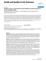
response to pegfilgrastim. As depicted in Figure 1, both
the WBC count and the ANC significantly increased on
day +11 compared with pre-treatment values (p =
0.0002 and p = 0.033, respectively) and returned to
baseline on day +21. Notably, filgrastim promoted a
greater increase of WBC and neutrophils compared with
pegfilgrastim, peaking on day +11 after the commencement of cytokine treatment (p = 0.0085 and p = 0.028
compared with baseline, respectively). Specifically, a
median of 16.5 × 10 3 WBC/μl of blood (range 7.7436.82) were counted in day +11 samples from filgrastim-treated patients compared with 11.64 × 10³ WBC/μl
of blood (range 6.88-15.78) in patients given pegfilgrastim (p < 0.05). Similarly, the ANC was significantly
higher on day +11 after filgrastim administration (13.6 ×
103/μl, range 5.54-31.81) compared with the pegfilgrastim group (7.91 × 10³/μl, range 3.39-13.6; p < 0.05).
It has been previously shown that unconjugated GCSF increases the number of lymphoid progenitors,
mature lymphocytes and monocytes when administered
to healthy HSC donors [20]. In our cohort of cancer
patients, both pegfilgrastim and filgrastim significantly
enhanced lymphocyte (p = 0.0002 and p = 0.0093,
respectively) and monocyte counts (p < 0.0001 and p =
0.013, respectively) compared with baseline, peaking on
day +11 from the commencement of cytokine treatment
(Figure 1). Again, monocyte counts were significantly
higher in patients treated with daily filgrastim (0.8 × 10³
Bonanno et al. Journal of Translational Medicine 2010, 8:114
/>
Page 5 of 15
Figure 1 Changes in leukocyte subsets in patients receiving growth factor support. Leukocytes, neutrophils, monocytes and lymphocytes
were enumerated with automated hematology analyzers before chemotherapy (day 0) and on days +7, +11 and +21 from G-CSF administration.
Bars depict median values. The results of statistical comparisons among baseline and post-treatment samples and between the two study
groups have been detailed in the main text.
cells/μl, range 0.47-1.85, on day +11) compared with
patients given pegfilgrastim (0.57 × 10³ cells/μl, range
0.21-0.93; p = 0.04). Neither lymphocyte nor monocyte
count at baseline differed significantly in the two patient
cohorts (lymphocyte count = 1.69 × 10³ cells/μl, range
0.8-2.24; and 1.21 × 10³ cells/μl, range 0.45-2.54, in the
filgrastim and pegfilgrastim group, respectively; monocyte count = 0.25 × 10 3 cells/μl, range 0.05-0.35; and
0.23 ± 0.06 × 10³ cells/μl, range 0.03-0.89, in the filgrastim and pegfilgrastim group, respectively), suggesting
that the sharper elevation of monocyte counts likely
reflected an intrinsic ability of filgrastim to mobilize
cells of the monocytic lineage. The observed changes in
leukocyte subsets were transient, as indicated by the
recovery of pre-treatment values by day +21 (Figure 1).
Importantly, both the absolute number and the frequency of lymphocytes and monocytes increased as a
result of pegfilgrastim administration (Figure 1), indicating the occurrence of mobilization and/or recruitment
from peripheral sites into the circulation. However, the
relative distribution of CD4 + T cells, CD8 + T cells,
CD19 + B cells and NK cells (defined as CD3 - CD16
+
CD56 + cells) within the lymphocyte population was
unaffected by pegfilgrastim administration (data not
shown). In sharp contrast to pegfilgrastim, filgrastim
was unable to affect the frequency of lymphocytic and
monocytic cells, as shown in Figure 1. The percentage
of lymphocytes within total leukocytes was even lower
on days +7 and +11 after filgrastim administration compared with baseline. Not unexpectedly, treatment with
pegfilgrastim was associated with the mobilization of
CD34-expressing HSC, which peaked on day +11 from
cytokine treatment (4.2 cells/μl, range 2-23.1, compared
with 0.9 cells/μl, range 0.5-10.4, at baseline; p < 0.05)
and declined to pre-treatment values by day +21 (0.8
cells/μl, range 0.25-2).
Mobilization of DC subsets and Treg cells
We next investigated whether pegfilgrastim induced
changes in the frequency of circulating DC precursors.
Cells were initially gated based on lack of expression of
surface antigens associated with lineage differentiation,
as detailed in Materials and Methods. A representative
flow cytometry profile is shown in Figure 2A. Lineagecells were then analyzed for their expression of HLADR in association with CD11c (DC1) or CD123 (DC2),
recognizing the IL-3 receptor a chain. Figure 2B depicts
the cumulative frequency of DC1 and DC2 cells within
Bonanno et al. Journal of Translational Medicine 2010, 8:114
/>
Page 6 of 15
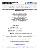
Figure 2 Mobilization of DC precursors and Treg cells in patients receiving growth factor support. The frequency of DC1 (lineage-HLA-DR
+
CD11c+) and DC2 (lineage-HLA-DR+CD123+) precursors and that of CD4+FoxP3+ Treg cells was estimated by flow cytometry, as detailed in
Materials and Methods. Panel A: Gating strategy for the enumeration of DC1 and DC2 precursors. Cells were initially gated based on lack of
surface antigens associated with blood cell lineages. The co-expression of HLA-DR and CD11c or CD123 is shown in one patient given
pegfilgrastim, and is representative of 12 independent experiments. Panel B: Cumulative frequency of DC1 (empty bars) and DC2 (black bars) in
patients given pegfilgrastim or filgrastim. Median values and interquartile range are shown. *p < 0.05 compared with baseline. **p < 0.01
compared with baseline. Panel C: Boxes and whiskers depicting median values and interquartile range. *p = 0.01 compared with healthy
controls (black bar); **p = 0.0009 compared with healthy controls (black bar). The Kruskal-Wallis test with Dunn’s correction for multiple
comparisons was used for statistical analyses. Panel D: Representative flow cytometry profile from one patient treated with pegfilgrastim.
Quadrants were set according to the proper isotypic control (not shown). The percentage of CD4+FoxP3+ T cells in indicated.
the total leukocyte population of patients treated with
either pegfilgrastim or daily filgrastim. In both cohorts
of patients, cytokine administration translated into
increased percentages of DC1 and DC2 cells, albeit with
a different kinetics. Specifically, DC1 precursor cells
were detected at higher frequency on day +7 after the
commencement of pegfilgrastim (p < 0.05) and declined
thereafter, whereas DC2 precursor cells reached a peak
value on day +11 (p < 0.05). In contrast, daily filgrastim
preferentially mobilized DC1 compared with DC2 cells,
and both DC populations peaked at day +11 (p < 0.01
and p < 0.05 for DC1 and DC2, respectively), corresponding to the day after drug discontinuation (Figure
2B).
Because FoxP3 + Treg cells are heterogeneous in
humans and FoxP3-expressing cells have been detected
both within CD4+CD25+ and within CD4+CD25- T-cell
populations [21], we measured the frequency of bona
fide Treg cells based on their CD4+FoxP3+ phenotype.
Treg cells at baseline were comparable in patients given
pegfilgrastim (5.2%, range 1.7-8.1) and in patients treated with daily unconjugated G-CSF (4.9%, range 3.2-
Bonanno et al. Journal of Translational Medicine 2010, 8:114
/>
7.7), and significantly exceeded those in healthy volunteers (2.9%, range 2.3-4; nr of subjects = 8; p < 0.01), in
agreement with other reports describing Treg expansion
in the immunosuppressive milieu of patients with gynecological malignancies [22]. As shown in Figure 2C, a
trend towards higher percentages of Treg cells was documented in samples collected after either pegfilgrastim or
filgrastim administration. In the pegfilgrastim group, a
median of 7.6% (range 5-9.6) CD4+ T cells co-expressed
FoxP3 on day +21 from cytokine administration compared with 5.2% (range 1.7-8.1) at baseline, but this difference failed to achieve statistical significance. Similarly,
5.8% (range 5.7-6.9) CD4+FoxP3+ T cells were detected
at late time-points after filgrastim administration compared with 4.97% (range 3.2-7.7) at baseline (p = NS).
Notably, the percentage of Treg cells at any time-point
after filgrastim treatment significantly exceeded that
measured in healthy controls (Figure 2C). A representative experiment aimed at detecting Treg cells for one
patient given pegfilgrastim is depicted in Figure 2D.
Cytokine measurements and Trp/Kyn ratio
It is now recognized that the balance between IL-12 and
IL-10 produced by the antigen presenting cell compartment dictates the outcome of an immune response, with
IL-12 release leading to robust T-cell priming and IL-10
secretion primarily mediating the induction of T-cell
unresponsiveness [23]. As shown in Figure 3A, serum IL12p40 levels significantly increased after pegfilgrastim
administration and returned to baseline on day +21. Conversely, IL-12p40 slightly declined in cancer patients
given daily G-CSF, and returned to pre-treatment values
by day +11. IL-10 serum levels were consistently below
the ELISA lowest standard (7.8 pg/ml), either in patients
treated with pegfilgrastim or in those given unconjugated
G-CSF (data not shown). TGF-b and HGF play significant roles as immune modulating growth factors both
physiologically and in pathological states such as cancer.
In order to gain further insights into the immune modulation exerted by G-CSF, we also measured TGF-b and
HGF levels before and after cytokine treatment. TGF-b
levels displayed minor fluctuations in the peripheral
blood of patients given either unconjugated G-CSF or
pegylated G-CSF (Figure 3A). In contrast, the administration of pegfilgrastim was associated with an increase of
serum HGF compared with baseline (Figure 3A). Importantly, serum HGF levels on day +11 were significantly
higher in patients receiving filgrastim than in those given
pegfilgrastim (p = 0.043). In both cohorts of patients,
HGF returned to pre-treatment values on day +21 from
the commencement of cytokine administration.
Because HGF may induce the expression of indoleamine 2,3-dioxygenase 1 (IDO1) [8], an enzyme implicated in the conversion of Trp into immune suppressive
Page 7 of 15
Kyn [24], we analyzed IDO1 mRNA expression in
patient monocytes and neutrophils and measured serum
Trp and Kyn levels after treatment with pegfilgrastim.
RT-PCR studies with purified monocytes and neutrophils indicated that mRNA signals for IDO1 were
unchanged after pegfilgrastim administration [see Additional file 1]. As shown in Additional file 1, serum Kyn
displayed minor fluctuations following pegfilgrastim
administration. It should be emphasized that Kyn levels
in 4 out of 5 patients, either at baseline or after the clinical provision of pegfilgrastim, were higher than those
measured in healthy controls. Finally, serum Trp levels
were significantly lower (< 40 μM) than in healthy controls (83.9 μM on average; data not shown) at any timepoint, in line with previous data on altered Trp catabolism in cancer patients [24].
In order to more accurately substantiate the assumption that pegfilgrastim alters the balance between IL12 and IL-10, monocytes, a prominent cellular source
of both IL-12 and IL-10, were magnetically purified on
day +11 from the peripheral blood of patients treated
with pegfilgrastim (24 hours before the anticipated
decline of serum pegfilgrastim concentration [16] and
coincident with maximal monocyte mobilization) and
from cancer patients treated with daily filgrastim (24
hours after the last G-CSF administration). Monocytes
were routinely > 95% pure, as evaluated by flow cytometry measurements of CD14 expression (data not
shown). Equal numbers of monocytes from pre-G-CSF
and post-G-CSF samples were cultured for up to 96
hours in the presence of LPS as a stimulus. The LPSinduced monocytic release of IL-10 increased after
pegfilgrastim administration (Figure 3B). Notably, postpegfilgrastim monocytes secreted considerable amounts
of IL-12p40 at any time-point in culture (Figure 3B).
In line with previous reports [25], monocytes from filgrastim-treated patients secreted low amounts of IL12p40. Intriguingly, IL-12p40 production by post-filgrastim monocytes was significantly lower than that
measured in post-pegfilgrastim monocyte cultures at
any time-point. To further reinforce the assumption
that pegfilgrastim, but not unconjugated G-CSF,
enhances the monocytic release of IL-12 on a per cell
basis, IL-12p70 levels were measured in supernatants
of monocytes purified from 3 patients given pegfilgrastim and 3 patients receiving unconjugated G-CSF. As
shown in Figure 4, post-pegfilgrastim monocytes
released significantly higher levels of IL-12p70 compared with monocytes isolated from cancer patients
treated with unconjugated G-CSF.
In vitro DC phenotype and function
It has been previously shown that filgrastim indirectly
affects DC number and function, skewing in vitro DC
Bonanno et al. Journal of Translational Medicine 2010, 8:114
/>
Page 8 of 15
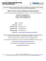
Figure 3 Ex vivo cytokine measurements and in vitro monocytic release of IL-10 and IL-12p40. Panel A: Patient serum was collected at
the indicated time-points and used to evaluate IL-12p40, TGF-b1 and HGF levels by ELISA. Bars depict median values and interquartile ranges
recorded in 12 independent experiments performed in duplicate. °p < 0.01 when comparing IL-12p40 levels on day +7 vs. day +21. °°p = 0.0036
when comparing IL-12p40 levels on day +11 vs. baseline and vs. day +21. *p = 0.0023 when comparing HGF levels on day +7 and day +11 vs.
baseline. §p = 0.0062 when comparing HGF levels on day +7 and day +11 vs. baseline and vs. day +21. Panel B: Monocytes were purified on
day +11 from the commencement of cytokine treatment, coincident with maximal mobilization into the peripheral blood. Cells (1 × 10⁶) were
stimulated with 1 μg/ml LPS in complete culture medium for up to 96 hours. Supernatants were harvested daily and used to measure IL-10 and
IL-12p40 by ELISA. IL-10 and IL-12p40 levels were also estimated in 7 patients with gynecological cancers treated with daily G-CSF. Median
values and interquartile range are shown. *p < 0.01 compared with IL-12p40 levels in supernatants of post-filgrastim monocytes.
differentiation towards a tolerogenic profile [10,11]. To
assess whether soluble factors induced by pegfilgrastim
hindered DC maturation, we cultured monocytes from
healthy controls with patient serum collected either
before or after G-CSF administration. At the end of the
6-day culture period, cells were recovered and labeled
with a panel of mAb recognizing DC activation/differentiation antigens. Control cultures consisted of immunogenic DC differentiated with GM-CSF and IL-4 under
serum-free conditions. The phenotypic and functional
features of the DC-like cells differentiated after the provision of filgrastim have been extensively reported elsewhere [11] and these experiments were not further
replicated in the present study.
For technical reasons, insufficient quantities of day +7
serum were obtained to be supplemented at 20% v/v to
the DC and monocyte cultures. Figure 5 thus illustrates
a representative experiment with day +11 and day +21
monocyte-derived DC preparations. Not unexpectedly,
monocytes cultured with GM-CSF and IL-4 under
serum-free conditions down-regulated CD14, were uniformly CD1a + , and up-regulated costimulatory molecules (CD80 and CD86) and DC maturation antigens
such as CD83 and CD209 (Figure 5A). In sharp contrast, monocytes cultured with either pre- or post-pegfilgrastim serum maintained a CD14+CD1a- phenotype, in
accordance with previous reports on the phenotype of
human serum-supplemented DC cultures [11].
Bonanno et al. Journal of Translational Medicine 2010, 8:114
/>
Page 9 of 15
Figure 4 In vitro monocytic release of bioactive IL-12p70. Monocytes (1 × 10⁶ ) purified from the peripheral blood of patients given
pegfilgrastim (n = 3) or filgrastim (n = 3) were stimulated with LPS as detailed in the legend to Figure 3B. Supernatants were harvested daily
and used to measure IL-12p70 by ELISA. Each point is representative of the mean value of triplicate IL-12p70 measurements.
Figure 5 Phenotypic features of DC-like cells from patients receiving pegfilgrastim. Monocytes were purified from the peripheral blood of
patients given pegfilgrastim and were cultured in the presence of either pre-G-CSF or post-G-CSF serum (20% v/v) for 6 days, as detailed in
Materials and Methods. Control cultures consisted of immunogenic DC preparations that were differentiated with GM-CSF and IL-4 without the
provision of additional maturation stimuli (GM4DC). Panel A: Percentage of cells staining positively for a given antigen in a representative
experiment out of 12 with similar results. Panel B: Relative expression of informative differentiation antigens. Median values and interquartile
range recorded in 12 independent experiments. *denotes a p value < 0.05 compared with the other time-points.
Bonanno et al. Journal of Translational Medicine 2010, 8:114
/>
Interestingly, monocyte cultures containing pre- and
post-pegfilgrastim serum differed in their expression of
costimulatory molecules. CD80 and CD86 were
expressed at significantly higher levels after culture with
post-pegfilgrastim serum, both in terms of percent positive cells and in terms of MFI (Figure 5A and 5B). In
addition, post-pegfilgrastim monocytes up-regulated the
DC maturation antigen CD209 compared with cells in
pre-G-CSF cultures (Figure 5B). ILT3 was also detected
on higher percentages of post-pegfilgrastim monocytic
cells, where its expression increased in terms of fluorescence intensity. Finally, CD83, CD11c and CD123 were
detected on comparable percentages of pre-G-CSF and
post-G-CSF monocytes. Taken together, phenotypic studies revealed that soluble factors contained in post-pegfilgrastim serum promoted the acquisition of a mature
DC-like phenotype, with high expression of costimulatory molecules and CD209, and preserved expression of
the monocyte/macrophage antigen CD14. In line with
this, monocytes nurtured with post-pegfilgrastim serum
possessed a diminished ability to endocytose FITC-conjugated dextran, a measure of DC maturation status,
compared with monocytes cultured with pre-pegfilgrastim serum and with immature DC differentiated with
GM-CSF and IL-4, used as control for optimal incorporation of FITC-dextran (Figure 6A and 6B).
Effect of post-G-CSF serum on alloantigen-induced T-cell
proliferation
We finally asked whether the DC-like preparations
obtained after culture of monocytes from G-CSF-treated
patients could differentially activate the proliferation of
naïve allogeneic CD4+CD25- T cells in comparison with
conventional immunogenic DC differentiated with GMCSF and IL-4. To this end, allogeneic naïve CD4+CD25T cells were pre-loaded with the fluorescent dye CFDASE and were then cultured with patient DC or monocytes at escalating ratios. As shown in Additional file 2,
T-cell proliferation as detected by the progressive halving of CFDA-SE fluorescence was superimposable under
the culture conditions here established, suggesting that
the alloantigen-presenting capacity of in vitro differentiated DC-like cells was unaffected by the in vivo exposure to pegfilgrastim. In a further set of experiments,
either pre- or post-pegfilgrastim serum were supplemented to allogeneic MLR cultures to assess whether soluble
factors in post-pegfilgrastim serum regulate an ongoing
T-cell response to monocytes from third-party healthy
donors. As shown in Figure 6C, the provision of postpegfilgrastim serum (days +7 and + 11) to an allogeneic
MLR culture translated into higher levels of T-cell proliferation compared with cultures supplemented with
post-filgrastim serum collected at the same time-points
(Figure 6C and 6D). Modeling of CFDA-SE profiles
Page 10 of 15
reinforced the concept that higher percentages of undivided, parental cells were contained within MLR cultures supplemented with serum from patients receiving
filgrastim [see Additional file 3], thus suggesting that
pegfilgrastim-induced soluble factors were less likely to
restrain T-cell proliferative responses in vitro than filgrastim-elicited immune suppressive mediators [18].
Discussion
It is conceivable that the G-CSF formulations currently
available for clinical use differentially affect WBC number and function. For instance, a direct comparison of
lenograstim (nonglycosylated G-CSF) and filgrastim or
pegfilgrastim with regard to neutrophil phenotype and
function indicated that neutrophils primed with lenograstim are less functional and structurally more immature compared with those primed with filgrastim and, to
a lesser extent, pegfilgrastim [26]. Importantly, randomized clinical trials evaluating single administration of
pegfilgrastim vs. daily filgrastim as an adjunct to chemotherapy in patients with hematological and solid
malignancies reported similar efficacy profiles [27] or
even a lower overall rate of febrile neutropenia in
patients treated with pegfilgrastim compared with those
given daily filgrastim [28].
The present study aimed to address whether pegfilgrastim given as prophylaxis for chemotherapy-induced
neutropenia affects the number and function of immune
cells, a finding with potential implications for the treatment of cancer patients. The immune modulating
actions of unconjugated G-CSF have been previously
described both in vitro and ex vivo [29]. This basic
knowledge has been translated into animal models of
autoimmune disorders to skew the immune response
and to promote tolerance. For instance, G-CSF ameliorated experimental autoimmune encephalomyelitis [30],
type 1 diabetes [31], experimental colitis [32] and lupus
nephritis [33] through effects on adaptive and innate
immune responses. A pilot clinical trial in Crohn’s disease provided proof of principle in favor of immune regulatory effects by filgrastim in the human setting [34].
In this study, daily treatment with G-CSF for 4 weeks
was correlated with an increase of IL-10-secreting type
1 Treg cells in the peripheral blood and with the accumulation of plasmacytoid DC in the gut lamina propria
[34].
In the present report, WBC and ANC recovery in
patients treated with pegfilgrastim occurred without the
fluctuations associated with daily filgrastim injections.
The administration of pegfilgrastim translated into a
transient but significant elevation of CD34-expressing
HSC, lymphocytes and monocytes. Lymphocyte recirculation is expected to favorably impact on the immune
control of the underlying malignancy, and the
Bonanno et al. Journal of Translational Medicine 2010, 8:114
/>
Page 11 of 15
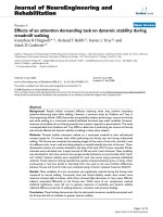
Figure 6 Functional features of Mo-DC from patients receiving pegfilgrastim. Monocytes were purified from the peripheral blood of
patients given pegfilgrastim and were cultured in the presence of either pre-G-CSF or post-G-CSF serum (20% v/v) for 6 days, as detailed in
Materials and Methods. Control cultures consisted of immunogenic DC preparations that were differentiated with GM-CSF and IL-4 without the
provision of additional maturation stimuli (GM4DC). Panel A: Uptake of FITC-conjugated dextran by monocytes cultured in vitro in the presence
of pre-pegfilgrastim serum (day 0) or post-pegfilgrastim serum (days +7 and +11). Median values and interquartile range are shown. *p < 0.05
compared with Mo-DC differentiated with GM-CSF and IL-4; §p < 0.05 compared with cells cultured with pre-G-CSF serum. Panel B:
Representative experiment; red histograms depict the uptake of FITC-conjugated dextran by monocytes kept at 4°C (negative control) and
empty histograms depict the uptake of FITC-conjugated dextran by the monocyte preparations kept at 37°C. Panel C: CD4+CD25- T cells and
monocytes were purified from the peripheral blood of healthy donors as detailed in the main text. After irradiation, monocytes were cultured
with CFDA-SE loaded, allogeneic T cells at a fixed monocyte-to-T cell ratio (1:27) for 7 days, either in the absence or presence of patient serum
(20% v/v). The proliferation index of T-cell cultures established in the presence of patient serum collected before and after G-CSF administration
is shown. The bars depict median and interquartile range recorded in 3 independent experiments performed in duplicate. Panel D: Results of a
representative experiment out of 3 with similar results. The percentage of parental, undivided cells (U; depicted in blue) is indicated. The analysis
of CFDA-SE fluorescence was performed with the proliferation wizard of the ModFit software package, as previously detailed [12].
observation that prompt lymphocyte recovery predicts a
higher relapse-free survival in cancer patients [35]
underpins the potential clinical significance of the pegfilgrastim-induced changes in WBC subsets. Pegfilgrastim
did not elicit any appreciable mobilization of Treg cells,
as documented by serial measurements of the frequency
of circulating CD4+FoxP3+ Treg cells. We cannot rule
out the possibility that any G-CSF-induced recirculation
of Treg cells was obscured by the high frequency of
Treg cells already measured at baseline. Of interest, filgrastim has been reported to increase the frequency of
CD4 + CD25 high Treg cells only when given to cancer
patients in combination with cyclophosphamide as HSC
mobilization regimen [36]. In healthy donors, the phenotype and frequency of CD4 + CD25 high FoxP3 + Treg
cells may be unaffected by G-CSF [37]. At variance with
human data, filgrastim recruited functional TGF-bexpressing Treg cells to the pancreatic lymph nodes of
NOD mice, with the likely aim to restrain the proliferation and function of diabetogenic T cells [31]. It remains
to be determined whether Treg recirculation and/or
recruitment to sites of inflammation and tissue injury
Bonanno et al. Journal of Translational Medicine 2010, 8:114
/>
may also occur in humans as a result of pegfilgrastim
administration.
We were also interested in evaluating whether pegfilgrastim induced the release of immune suppressive HGF
and TGF-b1. HGF is a pro-angiogenic and tumor-promoting cytokine. HGF reportedly skews DC function,
driving an IL-10-secreting tolerogenic profile both in
mice [38] and in humans [8]. We measured significantly
elevated levels of HGF in patients treated with either
pegfilgrastim or filgrastim. Furthermore, HGF secretion
was significantly lower after pegfilgrastim compared
with daily filgrastim administration. In contrast, serum
TGF-b1 levels were not modified by either G-CSF formulation. The observation that HGF induces functional
IDO1 in human monocyte-derived DC [8] raised the
previously unexplored possibility that pegfilgrastim may
indirectly activate IDO1-mediated Trp breakdown into
immune suppressive derivatives, collectively referred to
as Kyn. Interestingly, serum Kyn were not significantly
different when comparing samples at baseline with
those obtained from patients receiving pegfilgrastim. It
should be noted that baseline Kyn levels in our patient
cohort were higher than those measured in healthy controls (median Kyn concentration = 1.86 μM; number of
samples = 20), probably reflecting the expression of
functional IDO1 by the ovarian and endometrial cancer
cells [39]. Also, mRNA signals for IDO1 in monocytes
and granulocytes, a potential source of IDO1 activity
[40], were unchanged when comparing pre-G and postG samples. These observations are backed by a recent
study indicating that G-CSF-mobilized immature myeloid cells inhibit alloreactive responses in mice through
an IDO-independent mechanism, and that G-CSF signaling is incapable of directly inducing IDO [41].
The studies published so far suggest that the extent of
DC1/DC2 mobilization by filgrastim crucially depends
on the intensity of the mobilization regimen and on the
underlying neoplastic disorder. In this respect, filgrastim
preferentially mobilized DC2 in healthy donors [10] but
failed to impact on the DC1/DC2 ratio in patients with
hematological and solid malignancies [42]. In another
study with healthy donors, low-dose G-CSF (8-10 μg/
kg/day) increased the frequency of CD123+ blood DC
precursors but mobilized CD11c+ DC only occasionally
[43]. Furthermore, high-dose G-CSF (30 μg/kg/day)
mobilized CD123 + DC in patients with multiple myeloma but only occasionally in those affected by nonHodgkin’s lymphoma, and exerted varying effects on
CD11c+ DC [43]. We have shown herein that pegfilgrastim mobilized both DC1 and DC2 precursors into the
peripheral blood of patients with gynecological malignancies treated with carboplatin and paclitaxel, suggesting lack of DC skewing in vivo. The highest frequencies
of DC1 precursors were measured on day +7 from
Page 12 of 15
pegfilgrastim administration, whereas DC2 precursors
were higher in day +11 samples and declined thereafter.
It is conceivable that different chemotherapy/growth
factor combinations and doses and/or intrinsic characteristics of the underlying neoplastic disorder account
for differences in the relative proportion of DC1/DC2
precursors and in their mobilization kinetics. It is presently unknown whether the transient DC1 mobilization
induced by pegfilgrastim will impact on the host
immune system’s ability to control disease progression.
IL-12, a prototype member of a family of IL-12-related
cytokines that includes IL-23 and IL-27, is an instigator
of Th1 immune responses and possesses in vivo antitumor activities [44]. IL-12 is a heterodimer formed by a
35-kDa light chain (known as p35 or IL-12a) and a 40kDa heavy chain (known as p40 or IL-12b). Messenger
RNA encoding IL-12p35 is present in many cell types,
whereas mRNA encoding IL-12p40 is restricted to cells
that produce the biologically active heterodimer [45].
DC and monocytes have been reported to secrete a 101,000 fold excess of IL-12p40 compared with IL-12p75
[44]. A report on post-transplantation immune functions
in 43 patients receiving filgrastim has shown that cytokine administration delays the reconstitution of CD4+ T
cells and blunts anti-fungal T-cell responses [25]. These
abnormalities were correlated with the inability of DC
and monocytes from G-CSF-treated patients to release
IL-12p40 [25]. Interestingly, the in vivo immune modulating effects of G-CSF were replicated in vitro when
monocytes from normal volunteers were differentiated
along the DC lineage after their 24-hour pre-treatment
with exogenous G-CSF. Under these conditions, IL12p40 production was inhibited both at the mRNA and
protein level [25]. In our study, pegfilgrastim administration was associated with a significant increase of the
inducible IL-12p40 subunit in patient serum. In patients
given filgrastim, IL-12p40 slightly declined and returned
to baseline values by day +11 from the commencement
of cytokine treatment. Interestingly, neutrophil-derived
serine proteases have been reported to inactivate human
growth factors such as TNF-a at sites of inflammation
and to promote the formation of cytokine split products
[46]. It is tempting to speculate that immunoreactive IL12 in patients given filgrastim may have been degraded
as a result of sharp increases in circulating PMN capable
of releasing proteolytic enzymes. Intriguingly, monocytes
from patients treated with pegfilgrastim released higher
amounts of both IL-12p40 and IL-12p70 in vitro compared with monocytes from filgrastim-treated patients.
In contrast, the LPS-induced release of IL-10 increased
to a similar extent in cultures established with monocytes from patients given pegfilgrastim and filgrastim.
IL-12p40 homodimers may behave as IL-12 receptor
antagonists both in mice and in humans, inhibiting IL-
Bonanno et al. Journal of Translational Medicine 2010, 8:114
/>
12-induced T-cell proliferation [47,48]. Our observation
that post-pegfilgrastim monocytes release higher
amounts of bioactive IL-12p70 compared with post-filgrastim monocytes supports the conclusion that pegfilgrastim may not dampen in vivo anti-tumor immunity
and/or host defense against infectious agents, a response
that crucially depends on the balance between IL-12
and IL-10 production. It has been reported that 6-sulfo
LacNAc+ DC, a major source of IL-12 and potent inducers of T-cell responses in vitro, are efficiently mobilized
in healthy donors given G-CSF at 7.5 μg/kg of body
weight [49]. Conceivably, pegfilgrastim might also favor
the mobilization of 6-sulfo LacNAc+ DC or other as yet
unrecognized monocyte/DC populations with a unique
ability to produce bioactive IL-12.
It is known that unconjugated G-CSF promotes the
development of tolerogenic DC in vitro [11] and in vivo
[31]. We showed herein that pegfilgrastim-induced soluble factors promoted the emergence of mature DC-like
populations with high expression of costimulatory molecules (CD80, CD86), CD83 and CD209, and with low
endocytic capacity. Post-pegfilgrastim DC-like cells also
up-regulated ILT3, an inhibitory receptor detected on
anergizing DC preparations [50,51], and yet activated the
proliferation of allogeneic naïve T cells to a similar extent
as immunogenic DC. It should be noted that ILT3
expression may be dispensable for the induction of CD4
+
CD25 + Treg cells by 1,25-dihydroxyvitamin D3 [52],
indicating that molecular determinants of T-cell suppression other than ILT3 may be operational depending
upon the experimental system. Of potential interest, we
measured high levels of IL-10 in post-pegfilgrastim DC
cultures (317 ± 140 pg/ml compared with 27.1 ± 2.3 pg/
ml in control cultures of immunogenic GM4DC). IL-10
secretion may have been responsible for ILT3 up-regulation on post-pegfilgrastim monocytes, in line with the
effect of exogenous IL-10 on ILT3 expression by human
vascular endothelial cells [53]. We also evaluated the ability of post-pegfilgrastim DC to activate allogeneic T-cell
responses in vitro. Interestingly, monocytes from patients
given pegfilgrastim induced T-cell proliferation to a similar extent as immunogenic DC. In line with this, T-cell
proliferation in response to allogeneic monocytes was
not inhibited by the provision of post-pegfilgrastim
serum to the MLR culture. Our observations on in vitro
DC phenotype and function reinforce the view that pegfilgrastim and filgrastim differ in their ability to skew
monocyte function, with the former supporting the in
vitro development of activating DC and the latter favoring the emergence of tolerogenic DC preparations [18].
Conclusions
Taken together, the experimental evidence herein presented indicates that the administration of pegfilgrastim
Page 13 of 15
to hasten neutrophil recovery should not translate into
undesired immune suppression in cancer-bearing
patients, who might benefit from robust monocytic production of IL-12, in the absence of excessive induction
of immune suppressive IL-10 and HGF. A further implication of our findings pertains to HLA-matched stem
cell transplantation, a clinical context where pegfilgrastim administration may modulate the number of
immune cells and/or levels of immune regulatory soluble factors, thus ameliorating leukemia clearance. In this
respect, it has been shown that multiple pegylation of
G-CSF imparts an enhanced biological activity with
respect to immune cells and improves stem cell transplant in mice [54]. Intriguingly, multi-pegylated versions
of G-CSF separate GVHD from graft-versus-leukemia
(GVL) through the activation of invariant NKT cells,
thus contributing to leukemia eradication [55,56]. These
considerations add to the knowledge that pegfilgrastim
has advantages over filgrastim in terms of patient compliance, ease of administration and patient quality of life
[1]. Whether the pegfilgrastim-induced modulation of
immune function will favorably impact on disease control in cancer-bearing patients remains to be prospectively determined.
Additional material
Additional file 1: Expression of IDO1 mRNA and serum Kyn levels in
patients given pegfilgrastim. Panel A: Expression of IDO1 mRNA in
patient monocytes and granulocytes. Details on RNA extraction and
reverse-transcription were previously published [8]. The following primers
were used for mRNA amplification: 5’-ACTGCCCCTGTGATAAACTGTGG-3’
and 5’-GCGTGTGCCATTCTTGTAGTCTG-3’ (human IDO1; GI 156071492); 5’TGACATCAAGAAGGTGGTGA-3’ and 5’-TCCACCACCCTGTTGCTGTA-3’
(human GAPDH; GI 7669491). Primer sets were designed using the
Beacon Design Software (Version 3) and the sequences available in the
Gene Bank™ database. All nucleotide primers were synthesized by MWG
(Florence, Italy), and PCR products were analyzed on 3% agarose gel
(Agarose, type XII: low viscosity for beading, Sigma Aldrich) stained with
ethidium bromide. M = marker. + = normal endometrial tissue used as
positive control for IDO1 mRNA expression. Panel B: Quantitative
densitometry (Quantity One software; Bio-Rad, Hercules, CA) is shown
with monocytes and granulocytes isolated from 2 patients given
pegfilgrastim. Insufficient numbers of cells were available on day +7, and
PCR analyses were performed with patient material obtained on days 0,
+11 and +21. Normal endometrial tissue was used as positive control for
IDO1 mRNA expression (red column). Panel C: Serum Kyn levels were
measured by RP-HPLC in 5 patients before (day 0) and after pegfilgrastim
administration (days +7, +11 and +21), as detailed in Materials and
Methods. Data from each individual patient have been plotted using a
different color. The dotted line indicates the median serum Kyn
concentration measured in 50 healthy subjects (2.3 μM).
Additional file 2: T-cell stimulation by Mo-DC generated in vitro
after in vivo administration of pegfilgrastim. Mo-DC were
differentiated from patient monocytes in the presence of either pre-GCSF serum or post-G-CSF serum (collected on day + 11), as detailed in
Materials and Methods. Immunogenic DC were generated with IL-4 and
GM-CSF, in accordance with established DC differentiation protocols [17].
The Mo-DC preparations were co-cultured with CFDA-SE pre-loaded,
allogeneic naïve CD4+CD25- T cells at different T cell-to-DC ratio. The
percentage of CD25-expressing, CFDA-SEdim and CFDA-SEbright cells is
Bonanno et al. Journal of Translational Medicine 2010, 8:114
/>
indicated. One representative experiment out of 5 with similar results is
shown.
Additional file 3: Cell proliferation tracking after provision of postG-CSF serum to MLR cultures. MLR cultures were established as above
detailed. T cells and monocytes were plated at a fixed DC-to-T cell ratio
(1:27). The percentage of proliferating T cells residing within each cell
generation (G) was calculated with the proliferation wizard of the
ModFit™ software. Median values and interquartile range are shown.
*denotes a p value < 0.05 when comparing the percentage of parental
(P), undivided cells in MLR cultures established with serum from patients
given pegfilgrastim (black bars) or filgrastim (empty bars).
Page 14 of 15
8.
9.
10.
11.
Acknowledgements
GS and SR are supported by Fondazione Roma, Rome, Italy (Stem Cell
Project). SR receives an Investigator Grant (n. 8556) from Associazione Italiana
per la Ricerca sul Cancro (AIRC), Milan, Italy.
12.
Author details
1
Department of Gynecology and Obstetrics, Catholic University Med. School,
Rome, Italy. 2IRCCS in Gastroenterology, Istituto Clinico Humanitas, Milan,
Italy. 3Department of Medicine and Geriatrics, Hemostasis Research Centre,
Catholic University Med. School, Rome, Italy. 4Department of Hematology,
Catholic University Med. School, Rome, Italy. 5IRCCS San Raffaele Pisana,
Rome, Italy.
13.
Authors’ contributions
GB carried out the experiments and participated in the design of the study.
AM, AP and MC carried out the experiments. AP and GS participated in the
design of the study and were responsible for patient care and sample
procurement. RDC carried out the experiments and contributed to
manuscript drafting. SD gave intellectual input and advice. SR participated in
the design of the study, carried out the experiments, performed the
statistical analysis and drafted the manuscript. All authors read and approved
the final manuscript.
Competing interests
The authors declare that they have no competing interests.
Received: 11 May 2010 Accepted: 9 November 2010
Published: 9 November 2010
References
1. Molineux G: The design and development of pegfilgrastim (PEGrmetHuG-CSF, Neulasta). Curr Pharm Des 2004, 10:1235-1244.
2. Anderlini P, Champlin RE: Biologic and molecular effects of granulocyte
colony-stimulating factor in healthy individuals: recent findings and
current challenges. Blood 2008, 111:1767-1772.
3. Pan L, Delmonte J, Jalonen CK, Ferrara JL: Pretreatment of donor mice
with granulocyte colony-stimulating factor polarizes donor T
lymphocytes toward type-2 cytokine production and reduces severity of
experimental graft-versus-host disease. Blood 1995, 86:4422-4429.
4. Sloand EM, Kim S, Maciejewski JP, Van Rhee F, Chaudhuri A, Barrett J,
Young NS: Pharmacologic doses of granulocyte colony-stimulating factor
affect cytokine production by lymphocytes in vitro and in vivo. Blood
2000, 95:2269-2274.
5. Franzke A, Piao W, Lauber J, Gatzlaff P, Konecke C, Hansen W, SchmittThomsen A, Hertenstein B, Buer J, Ganser A: G-CSF as immune regulator in
T cells expressing the G-CSF receptor: implications for transplantation
and autoimmune diseases. Blood 2003, 102:734-739.
6. Rutella S, Pierelli L, Bonanno G, Sica S, Ameglio F, Capoluongo E, Mariotti A,
Scambia G, d’Onofrio G, Leone G: Role for granulocyte colony-stimulating
factor in the generation of human T regulatory type 1 cells. Blood 2002,
100:2562-2571.
7. Fujii K, Ishimaru F, Kozuka T, Matsuo K, Nakase K, Kataoka I, Tabayashi T,
Shinagawa K, Ikeda K, Harada M, Tanimoto M: Elevation of serum
hepatocyte growth factor during granulocyte colony-stimulating factorinduced peripheral blood stem cell mobilization. Br J Haematol 2004,
124:190-194.
14.
15.
16.
17.
18.
19.
20.
21.
22.
23.
24.
25.
26.
Rutella S, Bonanno G, Procoli A, Mariotti A, de Ritis DG, Curti A, Danese S,
Pessina G, Pandolfi S, Natoni F, et al: Hepatocyte growth factor favors
monocyte differentiation into regulatory interleukin (IL)-10++IL-12low/neg
accessory cells with dendritic-cell features. Blood 2006, 108:218-227.
Okunishi K, Dohi M, Fujio K, Nakagome K, Tabata Y, Okasora T, Seki M,
Shibuya M, Imamura M, Harada H, et al: Hepatocyte growth factor
significantly suppresses collagen-induced arthritis in mice. J Immunol
2007, 179:5504-5513.
Arpinati M, Green CL, Heimfeld S, Heuser JE, Anasetti C: Granulocytecolony stimulating factor mobilizes T helper 2-inducing dendritic cells.
Blood 2000, 95:2484-2490.
Rutella S, Bonanno G, Pierelli L, Mariotti A, Capoluongo E, Contemi AM,
Ameglio F, Curti A, De Ritis DG, Voso MT, et al: Granulocyte colonystimulating factor promotes the generation of regulatory DC through
induction of IL-10 and IFN-a. Eur J Immunol 2004, 34:1291-1302.
Rutella S, Pierelli L, Rumi C, Bonanno G, Marone M, Sica S, Capoluongo E,
Ameglio F, Scambia G, Leone G: T-cell apoptosis induced by granulocyte
colony-stimulating factor is associated with retinoblastoma protein
phosphorylation and reduced expression of cyclin-dependent kinase
inhibitors. Exp Hematol 2001, 29:401-415.
Tanaka J, Mielcarek M, Torok-Storb B: Impaired induction of the CD28responsive complex in granulocyte colony-stimulating factor mobilized
CD4 T cells. Blood 1998, 91:347-352.
Morris ES, MacDonald KP, Rowe V, Johnson DH, Banovic T, Clouston AD,
Hill GR: Donor treatment with pegylated G-CSF augments the generation
of IL-10-producing regulatory T cells and promotes transplantation
tolerance. Blood 2004, 103:3573-3581.
Bruns I, Steidl U, Fischer JC, Czibere A, Kobbe G, Raschke S, Singh R, Fenk R,
Rosskopf M, Pechtel S, et al: Pegylated granulocyte colony-stimulating
factor mobilizes CD34+ cells with different stem and progenitor subsets
and distinct functional properties in comparison with unconjugated
granulocyte colony-stimulating factor. Haematologica 2008, 93:347-355.
Yang BB, Kido A, Shibata A: Serum pegfilgrastim concentrations during
recovery of absolute neutrophil count in patients with cancer receiving
pegfilgrastim after chemotherapy. Pharmacotherapy 2007, 27:1387-1393.
Sallusto F, Cella M, Danieli C, Lanzavecchia A: Dendritic cells use
macropinocytosis and the mannose receptor to concentrate
macromolecules in the major histocompatibility complex class II
compartment: downregulation by cytokines and bacterial products. J Exp
Med 1995, 182:389-400.
Rutella S, Rumi C, Lucia MB, Sica S, Cauda R, Leone G: Serum of healthy
donors receiving granulocyte colony-stimulating factor induces T cell
unresponsiveness. Exp Hematol 1998, 26:1024-1033.
Laich A, Neurauter G, Widner B, Fuchs D: More rapid method for
simultaneous measurement of tryptophan and kynurenine by HPLC. Clin
Chem 2002, 48:579-581.
Imamura R, Miyamoto T, Yoshimoto G, Kamezaki K, Ishikawa F, Henzan H,
Kato K, Takase K, Numata A, Nagafuji K, et al: Mobilization of human
lymphoid progenitors after treatment with granulocyte colonystimulating factor. J Immunol 2005, 175:2647-2654.
Walker MR, Kasprowicz DJ, Gersuk VH, Benard A, Van Landeghen M,
Buckner JH, Ziegler SF: Induction of FoxP3 and acquisition of T regulatory
activity by stimulated human CD4+CD25- T cells. J Clin Invest 2003,
112:1437-1443.
Curiel TJ, Coukos G, Zou L, Alvarez X, Cheng P, Mottram P, EvdemonHogan M, Conejo-Garcia JR, Zhang L, Burow M, et al: Specific recruitment
of regulatory T cells in ovarian carcinoma fosters immune privilege and
predicts reduced survival. Nat Med 2004, 10:942-949.
Rutella S, Danese S, Leone G: Tolerogenic dendritic cells: cytokine
modulation comes of age. Blood 2006, 108:1435-1440.
Munn DH, Mellor AL: IDO and tolerance to tumors. Trends Mol Med 2004,
10:15-18.
Volpi I, Perruccio K, Tosti A, Capanni M, Ruggeri L, Posati S, Aversa F,
Tabilio A, Romani L, Martelli MF, Velardi A: Postgrafting administration of
granulocyte colony-stimulating factor impairs functional immune
recovery in recipients of human leukocyte antigen haplotypemismatched hematopoietic transplants. Blood 2001, 97:2514-2521.
Ribeiro D, Veldwijk MR, Benner A, Laufs S, Wenz F, Ho AD, Fruehauf S:
Differences in functional activity and antigen expression of granulocytes
primed in vivo with filgrastim, lenograstim, or pegfilgrastim. Transfusion
2007, 47:969-980.
Bonanno et al. Journal of Translational Medicine 2010, 8:114
/>
27. Vose JM, Crump M, Lazarus H, Emmanouilides C, Schenkein D, Moore J,
Frankel S, Flinn I, Lovelace W, Hackett J, Liang BC: Randomized,
multicenter, open-label study of pegfilgrastim compared with daily
filgrastim after chemotherapy for lymphoma. J Clin Oncol 2003,
21:514-519.
28. Holmes FA, O’Shaughnessy JA, Vukelja S, Jones SE, Shogan J, Savin M,
Glaspy J, Moore M, Meza L, Wiznitzer I, et al: Blinded, randomized,
multicenter study to evaluate single administration pegfilgrastim once
per cycle versus daily filgrastim as an adjunct to chemotherapy in
patients with high-risk stage II or stage III/IV breast cancer. J Clin Oncol
2002, 20:727-731.
29. Rutella S, Zavala F, Danese S, Kared H, Leone G: Granulocyte colonystimulating factor: a novel mediator of T cell tolerance. J Immunol 2005,
175:7085-7091.
30. Zavala F, Abad S, Ezine S, Taupin V, Masson A, Bach JF: G-CSF therapy of
ongoing experimental allergic encephalomyelitis via chemokine- and
cytokine-based immune deviation. J Immunol 2002, 168:2011-2019.
31. Kared H, Masson A, Adle-Biassette H, Bach JF, Chatenoud L, Zavala F:
Treatment with granulocyte colony-stimulating factor prevents diabetes
in NOD mice by recruiting plasmacytoid dendritic cells and functional
CD4+CD25+ regulatory T-cells. Diabetes 2005, 54:78-84.
32. Yoshimitsu M, Hayamizu K, Egi H, Okiyama J, Okajima M, Itamoto T,
Asahara T: The neutrophil/Th1 lymphocyte balance and the therapeutic
effect of granulocyte colony-stimulating factor in TNBS-induced colitis of
rat strains. J Interferon Cytokine Res 2006, 26:291-300.
33. Zavala F, Masson A, Hadaya K, Ezine S, Schneider E, Babin O, Bach JF:
Granulocyte-colony stimulating factor treatment of lupus autoimmune
disease in MRL-lpr/lpr mice. J Immunol 1999, 163:5125-5132.
34. Mannon PJ, Leon F, Fuss IJ, Walter BA, Begnami M, Quezado M, Yang Z,
Yi C, Groden C, Friend J, et al: Successful granulocyte-colony stimulating
factor treatment of Crohn’s disease is associated with the appearance of
circulating interleukin-10-producing T cells and increased lamina propria
plasmacytoid dendritic cells. Clin Exp Immunol 2009, 155:447-456.
35. Kumar S, Chen MG, Gastineau DA, Gertz MA, Inwards DJ, Lacy MQ, Tefferi A,
Litzow MR: Lymphocyte recovery after allogeneic bone marrow
transplantation predicts risk of relapse in acute lymphoblastic leukemia.
Leukemia 2003, 17:1865-1870.
36. Condomines M, Quittet P, Lu ZY, Nadal L, Latry P, Lopez E, Baudard M,
Requirand G, Duperray C, Schved JF, et al: Functional regulatory T cells are
collected in stem cell autografts by mobilization with high-dose
cyclophosphamide and granulocyte colony-stimulating factor. J Immunol
2006, 176:6631-6639.
37. Noel G, Bruniquel D, DeGuibert S, Birebent B, Grosset JM, Bernard M,
Dauriac C, Lamy-de-la-Chapelle T, Semana G, Brinster C: Regulatory CD4
+
CD25hi T cells conserve their function and phenotype after granulocyte
colony-stimulating factor treatment in human hematopoietic stem cell
transplantation. Hum Immunol 2008, 69:329-337.
38. Okunishi K, Dohi M, Nakagome K, Tanaka R, Mizuno S, Matsumoto K,
Miyazaki J, Nakamura T, Yamamoto K: A novel role of hepatocyte growth
factor as an immune regulator through suppressing dendritic cell
function. J Immunol 2005, 175:4745-4753.
39. Ino K, Yamamoto E, Shibata K, Kajiyama H, Yoshida N, Terauchi M, Nawa A,
Nagasaka T, Takikawa O, Kikkawa F: Inverse correlation between tumoral
indoleamine 2,3-dioxygenase expression and tumor-infiltrating
lymphocytes in endometrial cancer: Its association with disease
progression and survival. Clin Cancer Res 2008, 14:2310-2317.
40. Barth MC, Ahluwalia N, Anderson TJ, Hardy GJ, Sinha S, Alvarez-Cardona JA,
Pruitt IE, Rhee EP, Colvin RA, Gerszten RE: Kynurenic acid triggers firm
arrest of leukocytes to vascular endothelium under flow conditions. J
Biol Chem 2009, 284:19189-19195.
41. Joo YD, Lee SM, Lee SW, Lee WS, Park JK, Choi IW, Park SG, Choi I, Seo SK:
Granulocyte colony-stimulating factor-induced immature myeloid cells
inhibit acute graft-versus-host disease lethality through an indoleamine
dioxygenase-independent mechanism. Immunology 2009, 128:e632-640.
42. Gazitt Y, Akay C, Thomas C: No polarization of type 1 or type 2 precursor
dendritic cells in peripheral blood stem cell collections of non-Hodgkin’s
lymphoma patients mobilized with cyclophosphamide plus G-CSF, GMCSF, or GM-CSF followed by G-CSF. Stem Cells Dev 2006, 15:269-277.
43. Vuckovic S, Kim M, Khalil D, Turtle CJ, Crosbie GV, Williams N, Brown L,
Williams K, Kelly C, Stravos P, et al: Granulocyte-colony stimulating factor
Page 15 of 15
44.
45.
46.
47.
48.
49.
50.
51.
52.
53.
54.
55.
56.
increases CD123hi blood dendritic cells with altered CD62L and CCR7
expression. Blood 2003, 101:2314-2317.
Trinchieri G: Interleukin-12 and the regulation of innate resistance and
adaptive immunity. Nat Rev Immunol 2003, 3:133-146.
D’Andrea A, Rengaraju M, Valiante NM, Chehimi J, Kubin M, Aste M,
Chan SH, Kobayashi M, Young D, Nickbarg E, et al: Production of natural
killer cell stimulatory factor (interleukin 12) by peripheral blood
mononuclear cells. J Exp Med 1992, 176:1387-1398.
van Kessel KP, van Strijp JA, Verhoef J: Inactivation of recombinant human
tumor necrosis factor-a by proteolytic enzymes released from stimulated
human neutrophils. J Immunol 1991, 147:3862-3868.
Mattner F, Fischer S, Guckes S, Jin S, Kaulen H, Schmitt E, Rude E,
Germann T: The interleukin-12 subunit p40 specifically inhibits effects of
the interleukin-12 heterodimer. Eur J Immunol 1993, 23:2202-2208.
Ling P, Gately MK, Gubler U, Stern AS, Lin P, Hollfelder K, Su C, Pan YC,
Hakimi J: Human IL-12 p40 homodimer binds to the IL-12 receptor but
does not mediate biologic activity. J Immunol 1995, 154:116-127.
Baumeister SH, Holig K, Bornhauser M, Meurer M, Rieber EP, Schakel K: GCSF mobilizes slanDCs (6-sulfo LacNAc+ dendritic cells) with a high
proinflammatory capacity. Blood 2007, 110:3078-3081.
Chang CC, Ciubotariu R, Manavalan JS, Yuan J, Colovai AI, Piazza F,
Lederman S, Colonna M, Cortesini R, Dalla-Favera R, Suciu-Foca N:
Tolerization of dendritic cells by T(S) cells: the crucial role of inhibitory
receptors ILT3 and ILT4. Nat Immunol 2002, 3:237-243.
Rossetti M, Gregori S, Roncarolo MG: Granulocyte-colony stimulating
factor drives the in vitro differentiation of human dendritic cells that
induce anergy in naive T cells. Eur J Immunol 2010.
Penna G, Roncari A, Amuchastegui S, Daniel KC, Berti E, Colonna M,
Adorini L: Expression of the inhibitory receptor ILT3 on dendritic cells is
dispensable for induction of CD4+Foxp3+ regulatory T cells by 1,25dihydroxyvitamin D3. Blood 2005, 106:3490-3497.
Gleissner CA, Zastrow A, Klingenberg R, Kluger MS, Konstandin M, Celik S,
Haemmerling S, Shankar V, Giese T, Katus HA, Dengler TJ: IL-10 inhibits
endothelium-dependent T cell costimulation by up-regulation of ILT3/4
in human vascular endothelial cells. Eur J Immunol 2007, 37:177-192.
Banovic T, MacDonald KP, Markey KA, Morris ES, Kuns RD, Varelias A, Hill GR:
Donor treatment with a multipegylated G-CSF maximizes graft-versusleukemia effects. Biol Blood Marrow Transplant 2009, 15:126-130.
Morris ES, MacDonald KP, Hill GR: Stem cell mobilization with G-CSF
analogs: a rational approach to separate GVHD and GVL? Blood 2006,
107:3430-3435.
Morris ES, MacDonald KP, Rowe V, Banovic T, Kuns RD, Don AL,
Bofinger HM, Burman AC, Olver SD, Kienzle N, et al: NKT cell-dependent
leukemia eradication following stem cell mobilization with potent G-CSF
analogs. J Clin Invest 2005, 115:3093-3103.
doi:10.1186/1479-5876-8-114
Cite this article as: Bonanno et al.: Effects of pegylated G-CSF on
immune cell number and function in patients with gynecological
malignancies. Journal of Translational Medicine 2010 8:114.
Submit your next manuscript to BioMed Central
and take full advantage of:
• Convenient online submission
• Thorough peer review
• No space constraints or color figure charges
• Immediate publication on acceptance
• Inclusion in PubMed, CAS, Scopus and Google Scholar
• Research which is freely available for redistribution
Submit your manuscript at
www.biomedcentral.com/submit