Báo cáo hóa học: "VRα2 + ovalbumin" pdf
Bạn đang xem bản rút gọn của tài liệu. Xem và tải ngay bản đầy đủ của tài liệu tại đây (1.5 MB, 15 trang )
A
B
CD25
Foxp3
PBS
VRα2 + ovalbumin VRα2 + ECDα2
12.87
9.85
5.938.83
2.39
8.97
1.42
7.74
1.16
Gated on CD4+ cells
Spleen
C
Foxp3+/CD4+ (%)
Control
VR
α
2+ECD
α
2
P<0.001
0
10
20
30
40
Tumor
Control
CD4 Foxp3 Merge
VRα2+ECDα2
Interleukin-13 receptor α2 DNA prime boost
vaccine induces tumor immunity in murine tumor
models
Nakashima et al.
Nakashima et al. Journal of Translational Medicine 2010, 8:116
(10 November 2010)
RESEA R C H Open Access
Interleukin-13 receptor a2 DNA prime boost
vaccine induces tumor immunity in murine tumor
models
Hideyuki Nakashima, Toshio Fujisawa, Syed R Husain, Raj K Puri
*
Abstract
Background: DNA vaccines represent an attractive approach for cancer treatment by inducing active T cell and B
cell immune responses to tumor antigens. Previous studies have shown that interleukin-13 receptor a2 chain
(IL-13Ra2), a tumor-associated antigen is a promising target for cancer immunotherapy as high levels of IL-13Ra2
are expressed on a variety of human tumors. To enhance the effectiveness of DNA vaccine, we used extracellular
domain of IL-13Ra2 (ECDa2) as a protein-boost against murine tumor models.
Methods: We have developed murine models of tumors naturally expressing IL-13Ra2 (MCA304 sarcoma, 4T1
breast carcinoma) and D5 melanoma tumors transfected with human IL-13Ra2 in syngeneic mice and examined
the antitumor activity of DNA vaccine expressing IL-13Ra2 gene with or without ECDa2 protein mixed with CpG
and IFA adjuvants as a boost vaccine.
Results: Mice receiving IL-13R a 2 DNA vaccine boosted with ECDa2 protein were superior in exhibiting inhibition
of tumor growth, compared to mice receiving DNA vaccine alone, in both prophylactic and therapeutic vaccine
settings. In addition, prime-boost vaccination significantly prolonged the survival of mice compared to DNA
vaccine alone. Furthermore, ECDa2 booster vaccination increased IFN-g production and CTL activity against tumor
expressing IL-13Ra2. The immunohistochemical analysis showed the infiltration of CD4 and CD8 positive T cells
and IFN-g-induced chemokines (CXCL9 and CXCL10) in regressing tumors of immunized mice. Finally, the prime
boost strategy was able to reduce immunosuppressive CD4
+
CD25
+
Foxp3
+
regulatory T cells (Tregs) in the spleen
and tumor of vaccinated mice.
Conclusion: These results suggest that immunization with IL-13Ra2 DNA vaccine followed by ECDa2 boost mixed
with CpG and IFA adjuvants inhibits tumor growth in T cell dependent manner. Thus our results show an
enhancement of efficacy of IL-13Ra2 DNA vaccine with ECDa2 protein boost and offers an exciting approach in
the development of new DNA vaccine targeting IL-13Ra2 for cancer immunotherapy.
Background
It is widely known that cancer cells express cell surface
molecules su ch as specific anti gens or cytokine rece ptors
[1-3]. These molecules can be used as potential target for
immunotherapy, cytotoxin/immunotoxin, or gene thera-
pies. Among these various therapeutic approaches against
cancer, tumor vaccines are being developed based on the
understanding of the immunologic and genetic property of
tumors [1-3]. In contrast to conventional prophylactic vac-
cines for infectious diseases, thera peutic tumor vaccines
currently under development are designed to achieve an
active stimulation of the host immune system that induces
a non-specific or tumor antigen-specific immune response.
These tumor vaccines inc lude whole-cells; cell-lysates;
virus and bacteria; peptide or protein; antigen presenting
cells such as dendritic cells pulsed with antigen, mRNA or
gen e modified; tumor cells chemica lly and/or genet ically
modified; and tumor antigen peptide- and protein-based
vaccines mixed with adjuvant. These vaccines are being
tested in animal models and in the clinic [4]. In addition,
DNA vac cines are also bei ng t ested preclinically and in
* Correspondence:
Tumor Vaccines and Biotechnology Branch, Division of Cellular and Gene
Therapies, Center for Biologics Evaluation and Research, Food and Drug
Administration, NIH Building 29B, Room 2NN20, 29 Lincoln Drive MSC 4555,
Bethesda, MD, 20892, USA
Nakashima et al. Journal of Translational Medicine 2010, 8:116
/>© 2010 Nakashima et al; licensee BioMed Central Ltd. This is an Open Access article distributed under the terms of the Creative
Commons Attribution License (http://cr eativecommons.org/licenses/by/2.0), which permits unrestricted use, distribution, and
reproduction in any medium, provided the original work is properly cited.
clinical trials [5]. It has been shown that xenogeneic DNA
vaccines not only induce immune response against the
“foreign” protein but also generate autoreactive CTLs that
recognize the homologous host protein by cross-priming
[6,7]. To further enhance the effectiveness of DNA vac-
cines several strategies are being tested to enhance
immune response in patients [8-11].
Among numerous tumor cell surface-associated mole-
cules, the interleukin 13 r eceptor (I L-13R) a2chainis
overexpressed on certain types of human cancers includ-
ing glioblastoma, head and neck, kidney, ovarian, breast,
and Kaposi’s sarcoma [12-20]. This protein is one of the
two subunits of the receptor for IL-13, a Th2 cell-derived
pleiotropicimmuneregulatorycytokine[21].Wepre-
viously reported that over-expression of the IL-13Ra2
chain in pancreatic and breast cancer cells by stable trans-
fection induces reduced tumorigenicity in athymic nude
mice, indicating that the IL-13Ra2 chain is involved in
oncogenesis [22]. In addition, we recently de monstrated
that IL-13Ra2 is directly invol ved in cancer invasion and
metastasis in human pancreatic cancer models [23].
Because of the selective expression of IL-13Ra2insev-
eral types of tumors but not in normal t issues, we
hypothesized that IL-13Ra2 may be a potential target for
a cancer vaccine. In this context, we have demonstrated
that prophylactic and therapeutic vaccination of immu-
nocompetent mice with D5 melanoma with c DNA vac-
cine encoding human IL-13Ra2 caused significant
antitumor response [24]. Both T cells and B cells played
a significant role in i mmune response against these
tumors. Okano et al. [25] have identified a CTL epitope
in the IL-13Ra2 chain by in vitro stimulation of dendritic
cells with synthetic peptides, implying that this receptor
chain might serve as a tumor antigen inducing CTL.
In the p resent study, we evaluated prophylactic and
therapeutic effect of the IL-13Ra2 cDNA vaccina tion in
syngeneic animal models of D5 melano ma, MCA304 sar-
coma and 4T1 breast cancer cells expressing IL-13Ra2to
prime the immune system. After priming, we boosted
animals with extracellular domain of IL-13Ra2(ECDa2)
protein mixed with CpG adjuvant in IFA. This prime-
boost strategy resulted in a better tumor response in
three tumor mo dels. Tumors from vaccinated mice were
infiltrated wit h CD4
+
and CD8
+
T cells, resulting in the
production of chemokines, which were consistent with
the ability of effector cell s and molecules to play a role in
tumor regression mechanisms. This strategy with IL-
13Ra2cDNAboostedwithECDa2proteinwasableto
reduce Tregs in spleens and tumors of vaccinated mice.
Materials and methods
Cell lines, DNA vaccine, and reagents
D5 melanoma and MCA304 murine sarcoma cell lines
were kind gifts from Dr. Bernard A. Fox, Portland, OR,
and 4T1 breast carcinoma cell line [26] was purchased
fromtheAmericanTypeCultureCollection.Both
MCA304 and 4T1 tumors naturally express IL-13Ra2as
determined by RT-PCR analysis (Additional file 1, Fig-
ure S1). In contrast, D5 tumor cell line did not express
IL-13Ra2 and was stably transfected with human
IL-13Ra2 as previously described [24]. In D5a2model,
cDNA encoding the human IL-13Ra2 (termed VRa2)
was cloned into the VR1020 [24,27] mammalian expres-
sion vector (a kind gift from Vical, Inc., San Diego, CA).
For MCA304 and 4T1 model studies, cDNA vaccine
encoding the murine IL-13Ra2 was cloned into the
VR1012 mammalian expression vector (a kind gift from
Vical, Inc., San Diego, CA) using Kp nIandBglII sites,
and the sequences of the flanking regions of the junc-
tions were verified by direct sequencing (ABI Prism 310,
Applied Biosystems, Foster City, CA). As a negative con-
trol, we constructed the irrelevant cDNA plasmid vector,
which encoded human IL-2Rg chain. The resulting con-
structs were expan ded in Esche richia coli and purified
using an endotoxin-free EndoFree Giga kit (Qiagen, Inc.,
Valencia, CA). CpG 1826 [28] was synthesized at FDA/
CBER core facility. Incomplete Freund’sadjuvant(IFA)
was purchased from Sigma, St. Louis, Mo.
Animals and tumor models
All animal experiments were carried out in accordance
with the National Insti tutes of Health Guidelines for the
Care and Use of Laboratory Animals. Four-weeks-old
(~20 g in body weight) female C57BL/6 and BALB/c
mice were obtained from the Frederick Cancer Center
Animal Facilities (National Cancer Institute, Frederick,
MD). D5 and MCA304 tumor model s were established
in C57BL/6 and 4T1 tumor models in BALB/c mice by
s.c. injection of 0.5 × 10
6
cells in 150 μLofPBSinto
dorsal flank. Palpable tumors developed within 3 to 4
days. Tumor volumes were determined as previously
described [24]. Five to six mice were used for each
group.
Preparation of ECDa2
The ECDa2 protein was expressed and purified in our
laboratory [29]. The purity at each step was verified by
SDS-PAGE and Western blotting. The purity (>99%) of
the final recombinant protein (ECDa2-His6) was veri-
fied by SDS-PAGE.
Immunization with DNA vaccine followed by boost with
ECDa2 protein
Animals w ere immunized i.m. in right (50 μg) and left
(50 μg) thighs with VRa2 or control plasmid vector on
the indicated days by using a 50 μL Hamilton syringe
(total 100 μg/vaccination). Boost vaccination was admi-
nistrated by i.m. i njection of ECDa2 protein (50 μg) or
Nakashima et al. Journal of Translational Medicine 2010, 8:116
/>Page 3 of 15
ovalbumin control protein mixed with CpG (50 μg) in
IFA (100 μL) in a similar way as DNA vaccination. CpG
oligodeoxynucleotides (ODN) was chosen because it
acts as immune adjuvant, accelerating and boosting anti-
gen-specific immune responses by 5- to 500-fold [30]. In
some cases, IL-2Rg chain cDNA plasmid was used as an
irrelevant negative control.
IFN-g assay by ELISA
For IFN-g release, splenocytes harvested from each
group of mice were restimulated with mitomycin
C-treated MCA304 or 4T1 tumor cells for 48 h and
then the c ulture supernatant was collected and deter-
mined by ELISA kit (e-Bioscience, San Diego, CA)
according to the manufacture’s instructions.
CTL assay
Splenocytes from the immunized mice (4 × 10
6
per well)
were restimula ted with 2 × 10
5
mitomycin C-treated
MCA304 or 4T1 tumor cells in the presen ce of IL-2 (20
IU/mL) for 1 week in 24-well plates and then used as
effector cells for
51
Cr release assay according to the pro-
cedure described in an earlier study [24].
Immunohistochemistry and immunofluorescence assay
Tumor samples were harvested a nd fixed wit h 10% for-
malin or snap frozen with optimum cutting temperature
compound. Sections were then cut at 5 μm and analyzed
by immunostaining as previously described [24].
Flow cytometric analysis
To evaluate CD4
+
CD25
+
Foxp3
+
Tregs in splenocytes, cells
(1 × 10
6
) were first stained with FITC-conjugated anti-
CD4 and PE-conjugated anti-CD25 Abs (e-Bioscience).
Cells were then stained using Foxp3 Ab according to th e
manufacture’s instructions (e-Bioscience). A rat IgG2a PE-
Cy5 Ab was used as an isotype control. Cells were ana-
lyzed using a FACS caliber (Becton Dickinson Immunocy-
tometry Systems).
Statistical Analysis
The tumor volume in the treatment and control groups
was analyzed by ANOVA. Survival curves were gener-
ated by Kaplan-Meier method and compared using the
log-rank test.
Results
Protection from tumor development by prophylactic
IL-13Ra2 DNA vaccination boosted with ECDa2 protein
in MCA304 sarcoma, 4T1 breast carcinoma, and D5a2
melanoma models
We investigated the proph ylactic effect of the IL-13 Ra2
DNA vaccine followed by boost vaccination with
ECDa2 protein mixed with adjuvants on naturally
expressing IL-13Ra2 MCA304 sarcoma and 4T1 breast
carcinoma tumors in C57BL/6 and BALB/c mice,
respectively. We also tested prophylactic vaccination i n
D5 mela noma tumor transfected with human IL-13 Ra2
as D5 did not express IL-13Ra2. The vaccination sche-
dule is shown in Figure 1A. In MCA304 tumor model,
ECDa2 boost vaccine showed protection from tumor
growth compared to IL-13R a2 DNA vaccine alone
(Figure 1B). The tumor volume in ECDa2 boosted mice
at day 27 was significantly smaller (177 mm
3
) than that
of the IL-13Ra2 DNA vaccine alone mice (775 mm
3
,
P < 0.01). As shown in Figure 1C, overall sacrifice time
(OST) of animals (tumor-bearing mice were sacrificed
when tumor size reached 2 cm in diamete r according to
NIH animal g uidelines) was 23 days in VR mock vacci-
nated group, whereas OST of animals was significantly
increased to 33 and 51 days in the IL-13Ra2DNAvac-
cine alone (P < 0.05) and ECDa2 boosted group (P <
0.01), respectively. Compared with the IL-13Ra2DNA
vaccine alone group, significant prolonged OST was also
observed in the ECDa2 boosted group (P < 0.05). Pro-
longed sacrifice time in the ECDa2 b oosted group was
almost double compared with the VR moc k control
group.
Similarly, in 4T1 breast carcinoma and D 5a2 mela-
noma models, IL-13Ra2 DNA vaccine boosted with
ECDa2 protein showed significant (P < 0.05) antitumor
effect compared to the DNA vaccine alone. (Figure 1D
and 1F). OST of animals in 4T1 model was 30 days in
control groups, wherea s it was significantly (P < 0.05)
increased to 52 days in the ECDa2 boosted group
(Figure 1E). In D5a2 model, OST in prime boost mice
(45 days) was significantly longer than control mice
(21 days) (Figure 1G). These results demonstrate that
ECDa2 boost significantly enhances the efficacy of pro-
phylactic DNA vaccination against the target IL-13Ra2
antigen in MCA304, 4T1 and D5a2 tumor models.
Prophylactic IL-13Ra2 DNA and boost vaccinations
induce CTL activity and IFN-g release in MCA304 and 4T1
tumor models
To assess whether tumor protection caused by prophy-
lactic vacci nation was mediated by CD8
+
T cells, we per-
formed CTL a ssays and measured IFN-g release in two
tumor models. Splenocytes from the ECDa2 boosted
mice caused specific lysis of MCA304 target cells; 38%
lysis at an E/T ratio of 50:1, significantly (P < 0.001)
higher than that of control group (7% ) (Figure 2A). How-
ever, the % lysis of tumor cells in VRa2groupwasnot
much different from the control group. Furthermore,
IL-13Ra2 DNA vac cine alone group released more than
1,100 pg/mL of IFN- g .However,theECDa2 boosted
groups released 1,400 pg/mL of IFN-g.Incontrast,sple-
nocytes from the control mice showed l ow levels INF-g
Nakashima et al. Journal of Translational Medicine 2010, 8:116
/>Page 4 of 15
CB
A
Tumor implant
VRα
αα
α2 (IL-13Rα
αα
α2 DNA) ECDα
αα
α2 boost
-6 -4 -2 0 1 2
Timeline (weeks)
PBS
VR mock
VRα2
CpG in IFA
VRα2 + ECDα2
with CpG in IFA
Tumor volume (mm
3
)
Time (days)
MCA304
0
500
1000
1500
2000
0 6 9 1215182124273033
P<0.01
P<0.01
MCA304
Time (days)
Survival (%)
0 10 20 30 40 50 60
0
20
40
60
80
100
ED
4T1
2000
m
3
)
1500
4T1
PBS
VR mock
VR
α
2
Time (days)
P<0.05
Tumor volume (m
m
P<0.01
0
500
1000
0691215182124273033
4T1
Time (days)
Survival (%)
0 10 20 30 40 50
60
0
20
40
60
80
100
GF
Time (days)
Tumor volume (mm
3
)
0
500
1000
1500
0 6 9 1215182124273033
D5α2
P<0.01
P<0.01
D5α2
Time (days)
Survival (%)
0 10 20 30 40
50
0
20
40
60
80
100
VR
α
2
CpG in IFA
VRα2 + ECDα2
with CpG in IFA
PBS
VR mock
VRα2
VRα2 + ECDα2
with CpG in IFA
PBS + ECDα2
Figure 1 Prophylactic IL-13Ra2 DNA vaccination and post-tumor challenge boost with ECDa2. (A) prophylactic DNA vaccination of mice
with MCA304, 4T1 and D5a2 tumors. Three IL-13Ra2 DNA vaccine or control vector (100 μg) were injected at two week interval before MCA304
(B and C), 4T1 (D and E), or D5a2 tumor (F and G) challenge in mice (n = 6). The ECDa2 boost vaccinations were injected on week 1 and 2.
Tumor volumes were measured by Vernier caliper and Overall Sacrifice Time (OST) was calculated based on the sacrifice of mice when tumors
reached to >2 cm. Experiment were repeated twice; bars, SD.
Nakashima et al. Journal of Translational Medicine 2010, 8:116
/>Page 5 of 15
release of ~400 pg/mL (Figure 2B). Similar results were
observed with the 4T1 tumor model for CTL activity and
IFN-g release (Figure 2C and 2D). These results indicate
that IL-13Ra2 DNA prime and ECDa2 boost vaccination
induces specific CTL activity and IFN-g release in both
MCA304 and 4T1 tumor models. Vaccination with IL-
13Ra2 DNA alone also induced IFN-g release but it did
not show a difference in cytotoxicity compared to control
group most likely due to sensitivity of the assay.
Therapeutic IL-13Ra2 DNA and boost vaccination
inhibited established MCA304, 4T1, and D5a2
tumor growth
Having identified the e fficacy of the IL-13Ra2DNAand
ECDa2 boost vaccination in the prevention of MCA304,
4T1, and D5a2 tumor growth, we tested efficacy of this
vaccine in mice with established tumors to simulate a clin-
ical situation. Treatment schedule is shown in Figure 3A.
Mice with MCA304 tumors show ed inhibition of tumor
growth when vaccinated with IL-13Ra2DNAvaccine
alone (Figure 3B). Further boost with ECDa2 protein con-
tinued to show inhibition of tumor growth during the
treatment schedule. On day 30, the tumor volume of
MCA304 tumors in mice receiving the ECDa2 boost pro-
tein (252 mm
3
) was significantly smaller than that of mice
receiving the IL-13Ra2 DNA vacci ne alone (1334 mm
3
)
(P < 0.01). To confirm IL-13Ra2 specific immune
response, we used ovalbumin as an irrelevant protein for
boost vaccination. Ovalbumin boost did not inhibit tumor
growth as ECDa2 did (Figure 3B). This tumor growth
Figure 2 Measurement of CTL activity and IFN-g release in mice vaccinated with prophylactic IL-13Ra2 DNA and boosted with ECDa2.
Splenocytes restimulated with MCA304 (A) or 4T1 (C) tumor cells for 1 week in culture medium containing IL-2 (20 IU/mL) were used as effector
cells. MCA304 or 4T1 target cells labeled with
51
Cr for 2 hours, washed thrice, and then plated into 96 well plates with effector cells. Specific lysis
was calculated as described in materials and methods after 4 hours of culture. Culture supernatants of splenocytes restimulated with mitomycin
C-treated MCA304 (B) or 4T1 (D) tumor cells for 48 hours and were assessed by ELISA for murine IFN-g production. Experiments were repeated
twice; bars, SD.
Nakashima et al. Journal of Translational Medicine 2010, 8:116
/>Page 6 of 15
CB
MCA304
A
Tumor implant VRα
αα
α2 (IL-13Rα
αα
α2 DNA )
ECDα
αα
α2 boost
0 4 9 14 24 29 19
Timeline (days)
PBS
VR mock
VRα2
CpG in IFA
VRα2 + ECDα2
with CpG in IFA
VRα2 + Ovalbumin
with CpG in IFA
MCA304
P<0.05
Tumor volume (mm
3
)
Time (days)
0
500
1000
1500
2000
0 6 9 1215182124273033
P<0.01
P<0.01
Time (days)
survival (%)
0 10 20 30 40 50
0
20
40
60
80
100
3
)
2000
4T1
ED
PBS
Tumor volume (mm
3
0
500
1000
1500
0 6 9 1215182124273033
P<0.001
P<0.01
Time (days)
Time (days)
Survival (%)
0 10 20 30 40
50
0
20
40
60
80
100
4T1
VRα2
CpG in IFA
VR mock
VRα2 + ECDα2
with CpG in IFA
VRα2 + Ovalbumin
with CpG in IFA
G
Tumor volume (mm
3
)
0
500
1000
1500
2000
2500
0 6 9 121518212427
30
Time (days)
D5α2
P<0.05
P<0.01
D5α2
Time (days)
Survival (%)
0 10 20 30 40
0
20
40
60
80
100
F
D5mock tumor / VRmock
D5mock tumor / VRα2
D5α2 tumor / VRmock
D5α2 tumor / VRα2
D5α2 tumor / VRα2
+ECDα2
Figure 3 Therapeutic IL-13Ra2 DNA and boost vaccination inhibited established MCA304, 4T1, and D5a2 tumor growth.
(A) Therapeutic vaccination schedule in tumor bearing mice. Palpable tumors were established in 3 to 5 days. Mice (n = 6 per group) were
vaccinated as shown in Figure 3A. The ECDa2 boosted mice showed significant inhibition of tumor growth compared to IL-13Ra2 DNA vaccine
alone in MCA304 (B), 4T1 (D) and D5a2 (F) tumor models. Kaplan-Meier survival curves of MCA304 (C), 4T1 (E) and D5a2 (G) tumor models were
plotted. Ovalbumin, an irrelevant protein boost was used as a negative control. CpG in IFA served as negative control for ECDa2 protein.
Experiments were repeated twice; bars, SD.
Nakashima et al. Journal of Translational Medicine 2010, 8:116
/>Page 7 of 15
pattern was the same as the IL-13Ra2 DNA vaccine alone,
indicating that the boost with ECDa2 generated IL-13Ra2
specific immune response. OST of the mice was 21 days
in PBS treated group, whe reas it was significantly
increased to 32 and 43 days in the IL-13Ra2 DNA vaccine
alone group (P < 0.01) and ECDa2boostedgroup(P<
0.01), respectively (Figure 3C). Compared with DNA vac-
cine alone, significant prolonged survival time was
observed in ECDa2 boosted mice (P < 0.05). It is interest-
ing to note that ECDa2 boost prolonged survival time to
more than double (43 days) compared with the PBS group
(21 d ays). In addition, irrelevant cDNA plasmid vec tor
encoding human IL-2Rg
c
showed no inhibition on tumor
growth which was similar to the VR mock vaccinated
group (data not shown).
Similar results were observed in 4T1 breast cancer and
D5a2 melanoma models. Mic e receiving ECD a2 boost
protein showed significant antitumor effect as evident
by inhibition of tumor growth and increase in OST
compared to the mice receiving DNA vaccine alone in
both cancer models (Figure 3D-G). These results indi-
cate that therapeutic murine IL-13Ra2DNAprimeand
ECDa2 boost vaccination could be effective in reducing
tumor burdens in MCA304, 4T1, and D5a2 tumor bear-
ing mice, not only in the prophylactic but the therapeu-
tic setting too.
Therapeutic vaccination induces CTL activity against
established MCA304 and 4T1 tumor cells and antibody
production against IL-13Ra2
To assess whether the antitumor effect of the IL- 13Ra2
DNA and boost vaccination were associated with induc-
tion of CTL against two tumor MCA304 and 4T1 mod-
els, IFN-g production and CTL activity were examined.
For CTL, splenocytes from MCA304 tumor-be aring
mice were harvested on day 33 and restimulated wit h
mitomycin-c treated MCA304 tumor cells for one week.
The percent lysis of the ECDa2 boosted group was
~40% at an E/T ratio of 50:1 which was significantly
(P < 0.001) higher than that of the IL-13Ra2 DNA vac-
cine alone group (12%) (Figure 4A). In contrast, spleno-
cytes from the control mice showed much lower levels
of lysis of MCA304 target cells (5%).
The splenocytes from IL-13Ra2 DNA vaccine alone
group released over 1,100 pg/mL of IFN-g (Figure 4B).
Furthermore, the ECDa2 boosted mice released 1,300
pg/mL of IFN-g. In contrast, splenocytes from the con-
trol mice released low levels of IFN-g (200 pg/mL).
Similar results were observed with the 4T1 breast cancer
model (Figure 4C and 4D). T hese results suggest that
the treatment of MCA304 and 4T1 tumor-beari ng mice
with murine IL-13Ra2 DNA and the ECDa2 boost vac-
cination induced or amplified a specific CTL response
and IFN-g release against sarcoma and breast tumors in
the established tumor setting.
We have previously demonstrated that spleno cytes
from C57BL/6 mice challenged with mouse melanoma
(D5a2) when vaccinated with IL -13Ra2 DNA, mediated
a significant lysis of target cells (38% lysis at E/T 50:1)
[24]. However, in current study in sarcoma model
(MCA304), a significantly lower lysis was observed (13%
lysis at E/T 50:1) although this lysis was enhanced by
boosting mice with ECDa2 protein (38% lysis at E/T
50:1). Similar results were observed for IFN-g release in
both tumor models. The splenocyte culture supernatants
from mice treated with IL-13Ra2 DNA vaccine in D5a2
model released 1281 to 1541 pg/mL of IFN-g [24]. In
MCA304 model, it r eleased 1100 pg/mL of IFN-g in the
vaccinated mice (Figure 4B). In 4T1 tumor model, low-
est cytotoxicity of target cells and lowest amount of
IFN-g release was observed (Figure 4C and 4D). These
observations suggest that mice with melanoma tumors
with human IL-13Ra2(D5a2) elicit more robust
immune response compared to naturally expressing
murine MCA304 and 4T1 tumors. This difference may
beduetoxenoantigeninD5a2 tumors or differential
expression of IL-13Ra2 between tumors.
We also examined the effect of prime and boost vacci-
nation on IL-13Ra2 specific antibody production. Serum
samples collected from mice with MCA304 tumor on
days 33 in Figure 3B showed antibody response against
IL-13Ra2 as quantified by ELISA (See additional file 2,
Figure S2). The antibody against IL-13Ra2inmice
receiving I L-13Ra2DNAandECDa2 boost vaccination
was dramatically h igher than IL-13Ra2 DNA and ova l-
bumin vaccinated mice.
Infiltration of CD4
+
and CD8
+
T cells in tumors of
immunized mice
To examine whether CD4
+
and CD8
+
T cells were infil-
trated in tumors that produced chemokines is consistent
with the ability of effector cells and molecules to play a
role in tumor regression mechanisms, we assessed the
infiltration of CD4
+
and CD8
+
T cell s, as well as expres-
sion of IFN-g related chemokines (CXCL9 and CXCL10)
in established MCA304 tumors of mice receiving the IL-
13Ra2 DNA and boost vaccination. The tumor samples
were collected on day 33 from the mice of Figure 3B
and then i mmunohistochemistry and immunofluores-
cense microscopic analysis were done using specific
antibodies. The higher density of CD4
+
and CD8
+
T
cells were i dentified in t umor samples o f boost v acci-
nated mice compared to control tumors (Figure 5A).
The number of CD4
+
cells (results were average of three
view fields) w as 7 in control tumor and 44 in ECDa 2
boost ed mice (P < 0.05). The number of CD8
+
cell s was
Nakashima et al. Journal of Translational Medicine 2010, 8:116
/>Page 8 of 15
Figure 4 Induction of CTL activi ty and IFN-g production by therap eutic IL-13Ra2 DNA vaccination and boost in established MCA304
and 4T1 tumor models. CTL-mediated specific lysis for MCA304 (A) and 4T1 (C) tumor is measured as described in Figure 2. Splenocytes
harvested from mice (on day 33) were prepared for measurement of murine IFN-g production in MCA304 (B) and 4T1 (D) tumor group.
Experiments were repeated twice; bars, SD.
Nakashima et al. Journal of Translational Medicine 2010, 8:116
/>Page 9 of 15
9 in control tumor and 90 in ECDa2 boosted mice (P <
0.01) (Figure 5C).
Tumor samples were also stained with anti-MIG/
CXCL9 or anti-IP10/CXCL10 antibodies (Figure 5B).
These chemokines were selected because they have been
shown to be involved in the CTL-induced tumor regres-
sion [31-33]. Tumor samples of IL-13Ra2DNAand
ECDa2 boost vaccine-treated mice collected were
B
Control
VRα2+ECDα2
IgG CXCL9 CXCL10 IgG CXCL9 CXCL10
A
Control
CD4 CD8 IgG CD4 CD8IgG
VRα2+ECDα2
C
No. of the CD4
+
,CD8+ T cells/field
CD4
+
CD8
+
Control
VRα2+ECDα2
Control
0
50
100
150
P<0.05
P<0.01
VRα2+ECDα2
Figure 5 Detection of CD4
+
and CD8
+
T-cells and chemokines in regressin g tumors of vaccinated mice. The MCA304 tumor samples in
mice receiving PBS, the IL-13Ra2 DNA prime and ECDa2 boost vaccination were collected on days 33 from the experiment shown in Figure 3B
and the immunohistochemistry and immunofluorescense microscopic analyses were done using antibodies specific for CD4 and CD8 (A and C)
or CXCL9 and CXCL10 (B). IgG2 antibodies were used for isotype control (B). Three sections from each tumor samples were evaluated. The dark
brown stained cells in Panel A indicate CD4
+
and CD8
+
T-cells. Magnification X20. Representative data from experiments done twice with a total
6 mice per group.
Nakashima et al. Journal of Translational Medicine 2010, 8:116
/>Page 10 of 15
positive for CXCL9, whereas control tumor samples
were negative for this chemokine. However, CXCL10
was more strongly positive in tumor samples of vacci-
nated mice. These results suggest that therapeutic IL-
13Ra2 DNA prime and ECDa2 boost vaccine-induced
regression of MCA304 tumor involved infiltration of
CD4
+
and CD8
+
T cells and the production of certain
chemokines in tumors.
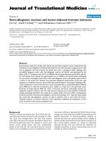
Therapeutic prime-boost vaccination decreased the
expression of regulatory T cells
We also investigated the effect of therapeutic vaccination
boosted wit h ECD a2onnumberofCD4
+
CD25
+
Foxp3
+
Tregs in the spleens and tumors of mice from Figure 3B .
Tregs were measured by flow cytometry in CD4
+
lym-
phocytes from splenocytes of t hese mice. The number of
Tregs in the PBS a nd VRa2 plus ovalb umin control
groups were 12.9% and 9.9%, respectively (Figure 6A).
However, the number of Tregs in the ECDa2boosted
group was 5.9%. To further confirm that the number of
Tregs infiltrated into tumor was also associated with the
population of Tregs in spleen, immunohistochemistry
was performed on same tumor samples obtained from
Figure 3B. Interestingly, tumor samples from ECDa2
boosted mice shown smaller number of the ratio
of Foxp3
+
/CD4
+
(12%) compared with that of cont rol
mice (30%, P < 0.001) (Figure 6B and 6C). These results
indicate that in addition to the generation of IL-13Ra2-
specific immune response, prime-boost vaccination strat-
egy decreased immunosuppressive Tregs in s pleen and
tumor to further enhance the efficacy of the vaccine.
Discussion
IL-13Ra2 is overexpressed on certain types of human
tumor tissues [15-22]. We now provide evidence that
IL-13Ra2 is highly expressed in a variety of murine
tumor cell lines (Additional file 1, Figure S1). Although
the significance of expression of IL-13Ra 2 in cancer is
not completely clear, our previous studies indicate that
IL-13Ra2 could be linked to oncogenesis and metastasis
and may provide a potential target for immunotherapy
[23,24]. We have extended our prior studies and
hypothesized that immunization with a DNA vaccine
encoding murine IL-13Ra2, boosted with ECDa2pro-
tein, may work more effectively in syngeneic murine
tumor models. We studied three murine tumor models,
MCA304 sarcoma, 4T1 breast cancer and D5a2 mela-
noma. Our results indicate that this strategy can pro-
duce significant anti tu mor effect in these tumor models
using both prophylactic and therapeutic vaccinations.
Toourknowledge,thisisthefirstreportofusing
ECDa2 protein in DNA prime-protein boost strategy to
enhance the efficacy of DNA vaccine. It has been
hypothesized that the use o f two versions of the same
immunogen may activate different subsets of immune
cells. It has been shown that DNA immunization is more
effective in inducing CD4
+
T-cell responses and priming
antigen-specific B cells, whereas protein immunization is
more effective in stimulating the proliferation of memory
B cells into antibody-secreting plasma cells [34]. In our
study, the IL-13Ra2DNAprimeandECDa2protein
boost activated CD4
+
and CD8
+
T cell responses and
enhanced antibody response against IL-13Ra2. The se T
and B cell responses induced by prime-boost strategy
correlated with tumor responses causing reduced tumor
burden and significantly prolonging mice survival, com-
pared with the IL-13Ra 2 DNA vaccine alone.
The involvement of systemic immunity in mediating
antitumor effects was confirmed by (a) induction of
tumor-specific CTL response, (b) IFN-g secretion by
splenocytes, and (c) infiltration of CD4
+
and CD8+
T cells in tumors that secreted tumor reactive chemo-
kines. Splenocytes collected from control mice produced
minimal level of IFN-g when they were restimulated
with MCA304 or 4T1 tumor cells. These splenocytes
also mediated low level of lysis of each target cells as
determined by CTL assays. However, each tumor cell-
restimulated splenocytes collected from mice receiving
the IL-13Ra2DNAvaccineboostedwithECDa2pro-
duced substantial levels of IFN-g in the culture superna-
tant and w as capable of mediating specific lysis of each
target cells. In contrast, ovalbumin, an irre levant protein
boost did not further induce CTL response, and thus,
we conclude that antitumor effects mediated by this vac-
cination strategy were murine IL-13Ra2 DNA specific.
It is reported that 4T1 breast tumor is highly metastatic
and weakly immunogenic [35,36]. Huang et al. showed
that pare ntal 4T1 tumor cells expressing only MHC class
I molecules ar e poorly immunogenic, and immunizations
of mice bearing 4T1 breast tumor with the irradiated
4T1 cells alone failed to induce the protective antitumor
immuneresponses[37].Ithasalsobeenreportedthat
4T1celllineelaboratesavarietyofimmunesuppressive
molecules including PGE- 2, T GF-b and other factors
[38]. These molecules are the reasons that 4T1 tumor is
poorly immunogenic to induce antitumor response.
Other examples of poorly immunogenic tumors have
been described in the literature. Kjaergaard et al.
explained several reasons for poor immunogenicity of
B16/D5 mouse m elanoma tum ors in response to th e
therapeutic effects of OX-40R mAb [39]. These authors
proposed that it is possible that B16/D5 tumor cells
either lack molecules that can serve sufficiently as tumor
antigens recognized by T cells or are deficient in the pro-
cessing, transportation or presentation of such molecules
by APCs. It may also be true for the poorly immunogenic
4T1 tumors of eliciting lower T cell responses. Indeed, in
our study, overall CTL act ivity and I FN-g production in
Nakashima et al. Journal of Translational Medicine 2010, 8:116
/>Page 11 of 15
CD25
Foxp3
PBS
VRα2 + ovalbumin VRα2 + ECDα2
12.87
9.85
5.938.83
2.39
8.97
1.42
7.74
1.16
Gated on CD4+ cells
A
B
Spleen
Control
CD4 Foxp3 Merge
Tumor
C
Foxp3+/CD4+ (%)
Tumor
Control VRα
αα
α2+ECDα
αα
α2
P<0.001
0
10
20
30
40
VRα2+ECDα2
Figure 6 Th erapeutic prime-boost vaccination decrea sed the expression of regulatory T cells. (A) FACS analysis of regulatory T cel ls in
splenocytes of vaccinated mice with MCA304 tumors. Splenocytes collected from Figure 3B on day 33 were stained with anti-CD4, CD25, Foxp3
and rat IgG2a (isotype control) antibodies and analyzed by FACS. Representative expression of CD25 and Foxp3 gated on CD4
+
cells is shown.
Results are representative of three independent experiments. (B) The MCA304 tumor samples from Figure 5 were stained with antibodies specific
for anti-CD4 and anti-Foxp3 and counterstained with 4’,6-diamidino-2-phenylindole. The number of positive cells were counted and plotted in C.
Tumor sections were quantitated for ≥2 mice per group with a minimum of two sections per tumor for each staining condition, with five fields
per section used for counting.
Nakashima et al. Journal of Translational Medicine 2010, 8:116
/>Page 12 of 15
4T1 tumor mode l were lower compared to MCA304
tumor model. However, the IL-13Ra2DNAandECDa2
boost vaccination could be effective in reducing tumor
burdens and induc e or amplify a specific CTL response
and IFN-g release against 4T1 tumors compared with the
IL-13Ra2 DNA vaccine alone.
It is noteworthy that the IFN-g-related chemokines
CXCL9 and CXCL10 were expressed in tumors derived
from mice receiving the IL-13Ra2 DNA vaccine boosted
with ECDa2. CXCL9 is known to function as a potent
chemoattrac tant for tumor infiltrating lymphocytes [31].
In addition, the CXCL10 displays antitumor properties
based on the attraction of monocytes and T lympho-
cytes [40]. Our results suggest that chemokines are most
likely produced by infiltrating immune cells causing
antitumor effect because these chemokines act as potent
T cell chemoattracants and angiogenesis inhibitors
through their interaction with CXCL3 [31-33].
DNA vaccination and IL-13Ra2 protein boost pro-
duced anti-IL-13Ra2 antibody in the serum of mice.
This antibody may be directly cytotoxic to tumor cells
or mediate growth inhibitory signal to target cells after
ligating with IL-13Ra2 antigen. We are currently exam-
ining the role of antibody in tumor rejection in the cur-
rent prime boost model. We have previously reported
that vaccination of human IL-13Ra2cDNAalonein
D5a2 model generated antibodies, which were modestly
cytotoxic to D5a2 tumor cells in vitro [24].
Interestingly, mice vaccinated with therapeutic IL-13Ra2
cDNA vaccine and boosted with ECDa2proteinshowed
lower percentage of Tregs in the spleen and tumor com-
pared to the PBS control in the MCA304 tumor model.
This is an interesting finding as Tregs play a prominent
role in the inhibition of anti-tumor immunity. It is possible
that the inhibitory effects of IL-13Ra2 DNA boosted with
ECDa2 protein vaccination on Tregs expansion will play a
potentially important role in clinical efficacy during the
treatment of immunocompromised patients, such as those
with cancer. The enhanced expansion of Tregs has been
repo rted in a number of solid and hematological cancers
[41-44]. Our results suggest that IL-13Ra2 cDNA boosted
with ECDa2 protein vaccination may enhance anti-tumor-
immunity by inhibiting the suppressive effects of Tregs.
We did not observe any visible toxicity in mice vacci-
nat ed with IL-13Ra2 DNA alone or in combination with
ECDa 2protein.Novisualchangesinanimalbehavior,
mobility, and body weight were observed after vaccina-
tion. Histopathological analyses of vital organs (liver, kid-
ney, lung, spleen, heart, and brain) manifested no
abnormal ities in vaccinated g roup compared to no treat-
ment group (data not shown). For future clinical trials,
we recommend to carefully observe patients by physical
exams, serum chemistry, complete blood count and any
sign of autoimmunity.
Many immunotherapy approaches, including therapeu-
tic tumor vaccines targeting specific tumor antigens are
being develop ed [1-3]. Our current results may be extra-
polated to the clinical setting, and it is possible that both
CD4
+
and CD8
+
T cells will be induced against IL-13Ra2
antigen by the DNA vaccine regimen as observed in this
animal study. Although the prime-boost vaccine
mediated regression of established tumor, complete
responses we re not observed in any of three tumor mod-
els tested. It is possible that the heterogeneous expression
of IL-13Ra2 in tumors is responsible for this effect.
Alternatively, a most effective dose of vaccine or schedule
of vaccination was not optimized. A more immunogenic
vector such as vaccinia virus and/or other virus expre s-
sing the IL-13Ra2and/oranIL-13Ra2 peptide vaccine
mixed with adjuvants may be neede d to g enerate robust
immune responses. These types of preclinical studies will
be needed to translate our observations to the clinic for
the treatment of patients with cancer.
Conclusion
Our results suggest that immunization with IL-13Ra2
DNA vaccine followed by ECDa2boostmixedwith
CpG and IFA adjuvants mediates significant antitumor
effects in T cell dependent manner. Thu s, IL-13Ra2 can
serve as a potent tumor antigen that can recruit
immune responses against IL-13Ra2 expressing solid
tumors.
Additional material
Additional file 1: Figure S1. Differential expression of IL-13Ra2
chain in murine tumor cell lines. The expression of IL-13Ra2 in murine
tumor cell lines was examined by analyzing expression of mRNA with
RT-PCR. Murine tumor cell lines were tested including three sarcoma cell
lines, MCA106, MCA304 and MCA310; two melanoma cell lines, B16 and
D5; one glioma cell line, GL261; and one breast cancer cell line, 4T1).
High levels of mRNA expression of IL-13Ra2 in three sarcoma cell lines
and 4T1 breast cancer cell line was observed. On the other hand, B16,
D5 melanoma and GL261 glioma cell lines showed low or undetectable
level of IL-13Ra2 mRNA. The primers formIL-13Ra2 used were: 5’-CGC-
ATT-TGT-CAG-AGC-ATT-GT-3’ (forward) and 5’-CCA-AGC-CCT-CAT-ACC-
AGA-AA-3’ (reverse).
Additional file 2: Figure S2. IL-13Ra2 DNA boosted with ECDa2
vaccination generated autoantibodies in serum. To measure the
antibody levels in mice, blood serum samples were periodically collected
on days 33 from the experiment shown in Figure 3B. Autoantibody
against IL-13Ra2 was quantified by ELISA using with standard
techniques. Briefly, 96-well plates were coated with a mouse IL-13Ra2Fc
recombinant protein (10 μg/ml; R&D Systems) for capture overnight at 4°
C. Serum samples (100 μl per well) diluted 1:1000 in blocking solution
were assayed in duplicate and incubated with the plate at room
temperature for 1 h. Wells were washed and then incubated with
biotinylated anti-mouse IL-13Ra2 Ab (0.5 μg/ml; R&D Systems) for
another 1 h. This was followed by streptavidin-HRP conjugated and
substrate solution (R&D systems) at room temperature for 20 min each.
Absorbance was read at 450 nm. These data demonstrate that
generation of antibody against IL-13Ra2 by the mice receiving IL-13Ra2
DNA and ECDa2 boost vaccination was dramatically increased compared
with IL-13Ra2 DNA and ovalbumin vaccinated mice.
Nakashima et al. Journal of Translational Medicine 2010, 8:116
/>Page 13 of 15
Abbreviations
IL-13: interleukin-13; IL-13Ra2: interleukin-13 receptor a2; ECDa2:
extracellular domain alpha 2; SC: subcutaneous; CTL: cytotoxic T
lymphocytes; IFN-g: Interferon-gamma; OST: overall sacrifice time.
Acknowledgements
We thank Dr. Cheng-Hong Wei and Dr. Ramjay Vatsan of CBER, FDA for
critical reading of manuscript. We are grateful to Dr. Norihisa Sakamoto for
technical assistance to perform FACS analysis; and Pamela Dover for general
support and procuring reagents.
Authors’ contributions
Conceived and designed the experiments: SRH, RKP. Performed the
experiments: HN, TF. Analyzed the data: HN, TF, SRH. Wrote the paper: HN,
SRH, RKP.
All authors have read and approved the final manuscript.
Competing interests
The authors declare that they have no competing interests.
Received: 16 July 2010 Accepted: 10 November 2010
Published: 10 November 2010
References
1. Berzofsky JA, Terabe M, Oh S, Belyakov IM, Ahlers JD, Janik JE, Morris JC:
Progress on new vaccine strategies for the immunotherapy and
prevention of cancer. J Clin Invest 2004, 113:1515-1525.
2. Ribas A, Butterfield LH, Glaspy JA, Economou JS: Current developments in
cancer vaccines and cellular immunotherapy. J Clin Oncol 2003,
21:2415-2432.
3. Finn OJ: Cancer vaccines: between the idea and the reality. Nat Rev
Immunol 2003, 3:630-641.
4. Ward S, Copier J, Dalgleish A: Technical challenges facing therapeutic
cancer vaccines. Curr Opin Drug Discov Devel 2008, 11:168-177.
5. Reichert JM, Paquette C: Therapeutic cancer vaccines on trial. Nat
Biotechnol 2002, 20:659-663.
6. Loftus DJ, Castelli C, Clay TM, Squarcina P, Marincola FM, Nishimura MI,
Parmiani G, Appella E, Rivoltini L: Identification of epitope mimics
recognized by CTL reactive to the melanoma/melanocyte-derived
peptide MART-1(27-35). J Exp Med 1996, 184:647-657.
7. Dyall R, Bowne WB, Weber LW, LeMaoult J, Szabo P, Moroi Y, Piskun G,
Lewis JJ, Houghton AN, Nikolic-Zugic J: Heteroclitic immunization induces
tumor immunity. J Exp Med 1998, 188:1553-1561.
8. Qin H, Cha SC, Neelapu SS, Lou Y, Wei J, Liu YJ, Kwak LW: Vaccine site
inflammation potentiates idiotype DNA vaccine-induced therapeutic T
cell-, and not B cell-, dependent antilymphoma immunity. Blood 2009,
114:4142-4149.
9. Davidson EJ, Faulkner RL, Sehr P, Pawlita M, Smyth LJ, Burt DJ,
Tomlinson AE, Hickling J, Kitchener HC, Stern PL: Effect of TA-CIN (HPV 16
L2E6E7) booster immunisation in vulval intraepithelial neoplasia patients
previously vaccinated with TA-HPV (vaccinia virus encoding HPV 16/18
E6E7). Vaccine 2004, 22:2722-2729.
10. Lindsey KR, Gritz L, Sherry R, Abati A, Fetsch PA, Goldfeder LC, Gonzales MI,
Zinnack KA, Rogers-Freezer L, Haworth L, et al: Evaluation of prime/boost
regimens using recombinant poxvirus/tyrosinase vaccines for the
treatment of patients with metastatic melanoma. Clin Cancer Res 2006,
12:2526-2537.
11. Perales MA, Yuan J, Powel S, Gallardo HF, Rasalan TS, Gonzalez C,
Manukian G, Wang J, Zhang Y, Chapman PB, et al: Phase I/II study of GM-
CSF DNA as an adjuvant for a multipeptide cancer vaccine in patients
with advanced melanoma. Mol Ther 2008, 16:2022-2029.
12. Caput D, Laurent P, Kaghad M, Lelias JM, Lefort S, Vita N, Ferrara P: Cloning
and characterization of a specific interleukin (IL)-13 binding protein
structurally related to the IL-5 receptor a chain. Journal of Biological
Chemistry 1996, 271:16921-16926.
13. Donaldson DD, Whitters MJ, Fitz LJ, Neben TY, Finnerty H, Henderson SL,
O’Hara RM Jr, Beier DR, Turner KJ, Wood CR, Collins M: The murine IL-13
receptor molecular cloning, characterization, and comparison with
murine IL-13 receptor a1.
Journal of Immunology 1998, 161:2317-2324.
14. David MD, Bertoglio J, Pierre J: Functional characterization of IL-13
receptor a2 gene promoter: A critical role of the transcription factor
STAT6 for regulated expression. Oncogene 2003, 22:3386-3394.
15. Kawakami M, Kawakami K, Takahashi S, Abe M, Puri RK: Analysis of
interleukin-13 receptor a2 expression in human pediatric brain tumors.
Cancer 2004, 101:1036-1042.
16. Kawakami M, Kawakami K, Kasperbauer JL, Hinkley LL, Tsukuda M,
Strome SE, Puri RK: Interleukin-13 Receptor a2 Chain in Human Head and
Neck Cancer Serves as a Unique Diagnostic Marker. Clinical Cancer
Research 2003, 9:6381-6388.
17. Puri RK, Leland P, Obiri NI, Husain SR, Kreitman RJ, Haas GP, Pastan I,
Debinski W: Targeting of interleukin-13 receptor on human renal cell
carcinoma cells by a recombinant chimeric protein composed of
interleukin-13 and a truncated form of Pseudomonas exotoxin A
(PE38QQR). Blood 1996, 87:4333-4339.
18. Husain SR, Obiri NI, Gill P, Zheng T, Pastan I, Debinski W, Puri RK: Receptor
for interleukin 13 on AIDS-associated Kaposi’s sarcoma cells serves as a
new target for a potent Pseudomonas exotoxin-based chimeric toxin
protein. Clinical Cancer Research 1997, 3:151-156.
19. Husain SR, Joshi BH, Puri RK: Interleukin-13 receptor as a unique target
for anti-glioblastoma therapy. Int J Cancer 2001, 92:168-175.
20. Joshi BH, Plautz GE, Puri RK: Interleukin-13 receptor alpha chain: a novel
tumor-associated transmembrane protein in primary explants of human
malignant gliomas. Cancer Res 2000, 60:1168-1172.
21. Kawakami K, Puri RK: Interleukin-13 and cancer. In Interleukin 13. Edited by:
Brombacher F. Texas. Landes Bioscience; 2003:65-78.
22. Kawakami K, Kawakami M, Snoy PJ, Husain SR, Puri RK: In vivo
overexpression of IL-13 receptor alpha2 chain inhibits tumorigenicity of
human breast and pancreatic tumors in immunodeficient mice. JExp
Med 2001, 194:1743-1754.
23. Fujisawa T, Joshi B, Nakajima A, Puri RK: A Novel Role of Interleukin-13
Receptor a2 in Pancreatic Cancer Invasion and Metastasis. Cancer Res
2009, 69:8678-8685.
24. Kawakami K, Terabe M, Kawakami M, Berzofsky JA, Puri RK: Characterization
of a novel human tumor antigen interleukin-13 receptor alpha2 chain.
Cancer Res 2006, 66:4434-4442.
25. Okano F, Storkus WJ, Chambers WH, Pollack IF, Okada H: Identification of a
novel HLA-A*0201-restricted, cytotoxic T lymphocyte epitope in a
human glioma-associated antigen, interleukin 13 receptor alpha2 chain.
Clin Cancer Res 2002, 8:2851-2855.
26. Aslakson CJ, Miller FR: Selective events in the metastatic process defined
by analysis of the sequential dissemination of subpopulations of a
mouse mammary tumor. Cancer Res 1992, 52:1399-1405.
27. Kawakami K, Kawakami M, Puri RK: IL-13 receptor-targeted cytotoxin
cancer therapy leads to complete eradication of tumors with the aid of
phagocytic cells in nude mice model of human cancer. J Immunol 2002,
169:7119-7126.
28. Ballas ZK, Krieg AM, Warren T, Rasmussen W, Davis HL, Waldschmidt M,
Weiner GJ: Divergent therapeutic and immunologic effects of
oligodeoxynucleotides with distinct CpG motifs. J Immunol 2001,
167:4878-4886.
29. Kioi M, Seetharam S, Puri RK: N-linked glycosylation of IL-13R alpha2 is
essential for optimal IL-13 inhibitory activity. FASEB J 2006, 20:2378-2380.
30. Klinman DM: Adjuvant activity of CpG oligodeoxynucleotides. Int Rev
Immunol 2006, 25:135-154.
31. Liao F, Rabin RL, Yannelli JR, Koniaris LG, Vanguri P, Farber JM: Human MIG
chemokine: biochemical and functional characterization. J Exp Med 1995,
182:1301-1314.
32. Di Carlo E, Comes A, Orengo AM, Rosso O, Meazza R, Musiani P,
Colombo MP, Ferrini S: IL-21 induces tumor rejection by specific CTL and
IFN-gamma-dependent CXC chemokines in syngeneic mice. J Immunol
2004, 172:1540-1547.
33. Inngjerdingen M, Rolstad B, Ryan JC: Activating and inhibitory Ly49
receptors modulate NK cell chemotaxis to CXC chemokine ligand (CXCL)
10 and CXCL12. J Immunol 2003, 171:2889-2895.
34. Lu S: Combination DNA plus protein HIV vaccines. Springer Semin
Immunopathol 2006, 28:255-265.
35. Ostrand-Rosenberg S, Baskar S, Patterson N, Clements VK: Expression of
MHC Class II and B7-1 and B7-2 costimulatory molecules accompanies
Nakashima et al. Journal of Translational Medicine 2010, 8:116
/>Page 14 of 15
tumor rejection and reduces the metastatic potential of tumor cells.
Tissue Antigens 1996, 47:414-421.
36. Pulaski BA, Terman DS, Khan S, Muller E, Ostrand-Rosenberg S:
Cooperativity of Staphylococcal aureus enterotoxin B superantigen,
major histocompatibility complex class II, and CD80 for immunotherapy
of advanced spontaneous metastases in a clinically relevant
postoperative mouse breast cancer model. Cancer Res 2000, 60:2710-2715.
37. Huang X, Wong MK, Yi H, Watkins S, Laird AD, Wolf SF, Gorelik E:
Combined therapy of local and metastatic 4T1 breast tumor in mice
using SU6668, an inhibitor of angiogenic receptor tyrosine kinases, and
the immunostimulator B7.2-IgG fusion protein. Cancer Res 2002,
62:5727-5735.
38. Muraoka RS, Dumont N, Ritter CA, Dugger TC, Brantley DM, Chen J,
Easterly E, Roebuck LR, Ryan S, Gotwals PJ, et al: Blockade of TGF-beta
inhibits mammary tumor cell viability, migration, and metastases. J Clin
Invest 2002, 109:1551-1559.
39. Kjaergaard J, Tanaka J, Kim JA, Rothchild K, Weinberg A, Shu S: Therapeutic
efficacy of OX-40 receptor antibody depends on tumor immunogenicity
and anatomic site of tumor growth. Cancer Res 2000, 60:5514-5521.
40. Luster AD, Leder P: IP-10, a -C-X-C- chemokine, elicits a potent thymus-
dependent antitumor response in vivo. J Exp Med 1993, 178:1057-1065.
41. Baecher-Allan C, Anderson DE: Immune regulation in tumor-bearing
hosts. Curr Opin Immunol 2006, 18:214-219.
42. Beyer M, Schultze JL: Regulatory T cells in cancer. Blood 2006, 108:804-811.
43. Sato E, Olson SH, Ahn J, Bundy B, Nishikawa H, Qian F, Jungbluth AA,
Frosina D, Gnjatic S, Ambrosone C, et al: Intraepithelial CD8+ tumor-
infiltrating lymphocytes and a high CD8+/regulatory T cell ratio are
associated with favorable prognosis in ovarian cancer. Proc Natl Acad Sci
USA 2005, 102:18538-18543.
44. Wolf AM, Wolf D, Steurer M, Gastl G, Gunsilius E, Grubeck-Loebenstein B:
Increase of regulatory T cells in the peripheral blood of cancer patients.
Clin Cancer Res 2003, 9:606-612.
doi:10.1186/1479-5876-8-116
Cite this article as: Nakashima et al.: Interleukin-13 receptor a2 DNA
prime boost vaccine induces tumor immunity in murine tumor models.
Journal of Translational Medicine 2010 8:116.
Submit your next manuscript to BioMed Central
and take full advantage of:
• Convenient online submission
• Thorough peer review
• No space constraints or color figure charges
• Immediate publication on acceptance
• Inclusion in PubMed, CAS, Scopus and Google Scholar
• Research which is freely available for redistribution
Submit your manuscript at
www.biomedcentral.com/submit
Nakashima et al. Journal of Translational Medicine 2010, 8:116
/>Page 15 of 15