Báo cáo hóa học: "Morphologic complexity of epithelial architecture for predicting invasive breast cancer survival" ppt
Bạn đang xem bản rút gọn của tài liệu. Xem và tải ngay bản đầy đủ của tài liệu tại đây (546.15 KB, 10 trang )
RESEARC H Open Access
Morphologic complexity of epithelial architecture
for predicting invasive breast cancer survival
Mauro Tambasco
1,2,3*
, Misha Eliasziw
1,4
, Anthony M Magliocco
1,2,5
Abstract
Background: Precise criteria for optimal patient selection for adjuvant chemotherapy remain controversial and
include subjective components such as tumour morphometry (pathological grade). There is a need to replace
subjective criteria with objective measurements to improve risk assessment and therapeutic decisions. We assessed
the prog nostic value of fractal dimension (an objective measure of morphologic complexity) for invasive ductal
carcinoma of the breast.
Methods: We applied fractal analysis to pan-cytokeratin stained tissue microarray (TMA) cores derived from 379
patients. Patients were categorized according to low (<1.56, N = 141), intermediate (1.56-1.75, N = 148), and high
(>1.75, N = 90) fractal dimension. Cox proportional-hazards regression was used to assess the relationship between
disease-specific and overall survival and fractal dimension, tumour size, grade, nodal status, estrogen receptor
status, and HER-2/neu status.
Results: Patients with higher fractal score had significantly lower disease-specific 10-year survival (25.0%, 56.4%, and
69.4% for high, intermediate, and low fractal dimension, respectively, p < 0.001). Overall 10-year survival showed a
similar association. Fractal dimension, nodal status, and grade were the only significant (P < 0.05) independent
predictors for both disease-specific and overall survival. Among all of the prognosticators, the fractal dimension
hazard ratio for disease-specific survival, 2.6 (95% confidence interval (CI) = 1.4,4.8; P = 0.002), was second only to
the slight ly higher hazard ratio of 3.1 (95% CI = 1.9,5.1; P < 0.001) for nodal status. As for overall survival, fractal
dimension had the highest hazard ratio, 2.7 (95% CI = 1.6,4.7); P < 0.001). Split-sample cross-validation analysis
suggests these results are generalizable.
Conclusion: Except for nodal status, morphologic complexity of breast epithelium as measured quantitatively by
fractal dimension was more strongly and significantly associated with disease-specific and overall survival than
standard prognosticators.
Background
The prognostic assessment of breast cancer is based on
factors that determine a patient’ srelapserisk,and
together with predictive factors (e.g., e strogen-receptor
status), it is used to make optimal therapeutic decisions
regarding adjuvant systemic therapy [1]. Such decisions
provide a balance between the potential benefit and
associated costs and side effects of treatment [1]. There-
fore, it is necessary to have sensitive and specific prog-
nosticators to accurately define risk category for breast
cancer.
Currently, the most significant p rognosticator for
women with breast cancer is axillary lymph node status
[1-4]. For node-positive patients, there is a direct rela-
tionship between the number of involved axillary nodes
and the risk for distant recurrence [4]. However, despite
the usefulness of l ymph node status, recommendations
for systemic adjuvant chemotherapy are not entirely
straightforward. For example, five-year survival rates
show that approximately 15% of all node-negative
patients with larger tumor sizes ( >1 cm) may benefit
from systemic adjuvant therapy, but about 85% would
survive without it [5]. F urthermore, approximately one-
third of node-positive patients are free of recurrence
after local-regional therapy [6-8].
* Correspondence:
1
Department of Oncology, University of Calgary, Calgary, Canada
Full list of author information is available at the end of the article
Tambasco et al. Journal of Translational Medicine 2010, 8:140
/>© 2010 Tambasco et al; licensee BioMed Central Ltd. This is an Open Access arti cle distributed under the terms of the Creative
Commons Attribution License ( which permits unrestricted use, distribution, and
reproduction in any medium, provided the original work is properly cited.
Other major prognostic risk factors, especially for
node-negative patients, are tumor size and histological
tumor grade [1-4,9,10]. For node-negative patients,
tumor size is a powerful prognostic factor that is used
routinely to make adjuvant treatment decisions [6,11],
and tu mor grade is primarily used to make decisions for
cases in which the tumor sizes are borderline [1,2,5].
Although tumor grade has prognostic value, significant
inter-observer variation in grading still exists [12-14]. as
pathologists are assessing complex histological charac-
teristics in a semi-quantitative manner.
It is known that invasive breast cancer (a malignant
neoplasm) demonstrates partial or complete lack of
structural organization and functional coordination with
surrounding normal tissue [ 15]. The idea central to th is
study is that this loss of structural organization and
functional coordination manifests itself in the form of
an increase in morphologic complexity of the epithelial
components at the sub-cellular, cellular, and multi-cellu-
lar levels, and the degree of this complexity can be
quantified and related to patient outcome. A method
that lends itself particularly useful for quantitatively
characterizing complex pathological structures at differ-
ent scales, is based on fractal analysis [16,17]. In this
study, we assess the prognostic value of a recently devel-
oped novel technique [18] to measure the fractal dimen-
sion of segmented histological structures of breast tissue
microarra y (TMA) cores stained with pan-cytokeratin to
highlight the morphology of epithelial architecture.
Methods
Patient Characteristics
A total of 408 patients with primary invasive ductal car-
cinoma (IDC) of the breast were selected retrospectively
from the Calgary Regional Hospitals after appropriate
ethics approval from the Institutional Review Board
(IRB). It should be noted that the IRB did not require
patient consent for this study as it was a retrospective
study in which many of the patients were deceased and
the risk of exposing patient confidentiality was extre-
mely low. Of these, 379 patients had at least one of
three TMA cores th at was sufficiently stained for fr actal
analysis. The age range of these patients at diagnosis
was 34 to 95 with a mean and median age of 65 and 66,
respectfully. Stage information was available for 375 of
379 pati ents with the following frequency distribution:
225 (60.0%) patients were Stage I, 99 (26.4%) were Stage
II, and 51 (13.6%) were Stage III. All patients selected
had received adjuvant tamoxifen treatment between
1988 and 2006. Cases were identified with Alber ta Can-
cer Board records of patients who had received tamoxi-
fen treatment without chemotherapy. In summary, the
inclusion criterion was any patient who had adequate
tissue for TMA construction, and had received adjuvant
tamoxifen treatment but no adjuvant chemotherapy.
Sample Preparation
Whole sections stained with Hemotoxylin and Eosin
(H&E) were used to select tumorareasfortheTMA
cores. Fourteen breast TMA blocks containing an average
of 94 tissue cores were constructed from formalin-fixed,
paraffin-embedded, previously untreated breast cancer
tissue. To ensure there was no selection bias, three
0.6 mm cores were chosen randomly from cancerous
areas of each donor b lock to construct the recipient
TMA core block, and the Leica RM2235 microtome
(Leica Microsystems Inc.) was used to cut 4 μmthick
sections from each TMA donor block. In a previous
study with prostate cancer specimens, we showed that
fractal analyses of specimens stained with pan-cytokera-
tin provide greater classification performance (benign
versus high grade) than serial sections of the same speci-
mens stained with H&E [18]. The reason for this is that
pan-cytokeratin isolates and highlights the morphology
of epithelial components and excludes structures that do
express pathological relevance in the form of morpholo-
giccomplexity(i.e.,connectivetissuecomponents).
Hence, we stained all the TMA sections with pan-
cytokeratin. This staining was performed using Ventana
Benchmark LT. Protease 1 antigen retrieval was used fol-
lowed by Ventana pre-diluted pan-cytokeratin (cat. N o.
760-2135) antibody with an incubation time o f 32 min-
utes. A Ventana ultraview™ DAB detection system was
used for detection.
Image Acquisition of TMA Cores
Microscopic images of the TMA cores were acquired
with an AxioCa m HR digital camera (Carl Zeiss, Inc.)
mounted on an optical microscope (Zeiss Axioscope) at
a magnification of 10 × objective. The AxioCam HR has
pixels of size 6.7 μm ×6.7μm, which are 1.06 μm ×
1.06 μm in apparent size at the combined magnifications
of 10 × objective and 0.63 × C-mount optical coupling
(optical interface between the microscope and digital
camera). T he images were taken at the camera’snative
resolution of 1300 × 1030 pixels, and saved in tagged
image file format (tif).
Fractal Analysis to Assess Morphologic Complexity
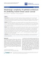
Unlike our intuitive notion of dimension (i.e., topologi-
cal dimension), fractal dimension can be a non-integer
value, and the greater the morphologic complexity of an
object, the higher its fractal dimension relative to its
topological dimension (Figure 1). Fractal dimension
quantifies the level of structural complexity by assessing
the variation in the level of detail in a structure as the
Tambasco et al. Journal of Translational Medicine 2010, 8:140
/>Page 2 of 10
structure is examined at different scales [19]. Hence, it
lends itself naturally to characterizing irregular struc-
tures that maintain a constant level of complexity over a
range of scales.
In this study, we applied an automated fractal analysis
technique we developed in previous work [18] to quan-
tify the morph olog ic complexity of breast epithelium, a
pathologically relevant histological feature. In summary,
this technique involves the following steps:
1. Application of a histological stain to tissue
specimens in order to highlight and isolate the histo-
logical structures of interest. In this case, these
structures include the outlines of the epithelial com-
ponents comprising the multi-cellular structures
(gland formations), cellular structures (individual cell
shapes), and sub-cellular structures (distribution of
keratin within the cells and nuclear shape).
2. Image acquisition and background correction of
stained specimens. The background correction was
done by acquiring a “blank” image (under the same
imaging conditions used to acquire the TMA
images), and using this “blank” image to subtract the
non-uniform background luminance [18]. The
resulting background corrected images are converted
to grey-scale (Figure 2).
3. Application of a series of intensity thresholds to
convert the grey-scale version of the image specimen
into a series of binary images from which histological
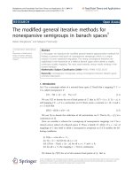
morphology outlines are derived (Figure 2). Figure 3
shows a sample magnified region of Figure 2A to
illustrate the segmented morphology outlines in more
detail.
4. Application of the box counting method [19]
(with appropriate spatial scale range - 10 to 50 μm)
[20] to compute the fractal dimension of each out-
line image obtained from step 3.
5. Identification of the glo bal maximum from a plot
of fractal dimension versus intensity threshold. This
maximum corresponds to the fractal dimension of
the pathological morphology.
In previous work, we showed that our method of find-
ing the fractal dimension is independent of changes in
microscope illumination setting or stain uniformity and
intensity [18]. Also, it should be noted that fractal
dimension is not affected by magnification as long as
the field of view of the specimen image still contains the
scale range of the structur es of inter est over which the
fractal dimension was found to be constant.
Our automated fractal analysis metho d was applied to
a total of 1224 TMA cores ( 3 cores for each of the 408
patient samples). For each patient, the T MA core with
the maximum fractal dimension was used for the statis-
ticalanalysisinthisstudy. The rationale for choosing
the maximum fractal dimension from the sampled tissue
cores is to reduce the possibility that the other TMA
cores from a given patient contain only benign or more
highly differentiated tissue. That is, it is expected that
the TMA core with the maximum fractal dimension is
Figure 1 Both the c ircle (left) and the Koch snowflake (right)
have a topological dimension of 1; however, the fractal
dimension (FD) of the Koch snowflake is greater than 1
because it has a more complex morphology than the circle.
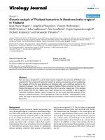
Figure 2 Pan-keratin stained TMA cores (left column)
representative of A: low (< 1.56), B: intermediate (1.56-1.75),
and C: high (> 1.75) fractal dimension categories, the
corresponding background corrected gray-scale images (center
column), and the corresponding outline morphology images
(right column) from which fractal dimensions are computed.
Figure 3 A: Original image (Figure 2C); B: Magnified portion of
A, the dashed rectangular region; C: Segmented outline
structures corresponding to the magnified image region.
Tambasco et al. Journal of Translational Medicine 2010, 8:140
/>Page 3 of 10
representative of the malignant neoplasm that has
deviated most from normal cellular/glandular breast
morphology, and therefore it is the most probable indi-
cator of abno rmal and/or aggre ssive tumor growth with
metastatic potential.
For 379 of the 408 pati ents (92.9%), fractal dimension
was successfully measured in at least one of the three
TMA cores generated per patient, and it could not be
determined for the remaining 29 patient specimens due
to insufficient staining (i.e., less than half of the speci-
men being staine d) or specimen folding. Eight of the 29
patients could not be assessed because all 3 of their
TMA cores resulted in a “ blank” slide. The breakdown
of the number of patients for which the TMA cores
were sufficiently stained for fractal analysis was as fol-
lows: 36 patients (9.5%) had one evaluable core, 105
patients (27.7%) had two evaluable cores, and 238
patients (62.8%) had three evaluable cores.
Statistical Analyses
For purposes of analyses, it is often useful to convert a
measured variable to a categor ical variable so as to place
patients into graded risk strata. As the particular fractal
analysis technique we developed is novel, there are no
established cutpoints available. Although several methods
exist to determine cutpoints, namely biological determina-
tion, data-oriented, and outcome-oriented, there is no sin-
gle method or criterion to specify which approach is best.
For the present analyses, we used a data-oriented
approach to select two cutpoints. The first cutpoint was
chosen to correspond to the upper quartile (75
th
percen-
tile) of the fractal dimension data, and the second cutpoint
was chosen as the median of the remaining lower three-
quarters of the data. Two cutpoints, rather than one, were
chosen to assess whether there was a graded relationship
between fractal dimension and patient prognosis.
Associations between categorized fractal dimension
scores and clinicopathological variables were assessed
for statistical significance using a chi-square test.
Kaplan-Meier methods were used to estimate 10-year
disease-specific and overall survival rates and the log-
rank test was used to compare the curves for statistical
significance. Disease-specific survival was measured
from the date of diagnosis to the date of death from
cancer or date of last follow-up. Overall survival was
measured fr om the date of diag nosis to the date of
death from any cause or date of l ast follow-up. The
above analyses were repea ted using Cox proportional
hazards regression modeling to assess whether any of
the clinicopat hological variables influenced the findings.
The proportionality assumption was assessed for all cov-
ariates using Log-Minus-Log Survival Plots and none
violated the assumption. Statistical analyses were
performed using SAS 9.2 software (SAS Institute Inc).
The prognostic accuracy of fractal dimension in pre-
dicting death from breast cancer and death from any
cause was quantified by the area under the curve (AUC)
from a receiver operating characteristic (ROC) analysis.
Values of AUC range from 0.5 (chance accuracy) to 1.0
(perfect accuracy), with the following intermediate
benchmarks: 0.6 (fair), 0.7 (good), 0.8 (excellent), and
0.9 (almost perfect). For the analysis, the predicted
probability of outcome from a Cox regression model
was considered as a continuum. The actual occurrence
of outcome was used as the comparative standard.
A split-sample cross-validation was performed to assess
the generalizability of the results [21]. The process con-
sisted of splitting the original sample of 379 patients into
a training set of 190 patients and a validation set of 189
patients using random sampling. A regression equation
was derived in the training set and the AUC between the
observed and predicted response values was calculated.
The regression coefficients from the training set were
then used to calculate predicted values in the validation
set. The AUC between these predicted values and
observed values in the validation set was calculated, and
is called the cross-validation coefficient. The shrinkage
coefficient was calculated as the difference between the
AUCs of the training and validation sets. The smaller the
shrinkage coefficient, the more confidence one can have
in the generalizability of the results. Although there are
no clear guidelines regarding the magnitude of shrinkage,
except that smaller is better, values less than 0.10 indicate
a generalizable model. Given a satisfactory shrinkage
coefficient, the d ata were combined from both sets and a
final regression equation was derived based upon the
entire sample.
Out of 379 evaluable patients, several had missing data:
15 (9.0%) tumor grades, 4 (1.1%) lymph node status, 15
(4.0%) estrogen-receptor status, and 12 (3.2%) HER-2/
neu status. Rather than excluding these patients from the
analyses and reducing the sample size, missing data were
imputed using t he predicted mean appro ach in SOLAS
3.0 software (Statistical Solutions, Ltd.). Imputation bias
was assessed by re-running all the analyses and excluding
any patient with missing data. As the estimates were
similar, the results are reported with the imputed data.
Results
Fractal Analysis of the TMA Cores
Fractal dimension scores ranged from 1.08 to 1.97, with
a median of 1.62, lower quartile 1.49, and upper quartile
1.75. There was moderate level of relatedness (intraclass
correlation = 0.51) among the cores. Using the
data-oriented approach to select two cutpoints, fractal
dimension values < 1.56 were considered low (N = 141),
1.56-1.75 as intermediate (N = 148), and > 1.75 as high
(N = 90). Figure 2 shows representative TMA cores
Tambasco et al. Journal of Translational Medicine 2010, 8:140
/>Page 4 of 10
from these fractal dimension categories. One can see
from this figure that the classification of TMA cores
into low, intermediate, and high fractal dimension cate-
gories (A-C) corresponds to the increasing c omplexity
of outline morphology.
Relationship between Fractal Dimension and Standard
Prognosticators
The baseline patient characteristics are shown in
Table 1. Higher fractal dimension was significantly asso-
ciated with traditional indicators of poor prognosis,
including older age, larger tumour sizes, higher tumour
grade, and positive lymph node status. However, fractal
dimension was not associated with either estrogen-
receptor status or HER-2/neu status.
Fractal Dimension as a Predictor of Outcome
The median patient follow-up was 5.2 years. The 10-yea r
disease-specific and overall survival rates for the entire
group of 379 patients were 52.5% and 42.5%, respectively.
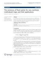
Patients with higher fractal scores had significantly worse
disease-specific survival than those with lower scores
(25.0% versus 56.4% versus 69.4%, p < 0.001; Table 2 and
Figure 4A). As well, patients with higher scores had sig-
nificantly worse overall survival (14.2% versus 39.9% ver-
sus 67.4%, p < 0.001; Table 2 and Figure 4B). T he AUCs
for fractal dimension w ere 0.66 and 0.67 for univariate
disease-specific and overall survival, respectively, indicat-
ing good levels of prognostic accuracy. As expected,
older age, higher grade, and positive lymph node status
were significantly predictive of worse outcome, but not
the size of the tumour, estrogen-receptor status, or
HER-2/neu status (Table 2).
Tumour Grade as a Predictor of Outcome
Tumour grade was derived from the original pathology
reports that included between 10 and 30 board-certified
cancer pathologists. In contrast to the distinct separation
of the disease-specific survival curves for the different
fractal dimension categories (Figure 4A), the disease-spe-
cific survival curves for grade 1 and 2 tumours virtually
overlaped each other over the entire 10-year follow-up
period (Figure 4 C). Also, there is virtual overlap in the
overall survival curves of tumour grades 1 and 2 for the
first 4-year period (Figure 4D). These results suggest that
tumour grades 1 and 2 do not discriminate patients with
respect to 10-year outcome.
Multivariate Analysis
Results from Cox proportional hazards regression
showed that fractal dimension remained statistically sig-
nificant even after adjusting for all clinicopathological
variables (Table 3). This result implies that fractal
dimension is a strong prognostic factor, even though the
multivariate hazard ratio (Table 3) is sma ller than the
univariate hazard ratio (Table 2). The AUCs for th e 7-
factor regression models were 0.73 and 0.75 for disease-
specific and overall survival, respectively. These AUCs
increased by onl y 0.07 and 0.08 when six clinical-patho-
logical factors were added to fractal dimension in the
multivariate regression model. The small increase in
AUCs incidate that the other clinical-pathological
Table 1 Patient Characteristics by Fractal Dimension Category
Number (%) < 1.56 (N = 141) % group 1.56 - 1.75 (N = 148) % group >1.75 (N = 90) % group P-value
Age
≤ 55 years 78 (20.6) 23.4 23.7 11.1 0.039
>55 years 301 (79.4) 76.6 76.3 88.9
Size of tumour
≤ 2 cm 272 (71.8) 78.7 69.6 64.4 0.047
>2 cm 107 (28.2) 21.3 30.4 35.6
Grade of tumour
1 & 2 338 (89.2) 92.9 91.9 78.9 0.001
3 41 (10.8) 7.1 8.1 21.1
Lymph node status
Negative 300 (79.2) 85.1 81.8 65.6 0.001
Positive 79 (20.8) 14.9 18.2 34.4
Estrogen-receptor status
Positive 355 (93.7) 93.6 93.9 93.3 0.98
Negative 24( 6.3) 6.4 6.1 6.7
HER-2/neu status
Negative 350 (92.4) 95.0 89.9 92.2 0.25
Positive 29 (7.6) 5.0 10.1 7.8
Tambasco et al. Journal of Translational Medicine 2010, 8:140
/>Page 5 of 10
factors contribute little to the prognostic accuracy
beyond fractal dimension. It is also worth noting that
even with the comparison of grades 1 and 2 as one cate-
gory versus grade 3 tumo urs, both disease-specific and
overall survival w ere more strongly and significantly
associated with fractal dimension than tumour grade.
Split-sample Cross-validation
The generalizability of the aforementioned results was
assessed by split-sample cross-validation as d escribed in
the statistical analysis section. The results, shown in
Table 4 are congruent, not only with each set but also
with the results of t he entire sample shown in Tables 2
and 3. Specifically, the frequency distribution of low,
moderate, and high fractal dimension is similar, as are
the 10-year disease-specific and overall survival rates in
these three catego ries. Even with smaller sample sizes,
both the training and validation sets still show a pattern
of doubling of hazards with higher levels of fractal
dimension. The shrinkage coefficients for disease-speci-
fic and overall survival we re -0.01 and -0.05, respec-
tively, both indicating that fractal dimension is
generalizable and that combining data from both sets i n
the analyses was justified.
Discussion
We previously developed a fractal analysis method to
quantitatively measure the mo rphologic complexity of
epithelial architecture [18], and showed a direct associa-
tion between fractal dimension and breast tumour
grade, suggesting that it may be a good surrogate mea-
sure of tumour differentiation [22]. In this study we
examined the prognostic value of fractal dimension by
analyzing 379 specimens from patients with invasive
breast cancer, and found that with the exception of
nodal status, fractal dimension showed a stronger asso-
ciation with disease-specific survival than standard clini-
cal prognosticators. The potential clini cal implications
of these results are substantial because to our knowl-
edge, this is the largest and only study of its kind inves-
tigating and demonstrating a positive association
between the morphologic complexity of breast epithelial
architecture (via the fractal dimension metric) and
patient outcome. The potential advantages of fractal
Table 2 Univariate Results from Kaplan-Meier Analysis and Cox Proportional Hazards Regression
Number of
Patients
10-year Disease-
Specific Survival (%)
Univariate Hazard
Ratio (95% CI)
P-value 10-year Overall
Survival (%)
Univariate Hazard
Ratio (95% CI)
P-value
Fractal
dimension
< 1.56 141 69.4 1.0 67.4 1.0
1.56 - 1.75 148 56.4 1.9 (1.1, 3.6) 0.03 39.9 2.1 (1.2, 3.6) 0.008
>1.75 90 25.0 3.5 (1.9, 6.4) < 0.001 14.2 3.6 (2.1, 6.1) < 0.001
Age
≤ 55 years 78 82.1 1.0 82.1 1.0
>55 years 301 40.8 3.3 (1.5, 7.2) 0.003 29.1 4.3 (2.0, 9.4) < 0.001
Size of tumour
≤ 2 cm 272 49.2 1.0 38.8 1.0
>2 cm 107 57.0 1.3 (0.8, 2.2) 0.21 47.9 1.3 (0.9, 2.0) 0.18
Grade of
tumour
1 & 2 338 56.1 1.0 45.4 1.0
3 41 22.1 3.4 (2.0, 5.7) < 0.001 19.3 2.8 (1.7, 4.6) < 0.001
Lymph node
status
Negative 300 57.6 1.0 47.8 1.0
Positive 79 32.2 4.0 (2.5, 6.3) < 0.001 21.3 3.4 (2.3, 5.1) < 0.001
Estrogen-
receptor status
Positive 355 53.8 1.0 43.1 1.0
Negative 24 40.1 1.6 (0.7, 3.4) 0.26 36.0 1.6 (0.8, 3.1) 0.19
HER-2/neu
status
Negative 350 51.6 1.0 42.3 1.0
Positive 29 60.6 1.2 (0.5, 2.7) 0.71 38.9 1.2 (0.6, 2.5) 0.59
Tambasco et al. Journal of Translational Medicine 2010, 8:140
/>Page 6 of 10
dimension over conventional tumour grading is that it is
a quanti tative and reprod ucible indicator t hat would be
able to provide pathologists with rapid and cost effective
high volume analysis from as few as three tissue micro-
array (TMA) cores per patient.
Ideally, a study investigating the value of a potential
prognosticator should only involve patients that have
not received any form of adjuvant systemic thera py.
However, as noted by Mirza et al. [5], such studies are
becoming increasingly difficult to pe rform because sys-
temic therapy is recommended for a n ever-wideni ng
range of breast cancer patients. Although none of the
patients in this study were treated with adjuvant che-
motherapy, they were all tre ated with adjuvant tamoxi-
fen therapy, including the 24 ER-negative patients
(note: cases selected for this study w here from as far
back as 1988 when tamoxifen was occasionally admi-
nistered to patients with ER-negat ive tumours). How-
ever, even though the patients received a form of
adjuvant systemic therapy, the sam e form of treatment
was received by all of the patients leading to the
expectation that fractal dimension will be independent
of the predictive factor related to tamoxifen therapy (i.
e., ER-positive status). Indeed, this appears to be the
case, since approximately the same percentage of ER-
positive patients are in t he low, intermediate, and high
fractal dimension groups (Table 1), which likely indi-
cates that tamoxifen therapy has put all o f these ER-
positive patients on an equal footing. However, another
possibility for this result may be that ER status does
not affect the morphologic complexity of epithelial
architecture. In either case, it may be argued that the
use of tamoxifen treated patients in a study investigat-
ing the value of a possible prognosticator, although not
ideal, does not detract from the ability to assess the
prognostic factor’s potential relative to other indepen-
dent prognosticators.
Previous studies have examined the application of
fractal analysis for characterizing cancer [23,24] and
have shown that fractal dimension can describe the
complex pathological structures seen in some cancers;
[18,22] however, to our knowledge, our results represent
Figure 4 Kaplan-Meier Disease-Specific and Overall Survival Curves by Fractal Dimension Category (Panels A and B, respectively);
Kaplan-Meier Disease-Specific Survival and Overall Survival Curves by Tumour Grade (Panels C and D, respectively).
Tambasco et al. Journal of Translational Medicine 2010, 8:140
/>Page 7 of 10
the largest and sole study relating fractal dimension of
epithelial architecture to patient outcome. Although we
did not use an external patient validation set in this
proof of principle study, we emplo yed a data-oriented
approach to minimize bias in the selection of cutpoints,
as well as, conducting a split-sample cross-validation
analysis.Thisanalysissuggeststhattheresultsare
generalizable, whereby higher f ractal dimensions are
associated with poorer outcome. This observation
demonstrates the high potential of fractal dimension as
an image-based prognostic marker, and it is congruent
with the notion that malignant breast neoplasms asso-
ciated with poorer outcome demonstrate partial or com-
plete lack of structural organization and functional
coordination with surrounding normal tissue [ 15].
Further more, it implies that changes in the morphologic
complexity of architectural components of the neoplasm
(i.e., the epithelium) that arise from changes in the
Table 4 Summary of Split Sample Training Set and Validation Set Results
Number of
Patients
10-year Disease-Specific
Survival (%)
Adjusted Hazard
Ratio (95% CI)
P-
value
10-year Overall
Survival (%)
Adjusted Hazard
Ratio (95% CI)
P-
value
Training Set
Patients
190
Fractal
dimension
< 1.56 68 69.4 1.0 66.2 1.0
1.56 - 1.75 76 52.3 2.4 (0.1, 5.8) 0.064 34.2 2.2 (1.0, 4.8) 0.050
>1.75 46 17.0 2.5 (1.0, 6.3) 0.056 16.5 1.8 (0.8, 4.1) 0.17
Validation Set
Patients
189
Fractal
dimension
< 1.56 73 71.6 1.0 70.6 1.0
1.56 - 1.75 72 60.5 1.3 (0.6, 3.3) 0.51 44.8 1.7 (0.8, 3.9) 0.18
>1.75 44 32.4 2.3 (1.0, 5.5) 0.06 11.2 3.2 (1.5, 6.9) 0.003
AUC adjusted disease-specific survival analysis, training set = 0.72, validation set = 0.73.
AUC adjusted overall survival analysis, training set = 0.68, validation set = 0.73.
Table 3 Adjusted Hazard Ratios (95% Confidence Intervals) from Cox Regression
Death from Breast Cancer P-value Death from Any Cause P-value
Fractal dimension
< 1.56 1.0 1.0
1.56 - 1.75 1.9 (1.1, 3.5) 0.043 2.0 (1.2, 3.5) 0.011
>1.75 2.6 (1.4, 4.8) 0.002 2.7 (1.6, 4.7) < 0.001
Age
≤ 55 years 1.0 1.0
>55 years 1.8 (0.8, 4.2) 0.14 2.7 (1.2, 5.9) 0.01
Size of tumour
≤ 2 cm 1.0 1.0
>2 cm 1.0 (0.6, 1.6) 0.96 1.0 (0.7, 1.6) 0.88
Grade of tumour
1 & 2 1.0 1.0
3 2.1 (1.1, 3.7) 0.01 1.7 (1.1, 3.0) 0.047
Lymph node status
Negative 1.0 1.0
Positive 3.1 (1.9, 5.1) < 0.001 2.6 (1.7, 4.1) < 0.001
Estrogen-receptor status
Positive 1.0 1.0
Negative 1.6 (0.7, 3.7) 0.27 1.5 (0.7, 3.2) 0.26
HER-2/neu status
Positive 1.0 1.0
Negative 1.1 (0.5, 2.5) 0.87 1.1 (0.5, 2.4) 0.70
Tambasco et al. Journal of Translational Medicine 2010, 8:140
/>Page 8 of 10
functional status of cells in malignan t neoplasms can be
quantified with fractal analysis.
Conclusions
In summary, the results of this retrospective study show
that fractal dimension is a promising image analysis mar-
ker for the prognosis of IDC of the breast. However, its’
prognostic value needs to be confirmed in external valida-
tion studies, and ultimately in the context of controlled
prospective clinical trials. As a step in this direction, in
future work, we will investigate the prognostic value of
fractal dimension for defining risk category for Stage I (i.e.,
lymph node-negative and tumour size ≤ 2cminmaxi-
mum diameter), IDC, ER-positive breast cancer patients
that have not received any form of adjuvant systemic ther-
apy. Such a study would be especially valuable because in
current clinical practice it is still difficult to identify this
subgroup of patients that would benefit most from adju-
vant chemotherapy. Also, in future work we will investi-
gate the prognostic and predictive value of combining
fractal dimension, a morphological index, with a quantita-
tive analysis of mitotic count, which is a cellular prolifera-
tion index t hat has been shown to be a significant
prognostic indicat or for node-negativ e breast cancer [5].
These investigations would provide validation of the sig-
nificance of morphologic complexity of epithelial architec-
ture in node-negative breast cancer, and explore the
possible synergy between morphologic complexity and cel-
lular proliferation. Also, they will bring us closer to the
realization of an objective prognosticator that can assist
clinicians in making optimal treatment decisions regarding
adjuvant systemic therapy for invasive breast cancer.
Abbreviations
AUC: Area under the curve; CI: Confidence interval; ER: Estrogen receptor;
FD: Fractal dimension; H&E: Hemotoxylin and eosin; HER-2/neu: Human
epidermal growth factor receptor 2; IDC: Invasive ductal carc inoma; IRB:
Institutional review board; ROC: Receiver operating characteristics; tif: tagged
image file format; TMA: Tissue microarray
Acknowledgements
This work was supported by the Alberta Heritage Foundation for Medical
Research (AHFMR) - ForeFront Block Grant. We want to thank Mie Konno
and Annie Yau for help with clinical data collection, and Chantelle Elson for
acquiring the breast specimen images.
Author details
1
Department of Oncology, University of Calgary, Calgary, Canada.
2
Tom
Baker Cancer Centre, Calgary, Canada.
3
Department of Physics & Astronomy,
University of Calgary, Calgary, Canada.
4
Department of Community Health
Science, University of Calgary, Calgary, Canada.
5
Department of Pathology &
Laboratory Medicine, University of Calgary, Calgary, Canada.
Authors’ contributions
MT performed the literature search, study design, fractal dimension analysis,
and drafted the manuscript and figures. ME participated in the study design,
performed the statistical analysis and interpretation, and drafted the
statistical analysis and results sections. AM participated in the study design,
the generation of the TMA cores and database, and the interpretation of the
data. All authors read and approved the final manuscript.
Authors’ information
MT is a board certified Medical Physicist with extensive expertise in radiation
oncology physics, and medical imaging and analysis. ME is a distinguished
Biostatistician with well over 150 publications, and expertise in the
application of statistics to medicine. AMM is a Molecular Pathologist with
extensive expertise in breast cancer pathology and the development and
clinical implementation of prognostic and predictive molecular biomarkers
of cancer.
Competing interests
With the help of University Technologies International (UTI), the authors are
exploring the possibility of commercializing the fractal analysis software used
to analyze the breast tissue microarray images in this study.
Received: 20 August 2010 Accepted: 31 December 2010
Published: 31 December 2010
References
1. Lonning PE, Knappskog S, Staalesen V, Chrisanthar R, Lillehaug JR: Breast
cancer prognostication and prediction in the postgenomic era. Ann
Oncol 2007, 18:1293-1306.
2. Cianfrocca M, Gradishar WJ: Controversies in the therapy of early stage
breast cancer. Oncologist 2005, 10:766-779.
3. Mori I, Yang Q, Kakudo K: Predictive and prognostic markers for invasive
breast cancer. Pathol Int 2002, 52:186-194.
4. Cianfrocca M, Goldstein LJ: Prognostic and predictive factors in early-
stage breast cancer. Oncologist 2004, 9:606-616.
5. Mirza AN, Mirza NQ, Vlastos G, Singletary SE: Prognostic factors in node-
negative breast cancer: a review of studies with sample size more than
200 and follow-up more than 5 years. Ann Surg 2002, 235:10-26.
6. Elledge RM, McGuire WL: Prognostic factors and therapeutic decisions in
axillary node-negative breast cancer. Annu Rev Med 1993, 44:201-210.
7. Polychemotherapy for early breast cancer: an overview of the
randomised trials. Early Breast Cancer Trialists’ Collaborative Group.
Lancet 1998, 352:930-942.
8. Tamoxifen for early breast cancer: an overview of the randomised trials.
Early Breast Cancer Trialists’ Collaborative Group. Lancet 1998,
351:1451-1467.
9. Elston CW, Ellis IO: Pathological prognostic factors in breast cancer. I. The
value of histological grade in breast cancer: experience from a large
study with long-term follow-up. Histopathology 1991, 19:403-410.
10. Henson DE, Ries L, Freedman LS, Carriaga M: Relationship among
outcome, stage of disease, and histologic grade for 22,616 cases of
breast cancer. The basis for a prognostic index. Cancer 1991,
68:2142-2149.
11. Saez RA, McGuire WL, Clark GM: Prognostic factors in breast cancer. Semin
Surg Oncol 1989, 5:102-110.
12. Meyer JS, Alvarez C, Milikowski C, Olson N, Russo I, Russo J, et al: Breast
carcinoma malignancy grading by Bloom-Richardson system vs.
proliferation index: reproducibility of grade and advantages of
proliferation index. Mod Pathol 2005, 18:1067-1078.
13. Robbins P, Pinder S, de Klerk N, Dawkins H, Harvey J, Sterrett G, et al:
Histological grading of breast carcinoma: a study of interobserver
agreement. Hum Pathol 1995, 26:873-879.
14. Chowdhury N, Pai MR, Lobo FD, Kini H, Varghese R: Interobserver variation
in breast cancer grading: a statistical modeling approach. Anal Quant
Cytol Histol 2006, 28:213-218.
15. Rizki A, Bissell MJ: Homeostasis in the breast: it takes a village. Cancer Cell
2004, 6:1-2.
16. Coffey DS: Self-organization, complexity and chaos: the new biology for
medicine. Nat Med 1998, 4:882-885.
17. Baish JW, Jain RK: Cancer, angiogenesis and fractals. Nat Med 1998, 4:984.
18. Tambasco M, Costello BM, Kouznetsov A, Yau A, Magliocco AM:
Quantifying the architectural complexity of microscopic images of
histology specimens. Micron 2009, 40:486-494.
19. Peitgen H, Jurgens H, Saupe D: Chaos and Fractals: New Frontiers of Science.
2 edition. New York: Springer-Verlag; 2004.
Tambasco et al. Journal of Translational Medicine 2010, 8:140
/>Page 9 of 10
20. Dixon V, Tambasco M: Effects of image resolution and noise on
estimating the fractal dimension of tissue specimens. Anal Quant Cytol
Histol 2010, 32:269-279.
21. Kleinbaum DG, Kupper LL, Muller KE, Nizam A: Applied Regression Analysis
and Other Multivariable Methods. 3 edition. Duxbury Press; 1998.
22. Tambasco M, Magliocco AM: Relationship between tumor grade and
computed architectural complexity in breast cancer specimens. Hum
Pathol 2008, 39:740-746.
23. Baish JW, Jain KJ: Fractals and cancer. Cancer Res 2000, 60:3683-3688.
24. Cross SS: Fractals in pathology. Journal of Pathology 1997, 182:1-8.
doi:10.1186/1479-5876-8-140
Cite this article as: Tambasco et al.: Morphologic complexity of
epithelial architecture for predicting invasive breast cancer survival.
Journal of Translational Medicine 2010 8:140.
Submit your next manuscript to BioMed Central
and take full advantage of:
• Convenient online submission
• Thorough peer review
• No space constraints or color figure charges
• Immediate publication on acceptance
• Inclusion in PubMed, CAS, Scopus and Google Scholar
• Research which is freely available for redistribution
Submit your manuscript at
www.biomedcentral.com/submit
Tambasco et al. Journal of Translational Medicine 2010, 8:140
/>Page 10 of 10