Báo cáo hóa học: "Evaluation of the anti-angiogenic properties of the new selective aVb3 integrin antagonist RGDechiHCit" potx
Bạn đang xem bản rút gọn của tài liệu. Xem và tải ngay bản đầy đủ của tài liệu tại đây (1015.26 KB, 10 trang )
RESEARC H Open Access
Evaluation of the anti-angiogenic properties of
the new selective a
V
b
3
integrin antagonist
RGDechiHCit
Gaetano Santulli
1
, Maria Felicia Basilicata
1
, Mariarosaria De Simone
2
, Carmine Del Giudice
1
, Antonio Anastasio
1
,
Daniela Sorriento
1
, Michele Saviano
3
, Annarita Del Gatto
4
, Bruno Trimarco
1
, Carlo Pedone
2
, Laura Zaccaro
4
,
Guido Iaccarino
1*
Abstract
Background: Integrins are heterodimeric receptors that play a critical role in cell-cell and cell-matrix adhesion
processes. Among them, a
V
b
3
integrin, that recognizes the aminoacidic RGD triad, is reported to be involved in
angiogenesis, tissue repair and tumor growth. We have recently synthesized a new and selective ligand of a
V
b
3
receptor, referred to as RGDechiHCit, that contains a cyclic RGD motif and two echistatin moieties.
Methods: The aim of this study is to evaluate in vitro and in vivo the effects of RGDechiHCit. Therefore, we
assessed its properties in cellular (endothelial cells [EC], and vascular smooth muscle cells [VSMC]) and animal
models (Wistar Kyoto rats and c57Bl/6 mice) of angiogenesis.
Results: In EC, but not VSMC, RGDechiHCit inhibits intracellular mitogenic signaling and cell proliferation.
Furthermore, RGDechiHCit blocks the ability of EC to form tubes on Matrigel. In vivo, wound healing is delayed in
presence of RGDechiHCit. Similarly, Matrigel plugs demon strate an antiangiogenic effect of RGDechiHCit.
Conclusions: Our data indicate the importance of RGDechiHCit in the selective inhibition of endothelial a
V
b
3
integrin in vitro and in vivo. Such inhibition opens new fields of investigation on the mechanisms of angiogenesis,
offering clinical implications for treatment of pathophysiological conditions such as cancer, proliferative retinopathy
and inflammatory disease.
Introduction
Angiogenesis is a complex multistep phenomenon con-
sisting of the sprouting and the growth of new capillary
blood vessels starting from the pre-existing ones. It
requires the cooperation of several cell types such as
endothelial cells (ECs), vascular smooth muscle cells
(VSMCs), macrophages, which should be activated, pro-
liferate and migrate to invade the extracellular matrix
and cause vascular remodeling [1,2]. The angiogenic
processisfinelytunedbyaprecisebalanceofgrowth
and inhibito ry factors and in mammalians it is normally
dormant except for some physiological conditions, such
as wound healing and ovulation. When this balance is
altered, excessive or defective angiogenesis occur and
the process becomes p athological. Excessive angiogen-
esis gives also rise to different dysfunctions, including
cancer, eye diseases, rheumatoid arthritis, atherosclero-
sis, diabetic nephropathy, inflammatory bowel disease,
psoriasis, endometriosis, vasculitis, and vascular malfor-
mations [3]. Therefore the discovery of angiogenesis
inhibitors would contribute to the development of thera-
peutic treatments for these diseases.
The integrins are cell adhesion receptors that mediat e
cell -cell and cell-matrix interactions and coordinate sig-
naling allowing a close regulation of physiol ogical phe-
nomena including cellular migration, proliferation and
differenti ation. In particular, the a
V
integrins, combined
with distinct b subunits, participate in the angiogenic
process. An extensively studied member of this receptor
class i s integrin a
V
b
3
, that is strongly overexpressed in
* Correspondence:
1
Department of Clinical Medicine, Cardiovascular & Immunologic Sciences,
“Federico II” University of Naples, Italy
Full list of author information is available at the end of the article
Santulli et al. Journal of Translational Medicine 2011, 9:7
/>© 2011 Santulli et al; licensee BioMed Central Ltd. This is a n Open Access article distributed under the terms of the Creative Commons
Attribu tion License ( which permits unrestricted use, distribution, and reproduction in
any medium, provided the original work is prope rly cited.
activated EC, melanoma, glioblastoma and prostat e can-
cers and in granu lation tissue, whereas is not detectable
in quiescent blood vessels or in the dermis and epithe-
lium of normal skin [4-6]. This integrin participates in
the activation of vascular endothelial growth factor
receptor-2 (VEGFR-2), providing a survival signal to the
proliferating vascular cells during new vessel growth
[7,8] and also seems to be essential in the step of vacuo-
lation and lumen formation [9]. It has been also
reported that a
V
b
3
is under the tight control of VEGF:
this integrin is not expressed in quiescent vessels [10],
but VEGF induces a
V
b
3
expression in vitro and, inter-
estingly, the VEGF and a
V
b
3
integrin expression are
highly correlated in vivo [11,12]. Therefore, a
V
b
3
should be considered a tumor and activated endothe-
lium marker.
a
V
b
3
is able of recognizing many proteins of the
extracellular matrix, bearing an exposed Arg-Gly-Asp
(RGD) tripeptide [5,13,14]. Even if different integrins
recognize different proteins containing the RGD triad,
many studies have demonstrated that the aminoa cids
flanking the RGD sequence of high-affinity ligands
appear to be critical in modulating their specificity of
interaction with integrin complexes [15,16].
Several molecules including peptides containing
RGD motif [11] have been recently developed as inhi-
bitors of a
V
b
3
integrin, in experiments concerning
tumor angiogenesis, showing a reduction of functional
vessel density associated with retardation of tumor
growth and metastasis formation [6,17]. So far, the
pentapeptide c(RGDf[NMe]V), also known as cilengi-
tide (EMD 121974), is the most active a
v
b
3
/a
v
b
5
antagonist reported in literature [18,19] and is in
phase III clinical trials as antiangiogenic drug for glio-
blastoma therapy [15]. The development of more
selective antiangiogenic molecule would help to mini-
mize the side-effects and increase the therapeutic
effectiveness.
We have recently designed and synthesized a novel
and s elective peptide antagonist, referred to as RGDe-
chiHCit, to visualize a
V
b
3
receptor on tumour cells [20].
It is a chimeric peptide containing a cyclic RGD motif
and tw o echistatin C-terminal moieties covalently linked
by spacer sequence. Cell adhesion assays have shown
that RGDechiHCit selectively binds a
V
b
3
integrin and
does no t cross-react with a
V
b
5
and a
IIb
b
3
integrins [20].
Furthermore, PET and SPECT imaging studies have
confirmed that the peptide localizes on a
V
b
3
expressing
tumor cells in xenograft animal model [21]. Since a
V
b
3
is also a marker of activated endothelium, the main pur-
pose of this study was to evaluate in vitro and in vivo
effects of RGDechiHCit on neovasculariz ation. Thus, we
first assessed the in vitro peptide properties on bovine
aortic ECs, and then in vivo, in Wistar Kyoto (WKY)
rats and c57BL/6 mice, the ability of this cyclic peptide
to inhibit angiogenesis.
Methods
Peptides
RGDechiHCit was prepared for the in vi tro and in vivo
stud ies as previously described [20]. To test the biologi-
cal effects of RGDechiHCit, we synthesized the cyclic
pentapeptide c(RGDf[NMe]V), also known as cilengitide
or EMD 121974 [14,19]. We also investigated RGDe-
chiHCit and c(RGDf[NMe]V ) peptides degradation in
serum. Both peptides were incubated and the resulting
solutions were analyzed by liquid chromatography/mass
spectrometry (LC/MS) at different times. 20μLof
human serum (Lonza, Basel, Switzerland) were added to
8 μL of a 1 mg/ml solution of either RGDechiHCit or c
(RGDf[NMe]V) at 37°C. After 1, 2, 4 and 24h, samples
were centrifuged for 1min at 10000g. Solutions w ere
analyzed by LCQ Deca XP Max LC/MS system
equ ipped with a diode-array detector combined with an
elctrospray ion source and ion trap mass analyzer (Ther-
moFinnigan, San Jose, CA, USA), using a Phenomene x
C
18
column (250× 2 mm; 5μm; 300 Ǻ) and a linear gra-
dient of H
2
O (0.1%TFA)/CH
3
CN(0.1%TFA)from10to
80% of CH
3
CN (0.1%TFA) in 30 min at flow rate of
200μL/min.
In vitro studies
In vitro studies were performed on cell cultures of ECs
or VSMCs, cultured in Dulbecco’ s modified Eagle’ s
medium (DMEM; Sigma-Aldrich, Milan, Italy) as pre-
viously described and validated [22,23]. Cell culture
plates were filled with 10 μg/cm
2
of human fibronectin
(hFN, Millipore
®
, Bedford, MA, USA) as desc ribed [24].
All experiments were performed in triplicate with cells
between passages 5 and 9.
Cell proliferation assay
Cell cultures were prepared as previously described [25].
Briefly, cells were seeded at density of 100000 per well
in six-we ll plates, serum starved, pre-incubated at 37°C
for 30’ wi th c(RGDf[NMe]V) or RGDechiHCit (10
-6
M).
Proliferation was induced using hFN (100 μg/ml). Cell
number was measured at 3, 6 and 20 h after stimulation
as previously described [26,27].
DNA synthesis
DNA synthesis was assessed as previously described
[27]. Briefly, cells were serum-starved for 24 h and then
incubated in DMEM with [
3
H]thymidine and 5% FBS.
After 3, 6 and 20 h, cells were fixed with trichloracetic
acid (0.05%) and dissolved in 1M NaOH. Scintillation
Santulli et al. Journal of Translational Medicine 2011, 9:7
/>Page 2 of 10
liquid was added and [
3
H]thymidine inco rporation was
assessed as previously described [27].
VEGF quantification
VEGF production was meas ured as previously described
[26]. Briefly, ECs were seeded at a density of 600000 per
well in six well plates, serum starved overnight, seeded
with c(RGDf[NMe]V) or RGDechiHCit (10
-6
M) and
then stimulated with hFN for 6 hours. Cultured medium
was collected and VEGF production was revealed by
western blot.
Endothelial Matrigel assay
The formation of network-like structures by ECs on an
extracellular matrix (ECM)-like 3D gel consisting of
Matrigel
®
(BDBiosciences,Bedford,MA,USA),was
performed as previously described and validated [27,28].
The six-w ell multidishes were coated with growth fac-
tor-reduced Matrig el in according to the manufacturer’s
instructions. ECs (5×10
4
) were seeded with c(RGDf
[NMe]V) or RGDechiHCit (10
-6
M), in the absence
(negative control) or presence (100 μg/ml) of hFN [24].
Cells were incubated at 37°C for 24h in 1 ml of DMEM.
After incubation, ECs underwent differentiation into
capillary-like tube structures. Tubule formation was
defined as a structure exhibiting a length four times its
width [27]. Network formation was observed using an
inverted phase- contrast microscope (Zei ss). Representa-
tive fields were taken, and the average of the total num-
ber of complete tubes formed by cells was counted in
15 random fields by two independent investigators.
Western blot
Immunoblot analyses were performed as previously
described and validated [23,28]. Mouse monoclonal
antibodies to extracellular signal regulated kinase
(ERK2) and phospho-ERK, anti-rabbit VEGF and actin
werefromSantaCruzBiotecnology(SantaCruz,CA,
USA). Levels of VEGF were determined using an anti-
body raised against VEGF-165 (Santa Cruz Biotechnol-
ogy) [26]. Experiments were performed in triplicate to
ensure reproducibility. Data are presented as arbitrary
densitometry units (ADU) after normalization for the
total c orresponding protein or actin as internal control
[24].
In vivo studies
Wound healing assay was performed on 14-week-old
(weight 293 ± 21 g) normotensive WKY male rats
(Charles River Laboratories, Calco (LC), Italy; n = 18),
and Matrigel plugs experiments were carried out on 16-
week-old (weight 33 ± 4 g) c57BL/6 mice (Charles River
Laboratories, Milan, Italy; n = 13). All animal proce-
dures were performed in accordance with the Guide for
the Care and Use of Laborator y Animals published by
the National Institutes of Health in the United States
(NIH Publication No. 85- 23, revised 1996) and
approved by the Ethics Committee for the Use of Ani-
mals in Research of “Federico II” University [23].
Wound Healing
The rats (n = 18) were anesthetized using vaporized iso-
flurane (4%, Abbott) and maintained by mask ventila-
tion (isoflurane 1.8%) [29]. The dorsum was shaved by
applying a depilatory creme (Veet, R eckitt-Benckiser,
Milano, Italy) and disinfected with povidone iodine
scrub. A 20 mm diameter open wound was excised
through the entire thickness of the skin, including the
panniculus carnosus layer, as described a nd validated
[1,28]. Pluronic gel (3 0%) containing (10
-6
M) c(RGDf
[NMe]V) (n = 6), RGDechiHCit (n = 7), or saline (n = 5)
was placed daily directly onto open wounds, then cov-
ered with a sterile dressing. Two operators blinded to the
identity of the sample examined and measured wound
areas every day, for 8 days. Direct measurements of
wound region were determined by digital planimetry
(pixel area), and subsequent analysis was performed
using a computer-assisted image analyzer (ImageJ soft-
ware, version 1.41, National Institutes of Health,
Bethesda, MD, USA). Wound healing was quantified as a
percentage of the original injury size. Eight days after
wounding, rats were euthanized. Wounds did not show
sign of infection. The lesion and adiacent normal skin
were excised, fixed by immersion in phosphate buffered
saline (PBS, 0.01 M, pH 7.2-7.4)/formalin and then
embedded in paraffin to be processed for immunohistol-
ogy, as described [1].
Matrigel Plugs
Mice (n = 13), anesthetized as described above, were
subcutaneously injected midw ay on the dorsal side,
using sterile conditions, with 0.2 ml of Matrigel
®
base-
ment matrix, pre-mixed with 10
-6
MVEGFand10
-5
Mc
(RGDf[NMe]V) (n = 4), 10
-6
M VEGF and 10
-5
M RGDe-
chiHCit (n = 5), or 10
-6
M VEGF alone (n = 4). After
seven days, mice were euthanized and the implanted
plugs were harvested from underneath the skin, fixed in
10% neutral-buffered formalinsolutionandthen
embedded in paraffin. Invading ECs were identified and
quantified by analysis of lectin immunostained sections,
as described [1,2].
Histology
All tissues were cut in 5 μm sections and slides were
counterstained with a standard mixture of hematoxylin
and eosin. For Masson’s trichrome staining of collagen
fibers, useful to assess the scar tissue formation, slides
were stained with Weigert Hematoxylin (Sigma-Aldrich,
Santulli et al. Journal of Translational Medicine 2011, 9:7
/>Page 3 of 10
St.Louis,MO,USA)for10minutes,rinsedinPBS
(Invitrogen) and then stained with Biebrich sca rlet-acid
fuchsin (Sigma-Aldrich) for 5 minutes. Sl ides were
rinsed in PBS and stained with phosp homolybdic/phos-
phot ungstic acid solution (Sigma-Aldrich) for 5 minutes
then stained with light green (Sigma-Aldrich) for 5 min-
utes [30]. ECs were identified by lectin immunohisto-
chemical staining (Sigma-Aldrich) [2] and quantitative
analysis was performed using digitized representative
high resolution photographic images, w ith a dedicated
software (Image Pro Plus; Media Cybernetics, Bethesda,
MD, USA) as previously described [28].
Data presentation and statistical analysis
All data are presented as the mean value ± SEM. Statis-
tical differences were determined by one-way or two-
wayANOVAandBonferroniposthoctestingwasper-
formed where applicable. A p value less than 0.05 was
considered to be significant. All t he statistical a nalysis
and the evaluation of data were performed using Graph-
Pad Prism version 5.01 (GraphPad Soft ware, San Diego,
CA, USA).
Results
Peptides
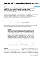
RGDechiHCit and c(RGDf[NMe]V) peptides stabilities
were evaluated in serum. The degradation of the pep-
tides were followed by LC/MS. The rev ersed-phase high
performance liquid chromatography (RP-HPLC) of
RGDechiHCit before the se rum incubation showed a
single peak at t
r
= 11.82 min corresponding to the com-
plete sequence (theoretical MW = 2100.1 g mol
-1
)as
indicated by the [M+H]
+
,[M+2H]
2+
and [M+3H]
+3
molecular ion adducts in the MS spectrum (Figure 1A).
Aft er 1h, chromatography showed two p eaks, ascribable
to RGDechiHCit and to a fragment of the complete
sequence (theoretical MW = 1929.1 g mol
-1
), respec-
tively, as confirmed by MS spectrum. Finally, after 24h a
further peak at t
r
= 10.93 min corresponding to another
RGDechiHCit degradation product (theoretical MW =
1775.8 g mol
-1
) appeared, as indicated by the molecular
ion adducts in the MS spectrum, although the peaks
attribut ed to the RGDechiHCit and to the first fragment
were still present (Figure 1B).
In contrast with RGDechiHCit, c(RGDf[NMe]V)
showed high stability in serum. The RP-H PLC profile of
the peptide before the incubation showed a single peak
at t
r
= 16.64 min, ascribable to the complete sequence
by the MS spectrum (Figure 1C). After 24h of incuba-
tion chromatogram and mass profiles failed to identify
any degradation product (Figure 1D).
Since RGDechiHCi t showed a low stability, we replen-
ished ant agonists every six hours in experiments invol-
ving chronic exposure.
In vitro experiments
Cell proliferation and DNA synthesis
Because angiogenesis is intimately associated to EC pro-
liferation, we explored the effects of RGDechiHCit and c
(RGDf[NMe]V) on hFN-stimulated E C. In this cellular
setting, after 6 hours, both a
v
b
3
integrin antagonists
inhibited in a comparable way the ability of hFN to
induce proliferation (hFN: +1.98 ± 0.6; hFN+RGDechiH-
Cit: +0.58 ± 0.24; hFN+c(RGDf[NMe]V): +0.6 ± 0.38
fold over basal; p < 0.05, ANOVA) as depicted in Figure
!"
!"
!"
!"
!"
!"
!"
!"
!"
!"
!"
!"
!"
!"
Figure 1 Reversed-phase high performance liquid
chromatography (RP-HPLC) chromatograms and mass spectra
at t = 0 and t = 24 h for RGDechiHCit (A and B) and c(RGDf
[NMe]V) (C and D), respectively. In panel B the chromatographic
peaks at tr = 11.70 (Black Star), 12.04 (Black Square) and 10.93 min
(Black Circle) are marked.
Santulli et al. Journal of Translational Medicine 2011, 9:7
/>Page 4 of 10
2A. After 20 hours such inhibitory effect was less
marked (Figure 2A). In VSMC there was only a trend of
an anti-proliferative effect for these peptides , due to the
less evident action of hFN in this specific cellular setting
(hFN: +1.21 ± 0.1; hFN+RGDechiHCit: +0.93 ± 0.07;
hFN+c(RGDf[NMe]V): +0.9 ± 0.09 fold over basal; NS;
Figure 3A).
The effects of RGDechiHCit and c(RGDf[NMe]V) on
EC and VSMC proliferation were also measured by asses-
sing the incorporation of [
3
H]Thymidine in response to
hFN. This assay confirmed the anti-proliferative action of
both these peptides, which is more evident after 6 hours
and in ECs (hFN : +1.84 ± 0.24; hFN+RGDechiHCit: +
1.02 ± 0.2; hFN+c(RGDf[NMe]V): + 1.09 ± 0.07 fold over
basal; p < 0.05, ANOVA; Figure 2B). On the contrary,
the effect of RGDechiHCit on VSMC did not reach sta-
tistical significance in comparison to the c(RGDf[NMe]V)
used as control (Figure 3B).
Effects on cellular signal transduction
Since hFN-mediated activation of ERK2 is linked to
angiogenesis [16,24,31], we analyzed the ability of
RGDechiHCit and c(RGDf[NMe]V) to inhib it hFN-
induced phosphorylation of ERK2 in EC and VSMC. In
accordance with the results on cell proliferation and
[
3
H]Thymidine incorporation, in EC both RGDechiHCit
and c(RGDf[NMe]V) significantly inhibited the hFN-
induced phosphorylation of mitogen-activated protein
ERK2 (Figure 2C). Also, in VSMC, there was no signifi-
cant inhibition of ERK2 phosphorylation by the RGDe-
chiHCit compund c(RGDf[NMe]V) (Figure 3C).
Evaluation of VEGF expression
Angiogenesis is largely dependent on ERK2 activation,
which in turn promotes cellular proliferation and
expression of VEGF. This cytokine promotes infiltr ation
of inflam matory cells, prolifer ation of ECs and VSMCs
and sustains the proangiogenic phenotype [12]. The
early release (6 hours) of the cytokine is therefore an
important readout when studying angiogenesis in vitro.
On these grounds, we assessed the expression levels of
this pivotal proangiogenetic factor in EC after 6 hours
of stimulation with hFN. h FN induces VEGF release
and such response was blunted by incubation with
either integrin antagonist, as depicted in Figure 4
Basa
l
RG
DechiHCit
h
FN
h
FN+RGDechiHCit
c
(RGDf[NMe]V)
h
FN+c(RGDf[NMe]V)
0
1
2
3
3h
6h 20h
*
*
#
#
Cell number
(Fold of Basal)
C
ell pr oliferation
Basa
l
RG
DechiHCit
h
FN
h
FN+RGDechiHCit
c
(RGDf[NMe]V)
h
FN+c(RGDf[NMe]V)
0
1
2
3
4
*
*
#
#
D
NA synthesis
[
3
H] thymidine
(Fold of Basal)
B
asal
RGD
echiHCit
hFN
hFN
+RGDechiHCit
c
(RGDf[NMe]V)
hFN
+c(RGDf[NMe]V)
0
2
4
6
8
10
*
#
#
pERK/ERK2 densitometry
(relative fold increase)
p
ERK
ERK2
hFN
++ -+
RGDe chiHCit
-+ - + - -
c(RGDf[NMe]V)
- - + +
C
A
B
Figure 2 In vitro effects of c(RGDf[NMe]V) and RGDechiHCit on
cell proliferation (Panel A) and DNA synthesis assessed by [
3
H]
thymidine incorporation (Panel B) in bovine aortic endothelial
cells (EC). Given alone, c(RGDf[NMe]V) or RGDechiHCit did not
affect EC proliferation. Neverteless, incubation with these a
V
b
3
integrin antagonists inhibited in a comparable way EC proliferation
in response to the mitogenic stimulus, hFN. All experiments
depicted in this figure were performed from three to six times in
duplicate (* = p < 0.05 vs Basal, # = p < 0.05 vs hFN). Panel C. In
vitro effects of c(RGDf[NMe]V) and RGDechiHCit on EC signal
transduction. Extracellular signal regulated kinase (ERK)/mitogen-
activated protein kinase activation: western blot of activated
(phosphorylated: pERK) ERK2 after hFN-stimulation. Equal amounts
of proteins were confirmed via blotting for total ERK. Densitometric
analysis (bar graph) showed that hFN stimulation caused ERK
activation (* = p < 0.05 vs Basal) and that treatment with a
V
b
3
antagonists blunted such activation (# = p < 0.05 vs hFN). Error bars
show SEM. Representative blots are shown in the inset.
Basal
RGDechiHCit
hFN
hFN+RGDechiHCit
c(RGDf[NMe]V)
hFN+c(RGDf[NMe]V)
0.0
0.5
1.0
1.5
2.0
3 h
6 h
20 h
*
*
#
Cell number
(Fold of Basal)
Cell proliferation
Basal
RGDechiHCit
hFN
hFN+RGDechiHCit
c(RGDf[NMe]V)
hFN+c(RGDf[NMe]V)
0
1
2
3
4
*
*
#
[
3
H] thymidine
(Fold of Basal)
DNA synthesis
Basal
RGDechiHCit
hFN
hFN+RGDechiHCit
c(RGDf[NMe]V)
h
FN+c(RGDf[NMe]V)
0
1
2
3
4
*
#
pERK/ERK2 densitometry
(relative fold increase)
pERK
ERK2
hFN
++ - +
RGDe chiHCit
-+ - + - -
c(RGDf[NMe]V)
- - ++
A
B
C
Figure 3 In vitro effects of c(RGDf[NME]V) and RGDechiHCit on
vascular smooth muscle cell (VSMC) cell proliferation (Panel A)
and DNA synthesis assayed by [
3
H]thymidine incorporation
(Panel B). In this cellular setting, hFN induced a mitogenic stimulus,
appreciable especially at 20h. c(RGDf[NMe]V) but not RGDechiHCit
at that time-point induced an attenuation of such proliferative
response. All experiments were performed from three to five times
in triplicate (* = p < 0.05 vs Basal; # = p < 0.05 vs hFN). In vitro
effects of c(RGDf[NMe]V) and RGDechiHCit on VSMC signal
transduction were represented in Panel C. Extracellular signal
regulated kinase (ERK)/mitogen-activated protein kinase activation:
western blot of activated (phosphorylated: pERK) ERK2 after hFN-
stimulation. Blots were then stripped and reprobed for either total
ERK as a loading control. Densitometric analysis (bar graph) showed
that hFN induced ERK phosphorylation (* = p < 0.05 vs Basal) and
that treatment with c(RGDf[NMe]V) but not RGDechiHCit decreased
such activation (# = p < 0.05 vs hFN). Error bars show SEM.
Representative blots are presented in the inset.
Santulli et al. Journal of Translational Medicine 2011, 9:7
/>Page 5 of 10
(hFN: +18.9 ± 1.02; hFN+RGDechiHCit: +2.44 ± 0.76;
hFN+c(RGDf[NMe]V): +3.19 ± 0.73 fold over basal,
ADU; p < 0.05, ANOVA).
Endothelial Matrigel assay
The formation of ca pillary-like tube structures in the
ECM by ECs is a pivotal step in angiogenesis and is also
involved in cell migration and inv asion [26]. To evaluate
any potential antiangiogenic activity of our novel integ-
rin antagonist, in vitro angiogenesis assays were con-
ducted by evaluating hFN-induced angiogenesis of ECs
on Matrigel.
As shown in Figur e 5, when ECs were plated on wells
coated with Matrigel without the addition of hFN, they
showed formation of only a few spontaneous tube struc-
tures (17.4 ± 1.2 branches per 10000 μm
2
). On the
other hand, when the cells were plated on Matrigel with
the addiction of hFN, cells formed a characteristic
capillary-like network (42.8 ± 4.4 branches per 10000
μm
2
; p < 0.05 vs Basal, ANOVA). In the presence
of RGDechiHCit or c(RGDf[NMe]V), the extent of
tube formation hFN-induced was significantly reduced
(10.03 ± 1.44; 14.11 ± 3.9, respect ively; p < 0.05 vs hFN
alone, ANOVA; Figure 5).
In vivo experiments
Wound healing
The examination of full-thickness wounds in the back
skin showed that both RGDechiHCit and c(RGDf
[NMe]V) slowed down healing (Figure 6). At a macro-
scopic observation, the delay in the wound healing in
treated rats was evident, with raised margins, more
extensive wound debris and scab, that persisted for at
least 7 days after surgery. Moreover, histological
hFN
++ - +
RGDechiHCit
-+ - + - -
c(RGDf[NMe]V )
- - ++
VEGF
actin
Basal
RGDechiHCit
hFN
hFN+RGDechiHCit
c(RGDf[NMe]V)
h
FN+c(RGDf[NMe]V)
0
5
10
15
20
25
*
#
#
ADU
(relative fold increase)
Figure 4 VEGF production i n bovine aortic endothelial cells
(ECs) measured by Western blot (inset). Shown are VEGF levels
after 6 hours of serum starvation. Equal amount of proteins were
verified by blotting for actin. Quantification of western blot from all
experiments demonstrated that hFN was able to increase VEGF
production (* = p < 0.05 vs Basal), while after c(RGDf[NMe]V) or
RGDechiHCit treatment VEGF levels returned to basal conditions (#
= p < 0.05 vs hFN). All data derived from three different
experiments performed in duplicate. The results were expressed as
fold increased with respect to the basal condition in untreated
samples. Error bars show SEM.
0
10
20
30
40
50
*
hFN
hFN
+
RGDechiHCit
hFN
+
c
(RGDf[NMe]V)
*
Basal
#
Branches per 10000
P
m
2
Basal
hFN
hFN+RGDechiHCit
hFN+c(RGDf[NMe]V)
____
____
Figure 5 Representative phase contrast ph otomicrographs of
bovine aortic endothelial cells (ECs) are shown plated on
Matrigel. Both c(RGDf[NMe]V) and RGDechiHCit inhibited hFN-
induced tube formation. Microscopy revealed numbers of network
projections (branches) formed in each group after 12 h of
incubation. Data from three experiments in triplicate are
summarized in the graph (* = p < 0.05 vs Basal; # = p < 0.05 vs
hFN). Error bars show SEM. The black bar corresponds to 100 μm.
Santulli et al. Journal of Translational Medicine 2011, 9:7
/>Page 6 of 10
Figure 6 Both c(RGDf[NMe]V) and RGDechiHCit slowed down the closure of full thickness punch biopsy wounds. Three to five rats were
analyzed at each time point. Gross appearance (representative digital photographs, light blue bar: 1 cm) after 5 days of the wound treated with
pluronic gel containing c(RGDf-[NMe]V), RGDechiHCit (10
-6
M) or saline. Diagram of the kinetics of wound closure; * = p < 0.05 vs Control; # =
p < 0.05 vs c(RGDf-[NMe]V, ANOVA). Error bars show SEM. Representative sections (5 μm) of wounds excised 8 days after surgery (see Methods):
Hematoxylin & Eosin, Lectin immunohistochemistry, Masson’s trichrome; black bar: 100 μm. Histological analysis revealed a retarded repair
pattern in treated rats, which is consistent with inhibition of angiogenesis in the granulation tissue. In particular, in control animals, epidermal
cell growth achieved complete re-epitalization (green arrowheads) and there was a well defined and organized fibrous core of scar tissue. Both
in c(RGDf[NMe]V) and RGDechiHCit treated rats there was a chronic inflammatory infiltrate (red arrows) and lectin staining showed (in brown)
the presence of vessels in the granulation tissue.
Santulli et al. Journal of Translational Medicine 2011, 9:7
/>Page 7 of 10
analysis showed that while control rats pre sented a
dermal scar tissue consisting of a well defined and
organized fibrous core with minimal chronic inflam-
matory cells, skin wounds exposed to RGDechiHCit or
c(RGDf[NMe]V) exhibited a retarded repair pattern.
Indeed, there was an intense inflammatory infiltrate,
extended from the wo und margin into the region of
the panniculus carnos us muscle and hypodermis. More-
over, the basal epidermis was disorganized and epidermal
cell growth failed to achieve re-epithelialization, as shown
in Figure 6.
Matrigel plugs
After injection, Matrigel implants containing the angio-
genic stimulant VEGF (10
-5
M) form ed a plug into
which ECs can migrate. Matrigel pellets evidenced a sig-
nificant lower EC infiltration, identified through means
of immunohistological lectin staining, in c(RGDf[NMe]
V) and RGDechiHCit treated plugs respect to VEGF
alone (VEGF+RGDechiHCit: 0.211 ± 0.034; VEGF+c
(RGDf[NMe]V): 0.185 ± 0.027 fold over VEGF alone;
p < 0.05, ANOVA), as depicted in Figure 7.
Discussion
In the present study, we evaluated t he anti-angiogenic
properties of RGDechiHCit peptide in vitro on EC and
VSMC cells and in vivo on animal models of rats and
mice. The data here reported recapitulate the well-
known antiangiogenic properties of c(RGDf[NMe]V),
that was used as control. We p reviously described the
design and synthesis of RGDechiHCit, a novel and selec-
tive ligand for a
V
b
3
integrin, containing a cyclic RGD
motif and two echistatin C-terminal moieties [20]. In
vitro studies showed that this molecule is able to selec-
tively bind a
V
b
3
integrin and not to cross-react with
other type of integrins. Furthermore, PET a nd SPECT
imaging studies have confirmed that the peptide loca-
lizes on a
V
b
3
expressing tumor cells in xenograft animal
model [ 21]. Given the presence in the molecule of the
RGD s equence it was obvious to speculate that RGDe-
chiHCit acted as an antagonist. Our report is the first
evidence that our peptide acts as antagonist for a
V
b
3
integrin. I ts ability to inhibit hFN-induced cell pro lifera-
tion is comparable to that of c(RGDf[NMe]V), although
the half-life is quite reduced.
A major evidence that is brought up by our results is
the peculiar selectivity of RGDechiHCit towards EC, as
compared to c(RGDf[NMe]V). Indeed, RGDechiHCit
fails to inhibit VSMC proliferation in vitro, opposite to c
(RGDf[NMe]V). We believe that this feature is due to
the selectivity of such a novel compound toward a
V
b
3
.
Indeed, VSMCs express a
V
b
3
only during embryogenesis
[31], but express other integrins which may be blocked
by c(RGDf[NMe]V). On the contrary, a
V
b
3
is expressed
by ECs [8], t hus conferring RGDechiHCit selectivity
toward this cell type. This issue is relevant cause the
effect in vivo is similar between the two antagonists on
wound healing and Matrigel plugs invasion. Indeed, our
data suggest that inhibition of the endothelial integrin
system is sufficient to inhibit angiogenesis. It is possible
to speculate that the higher specificity of RGDechiHCit
for the endothelium would result in a lower occurrence
of side effects than the use of less selective inhibitors.
This is only an indirect evidence, that needs further
investigation in more specific experimental setups.
Indeed, of the wide spectrum of integrins that are
expressed on the surface of ECs, a
V
b
3
receptor has been
identified as having an especially interesting expression
pattern among vascular cells during angiogenesis, vascu-
lar remodeling, tumor progression and metastasis
[6,32,33]. What is more, two pathways of angiogenesis
have been recently identified based on the related
butdistinctintegrinsa
V
b
3
and a
V
b
5
[4]. In particular,
a
V
b
3
integrin activates VEGF receptors and inhibition
of b
3
subunit has been shown to reduce phosphorylation
of VEGF receptors [7], thereby limiting the biological
Figure 7 Representative immunohis tochemical sections (5 μm)
of subcutaneously injected Matrigel plugs. ECs were identified
(light blue arrowheads) by lectin staining, which gave a brown
reaction product, as described in Methods. Both c(RGDf[NMe]V) and
RGDechiHCit treatment reduced the number of invading cells from
the edge (black arrows) to the core of implanted Matrigel plug.
Analysis was conducted in 20 randomly chosen cross-sections
per each group. Bar: 400 nm. * = p < 0.05 vs VEGF. Error bars
show SEM.
Santulli et al. Journal of Translational Medicine 2011, 9:7
/>Page 8 of 10
effects of VEGF [1]. Further, Mahabeleshwar and cowor-
kers have sh own the intimate interaction occurring
between a
V
b
3
integrin and the VEGFR-2 in primary
human EC [12]. The relevance of this molecule to
angiogenesis and its potential as a therapeutic target
has, therefo re, been well established [34,35] and in this
report we show that its activity is highly critical for both
hFN or VEGF-stimulated ECs proliferation.
Our results concerning RGDechiHCit in angiogenic
processes are of immediate translational importance,
because deregulation of angiogenesis is involved in sev-
eral clinical conditions including cancer, ischemic, and
inflammatory diseases (atherosclerosis, rheumatoid
arthritis, or age-related macular degeneration) [34-36].
Therefore, the research for drugs able to modulate
angiogenesis constitutes a crucial investigation field.
Since RGDechiHCit is rapidly removed in serum it is
possible to increase its effect by engineering the mole-
cule to elongate its lifespan. In the present paper we cir-
cumvented this issue by i ncreasing the times o f
application of the drug both in vitro and in vivo,orby
reducing the times of observation. This issue can be
solved by the use of a more stable aromatic pharmaco-
phore that recapitulates the binding properties of RGDe-
chiHCit. Clearly, further investigations are also needed
to fully understand the basic cell biologica l mechanism s
underlying growth factor receptors and integrin function
during angiogenesis. The knowledge of molecular basis
of this complex mechanism remains a challenge of fasci-
nating interest, with clinical implications for treatment
of a large number of pathophysiological conditions
including but not limited to solid t umors [17,37], dia-
betic retinopathy [38,39] and inflammatory disease [36].
Conclusions
The present study indicates the importance of RGDe-
chiHCit i n the selective inhibition of endothelial a
V
b
3
integrin. Such inhibition opens new fields of investiga-
tion on the mechanisms of angiogenesis, offering clinical
implications for the treatment of several conditions such
as proliferative retinopathy, inflammatory disease and
cancer.
Author details
1
Department of Clinical Medicine, Cardiovascular & Immunologic Sciences,
“Federico II” University of Naples, Italy.
2
Department of Biological Sciences,
“Federico II” University of Naples, Italy.
3
Institute of Crystallography (Consiglio
Nazionale delle Ricerche, CNR), Bari, Italy.
4
Institute of Biostructures and
Bioimaging (Consiglio Nazionale delle Ricerche, CNR), Naples, Italy.
Authors’ contributions
GS and GI designed research; GS, MFB, MDS, CDG, AA, and DS carried out
the experiments; GS and GI performed the statistical analysis; GS, GI and LZ
drafted the manuscript; GS, MS, ADG, BT, CP and GI supervised the project;
GS and MFB equally contributed to this work. All authors read and approved
the final manuscript.
Competing interests
We have no financial or personal relationships with other people or
organizations that would bias our work. No benefits in any form have been
received or will be received from a commercial party related directly or
indirectly to the subject of our article.
Received: 28 June 2010 Accepted: 13 January 2011
Published: 13 January 2011
References
1. Santulli G, Ciccarelli M, Palumbo G, Campanile A, Galasso G, Ziaco B,
Altobelli GG, Cimini V, Piscione F, D’Andrea LD, et al: In vivo properties of
the proangiogenic peptide QK. J Transl Med 2009, 7:41.
2. Bonauer A, Carmona G, Iwasaki M, Mione M, Koyanagi M, Fischer A,
Burchfield J, Fox H, Doebele C, Ohtani K, et al: MicroRNA-92a controls
angiogenesis and functional recovery of ischemic tissues in mice. Science
2009, 324:1710-1713.
3. Desgrosellier JS, Cheresh DA: Integrins in cancer: biological implications
and therapeutic opportunities. Nat Rev Cancer 2010, 10:9-22.
4. Hood JD, Frausto R, Kiosses WB, Schwartz MA, Cheresh DA: Differential
alphav integrin-mediated Ras-ERK signaling during two pathways of
angiogenesis. J Cell Biol 2003, 162:933-943.
5. Takahashi S, Moser M, Montanez E, Nakano T, Seo M, Backert S, Inoue I,
Awata T, Katayama S, Komoda T, Fassler R: The fibronectin RGD motif is
required for multiple angiogenic events during early embryonic
development. Arterioscler Thromb Vasc Biol 2010, 30:e1.
6. Castel S, Pagan R, Garcia R, Casaroli-Marano RP, Reina M, Mitjans F, Piulats J,
Vilaro S: Alpha v integrin antagonists induce the disassembly of focal
contacts in melanoma cells. Eur J Cell Biol 2000, 79:502-512.
7. Soldi R, Mitola S, Strasly M, Defilippi P, Tarone G, Bussolino F: Role of
alphavbeta3 integrin in the activation of vascular endothelial growth
factor receptor-2. Embo J 1999, 18:882-892.
8. Lu H, Murtagh J, Schwartz EL: The microtubule binding drug laulimalide
inhibits vascular endothelial growth factor-induced human endothelial
cell migration and is synergistic when combined with docetaxel
(taxotere). Mol Pharmacol 2006, 69:1207-1215.
9. Bayless KJ, Salazar R, Davis GE: RGD-dependent vacuolation and lumen
formation observed during endothelial cell morphogenesis in three-
dimensional fibrin matrices involves the alpha(v)beta(3) and alpha(5)
beta(1) integrins. Am J Pathol 2000, 156:1673-1683.
10. Brooks PC, Stromblad S, Sanders LC, von Schalscha TL, Aimes RT, Stetler-
Stevenson WG, Quigley JP, Cheresh DA: Localization of matrix
metalloproteinase MMP-2 to the surface of invasive cells by interaction
with integrin alpha v beta 3. Cell 1996, 85:683-693.
11. Abumiya T, Lucero J, Heo JH, Tagaya M, Koziol JA, Copeland BR, del
Zoppo GJ: Activated microvessels express vascular endothelial growth
factor and integrin alpha(v)beta3 during focal cerebral ischemia. J Cereb
Blood Flow Metab 1999, 19:1038-1050.
12. Mahabeleshwar GH, Feng W, Reddy K, Plow EF, Byzova TV: Mechanisms of
integrin-vascular endothelial growth factor receptor cross-activation in
angiogenesis. Circ Res 2007, 101:570-580.
13. Xiong JP, Stehle T, Zhang R, Joachimiak A, Frech M, Goodman SL,
Arnaout MA: Crystal structure of the extracellular segment of integrin
alpha Vbeta3 in complex with an Arg-Gly-Asp ligand. Science 2002,
296:151-155.
14.
Aumailley M, Gurrath M, Muller G, Calvete J, Timpl R, Kessler H: Arg-Gly-Asp
constrained within cyclic pentapeptides. Strong and selective inhibitors
of cell adhesion to vitronectin and laminin fragment P1. FEBS Lett 1991,
291:50-54.
15. Schottelius M, Laufer B, Kessler H, Wester HJ: Ligands for mapping
alphavbeta3-integrin expression in vivo. Acc Chem Res 2009, 42:969-980.
16. Eliceiri BP, Klemke R, Stromblad S, Cheresh DA: Integrin alphavbeta3
requirement for sustained mitogen-activated protein kinase activity
during angiogenesis. J Cell Biol 1998, 140:1255-1263.
17. Bai J, Zhang J, Wu J, Shen L, Zeng J, Ding J, Wu Y, Gong Z, Li A, Xu S, et al:
JWA regulates melanoma metastasis by integrin alpha(V)beta(3)
signaling. Oncogene 2010, 29:1227-1237.
18. Eskens FA, Dumez H, Hoekstra R, Perschl A, Brindley C, Bottcher S,
Wynendaele W, Drevs J, Verweij J, van Oosterom AT: Phase I and
pharmacokinetic study of continuous twice weekly intravenous
administration of Cilengitide (EMD 121974), a novel inhibitor of the
Santulli et al. Journal of Translational Medicine 2011, 9:7
/>Page 9 of 10
integrins alphavbeta3 and alphavbeta5 in patients with advanced solid
tumours. Eur J Cancer 2003, 39:917-926.
19. Dechantsreiter MA, Planker E, Matha B, Lohof E, Holzemann G, Jonczyk A,
Goodman SL, Kessler H: N-Methylated cyclic RGD peptides as highly
active and selective alpha(V)beta(3) integrin antagonists. J Med Chem
1999, 42:3033-3040.
20. Del Gatto A, Zaccaro L, Grieco P, Novellino E, Zannetti A, Del Vecchio S,
Iommelli F, Salvatore M, Pedone C, Saviano M: Novel and selective alpha
(v)beta3 receptor peptide antagonist: design, synthesis, and biological
behavior. J Med Chem 2006, 49:3416-3420.
21. Zannetti A, Del Vecchio S, Iommelli F, Del Gatto A, De Luca S, Zaccaro L,
Papaccioli A, Sommella J, Panico M, Speranza A, et al: Imaging of
alphavbeta3 expression by a bifunctional chimeric RGD peptide not
cross-reacting with alphavbeta5. Clin Cancer Res 2009, 15:5224-5233.
22. Ciccarelli M, Cipolletta E, Santulli G, Campanile A, Pumiglia K, Cervero P,
Pastore L, Astone D, Trimarco B, Iaccarino G: Endothelial beta2 adrenergic
signaling to AKT: role of Gi and SRC. Cell Signal 2007, 19:1949-1955.
23. Iaccarino G, Ciccarelli M, Sorriento D, Cipolletta E, Cerullo V, Iovino GL,
Paudice A, Elia A, Santulli G, Campanile A, et al: AKT participates in
endothelial dysfunction in hypertension. Circulation 2004, 109:2587-2593.
24. Illario M, Cavallo AL, Monaco S, Di Vito E, Mueller F, Marzano LA,
Troncone G, Fenzi G, Rossi G, Vitale M: Fibronectin-induced proliferation in
thyroid cells is mediated by alphavbeta3 integrin through Ras/Raf-1/
MEK/ERK and calcium/CaMKII signals. J Clin Endocrinol Metab 2005,
90:2865-2873.
25. Iaccarino G, Smithwick LA, Lefkowitz RJ, Koch WJ: Targeting Gbeta gamma
signaling in arterial vascular smooth muscle proliferation: a novel
strategy to limit restenosis. Proc Natl Acad Sci USA 1999, 96:3945-3950.
26. Iaccarino G, Ciccarelli M, Sorriento D, Galasso G, Campanile A, Santulli G,
Cipolletta E, Cerullo V, Cimini V, Altobelli GG, et al: Ischemic
neoangiogenesis enhanced by beta2-adrenergic receptor
overexpression: a novel role for the endothelial adrenergic system. Circ
Res 2005, 97:1182-1189.
27. Ciccarelli M, Santulli G, Campanile A, Galasso G, Cervero P, Altobelli GG,
Cimini V, Pastore L, Piscione F, Trimarco B, Iaccarino G: Endothelial alpha1-
adrenoceptors regulate neo-angiogenesis. Br J Pharmacol 2008,
153:936-946.
28. Sorriento D, Ciccarelli M, Santulli G, Campanile A, Altobelli GG, Cimini V,
Galasso G, Astone D, Piscione F, Pastore L, et al: The G-protein-coupled
receptor kinase 5 inhibits NFkappaB transcriptional activity by inducing
nuclear accumulation of IkappaB alpha. Proc Natl Acad Sci USA 2008,
105:17818-17823.
29. Sorriento D, Santulli G, Fusco A, Anastasio A, Trimarco B, Iaccarino G:
Intracardiac Injection of AdGRK5-NT Reduces Left Ventricular
Hypertrophy by Inhibiting NF-{kappa}B-Dependent Hypertrophic Gene
Expression. Hypertension 2010, 56
:696-704.
30. Santulli G, Illario M, Palumbo G, Sorriento D, Cipolletta E, Trimarco V, Del
Giudice C, Ciccarelli M, Trimarco B, Iaccarino G: CaMK4 partecipates in the
settings of the hypertensive phenotype: a human genome wide analysis
supported by animal model. Eur Heart J 2009, 30(Suppl.1):161.
31. Astrof S, Hynes RO: Fibronectins in vascular morphogenesis. Angiogenesis
2009, 12:165-175.
32. Zaccaro L, Del Gatto A, Pedone C, Saviano M: Peptides for tumour therapy
and diagnosis: current status and future directions. Curr Med Chem 2009,
16:780-795.
33. Verbisck NV, Costa ET, Costa FF, Cavalher FP, Costa MD, Muras A, Paixao VA,
Moura R, Granato MF, Ierardi DF, et al: ADAM23 negatively modulates
alpha(v)beta(3) integrin activation during metastasis. Cancer Res 2009,
69:5546-5552.
34. Laitinen I, Saraste A, Weidl E, Poethko T, Weber AW, Nekolla SG,
Leppanen P, Yla-Herttuala S, Holzlwimmer G, Walch A, et al: Evaluation of
alphavbeta3 integrin-targeted positron emission tomography tracer 18F-
galacto-RGD for imaging of vascular inflammation in atherosclerotic
mice. Circ Cardiovasc Imaging 2009, 2:331-338.
35. Furundzija V, Fritzsche J, Kaufmann J, Meyborg H, Fleck E, Kappert K,
Stawowy P: IGF-1 increases macrophage motility via PKC/p38-dependent
alphavbeta3-integrin inside-out signaling. Biochem Biophys Res Commun
2010, 394:786-791.
36. Vanderslice P, Woodside DG: Integrin antagonists as therapeutics for
inflammatory diseases. Expert Opin Investig Drugs 2006, 15:1235-1255.
37. Tani N, Higashiyama S, Kawaguchi N, Madarame J, Ota I, Ito Y, Ohoka Y,
Shiosaka S, Takada Y, Matsuura N: Expression level of integrin alpha 5 on
tumour cells affects the rate of metastasis to the kidney. Br J Cancer
2003, 88:327-333.
38. Crawford TN, Alfaro DV, Kerrison JB, Jablon EP: Diabetic retinopathy and
angiogenesis. Curr Diabetes Rev 2009, 5:8-13.
39. Santulli RJ, Kinney WA, Ghosh S, Decorte BL, Liu L, Tuman RW, Zhou Z,
Huebert N, Bursell SE, Clermont AC, et al: Studies with an orally
bioavailable alpha V integrin antagonist in animal models of ocular
vasculopathy: retinal neovascularization in mice and retinal vascular
permeability in diabetic rats. J Pharmacol Exp Ther 2008, 324:894-901.
doi:10.1186/1479-5876-9-7
Cite this article as: Santulli et al.: Evaluation of the anti-angiogenic
properties of the new selective a
V
b
3
integrin antagonist RGDechiHCit.
Journal of Translational Medicine 2011 9:7.
Submit your next manuscript to BioMed Central
and take full advantage of:
• Convenient online submission
• Thorough peer review
• No space constraints or color figure charges
• Immediate publication on acceptance
• Inclusion in PubMed, CAS, Scopus and Google Scholar
• Research which is freely available for redistribution
Submit your manuscript at
www.biomedcentral.com/submit
Santulli et al. Journal of Translational Medicine 2011, 9:7
/>Page 10 of 10