báo cáo hóa học:" Regression of orthotopic neuroblastoma in mice by targeting the endothelial and tumor cell compartments" pdf
Bạn đang xem bản rút gọn của tài liệu. Xem và tải ngay bản đầy đủ của tài liệu tại đây (1.5 MB, 11 trang )
BioMed Central
Page 1 of 11
(page number not for citation purposes)
Journal of Translational Medicine
Open Access
Research
Regression of orthotopic neuroblastoma in mice by targeting the
endothelial and tumor cell compartments
Dieter Fuchs*
1
, Rolf Christofferson
1,2
, Mats Stridsberg
3
, Elin Lindhagen
3
and
Faranak Azarbayjani
1
Address:
1
Department of Medical Cell Biology, Uppsala University, 75123 Uppsala, Sweden,
2
Department of Woman and Child Health, Uppsala
University Hospital, 75185 Uppsala, Sweden and
3
Department of Medical Sciences, Uppsala University Hospital, 75185 Uppsala, Sweden
Email: Dieter Fuchs* - ; Rolf Christofferson - ;
Mats Stridsberg - ; Elin Lindhagen - ;
Faranak Azarbayjani -
* Corresponding author
Abstract
Background: High-risk neuroblastoma has an overall five-year survival of less than 40%, indicating
a need for new treatment strategies such as angiogenesis inhibition. Recent studies have shown that
chemotherapeutic drugs can inhibit angiogenesis if administered in a continuous schedule. The aim
of this study was primarily to characterize tumor spread in an orthotopic, metastatic model for
aggressive, MYCN-amplified neuroblastoma and secondarily to study the effects of daily
administration of the chemotherapeutic agent CHS 828 on tumor angiogenesis, tumor growth, and
spread.
Methods: MYCN-amplified human neuroblastoma cells (IMR-32, 2 × 10
6
) were injected into the
left adrenal gland in SCID mice through a flank incision. Nine weeks later, a new laparotomy was
performed to confirm tumor establishment and to estimate tumor volume. Animals were
randomized to either treatment with CHS 828 (20 mg/kg/day; p.o.) or vehicle control. Differences
between groups in tumor volume were analyzed by Mann-Whitney U test and in metastatic spread
using Fisher's exact test. Differences with p < 0.05 were considered statistically significant.
Results: The orthotopic model resembled clinical neuroblastoma in respect to tumor site, growth
and spread. Treatment with CHS 828 resulted in tumor regression (p < 0.001) and reduction in
viable tumor fraction (p < 0.001) and metastatic spread (p < 0.05) in correlation with reduced
plasma levels of the putative tumor marker chromogranin A (p < 0.001). These effects were due
to increased tumor cell death and reduced angiogenesis. No treatment-related toxicities were
observed.
Conclusion: The metastatic animal model in this study resembled clinical neuroblastoma and is
therefore clinically relevant for examining new treatment strategies for this malignancy. Our results
indicate that daily scheduling of CHS 828 may be beneficial in treating patients with high-risk
neuroblastoma.
Published: 12 March 2009
Journal of Translational Medicine 2009, 7:16 doi:10.1186/1479-5876-7-16
Received: 29 September 2008
Accepted: 12 March 2009
This article is available from: />© 2009 Fuchs et al; licensee BioMed Central Ltd.
This is an Open Access article distributed under the terms of the Creative Commons Attribution License ( />),
which permits unrestricted use, distribution, and reproduction in any medium, provided the original work is properly cited.
Journal of Translational Medicine 2009, 7:16 />Page 2 of 11
(page number not for citation purposes)
Background
Neuroblastoma (NB) is the most common extracranial
solid tumor of childhood. High-risk NB has a long-term
survival rate of less than 40% despite intensive treatment
protocols involving high-dose chemotherapy, usually
with bone marrow rescue, aggressive surgery, and radio-
therapy [1,2]. Therefore, new treatment strategies, evalu-
ated in clinically relevant, reliable, and reproducible
animal models, are needed for this malignancy.
Angiogenesis inhibition is a novel treatment strategy,
where the formation of new blood vessels is inhibited,
thereby reducing both the metabolic exchange of the
tumor and its vascular access for metastatic spread. In NB,
a high tumor angiogenesis correlates with metastatic dis-
ease and poor outcome [3]. Furthermore, increased
microvascular proliferation has recently been shown to
correlate with poor survival in children with NB [4]. There
are many ways for angiogenesis inhibition, e.g. specific
inhibition of an angiogenic growth factor. In s.c. models
for NB, this approach resulted in a significantly reduced
tumor growth rate [5,6]. Another way for angiogenesis
inhibition is based on modified schedules and doses of
chemotherapeutic drugs, namely, switching from the cur-
rent maximum tolerable dose (MTD) to a continuous dos-
ing scheme [7]. Even though endothelial cells are
damaged by MTD, the beneficial antiangiogenic effects of
MTD schedules are compromised by treatment breaks
between cycles. These breaks are required for patient
recovery but allow endothelial cell repair and regrowth
[8,9]. Chemotherapy given at frequent intervals without
extended rest periods, has been shown to target endothe-
lial cells and tumor vessels in vivo [10]. The benefits of
continuous therapy, e.g. reduced host toxicity together
with continuous drug exposure resulting in a sustained
antiangiogenic effect, are investigated in a number of clin-
ical trials [11].
The chemotherapeutic drug CHS 828 is a pyridylguani-
dine that potently inhibits nicotinamide phosphoribosyl
transferase (NAMPT) in a time dependent manner
[12,13]. NAMPT is an enzyme involved in the biosynthe-
sis of oxidized nicotinamide adenine dinucleotide
(NAD
+
). In eukaryotic cells NAD
+
has been shown to play
a pivotal role as an essential coenzyme/transmitter mole-
cule for the generation of ATP. Due to the higher prolifer-
ation rate, cancer cells demand higher ATP synthesis and
therefore have higher turnover of NAD
+
and an upregu-
lated NAMPT enzyme to meet this energy demand. In fact,
NAMPT inhibition with CHS 828 has shown significant
antitumor activity in many preclinical in vitro and in vivo
models [14-17]. In clinical phase I studies conducted with
CHS 828, doses up to 500 mg were administered to
patients. Based on the observed dose limiting toxicities at
500 mg (228 mg/m
2
), Ravaud et al. suggested administra-
tion of 420 mg CHS 828 every 3 weeks for clinical phase
II studies [18] whereas the results of another clinical phase
I study recommended more frequent administration at 20
mg once a day for 5 days in cycles of 28 days duration
[19].
In preclinical studies in mice, CHS 828 could reduce
growth of s.c. NB without any signs of toxicity [17]. In
order to investigate this finding in a clinically more rele-
vant setting, we developed and characterized a relevant
orthotopic mouse models for high-risk NB. Generally,
orthotopic tumor models resemble clinical disseminated
disease more closely and have a more realistic tumor-host
interaction than heterotopic, s.c. models. To be able to
evaluate and to make a direct comparison between these
models in treating NB, mice bearing orthotopic tumors
were treated with the same dose and route of administra-
tion as in [17].
We found that the orthotopic growth and spread of NB
cells in SCID mice resembled the patterns observed in
high-risk NB patients. Daily oral administration of a non-
toxic dose of CHS 828 to the host animal induced tumor
regression and reduced bone marrow and liver metastases
by a dual mechanism of action, restraining growth of both
tumor cells and tumor vasculature.
Methods
CHS 828
The chemotherapeutic drug CHS 828 (N-(6-chlorophe-
noxyhexyl)-N'-cyano-N"-4-pyridylguanidine) was sup-
plied by LEO Pharma (Ballerup, Denmark). For in vitro
use, CHS 828 was dissolved to 5 mM in dimethyl sulfox-
ide (DMSO) (Merck, Darmstadt, Germany) and further
diluted in serum-free culture medium. For the in vivo
study, the drug was suspended in peanut oil (5 μg/μl) at
least once a week and stored at 4–8°C.
Cells
The human NB cell line IMR-32 (ATCC, Rockville, MD),
isolated from an abdominal NB in a 13-month-old boy, is
MYCN amplified and has a 1p deletion and a 47 + XY
karyotype [20]. SH-SY5Y (kindly provided by Dr. June
Biedler, The Memorial Sloan-Kettering Cancer Centre,
NY) was derived from a poorly differentiated, non-
MYCN-amplified human NB [21]. SK-N-SH, a kind gift of
Dr. Fredrik Hedborg, Uppsala University, Sweden, was
isolated from a bone marrow metastasis of a 4 year old
female NB patient. Cells were cultured as described previ-
ously [5]. Non-essential amino acids (Sigma Chemical
Co., St. Louis, MO) were added to IMR-32 cells. Human
foreskin fibroblasts (CCD-1064SK, a kind gift of Dr. Mag-
nus Essand, Uppsala University, Sweden) were cultured
under the same conditions as SH-SY5Y [5]. Immortalized
bovine endothelial cells (hTERT-BCE [22], a kind gift
Journal of Translational Medicine 2009, 7:16 />Page 3 of 11
(page number not for citation purposes)
from Dr. Yihai Cao, Karolinska Institute, Stockholm, Swe-
den), were cultured as described previously [22].
All cells tested negative for mycoplasms and were grown
in humidified air (95%) and 5% CO
2
at 37°C. All in vitro
experiments were performed under optimal culture con-
ditions (i.e. with serum).
Fluorometric microculture cytotoxicity assay
Drug cytotoxicity was determined using the fluorometric
microculture cytotoxicity assay (FMCA) method [23].
Briefly, CHS 828 stock solution, dissolved to 5 mM in
DMSO, was diluted in medium to final concentrations
ranging from 0.1 nM to 10 μM. Triplicates of drug solu-
tions (10 × final concentration; 20 μl) were added to v-
bottomed 96-well microtiter plates (Nunc, Roskilde, Den-
mark). NB cells (20,000/well), fibroblasts (15,000/well)
and endothelial cells (5,000/well) (cultured in medium
containing 10% serum) were added to the wells, and the
cell survival index, defined as fluorescence in percent of
control cultures, was calculated after a 24, 48, and 72 h
incubation period. IC
50
values were determined as CHS
828 concentrations with a survival index below 50%.
Cell morphology and cell death in vitro
Morphological changes in NB cells due to exposure to
CHS 828 were assessed by phase-contrast microscopy.
IMR-32 (1.5 × 10
5
/ml) were allowed to set overnight
before replacing the medium with fresh medium contain-
ing 1 nM CHS 828. The cell morphology was recorded
after 0, 4, 24, 48, 72, and 96 h with a digital phase-con-
trast microscope at × 100.
Quantification of cell death was performed by propidium
iodine (PI) and DAPI (4',6-diamino-2-phenylindole)
staining [24]. IMR-32 cells (1.5 × 10
5
/ml) were stained
with 10 μg/ml PI and DAPI after 24, 48, and 72 h exposure
to 1 nM CHS 828. Disintegration of the plasma mem-
brane results in red fluorescence, which is a marker of cell
death (determined by evaluation of at least 2,000 cells per
well by UV microscopy).
Animals
Female SCID mice (B&M, Ry, Denmark) were xenografted
at the age of 6 weeks (mean body weight, 17.3 g). The ani-
mals were housed in an isolated room at 24°C with a 12-
h day/night cycle. They were fed ad libitum with water and
food pellets. Animal weight and general appearance were
recorded daily throughout the experiment. The experi-
ment was approved by the regional ethics committee for
animal research.
Xenografting and confirmation of tumor establishment
Subconfluent IMR-32 cells were harvested and kept on ice
until xenotransplantation. The recipient mice were shaved
and cleansed with 70% ethanol at the site of incision and
anesthetized with 2% Fluothane (Zeneca Ltd., Maccles-
field, UK) supplemented with 50% N
2
O in oxygen. IMR-
32 cells (20 μl; 2 × 10
6
cells) were injected into the left
adrenal gland through a left flank incision, which was
closed with interrupted sutures in 2 layers. Buprenorphine
(10 μg/kg; s.c.; Schering-Plough Europe, Brussels, Bel-
gium) was administered once as postoperative analgesia.
All handling of the animals was performed under aseptic
conditions.
Nine weeks after xenografting, all animals (n = 35)
showed establishment of primary adrenal gland tumors
which was verified by re-laparotomy. Tumor volume
(mean volume: 0.77 ml), was estimated as described in
[25].
Measurement of tumor volume, drug administration,
perfusion fixation, and autopsy
Mice were randomized to 1 of the 3 groups: controls (pea-
nut oil, daily, p.o., 10 days; n = 10) and CHS 828 treat-
ment (20 mg/kg, daily, p.o.) for 10 (n = 13) or 30 days (n
= 10). Administration of 20 mg/kg/day has previously
been shown to be non-toxic to mice. At the study end-
points, animals were subjected to perfusion fixation [17].
After perfusion fixation, the tumors were dissected out,
and their absolute weights and volumes were recorded.
The internal organs were examined for macroscopic
metastases (see below).
Chromogranin A analyses
Chromogranin A (CgA) serum levels were analyzed as a
marker for tumor burden and treatment efficacy. Venous
blood was drawn from the right atrium before perfusion
fixation. The blood was stored at 4°C overnight and spun
at 135 × g for 10 min. The serum was removed and stored
at -20°C. Serum levels of human CgA were measured by a
commercial radioimmunoassay (Eurodiagnostica,
Malmö, Sweden) according to the manufacturer's instruc-
tions. Only tumor-derived CgA was detected since the
assay distinguishes between human and murine CgA.
Tissue analyses
At autopsy, the organs were examined for macroscopic
metastases, sliced in ~1-mm sections, and examined with
a dissection microscope (× 20). Orthotopic tumors, the
iliac crest, and organ biopsies with suspected metastases
were dehydrated and embedded in paraffin. Tissue sec-
tions were cut at 3 μm, placed on diaminoalkyl-silane-
treated glass slides, dewaxed, rehydrated, and stained
immunohistochemically as described below. All these
steps were performed in humid chambers at room tem-
perature, unless otherwise indicated. After immunohisto-
chemistry, the sections were counterstained with Harris'
Journal of Translational Medicine 2009, 7:16 />Page 4 of 11
(page number not for citation purposes)
hematoxylin and mounted with Kaiser's glycerol gelatin
(Merck).
For the quantification of angiogenesis, Bandeiraea sim-
plicifolia-1 (BS-1) lectin was used to mark endothelial
cells [25]. BS-1 (L3759; Sigma) was used at 1:50 dilution,
and the sections were incubated for 2 h. Endothelial cells
were used as positive controls, and the omission of the
neuraminidase solution served as a negative control.
Immunohistochemical staining for DNA strand breaks
(i.e. cell death) was performed by the TUNEL assay using
an "In Situ Cell Death Detection Kit, POD" (Roche, Indi-
anapolis, IN) according to the manufacturer's instruc-
tions. Murine ileum was used as a positive control, and
the replacement of TdT with water served as a negative
control.
Apoptosis was detected by staining for cleaved caspase-3
[6]. Sections were developed using Vector
®
NovaRED™
(SK-4800, Vector Laboratories, Inc., Burlingame, CA).
Human tonsil or murine colon served as a positive con-
trol, and the omission of the primary antibody served as a
negative control.
Staining specific for neuroendocrine and adrenergic cells,
i.e. NB cells, was performed by CgA immunohistochemis-
try. Before dehydration and embedding in paraffin, iliac
crest biopsies were decalcified in Parengy's decalcification
solution (University Hospital Pharmacy, Uppsala, Swe-
den) for 1 week. Tissue sections on glass slides were
treated with Target Retrieval Solution (S3308, Dako) and
blocked in 0.3% H
2
O
2
for 30 min and in 1% BSA and 10%
rabbit serum for 20 min. Primary antibody (M0869,
Dako) was applied at 1:100 dilution for 30 min. The bioti-
nylated secondary antibody (K335, Dako A/S) was
applied at 1:80 dilution for 30 min. For detection, ABC/
HRP (K355, Dako) was applied at 1:100 dilution for 30
min. The sections were developed using DAB (SK-4100,
Vector). NB cell pellets were used as positive controls, and
the omission of the primary antibody served as a negative
control. To detect NB cells in the bone marrow of the iliac
crest, 3 CgA-stained sections were examined in a blinded
fashion by 2 independent investigators. Two to 3 CgA-
positive cells in one section were classified as metastasis.
Stereologic quantification
All sections were quantified at × 400 magnification in a
blinded fashion [5,26]. Vascular parameters from up to 35
grids, depending on tumor size, were quantified for each
tumor. Only stereologic estimates from grids with a viable
upper right corner and in which the entire grid covered
tumor tissue were used for quantification. If more than
50% of the upper right corner covered densely packed
nuclei with sparse cytoplasm (i.e. NB cells), the grid was
assigned 'viable'.
The percentage of TUNEL- and caspase-3-positive cells
was calculated among ~2,000 cells in each tumor by using
the upper right quarter of the counting grid mentioned
above.
Treatment-related bone marrow toxicity was investigated
in hematoxylin-eosin stained sections of the iliac crest.
The percentage of megakaryocytes was calculated among
at least 2,000 bone marrow cells.
Statistical methods
All the data were processed in GraphPad Prism 4 for Win-
dows (GraphPad Software Inc.). Differences between
tumor volumes were analyzed with Mann-Whitney U test
and differences in organ weight were analyzed using the
Kruskal-Wallis test. Statistical differences between metas-
tases in CHS 828-treated animals and control animals
were analyzed using Fisher's exact test. Differences with p
< 0.05 were considered statistically significant.
Results
CHS 828 is toxic to NB cells but not to fibroblasts in vitro
CHS 828 was more toxic to NB cells than to endothelial
cells or fibroblasts in vitro. IC
50
values for fibroblasts were
above 10 μM CHS 828 (the highest concentration tested).
Drug activity was time dependent with the first signs of
toxicity after 48 h and high NB cell-specific toxicity after
72 h of continuous drug exposure (Table 1).
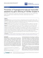
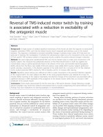
IMR-32 viability remained unaffected during the first 48 h
of exposure to 1 nM CHS 828 but showed a 560%
increase in cell death after 72 h of exposure as compared
to controls (Figure 1A, B).
Table 1: CHS 828 toxicity profile
IC
50
24 h 48 h 72 h
htertBCE >10 μM 200 – 500 nM 50 – 100 nM
SH-SY5Y >10 μM>10 μM2 – 5 nM
IMR-32 >10 μM>10 μM 0.2 – 0.5 nM
SK-N-SH >10 μM>10 μM2 – 5 nM
CCD-1064SK n.d. n.d. >10 μM
Triplicates of the NB cell lines IMR-32, SH-SY5Y, SK-N-SH cells (1 ×
10
5
/ml), human foreskin fibroblasts CCD-1064SK (7.5 × 10
4
/ml) and
endothelial cells htertBCE (2.5 × 10
4
/ml) were incubated with 16
different concentrations of CHS 828 for 24, 48 and 72 h
(concentration range: 0.1 nM – 10 μM). Cell survival was measured by
FMCA. Survival index was calculated as the percentage of viable cells
at the actual concentration divided by percentage of viable cells in
wells incubated without drug. Concentration intervals for IC
50
.
Journal of Translational Medicine 2009, 7:16 />Page 5 of 11
(page number not for citation purposes)
CHS 828 induces regression of rapidly growing orthotopic
NB in vivo
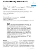
Tumors from vehicle-treated animals grew significantly
within 10 days from randomization (p < 0.05) (Figure 2,
Figure 3). Despite this rapid growth, no tumor rupture or
intraperitoneal bleeding was observed. Daily treatment
with CHS 828 (20 mg/kg; p.o.) for 10 days significantly
reduced mean tumor volume (-89%) and weight (-92%)
compared to untreated littermates (p = 0.0002 and p =
0.0001, respectively). An additional 20 days of treatment
(total of 30 days) further reduced tumor volume (-92%)
and weight (-86%) compared to short term treatment (p
= 0.0005 and p = 0.0006, respectively) (Figure 2, Figure
3). Administration of CHS 828 resulted in tumor regres-
sion (final tumor volume compared to starting volume)
after 10 (-81%; p < 0.0001) and 30 days (-98%; p <
0.0001). A detailed summary of tumor data is provided in
Additional file 1 (see Additional file 1: Observation
parameters of tumor-bearing SCID mice during the exper-
iment).
In addition to the reduction in tumor volume, treatment
with CHS 828 for 10 days also significantly reduced the
percentage of viable tumor tissue from 75.5% to 15.4% (p
< 0.0001) and increased the fraction of dead (i.e. TUNEL
positive) cells from 26.8% to 78.2% (p < 0.0001). The
fraction of apoptotic cells was not different compared to
controls when quantified by caspase-3 immunohisto-
chemistry.
There were no adverse effects of CHS 828 on the general
status of the animals. CHS 828 did not affect the body or
organ weight (liver, spleen, lung and kidney) in any of the
treated animals compared with controls (see Additional
file 2: Organ weight of healthy and tumor-bearing SCID
mice). Furthermore, no treatment-related diarrhea or
vomiting was observed, and the percentage of megakaryo-
In vitro morphology of NB cells cultured with or without CHS 828Figure 1
In vitro morphology of NB cells cultured with or with-
out CHS 828. IMR-32 (1.5 × 10
5
/ml) were cultured in 24-
well plates in the absence (control) or presence of 1 nM CHS
828 for 0 to 72 h. Cell morphology, investigated by phase-
contrast microscopy, revealed signs of cell death in NB cells
exposed to CHS 828 (A). Viability of NB cells (IMR-32) was
quantified in DAPI (4',6-diamino-2-phenylindole)-propidium
iodine (10 μg/ml)-stained cells by fluorescence microscopy
(B). Cells with intact plasma membrane (blue; DAPI staining)
and cells with disrupted membrane (red; propidium iodine
staining), magnification in A-B: × 100.
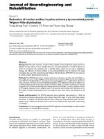
Orthotopic NB growth in SCID miceFigure 2
Orthotopic NB growth in SCID mice. SCID mice carry-
ing orthotopic NB xenotransplants were randomized at an
estimated tumor volume of 0.8 ml (h n = 10, controls; n n =
23, for CHS 828 treatment). After randomization, mice were
treated daily with either vehicle (h n = 9; 10 days) or with
CHS 828 (20 mg/kg; p.o.) for 10 (n n = 13) or 30 (n n = 10)
days. Mann-Whitney U test was used to evaluate differences
between the groups.
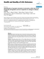
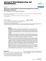
Orthotopic NB tumors at autopsy after treatment with CHS 828 or vehicleFigure 3
Orthotopic NB tumors at autopsy after treatment
with CHS 828 or vehicle. Orthotopic tumors at autopsy
treated with vehicle or CHS 828 (20 mg/kg/day) for 10 or 30
days. Note the brown color of the tumor after 10 days of
treatment, indicating areas of resorbed hemorrhage. T =
tumor, RK = right kidney, LK = left kidney; arrows indicate
the normal right adrenal gland.
Journal of Translational Medicine 2009, 7:16 />Page 6 of 11
(page number not for citation purposes)
cytes in the bone marrow of the iliac crest did not differ
between treated and healthy animals (2.46% ± 0.36% and
2.57% ± 0.40%, respectively; n.s.). Three mice were
excluded from the study: 2 mice before (1 due to inexpli-
cable weight loss and 1 due to paraplegia) and 1 mouse
after randomization (paraplegia; control group). The 2
cases of paraplegia were caused by orthotopic NB growth
extending into the spinal canal.
Metastatic pattern of orthotopic NB mimics disseminated
disease in high-risk NB patients
Few large, macroscopic organ metastases were observed at
autopsy. Examination of the lung, liver, spleen, bone mar-
row, and both kidneys under a dissection microscope
revealed NB spread to many of these organs. This was con-
firmed by either hematoxylin-eosin staining or CgA
immunohistochemistry. Table 2 summarizes NB spread
in this orthotopic model compared to clinical NB.
The frequency of NB spread was reduced by CHS 828
compared with controls. Postmortem classification
according to the INSS (International Neuroblastoma Stag-
ing System) showed that all control animals were classi-
fied as stage 4. Metastases detected in the treatment
groups were smaller and showed morphological signs of
regression (tumor necrosis) compared with metastases
detected in controls (Figure 4).
In 2 control animals, there was NB growth in the thymus,
thoracic lymph nodes, and along the thoracic vertebrae,
whereas no NB spread to these sites could be detected in
CHS 828-treated animals. Postmortem evaluation of
treatment efficiency by applying the INSS revealed a trend
toward lower stages (better resectability) when tumors
were treated with CHS 828 (Table 3). No peritoneal
metastases were detected, indicating that no free tumor
cells were seeded onto the peritoneal surface during
xenotransplantation.
Reduced CgA-levels in serum of CHS 828-treated animals
Human CgA was detected in the serum of all vehicle-
treated controls (n = 9) (10.7 ± 4.0 nmol/L). However, in
10 days study with CHS 828, only 1/13 mice showed a
detectable concentration of CgA (1 nM/L), and no ani-
mals receiving long-term treatment (n = 10) or healthy lit-
termates without tumors (n = 5) had detectable CgA
concentrations in serum (detection limit: 0.8 nmol/L) (p
< 0.001).
CHS 828 reduces tumor angiogenesis
Daily administration of CHS 828 altered vascular param-
eters as determined by stereology (Table 4). Vessel den-
sity, vessel length density (L
v
), and surface density (S
v
)
were significantly reduced in these tumors compared to
vehicle-treated controls. The vessel volumetric density
(V
v
) was reduced in CHS 828-treated tumors but the
reduction was not significant (p = 0.09) (Table 4). A single
layer of endothelial cells encircled the lumen of vessels in
untreated tumors (Figure 5A and Figure 5C) whereas in
CHS 828 treated tumors, endothelial cells were frequently
not entirely surrounding the lumen (Figure 5B, D, E) or
detaching from the basement membrane (Figure 5E).
Despite the incomplete endothelial cell lining, only 1/13
(8%) of the animals treated with CHS 828 for 10 days
showed intra-tumor hemorrhage, defined as erythrocytes
outside vessel lumen, whereas 9/9 (100%) of the tumors
in control animals had erythrocytes in the tumor tissue.
Table 2: Invasive pattern of orthotopic NB in SCID mice
Orthotopic mouse model Clinical NB
Site Control
at 10 days
CHS 828
at 10 days
CHS 828
at 30 days
[28]
Animals with metastases 100% (9/9) 46% (6/13)* 40% (4/10)*
Lung 11% (1/9) 0% (0/13) 0% (0/10) 34%
Liver 78% (7/9) 23% (3/13)* 0% (0/10)*** 30%
Spleen 22% (2/9) 8% (1/13) 30% (3/10) n.d.
Bone marrow iliac crest
spine
a
78% (7/9)
22% (2/9)
23% (3/13)*
8% (1/13)
10% (1/10)**
10% (1/10)
71%
Bone n.d. n.d. n.d. 56%
Lymph nodes n.d. n.d. n.d. 31%
Kidney invasion
†
22% (2/9) 0% (0/13) 0% (0/10) n.d.
Metastatic spread in SCID mice carrying orthotopic NB xenotransplants treated either with CHS 828 (20 mg/kg/day) or vehicle compared to
metastatic incidence of NB at INSS stages 4 and 4S in clinic [28]. Data shows microscopic metastases in the marrow of the iliac crest, and
composite data of macro- and microscopic metastases to the organs.
* < 0.05; ** < 0.01; *** < 0.001 (compared with controls); Fisher's exact test.
a
NB cells in the spine and kidney were regarded as continuous tumor growth.
n.d., not determined; NB, neuroblastoma; INSS, International Neuroblastoma Staging System
Journal of Translational Medicine 2009, 7:16 />Page 7 of 11
(page number not for citation purposes)
Discussion
In this study, we developed an orthotopic model for high-
risk NB and characterized tumor spread in this model.
Our results showed that orthotopic implantation of
MYCN amplified NB cells into the adrenal gland favors
metastatic spread since all the control animals developed
macroscopic metastasis. Postmortem NB staging accord-
ing to the INSS criteria was performed to address the met-
astatic pattern of NB [27]; the result showed that the
metastatic pattern of MYCN-amplified NB cells in this
model resembled high-risk NB. However, we observed a
higher incidence of liver metastases in our model as com-
pared to children with INSS stage 4 and older than 1 year.
A possible explanation is the MYCN amplification status
of the NB cells (IMR-32) used for orthotopic xenotrans-
plantation in this study. MYCN amplification in NB
increases risk for tumor spread to the liver, which in turn
significantly decreases 3 year event-free survival in the
patient group of INSS stage 4 and age over 1 year [28].
Using this orthotopic model for high-risk NB, we exam-
ined the effect of daily administration of the cyanoguani-
dine CHS 828 (20 mg/kg/day; equal to 60 mg/m
2
/day) on
the growth and metastatic potential of this highly malig-
nant neuroendocrine tumor. The dose chosen is consid-
ered low since the lethal dose mice has been shown to be
853 mg/m
2
and MTD in phase I studies was 228 mg/m
2
[18]. The dose is also lower when compared to another
preclinical study where CHS 828 was administered to
mice at 100 mg/kg/week (300 mg/m
2
/week) and 250 mg/
kg/week (750 mg/m
2
/week) (designated "low" and
"high" dose, respectively) [14]. Interestingly, the 300 mg/
m
2
/week dose only reduced neuroendocrine tumor
growth.
In our study we showed that CHS 828 induced tumor
regression, reduced the viable tumor tissue fraction, and
reduced the number of animals with metastases and
number of metastases per animal without causing toxic-
ity. This finding is of considerable importance since CHS
828 successfully treated large, established tumors that
were more than twice the size of s.c. tumors in the study
of Svensson et al. [17]. Additionally, we observed tumor
regression whereas s.c. tumors showed reduced growth
compared to controls [17]. The more pronounced treat-
ment efficacy in the metastatic model mimicking clinical
disseminated disease compared to heterotopic, s.c. mod-
els indicates that orthotopic models should be considered
in preclinical drug screening programs.
Postmortem staging of treated animals showed a trend
toward lower INSS stages compared to controls. In addi-
tion to tumor staging, we investigated the potential value
of CgA serum levels for predicting treatment outcome.
CgA is an acidic, monomeric protein and is co-stored and
co-released with catecholamines from secretory granules
in neural, endocrine, and neuroendocrine cells [29]. CgA
was almost exclusively detected in serum from INSS stage
4 mice. Thus, our results support the concept of NB as a
neuroendocrine tumor and the suitability of CgA as a NB
tumor marker [30-32] and as an indicator of treatment
efficacy.
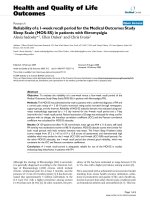
Metastatic NB growth in the liverFigure 4
Metastatic NB growth in the liver. Liver metastases
were smaller and exhibited large necrotic areas after 10 days
of CHS 828 treatment (B) compared to controls (A). "C"
and "D" are magnifications of "A" and "B", respectively. In C
and D the border between healthy liver tissue and either via-
ble tumor tissue (densely packed nuclei with sparse cyto-
plasm) (C) or areas of tumor necrosis (D) is outlined.
Hematoxylin-eosin staining; bars = 20 μm.
Table 3: Staging of orthotopic NB in SCID mice
Orthotopic mouse model
Control
at 10 days
CHS 828
at 10 days
CHS 828
at 30 days
INSS stage 1 0% (0/9) 46% (6/13) 60% (6/10)
stage 2
a
0% (0/9) 8% (1/13) 0% (0/10)
stage 3 0% (0/9) 0% (0/13) 0% (0/10)
stage 4
b
100% (9/9) 46% (6/13) 40% (4/10)
Postmortem classification of control and CHS 828-treated animals
using the INSS criteria for staging [27].
a
No extensive lymph node investigation was performed; therefore,
"stage 2" was not divided into "2A" and "2B"
b
INSS stage 4 was not separated into stages 4 and 4S since 4S is for
infants
INSS (International Neuroblastoma Staging System) according to
Simpson and Gaze [27] at autopsy
Journal of Translational Medicine 2009, 7:16 />Page 8 of 11
(page number not for citation purposes)
Immunohistological studies of tumor sections showed
morphological signs of cell death, i.e. condensed and frag-
mented nuclei, after CHS 828 treatment for 10 days, caus-
ing a reduction of the viable tumor fraction by more than
a factor of 5.7 compared to controls. The decrease in via-
ble tissue fraction was independent of activated caspases-
3. This observation is supported by studies reporting that
CHS 828 induces late programmed cell death with fea-
tures not related to classical apoptosis [33,34]. In fact,
CHS 828 has been reported to inhibit cellular synthesis of
NAD resulting in energy depletion, and subsequent cell
death [12]. NAD is produced primarily through biochem-
ical salvage pathway using nicotinamide as a substrate.
CHS 828 inhibits NAD synthesis from nicotinamide only
after continuous and long time exposure [12]. Delayed
cell death was confirmed in our in vitro studies in which
the viability of human NB cells (IMR-32, SK-N-SH and
SH-SY5Y) was affected only after prolonged exposure to
CHS 828.
CHS 828 caused cell death in all three NB cell lines in vitro
with IC
50
values 20 × below values of endothelial cells.
Human fibroblasts never reached IC
50
values at concentra-
tions tested (0.1 nM – 10 μM).
Compared to results from Åleskog et al. who tested CHS
828 toxicity on human lymphocytes in the same FMCA
protocol described here, NB cells in our study had lower
IC
50
values [35]. This indicates a higher drug sensitivity of
NB cells. We speculate that the high CHS 828 sensitivity
of the NB cell lines might be due to an active uptake of
CHS 828 in NB cells, mediated by the noradrenalin trans-
port transmembrane protein in analogy with MIBG [36].
It has been shown that the human NB cells used in this
study are so-called MIBG-positive cell lines (a characteris-
tic shared with 85% of NB cells in patients) in which there
is an apparent noradrenalin transporter gene expression
[37,38]. MIBG is a molecule that is specifically taken up
by most NB cells [39] and cytotoxic drugs with structural
homology to MIBG (e.g. CHS 828) may have a similar
selectively for NB cells. To address the question whether
CHS 828 was less active in cell lines with greater avidity
for MIBG, we included the NB cell line SK-N-SH in our in
vitro toxicity studies. CHS caused cell death in all NB cell
lines without any correlation to their avidity in taking up
MIBG. We therefore conclude that CHS 828 could be
taken up by different NB cells despite presence of chlo-
rophenoxyhexyl and cyano groups in the chemical struc-
ture of this drug.
As rodents have been shown to tolerate higher CHS 828
levels than man both in vitro [40] and in vivo [41], the dose
chosen in the current study can be considered low for the
host cells (including the endothelial cells) but higher for
the human tumor cells. Despite this, both tumor vessels of
murine origin and human tumor cells were affected by
treatment with CHS 828. Thus, we believe that the current
administration of CHS 828 represents a dual targeting
approach involving the inhibition of angiogenesis, and
direct tumor cell toxicity. The two processes (angiogenesis
inhibition and tumor cell toxicity) may have different
kinetics and may vary in proportion with the distance
from the nearest vessel. Furthermore, treated animals
showed less intra-tumor hemorrhage than controls.
Therefore, the vasculature in tumors treated with CHS 828
was more stable than vessels in rapidly growing, untreated
tumors indicating vessel normalization [42].
More prolonged schedules of CHS 828 have previously
been shown to increase antitumor activity as well as toxic-
ity in vitro [13], in vivo [41] and clinically [18,19]. In the
current study, no bone marrow toxicity due to prolonged
exposure to low doses of CHS 828 was found. This was
investigated by quantifying the percentage of megakaryo-
cytes in the bone marrow of the iliac crest. Megakaryo-
cytes, the precursors of platelets, were easily identified
despite the disorderly arranged cells in the bone marrow.
We found that the frequency of megakaryocytes in the
bone marrow was not affected by CHS 828 treatment.
In clinical phase I studies, CHS 828 showed a large varia-
tion in drug uptake both between and within patients
Table 4: Quantification of tumor angiogenesis by stereology
vessel density
(mm
-2
)
Lv
(mm
-2
)
Vv
(10
-3
)
Sv
(mm
-1
)
control (n = 9) 39.9 ± 18.5 77.2 ± 37.1 5.1 ± 2.5 2.6 ± 1.3
CHS 828
a
(n = 13) 12.8 ± 10.3 25.6 ± 20.5 2.8 ± 2.1 1.0 ± 0.7
Change
b
(%) -67.1% ** -67.1% ** -44.7% -62.7% *
CHS 828 was administered at 20 mg/kg/day by oral gavage.
Lv, length of vessels per tumor volume (length density); Vv, volume of vessels per tumor volume (volumetric density); Sv, surface area of vessels per
tumor volume (surface density). Mean ± 1SD, Mann-Whitney U test.
a
CHS 828 treatment for 10 days
b
Change compared to control
*p < 0.05 **p < 0.01
Journal of Translational Medicine 2009, 7:16 />Page 9 of 11
(page number not for citation purposes)
[18,19] which was also observed in a previous study in
nude mice [17]. This inter-individual variability has partly
been explained by variations of hepatic and intestinal
CYP3A4 activity, an enzyme important for metabolizing
cyanoguanidines such as CHS 828 [19,43]. Another expla-
nation for this variability might be related to the low sol-
ubility of CHS 828 hampering uptake in the
gastrointestinal tract. Clinical trials using orally adminis-
tered CHS 828 were discontinued due to the variation in
exposure levels and dose limiting toxicities. Hence a water
soluble prodrug, EB1627 (GMX1777) was synthesized by
adding a tetraethylenglycol moiety to the parent drug CHS
828 (GMX1778). This compound could be administered
i.v. thus allowing a controlled dosing to the patient. After
intravenous administration, the tetraethylenglycol moiety
rapidly dissociates and releases CHS 828 without reduc-
ing antitumor activity [44].
Conclusion
We believe that the metastatic and clinically relevant
model evaluated here provides an excellent tool for exam-
ining new treatment strategies in children with high-risk
NB. Based on data derived from this model, we suggest
that the active compound CHS 828 might provide clinical
benefits in treating children with high-risk NB.
Competing interests
The authors declare that they have no competing interests.
Authors' contributions
DF, RC and FA designed the study. DF acquired data
which was analyzed by DF and FA, except for CgA data
which was analyzed by MS. RC contributed with data
interpretation and drafting of the manuscript written by
DF and FA. EL and MS provided input in writing of the
manuscript. All authors read and approved the manu-
script.
Additional material
Acknowledgements
Barbro Einarsson provided excellent technical assistance. CHS 828 was
kindly provided by LEO Pharma (Ballerup, Denmark). This work was sup-
ported by a grant from the Children's Cancer Foundation of Sweden and
the Gillbergska Foundation.
References
1. Cotterill SJ, Pearson AD, Pritchard J, Foot AB, Roald B, Kohler JA,
Imeson J: Clinical prognostic factors in 1277 patients with neu-
roblastoma: results of The European Neuroblastoma Study
Group 'Survey' 1982–1992. Eur J Cancer 2000, 36:901-908.
Additional File 1
Observation parameters of tumor-bearing SCID mice during the
experiment. A table summarizing individual follow-up of body weight and
tumor development for each individual mouse in the study. Statistical
analysis (Mann-Whitney U test) indicates group differences in tumor vol-
ume, tumor weight and tumor index (tumor weight/final body weight ×
100).
Click here for file
[ />5876-7-16-S1.doc]
Additional File 2
Organ weight of healthy and tumor-bearing SCID mice. A table sum-
marizing organ weight for each individual mouse in the study, including
healthy littermates. Statistical analysis (Kruskal Wallis test).
Click here for file
[ />5876-7-16-S2.doc]
Representative morphology and vessel profile in orthotopic NB xenograftsFigure 5
Representative morphology and vessel profile in
orthotopic NB xenografts. Vehicle-treated tumors (con-
trol) contained a larger number of small vessels (stained in
brown) (A, C) compared to CHS 828 (20 mg/kg/day; p.o.)
treated tumors (B, D-E) already 10 days after randomiza-
tion. Vessels of control tumors had a thin endothelial cell lin-
ing (brown) (A, C). CHS 828 treated tumors revealed
vessels only partly surrounded by endothelial cells (arrow-
heads) (B, D-E) or endothelial cells detaching from the base-
ment membrane (arrows) (E). C and D are magnifications of
A and B, respectively. Bandeiraea simplicifolia-1 (BS-1) lectin
staining (brown); bar = 40 μm.
Journal of Translational Medicine 2009, 7:16 />Page 10 of 11
(page number not for citation purposes)
2. De Bernardi B, Nicolas B, Boni L, Indolfi P, Carli M, Cordero Di Mon-
tezemolo L, Donfrancesco A, Pession A, Provenzi M, di Cataldo A, et
al.: Disseminated neuroblastoma in children older than one
year at diagnosis: comparable results with three consecutive
high-dose protocols adopted by the Italian Co-Operative
Group for Neuroblastoma. J Clin Oncol 2003, 21:1592-1601.
3. Meitar D, Crawford SE, Rademaker AW, Cohn SL: Tumor angio-
genesis correlates with metastatic disease, N-myc amplifica-
tion, and poor outcome in human neuroblastoma. J Clin Oncol
1996, 14:405-414.
4. Peddinti R, Zeine R, Luca D, Seshadri R, Chlenski A, Cole K, Pawel B,
Salwen HR, Maris JM, Cohn SL: Prominent microvascular prolif-
eration in clinically aggressive neuroblastoma. Clin Cancer Res
2007, 13:3499-3506.
5. Backman U, Svensson A, Christofferson R: Importance of vascular
endothelial growth factor A in the progression of experi-
mental neuroblastoma. Angiogenesis 2002, 5:267-274.
6. Segerstrom L, Fuchs D, Backman U, Holmquist K, Christofferson R,
Azarbayjani F: The Anti-VEGF Antibody Bevacizumab
Potently Reduces the Growth Rate of High-Risk Neuroblas-
toma Xenografts. Pediatr Res 2006, 60:576-581.
7. Kerbel RS: Antiangiogenic therapy: a universal chemosensiti-
zation strategy for cancer? Science 2006, 312:1171-1175.
8. Bertolini F, Paul S, Mancuso P, Monestiroli S, Gobbi A, Shaked Y, Ker-
bel RS: Maximum tolerable dose and low-dose metronomic
chemotherapy have opposite effects on the mobilization and
viability of circulating endothelial progenitor cells. Cancer Res
2003, 63:4342-4346.
9. Pietras K, Hanahan D: A multitargeted, metronomic, and max-
imum-tolerated dose "chemo-switch" regimen is antiang-
iogenic, producing objective responses and survival benefit
in a mouse model of cancer. J Clin Oncol 2005, 23:939-952.
10. Klement G, Baruchel S, Rak J, Man S, Clark K, Hicklin DJ, Bohlen P,
Kerbel RS: Continuous low-dose therapy with vinblastine and
VEGF receptor-2 antibody induces sustained tumor regres-
sion without overt toxicity. J Clin Invest 2000,
105:R15-24.
11. Kerbel RS, Kamen BA: The anti-angiogenic basis of metro-
nomic chemotherapy. Nat Rev Cancer 2004, 4:423-436.
12. Olesen UH, Christensen MK, Bjorkling F, Jaattela M, Jensen PB,
Sehested M, Nielsen SJ: Anticancer agent CHS-828 inhibits cel-
lular synthesis of NAD. Biochem Biophys Res Commun 2008,
367:799-804.
13. Hassan SB, Jonsson E, Larsson R, Karlsson MO: Model for time
dependency of cytotoxic effect of CHS 828 in vitro suggests
two different mechanisms of action. J Pharmacol Exp Ther 2001,
299:1140-1147.
14. Johanson V, Arvidsson Y, Kolby L, Bernhardt P, Sward C, Nilsson O,
Ahlman H: Antitumoural effects of the pyridyl cyanoguanidine
CHS 828 on three different types of neuroendocrine
tumours xenografted to nude mice. Neuroendocrinology 2005,
82:171-176.
15. Hovstadius P, Lindhagen E, Hassan S, Nilsson K, Jernberg-Wiklund H,
Nygren P, Binderup L, Larsson R: Cytotoxic effect in vivo and in
vitro of CHS 828 on human myeloma cell lines. Anticancer
Drugs 2004, 15:63-70.
16. Hjarnaa PJ, Jonsson E, Latini S, Dhar S, Larsson R, Bramm E, Skov T,
Binderup L: CHS 828, a novel pyridyl cyanoguanidine with
potent antitumor activity in vitro and in vivo. Cancer Res 1999,
59:5751-5757.
17. Svensson A, Backman U, Jonsson E, Larsson R, Christofferson R:
CHS 828 inhibits neuroblastoma growth in mice alone and in
combination with antiangiogenic drugs. Pediatr Res 2002,
51:607-611.
18. Ravaud A, Cerny T, Terret C, Wanders J, Bui BN, Hess D, Droz JP,
Fumoleau P, Twelves C: Phase I study and pharmacokinetic of
CHS-828, a guanidino-containing compound, administered
orally as a single dose every 3 weeks in solid tumours: An
ECSG/EORTC study. Eur J Cancer 2005, 41:702-707.
19. Hovstadius P, Larsson R, Jonsson E, Skov T, Kissmeyer AM, Krasiln-
ikoff K, Bergh J, Karlsson MO, Lonnebo A, Ahlgren J: A Phase I
study of CHS 828 in patients with solid tumor malignancy.
Clin Cancer Res 2002, 8:2843-2850.
20. Zaizen Y, Taniguchi S, Suita S: The role of cellular motility in the
invasion of human neuroblastoma cells with or without N-
myc amplification and expression.
J Pediatr Surg 1998,
33:1765-1770.
21. Biedler JL, Roffler-Tarlov S, Schachner M, Freedman LS: Multiple
neurotransmitter synthesis by human neuroblastoma cell
lines and clones. Cancer Res 1978, 38:3751-3757.
22. Veitonmaki N, Fuxe J, Hultdin M, Roos G, Pettersson RF, Cao Y:
Immortalization of bovine capillary endothelial cells by
hTERT alone involves inactivation of endogenous
p16INK4A/pRb. Faseb J 2003, 17:764-766.
23. Larsson R, Nygren P, Ekberg M, Slater L: Chemotherapeutic drug
sensitivity testing of human leukemia cells in vitro using a
semiautomated fluorometric assay. Leukemia 1990, 4:567-571.
24. Welsh N: Assessment of apoptosis and necrosis in isolated
islets of Langerhans: methological considerations. Curr Top
Biochem Res 2000, 3:189-200.
25. Backman U, Christofferson R: The selective class III/V receptor
tyrosine kinase inhibitor SU11657 inhibits tumor growth and
angiogenesis in experimental neuroblastomas grown in
mice. Pediatr Res 2005, 57:690-695.
26. Wassberg E, Hedborg F, Skoldenberg E, Stridsberg M, Christofferson
R: Inhibition of angiogenesis induces chromaffin differentia-
tion and apoptosis in neuroblastoma. Am J Pathol 1999,
154:395-403.
27. Simpson JK, Gaze MN: Current Management of Neuroblast-
oma. Oncologist 1998, 3:253-262.
28. DuBois SG, Kalika Y, Lukens JN, Brodeur GM, Seeger RC, Atkinson
JB, Haase GM, Black CT, Perez C, Shimada H, et al.: Metastatic sites
in stage IV and IVS neuroblastoma correlate with age, tumor
biology, and survival. J Pediatr Hematol Oncol 1999, 21:181-189.
29. Hendy GN, Bevan S, Mattei MG, Mouland AJ: Chromogranin A.
Clin Invest Med 1995, 18:47-65.
30. Hsiao RJ, Seeger RC, Yu AL, O'Connor DT: Chromogranin A in
children with neuroblastoma. Serum concentration parallels
disease stage and predicts survival.
J Clin Invest 1990,
85:1555-1559.
31. Wassberg E, Stridsberg M, Christofferson R: Plasma levels of
chromogranin A are directly proportional to tumour burden
in neuroblastoma. J Endocrinol 1996, 151:225-230.
32. Seregni E, Ferrari L, Bajetta E, Martinetti A, Bombardieri E: Clinical
significance of blood chromogranin A measurement in neu-
roendocrine tumours. Ann Oncol 2001, 12(Suppl 2):S69-72.
33. Frost BM, Lonnerholm G, Nygren P, Larsson R, Lindhagen E: In vitro
activity of the novel cytotoxic agent CHS 828 in childhood
acute leukemia. Anticancer Drugs 2002, 13:735-742.
34. Martinsson P, Liminga G, Dhar S, de la Torre M, Lukinius A, Jonsson
E, Bashir Hassan S, Binderup L, Kristensen J, Larsson R: Temporal
effects of the novel antitumour pyridyl cyanoguanidine (CHS
828) on human lymphoma cells. Eur J Cancer 2001, 37:260-267.
35. Aleskog A, Bashir-Hassan S, Hovstadius P, Kristensen J, Hoglund M,
Tholander B, Binderup L, Larsson R, Jonsson E: Activity of CHS 828
in primary cultures of human hematological and solid
tumors in vitro. Anticancer Drugs 2001, 12:821-827.
36. Montaldo PG, Lanciotti M, Casalaro A, Cornaglia-Ferraris P, Ponzoni
M: Accumulation of m-iodobenzylguanidine by neuroblast-
oma cells results from independent uptake and storage
mechanisms. Cancer Res 1991, 51:4342-4346.
37. Boyd M, Cunningham SH, Brown MM, Mairs RJ, Wheldon TE:
Noradrenaline transporter gene transfer for radiation cell
kill by 131I meta-iodobenzylguanidine. Gene Ther 1999,
6:1147-1152.
38. Lode HN, Bruchelt G, Seitz G, Gebhardt S, Gekeler V, Niethammer
D, Beck J: Reverse transcriptase-polymerase chain reaction
(RT-PCR) analysis of monoamine transporters in neuroblas-
toma cell lines: correlations to meta-iodobenzylguanidine
(MIBG) uptake and tyrosine hydroxylase gene expression.
Eur J Cancer 1995, 31A:586-590.
39. Kushner BH: Neuroblastoma: a disease requiring a multitude
of imaging studies. J Nucl Med 2004, 45:1172-1188.
40. Lindhagen E, Hjarnaa PJ, Friberg LE, Latini S, Larsson R: Pharmaco-
dynamic differences between species exemplified by the
novel anticancer agent CHS 828.
Drug Dev Res 2004,
61:218-226.
41. Friberg LE, Hassan SB, Lindhagen E, Larsson R, Karlsson MO: Phar-
macokinetic-pharmacodynamic modelling of the schedule-
dependent effect of the anti-cancer agent CHS 828 in a rat
hollow fibre model. Eur J Pharm Sci 2005, 25:163-173.
42. Jain RK: Normalization of tumor vasculature: an emerging
concept in antiangiogenic therapy. Science 2005, 307:58-62.
Publish with Bio Med Central and every
scientist can read your work free of charge
"BioMed Central will be the most significant development for
disseminating the results of biomedical research in our lifetime."
Sir Paul Nurse, Cancer Research UK
Your research papers will be:
available free of charge to the entire biomedical community
peer reviewed and published immediately upon acceptance
cited in PubMed and archived on PubMed Central
yours — you keep the copyright
Submit your manuscript here:
/>BioMedcentral
Journal of Translational Medicine 2009, 7:16 />Page 11 of 11
(page number not for citation purposes)
43. Lown KS, Kolars JC, Thummel KE, Barnett JL, Kunze KL, Wrighton
SA, Watkins PB: Interpatient heterogeneity in expression of
CYP3A4 and CYP3A5 in small bowel. Lack of prediction by
the erythromycin breath test. Drug Metab Dispos 1994,
22:947-955.
44. Binderup E, Bjorkling F, Hjarnaa PV, Latini S, Baltzer B, Carlsen M,
Binderup L: EB1627: a soluble prodrug of the potent antican-
cer cyanoguanidine CHS828. Bioorg Med Chem Lett 2005,
15:2491-2494.