Báo cáo hóa học: " Human saliva, plasma and breast milk exosomes contain RNA: uptake by macrophages" pdf
Bạn đang xem bản rút gọn của tài liệu. Xem và tải ngay bản đầy đủ của tài liệu tại đây (1.26 MB, 8 trang )
RESEARC H Open Access
Human saliva, plasma and breast milk exosomes
contain RNA: uptake by macrophages
Cecilia Lässer
1
, Vesta Seyed Alikhani
1
, Karin Ekström
1
, Maria Eldh
1
, Patricia Torregrosa Paredes
2
, Apostolos Bossios
1
,
Margareta Sjöstrand
1
, Susanne Gabrielsson
2
, Jan Lötvall
1*
, Hadi Valadi
3
Abstract
Background: Exosomes are 30-100 nm membrane vesicles of endocytic origin produced by numerous cells. They
can mediate diverse biological functions, including antigen presentation. Exosomes have recently been shown to
contain functional RNA, which can be delivered to other cells. Exosomes may thus mediate biological functions
either by surface-to-surface interactions with cells, or by the delivery of functional RNA to cells. Our aim was
therefore to determine the presence of RNA in exosomes from human saliva, plasma and breast milk and whether
these exo somes can be taken up by macrophages.
Method: Exosomes were purified from human saliva, plasma and breast milk using ultracentrifugation and filtration
steps. Exosomes were detected by electron microscopy and examined by flow cytometry. Flow cytometry was
performed by capturing the exosomes on anti-MHC class II coated beads, and further stain with anti-CD9, anti-
CD63 or anti-CD81. Breast milk exosomes were further analysed for the presence of Hsc70, CD81 and calnexin by
Western blot. Total RNA was detected with a Bioanalyzer and mRNA was identified by the synthesis of cDNA using
an oligo (dT) primer and analysed with a Bioanalyzer. The uptake of PKH67-labelled saliva and breast milk
exosomes by macrophages was examined by measuring fluorescence using flow cytometry and fluorescence
microscopy.
Results: RNA was detected in exosomes from all three body fluids. A portion of the detected RNA in plasma
exosomes was characterised as mRNA. Our result extends the characterisation of exosomes in heal thy humans and
confirms the presence of RNA in human saliva and plasma exosomes and reports for the first time the presence of
RNA in breast milk exosomes. Our results also show that the saliva and breast milk exosomes can be taken up by
human macrophages.
Conclusions: Exosomes in saliva, plasma and breast milk all contain RNA, confirming previous findings that
exosomes from several sources contain RNA. Furthermore, exosomes are readily taken up by macrophages,
supporting the notion that exosomal RNA can be shuttled between cells.
Background
Exosomes are small membrane vesicles (30-100 nm) of
endocytic origin that are released from the producing
cell into the extracellular environment [1]. Many cells in
thebodyhavethecapacitytoproduceandreleaseexo-
somes to their surrounding environment, including den-
dritic cells, B cells, T cells, mast cells, tumour cells and
epithelial cells [2-7]. Exosomes are also present in
body fluids including plasma, urine, saliva, malignant
effusions, synovial fluid, breast milk, bronchoalveolar
lavage fluid and epididymal fluid [8-15] indicating
importance in vivo. Until now, exosomes have been
implicated primarily in antigen presentation, as they
often e xpress several proteins involved in cell adhesion
and co-stimulation including ICAM-1, CD86, CD63 and
CD82, MHC class I and MHC class II [1]. These immu-
nological functions have led to the development of
anti-tumour vaccines based on exosomes, which are
currently in early clinical development [16,17].
Exosomes have b een proposed to signal by both the
bindi ng to cell surface receptors throu gh adhesion mole-
cules [3] and by fusion with or internalisation by the
* Correspondence:
1
Krefting Research Centre, Sahlgrenska Academy, University of Gothenburg,
Box 424, 405 30 Gothenburg, Sweden
Full list of author information is available at the end of the article
Lässer et al. Journal of Translational Medicine 2011, 9:9
/>© 2011 Lässer et al; licensee BioMed Central Ltd . This is an Open Access article distributed under the terms of the Creative Commons
Attribu tion License ( which permits unrestricted use, distribution, and re production in
any medium, provided the original work is p roperly cited.
recipient cell, potentially donating their own cytoplasm to
the recipient cell [18,19]. The latter implies that exosomes
may have mechanisms that are different to their function
in the immune system. We have recently discovered sub-
stantial amounts of RNA in exosomes deri ved from mast
cells [20], which have the capacity to donate their RNA to
other cells and can subsequently affect the protein produc-
tion of a recipient cell. This argues that RNA can be trans-
ferred between mammalian cells by an extracellular
exosome based transport mechanism, which has vast
implications in the understanding of cell communication,
regulation and signalling, in addition to extensive thera-
peutic potential in many diseases. Therefore, studies to
determine the presence of RNA in exo somes harvest ed
from humans in vivo are of high priority.
As human plasma, saliva and breast milk all contain
exosomes [8,12,15], the aims of the current study were
to determine whether these exosomes contain RNA and
whether they can be taken up by other cells, which
would support the concept that shuttling of RNA may
occur in humans.
Methods
Exosome purification from saliva
Saliva from healthy humans was collected in Falcon tubes
on ice, during a period of no eating or drinking and pooled
together. For the RNA isolation experiment, 100 μl of the
protease inhibitor Complete Mini ( Roche D iagnostics
Scandinavia AB, Bromma, Sweden) and 800 units of
RNase inhibit or R iboLock R ibonuclease Inhibitor (Fer-
mentas, St. Leon-Rot, Germany) were added per 20 ml of
saliva. For the flow cytometry, electron microscopy and
uptake experiments no inhibitors were added to the tubes.
The saliva was diluted 1:1 with phosphate buffered saline
(PBS) and centrifuged at 16 500 × g for 20 min to remove
cells and debris. The supernatant was filtered through a
0.2 μm VWR
®
Vacuum Filtration System (VWR Interna-
tional, West Chester, PA, USA), before ultracentrifugation
(Ti70 or Ti45 rotor, Beckman Coulter, Brea, CA, USA) at
120 000 × g for 7 0 min to pe llet the exosomes.
Exosome purification from blood plasma
A volume of 450-500 ml of blood was collected from
donors. Plasma was derived from heparinised blood by
centrifugation at 1 800 × g for 10 min. Further centrifu-
gation at 29 500 × g for 20 min was performed to pellet
any remaining cells and debris. The superna tant was
then filtered through a 0.2 μm VWR
®
Vacuum Filtration
System, followed by ultracentrifugation at 120 000 × g for
90 min to pellet the exosomes.
Exosome purification from breast milk
Human breast milk was collected from healthy mothers,
immediately stored at -20ºC and later transferred to the
laboratory and stored at -80 ºC. To remove cells and
debris, t he breast milk was first centrifuged at 300 × g
for 10 min, followed by centrifugation at 16 500 × g for
20 min. The supernatant was then filtered through a
0.2 μmVWR
®
Vacuum Filtration System, followed by
ultracentrifugation at 120 000 × g for 70 min to pellet
the exosomes.
Electron microscopy
Exosomes from saliva, plasma and breast milk were
isolated as described above, washed in PBS to further
purify the sample, filtered, and ultracentrifuged again at
120 000 × g for 70 min to re-pellet the exosomes. T he
exosome pellet was resuspended in PBS an d loaded onto
formvar carbon coated grids (Ted Pella Inc, Redding,
USA). Next, the exosomes were fixed in 2% paraformalde-
hyde and was hed. The e xosomes w ere immunostained
with anti-CD63 antibody (BD Bioscience, E rembodegem,
Belgium) or isotype control (Sigma-Aldrich, St Louis, MO,
USA), followed by staining with a 10 nm gold-labelled
secondary antibody (Sigma-Aldrich). The exosomes were
subsequently fixed in 2.5% glutaraldehyde, washed, con-
trasted in 2% uranyl acetate and embedded in a mixture
of uranyl ace tate (0.8%) and methyl cellulose (0.13%).
The preparations were examined in a LEO 912AB
Omega electron microscope (Carl Zeiss NTS, Jena,
Germany).
Flow cytometry of exosomes
Isolated saliva, plasma or breast milk exosomes were
resuspended in PBS and loaded onto anti-MHC class II
coated beads (custom-made by Dynal, part of Invitrogen
Ltd, Paisley, UK). The anti-MHC class II coated beads
(8 × 10
4
) were mixed with a minimum of 50 μgofexo-
somal pro tein, before being incubated overnight at 4ºC
with gentle agitation. The bead-exo some complexes
were washed twice in PBS with 3% Fetal Bovine Serum
(FBS). Prior to use, the FBS was ultracentrifuged at
120 000 × g for 1.5 hours, to eliminate serum exosomes.
The bead-exosom e complexes were resuspended in Ig G
(Sigma-Aldrich) and incubated for 15 min at room tem-
perature, before being washed twice more, as above.
The tetraspanins CD9, CD63 and CD81, known to be
enriched in exosomes, were used as markers for exo-
somes. The bead-exosome complexes were incubated
with PE-labelled anti-CD9 (clone M-L13), anti-CD63
(clone H5C6), anti-CD81 (clone JS-81) or the c orre-
sponding isotype control (all antibodies were from BD
Biosciences) for 40 min at room temperature with agita-
tion and washed t hree times before analysis. As a con-
trol for unspecific binding of the antibodies to the
beads, beads were stained with all three antibodies with-
out the addition of exosomes and showed no difference
when compared to exosome coated beads stained with
Lässer et al. Journal of Translational Medicine 2011, 9:9
/>Page 2 of 8
the isotype control. The samples were then acquired in
a FACScan or FACSAria (BD Biosciences) and analysed
using the FlowJo Software (Tri St ar Inc , Ashlan d,
OR, USA).
Western blot analysis of breast milk exosomal proteins
Isolated breast milk exosomes were re-suspended in PBS
and ultracentrifuged at 120 000 × g for 70 min to be re-
pelleted befo re dissolved in ProteoJET Mammalian C ell
Lysis Reagent (Fermentas). For extraction of total pro-
tein, the sample was incubat ed at room tempe rature for
10 min on a shaker, sonicated for 5 min and vortexed,
before being centrifuged at 13 000 × g for 10 min. The
protein content of the supernatant was measured with a
spectrophotometer at 750 nm utilising the D
c
Protein
Assay reagent A and B (Bio-Rad Laboratories, Hercules,
CA, USA). 100 μg proteins from the supernatant were
loaded per well onto a 10% acrylamide gel. Monocyte
derived macrophages from buffy coat were used as a
control. The proteins were blotted onto a nitrocellulo se
membrane (Bio-Rad Laboratories) overnight at 4°C. The
membrane was blocked with 0.5% Blotting Grade
Blocker Non-Fat Dry Milk (Bio-Rad Laboratories) in
TBS for 2 h, before washed 3 × 5 min in TBS-Tween
(used for all the washes throughout the Western blot
experiment). The membrane was then incubated with
either anti-calnexin (1:1000) (Santa Cruz Biotechnology,
Santa Cruz, CA, USA), anti-Hsc70 (1:1000) (Enzo Life
Science, Farmingdale, NY, USA) or anti-CD81 (1:800)
(Santa Cruz) diluted in 0.25% non-fat dry milk in TBS-
Tween for 2 h. The membrane was washed 3 × 5 min
before incubated with the secondary antibody for 2 h.
The secondary antibodies used were goat F(ab)
2
anti-
rabbi t IgG (HRP conjugated ) for the calnexin and CD81
(1:5000 ) (Harlan Sera-Lab, Loughborough, UK) and rab-
bit F(ab)
2
anti-Rat IgG (HRP conjugated) for the Hsc70
(1:4000) (Southern Biotech, Birmingham, A L, USA)
diluted in 0.25% non- fat dry milk powder in TBS-
Tween. The membrane was washed 3 × 5 min, before
being analysed with the Amersham™ ECL Plus™
Western Blotting Detection System (GE Healthcare,
Uppsala, Sweden) and a VersaDoc 4000 MP (Bio-Rad
Laboratories).
RNA isolation and detection
RNA was isolated using Trizol
®
(Invitrogen) according
to the manufacturer’s protocol and dissolved in DEPC
H
2
O (Fermentas). For detection of RNA, an Agilent
2100 Bioanalyzer (Agilent Technologies Sweden AB,
Kista, Sweden) was utilised for all samples. The exoso-
mal RNA was compared with cellular RNA from the
human mast cell line HMC-1. The HMC-1 cells (Dr J.
Butterfield, Mayo Clinic, Rochest er, MN, USA) were
cultured in a 37ºC humidified incubator wi th 5% CO
2
,
in complete medium consisting of Iscove ’s Modified
Dulbecco’ s Medium (IMDM) supplemented with 10%
FBS, 100 units/ml penici llin, 100 μg/ml streptomycin,
2 mM L-glutamine and 1.2 mM/ml alfa-t hiogl ycerol (all
reagents from Sigma-Aldrich).
For the detection of mRNA in exosomes, the total
RNA isolated was converted to cDNA using Rever-
tAid™ H Minus First Strand cDNA Synthesis Kit (Fer-
mentas) and the oligo (dT) primer. The second strand
of the cDNA was synthesised by adding 10 μlof10×
DNA polymerase 1 reaction buffer, 4 μlofDNApoly-
merase 1, 5 μlofT4DNAligaseand61μlofDEPC
water (all reagents were from Fermentas) to the first
strand of cDNA product. The sample was incubated at
14ºC for 2 h before the reaction was stopped by incuba-
tion at 70ºC for 10 min. The detection of cDNA was
performed using a Bioanalyzer.
Exosome staining
Saliva and breast milk exosomes were isolated as
described abov e, and furth er pur ified by being disso lved
in PBS and ultracentrifuged at 120 000 × g for 70 min.
The exosomes were labelled with PKH67 Green Fluores-
cent C ell Linker Kit for General Cell Membrane Label-
ling (Sigma-Aldrich) according to the manufacturer’s
protocol, with minor modifications in the washing pro-
cess. Briefly, the exosomes were diluted in PBS before
1 ml of Diluent C was added. As a control, 1 ml of
Diluent C with the same volume of PBS was used. 4 μl
of PKH67 dye was added to 1 ml of Diluent C befo re
being added to the exosomes and the control. The sam-
ples were mixed gently for 4 min before 2 ml of 1%
BSA was added to bind the excess dye. The samples
were then transferred to 300 kDa Vivaspin filters (Sar-
torius Stedim Biotech GmbH, Goettingen, Germany)
and centrifuged at 4000 × g. The sample were washed
3 times with 5 ml of PBS before being transferred to
new 300 kDa Vivaspin filters and washed twice with
5 ml IMDM (Sigma-Aldrich).
Uptake of saliva and breast milk exosomes by
macrophages
Peripheral mononuclear cells (PBMCs) were isolated
from buffy coat using Leucosep
®
Tubes (Greiner Bio-
One GmbH, Frickenhausen, Germany), according to the
manufacturer’ s protocol. The PBMCs were washed
repeatedly with 2 mM EDTA in PBS, b efore being dis-
solved in 0.5% BS A and 2 mM EDTA in PBS. Mono-
cytes were isolated from PBMCs using a Monocyte
Isolation Kit II (Miltenyi Biotec Gmbh, Bergisch Glag-
bach, Germany) according to the manufacturer’sproto-
col. The purity of the monocytes was determined with a
FACSAria by the detection of CD14 (clone MFP9,
BD Biosciences). To allow for differentiation into
Lässer et al. Journal of Translational Medicine 2011, 9:9
/>Page 3 of 8
macrophages, the monocytes were cultured for 7 days in
a 37ºC humidified incubator with 5% CO
2
, in complete
medium consisting o f IMDM supplemented with 10%
FBS, 100 units/ml penici llin, 100 μg/ml streptomycin,
2 mM L-glutamine, 110 μg/ml sodium pyruvate
(all reagents were from Sigma-Aldrich) and 10 ng/ml
GM-CSF (R&D Systems, Minneapolis, MN, USA). The
FBS was ultracentrifuged prior to use t o eliminate
serum e xosomes. For analysis with flow cytometry cells
were cultured in 96-well plates and for fluorescence
microscopy, the cells were cultured in 8-well Perma nox
Slides (Thermo Fisher Scientific, New York, USA).
10 μgofthePKH67labelledexosomesorthesame
volume of the PKH67-PBS control was a dded p er
200 000 macrophages and incubated for 2 h at either
37ºC or 4ºC. The binding of the exosomes to the macro-
phages was analysed with a FACSAria and visualised
with fluorescence microscope (Zeiss Axioplan 2, Carl
Zeiss, Jena, Germany). For analysis with flow cytometry
the cells were washed twice with PBS, treated with a
0.25% trypsin-EDTA solution (Sigma-Aldrich) and
washed twice with 1% FBS in PBS before acquired in
FACSAria and analysed with FlowJo software. For fluor-
escence microscopy, the cells were wa shed twice with
PBS, fixed with 4% formaldehyde for 15 min and washed
twice with PBS before being mounted with Vectashield
(Vector Laboratories Inc., Burlingame, USA) with 3%
7-ADD (BD Biosciences) to label nuclei.
Results
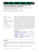
Human saliva, plasma and breast milk contain exosomes
Exosomes from saliva, plasma and breast milk were
identified using electron microscopy (Figure 1A-D) and
exosomes from all sources were positive for CD63,
using immunogold staining (Figure 1B-D). Furthermore,
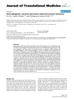
flow cytometry of saliva, plasma and breast milk exo-
somes captured on anti -MHC class II coat ed beads
rev ealed the presence of CD9, CD63 and CD81 on exo-
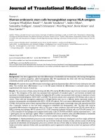
somes from all three sources (Figure 2). Breast milk exo-
somes were further characterised by Western blotting
and was shown to be positive for Hsc70 and CD81, but
negative for the endoplasmic reticulum marker calnexin
(Figure 3).
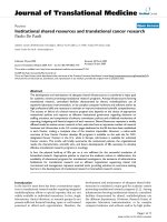
Human exosomes contain RNA
The RNA content of the saliva, plasma and breast milk
exosomes was analysed using a Bioanalyzer instrument,
which revealed that all three types of exosomes contain
RNA, with little or no ribosomal RNA (18S- and 28S-
rRNA) (Figure 4). The pattern of exosomal RNA visua-
lised in the Bioanalyzer differed substantially from
HMC-1 cell RNA, which contain substantial am ounts of
ribosomal RNA (Figure 4).
A)
C)
B)
D)
Figure 1 Exosomes from saliva, plasma and breast milk
detected with electron microscopy. Exosomes from human saliva
(A, B), plasma (C) and breast milk (D) were examined in the electron
microscope. No isotype control antibody (A), but anti-CD63
antibody (B-D), was detected by 10 nm gold labelled secondary
antibody. The scale bars represent 100 nm.
Saliva
exosomes
Plasma
exosomes
Breast milk
exosomes
10
2
10
0
10
1
10
3
CD9-PE
CD81-PE
CD63-PE
10
2
10
0
10
1
10
3
10
2
10
0
10
1
10
3
10
2
10
0
10
1
10
3
10
2
10
0
10
1
10
3
10
2
10
0
10
1
10
3
1
0
2
1
0
1
1
0
3
1
0
4
1
0
2
1
0
1
1
0
3
1
0
4
1
0
2
1
0
1
1
0
3
1
0
4
20
40
60
80
100
20
40
60
80
100
20
40
60
80
100
Events EventsEvents
Figure 2 Flow cytomet ry detection of surface molecules on
exosomes from saliva, plasma and breast milk. Exosomes from
saliva, plasma and breast milk captured on anti-MHC class II beads
were immunostained by using monoclonal antibodies against the
tetraspanins CD9, CD63 and CD81 and analysed by flow cytometry.
The antibodies (open peaks) were compared with their appropriate
isotype controls (filled peaks).
Lässer et al. Journal of Translational Medicine 2011, 9:9
/>Page 4 of 8
We also confirmed the presence of polyadenylated
RNA in exosomes from plasma, by synthesising cDNA
using an oligo (dT) primer (Figure 5). However, cDNA
could not be synthesised from exosomal RNA extracted
from saliva or breast milk, using the same method (data
not shown).
Human macrophages take up human saliva and breast
milk exosomes
To examine whether exosomes from human body fluids
can be taken up by recipient cells, human saliva and
breast milk exosomes were labelled with PKH67 dye
(green) and added to cultures of human macrophages,
derived f rom buffy coat monocytes (purity >94%). Flow
cytometry showed an uptake of the exos omes by macro-
phages, shown by an increase of mean fluore scence
intensity (MFI) for PKH67, compared with macrophages
cultured with the PBS control, or cultured with exo-
somes at 4˚C (Figure 6A-B). The uptake of the fluores-
cent exosomes by the macrophages was also visualised
using fluorescence microscopy (Figure 6C-D).
Discussion
This study confirms the presence of exosomes in human
saliva, plasma and breast milk, shown by b oth electron
microscopy and flow cytometry. We demonstrate that
exosomes from all three biological sources contain sig-
nificant amounts of primarily short RNA, of which a
portion is identified as mRN A in plasma exosomes. The
study also shows uptake of saliva and breast milk exo-
somes by macrophages.
The vesicles isolated from saliva, plasma and breast
milk, were shown by electron mic roscopy to have a
Hsc70
CD81
Calnexin
Exosome
s
Cell
s
Figure 3 Characterisation of breast milk exosomes by Western
blot. The exosomal proteins from breast milk exosomes were
loaded onto a 10% acrylamide gel and transferred to a
nitrocellulose membrane. The breast milk exosomes are positive for
Hsc70 and CD81, but negative for the endoplasmic reticulum
protein, calnexin. Macrophage protein ("Cells”) was used as positive
control.
25 35 45 55 65
Cellular RNA
Plasma exosomal RNA
25
20
15
10
5
0
60
40
20
0
20
15
10
5
0
Saliva exosomal RNA
[FU]
Breast milk exosomal RNA donor 1
Breast milk exosomal RNA donor 2
Breast milk exosomal RNA donor 3
Breast milk exosomal RNA donor 4
Breast milk exosomal RNA donor 5
Breast milk exosomal RNA donor 6
60
45
30
15
0
40
30
20
10
0
150
120
90
60
30
0
15
10
5
0
15
10
5
0
50
40
30
20
10
0
[sec]
25
35 45 55 65 25 35 45 55 65
Figure 4 Exosomal RNA analysed using a Bioanalyzer . Total RNA was isolated from saliva, plasma and breast mil k exosomes using Trizol
®
and analysed with a Bioanalyzer. The results show that exosomes from human saliva, plasma and breast milk contain a dissimilar RNA content
compared to cellular RNA from HMC-1 cells, as exosomes contain little or no ribosomal RNA.
Lässer et al. Journal of Translational Medicine 2011, 9:9
/>Page 5 of 8
diameter of 50-80 nm, which is comparable with pre-
viously identified exosomes [2-4]. Furthermore, immuno-
gold staining showed that the exosomes were positive for
the tetraspanin CD63, a commonly used exosome mar-
ker. Flow cyto metry analysis further indirectly showed
the p resence of MHC class II o n saliva, plasma and
breast milk derived vesicles, as well as the presence of
CD9, CD63 and CD81. While we acknowledge that
viruses below 200 nm may constitute a small fraction of
the exosome preparation, the EM analysis and detection
of multiple exosomal proteins strongly suggests that the
vesicles identified are exosomes and not other nano
particles.
The c urrent study confirms our original finding, t hat
exosomes contain RNA [20] by clarifying that exosomes
in different body fluids from healthy individuals also
contain RNA. It was recently reported that exosomes
from human plasma and saliva contain RNA [21-23],
which further supports this conclusion. This study
reports, for the first time, the presence of RNA in
human breast milk exosomes, which implies that exo-
somes could deliver RNA from cells of the mo ther, to
cells in the offspring.
Many compartments of the cell, besides the multivesicu-
lar bodies, can release vesicles. As the finding of RNA-
containing exosomes in breast milk is novel, we confirmed
that these were truly exosomes by showing the presence of
Hsc70 and CD81, and the absence of the endoplasmatic
reticulum protein, calnexin. As no calnexin was detected,
this indicates that there is little, or no, contamination by
endoplasmic reticulum-derived vesicles in the breast milk
derived exosomes. Furthermore, breast milk exosomes has
previously been shown to contain Hs c70 and CD81 [12],
the detection of these molecules by Western blot on the
breast milk derived exosomes isolated in this study served
to further confirm their ident ification as ex osomes. We
therefore c onclude that the RNA-containing vesicles
found in breast milk are exosomes. We also confirmed
our f inding by detecting RNA-containing exosomes in
breast milk from six different donors.
PBS Exo Exo
3
7
°
C
3
7
°
C
4°
C
PBS 37°C Exo 37°C
Exo 4°C
PKH
6
7
(MFI)
Saliva
Breast
milk
14 000
12 000
10 000
8 000
6 000
4 000
2 000
0
3 000
2 500
2 000
1 500
1 000
500
0
A) C)
B) D)
Figure 6 Uptake of saliva and breast milk exosomes by human macrophages. 10 μg of the PKH67-labelled saliva exosomes, PKH67-labelled
breast milk exosomes or a PKH67-PBS control were added per 200 000 macrophages and incubated at 37ºC or 4ºC for 2 h. The uptake of the
fluorescently labelled saliva and breast milk exosomes by macrophages was detected with both flow cytometry (A and B respectively) and
fluorescence microscopy (C and D respectively). The uptake was reduced at 4ºC, indicating a biologically active uptake. In the fluorescence
microscopy pictures (C and D), 7-AAD was used to detect the nucleus of the macrophages (red) and PKH67 was used to label the exosomes
(green). MFI data are shown as mean ± SEM for saliva exosomes n = 3 and for breast milk exosomes n = 4.
Pl
asma
exosomes
Figure 5 Detection of mRNA in plasma exosomes using a
Bioanalyzer. The exosomal RNA was transcribed to cDNA using an
oligo (dT) primer. The results show that a portion of the RNA in
plasma exosomes is mRNA. Arrows show the peaks for the lower
and upper markers. The peaks in between these markers indicate
the presence of cDNA synthesised from plasma exosomal RNA.
Lässer et al. Journal of Translational Medicine 2011, 9:9
/>Page 6 of 8
Exosomes from saliva and breast milk can be taken up
by human macrophages, as shown by the uptake of
fluorescently stained exosomes. It has been shown that
other cells can take up exosomes in a similar way to
macrophages [ 24,25], which indicates that this is a com-
mon feature of exosomes. The active uptake of the body
fluid derived exosomes by recipient cells indicates
in vivo relevance of exosome transfer. It has recently
been shown that acidic c onditions increases the uptake
of tumour exosomes [19]. This could be important, as
saliva exosomes may be taken up by cells in the acidic
environment of the gastrointestinal tract.
The presences of RNA in exosomes from the three
different human body fluids invest igated, raises specula-
tion about its importance in human biology. As exo-
somes can shuttle RNA between cells, it is not
unreasonable to suggest that exosomes in plasma may
be a vector for genetic communication between cells in
different organs and that exosomes in brea st milk may
be an important vector for communication between
motherandchildviabreastfeeding.Wehavepreviously
found that the mRNA delivered from one mast cell to
another mast cell via exosomal shuttle is functional [20].
However, it is possible that exosomal microRNA may
have an exte nded capacity to affec t a recipien t cell by
RNA interference [26]. It has also been shown in several
studies of cancer patients, that plasma exosomes and/or
similar vesicles, contain RNA [21,27,28]. Putatively, the
RNA content in exosomes could be utilised as biological
markers in different diseases. However, to reach that
goal, extensive characterisation of the exosomal RNA
from di ffere nt diseases w ould be required, as well as in
healthy humans.
In exosomes from plasma, we could detect the pre-
sence of mRNA, confirming our previous study showing
presence of mRNA in ma st cell exosomes [20], as well
as confirming the studies showing the presence of
mRNA in ex osomes from human samples such as sa liva
and plasma [23,28]. Despite using the same method, the
current study was unable to identify mRNA in the
human saliva and breast milk exosomes. Importantly,
the yield of RNA isolated from exosomes varies substan-
tially, which strongly emphasises the need to optimise
and standardise exosomal RNA isolation, which would
then allow comparison between different exosome
studies.
The biological significance of the shuttle of RNA
between cells by exosomes has been previously deter-
mined in our original study [20], which showed that
human mast cells can take up mouse mast cell exo-
somes and subsequently produce mouse proteins from
the mRNA delivered in the exosomes. It is unclear
whether biologically important shuttling of RNA is actu-
ally occurring in the human body, but our current study
indirectly suggests that the potential for such a mechan-
ism exists. It is likely th at the most extensive shuttling
of RNA w ould be occurring in the micro environment
around the cells producing and releasing the RNA-
containing exosomes. However, the finding of RNA-
containing exosomes in plasma implies that these at
least theoretically could deliver RNA to distant cells.
Our novel discovery of RNA-containing exosomes in
breast milk, suggests that these exosomes may transfer
genetic signals from mother to child during breastfeed-
ing. This increases both the complexity of the mo ther-
to-child interaction and the complexity by which
exosomes can function. Breast milk provides many
health advantages to the child [29], but it has not yet
been determined whether any such effect could be
attributed to the exosome content in the br east milk.
One eff ect of breast milk exosomes observed in vitro is
the induction of T-regulatory (FOXP3 positive) cells
[12], which leads to the speculation that exosomes could
help the child develop immunological tolerance.
Wecannotignorethepossibilitythatonlyasub-
population of saliva, plasma and breast milk exosomes
contain RNA and extensive investigations will be
required to determine exactly which cells produce exo-
somes containing functional RNA. The cellular sources
of the exosomes in human plasma and breast milk are
not clear, but the isolated exosomes are most likely
released by a mixture of the immune comp etent cells
present in the fluid and epithelial cells [2,3,7]. The ori-
gin of saliva exosome s has also not been determined,
but it has been shown that primary cultures of salivary
glands can release exosomes [30] which suggests that
exosomes in saliva are at least partly derived from sali-
vary gland epithelial cells.
Conclusions
We have confirmed the presence of RNA in human
plasma, saliva and breast milk exosomes, and have docu-
mented that exosomes from human saliva and b reast
milk can be taken up by human cells. As exosomes can
deliver their RNA to the recipient cells, we suggest that
human exosomes can deliver functional genetic signals
to other cells. The fi nding of RNA-containing exosomes
in saliva and breast milk, suggests that the shuttling of
RNA via exosomes may occur between individuals, dur-
ing kissing or breastfeeding.
Acknowledgements
We thank the blood bank at Sahlgrenska University Hospital, Gothenburg for
acquiring the blood. We also want to acknowledge all of the blood, saliva
and breast milk donors for their contribution. The human mast cell line,
HMC-1, was kindly provided by G. Nilsson (Uppsala University). This study
was financed by the Swedish Research Council (K2008-57X-20 676-01-3), the
Swedish Heart and Lung Foundation, the Swedish Asthma- and Allergy
Foundation and the VBG Centre for Asthma and Allergy Research. Jan Lötvall
Lässer et al. Journal of Translational Medicine 2011, 9:9
/>Page 7 of 8
is financed by the Herman Krefting Foundation against Asthma/Allergy.
Gothenburg University is a part of the EU funded GA
2
LEN Network of
Excellence.
Author details
1
Krefting Research Centre, Sahlgrenska Academy, University of Gothenburg,
Box 424, 405 30 Gothenburg, Sweden.
2
Department of Medicine, Clinical
Allergy Research Unit, Karolinska University Hospital Solna, Stockholm,
Sweden.
3
Dept. of Rheumatology and Inflammation Research, Sahlgrenska
Academy, University of Gothenburg, Guldhedsgatan 10A, 413 46
Gothenburg, Sweden.
Authors’ contributions
CL designed and carried out the flow cytometry and RNA work for the saliva
and breast milk exosomes, conducted the electron microscopy and Western
blot experiments for breast milk exosomes and performed the uptake
experiments and prepared the manuscript; VSA carried out the flow
cytometry and RNA work for plasma exosomes and prepared the
manuscript; KE designed the flow cytometry and designed and conducted
the electron microscopy for saliva and plasma exosomes; ME and PTP
conducted RNA work for breast milk exosomes; AB and MS participated in
the planning and designing of the experiment; SG provided knowledge
regarding breast milk exosomes; JL conceived of the study and participated
in the preparation of the manuscript; HV designed and coordinated
experiments and helped prepare sections of the manuscript. All authors read
and approved the final manuscript.
Competing interests
The authors declare no competing financial interests. JL, KE, AB, MS and HV
are co-owners of a patent for the use of exosomes as vectors for gene
therapy.
Received: 15 January 2010 Accepted: 14 January 2011
Published: 14 January 2011
References
1. Théry C, Zitvogel L, Amigorena S: Exosomes: composition, biogenesis and
function. Nat Rev Immunol 2002, 2:569-579.
2. Théry C, Regnault A, Garin J, Wolfers J, Zitvogel L, Ricciardi-Castagnoli P,
Raposo G, Amigorena S: Molecular Characterization of Dendritic Cell-
derived Exosomes: Selective Accumulation of the Heat Shock Protein
hsc73. J Cell Biol 1999, 147:599-610.
3. Raposo G, Nijman HW, Stoorvogel W, Liejendekker R, Harding CV,
Melief CJM, Geuze HJ: B Lymphocytes Secrete Antigen-presenting
Vesicles. J Exp Med 1996, 183:1161-1172.
4. Blanchard N, Lankar D, Faure F, Regnault A, Dumont C, Raposo G, Hivroz C:
TCR Activation of Human T Cells Induces the Production of Exosomes
Bearing the TCR/CD3/ζ Complex. J Immunol 2002, 168:3235-3241.
5. Raposo G, Tenza D, Mecheri S, Peronet R, Bonnerot C, Desaymard C:
Accumulation of Major Histocompatibility Complex Class II Molecules in
Mast Cell Secretory Granules and Their Release upon Degranulation. Mol
Biol Cell 1997, 8:2631-2645.
6. Wolfers J, Lozier A, Raposo G, Regnault A, Théry C, Masurier C, Flament C,
Pouzieux S, Faure F, Tursz T, et al: Tumor-derived exosomes are a source
of shared tumor rejection antigens for CTL cross-priming. Nat Med 2001,
7:297-303.
7. Van Niel G, Raposo G, Candalh C, Boussac M, Hershberg R, Cerf-
Bensussan N, Heyman M: Intestinal Epithelial Cells Secrete Exosome-like
Vesicles. Gastroenterology 2001, 121:337-349.
8. Caby MP, Lankar D, Vincendeau-Scherrer C, Raposo G, Bonnerot C:
Exosomal-like vesicles are present in human blood plasma. Int Immunol
2005, 17:879-887.
9. Pisitkun T, Shen R-F, Knepper MA: Identification and proteomic profiling
of exosomes in human urine. PNAS 2004, 101:13368-13373.
10. Andre F, Schartz NEC, Movassagh M, Flament C, Pautier P, Morice P,
Pomel C, Lhomme C, Escudier B, Le Chevalier T, et al: Malignant effusions
and immunogenic tumour-derived exosomes. Lancet 2002, 360:295-305.
11. Skriner K, Adolph K, Jungblut PR, Burmester GR: Association of Citrullinated
Proteins With Synovial Exosomes. Arthritis Rheum 2006, 54:3809-3814.
12. Admyre C, Johansson SM, Qazi KR, Filen J-J, Lahesmaa R, Norman M,
Neve EPA, Scheynius A, Gabrielsson S: Exosomes with Immune
Modulatory Features Are Present in Human Breast Milk. J Immunol 2007,
179:1969-1978.
13. Admyre C, Grunewald J, Thyberg J, Gripenbäck S, Tornling G, Eklund A,
Scheynius A, Gabrielsson S: Exosomes with major histocompatibility
complex class II and co-stimulatory molecules are present in human BAL
fluid.
Eur Respir J 2003, 22:578-583.
14. Gatti J-L, Métayer S, Belghazi M, Dacheux F, Dacheux J-L: Identification,
Proteomic Profiling, and Origin of Ram Epididymal Fluid Exosome-Like
Vesicles. Biol Reprod 2005, 72:1452-1465.
15. Ogawa Y, Kanai-Azuma M, Akimoto Y, Kawakami H, Yanoshita R: Exosome-
Like Vesicles with Dipeptidyl Peptidase IV in Human Saliva. Biol Pharm
Bull 2008, 31:1059-1062.
16. Chaput N, Schartz NEC, Andre F, Zitvogel L: Exosomes for immunotherapy
of cancer. Adv Exp Med Biol 2003, 532:215-221.
17. Morse MA, Garst J, Osada T, Khan S, Hobeika A, Clay TM, Valente N,
Shreeniwas R, Sutton MA, Delcayre A, et al: A phase I study of dexosome
immunotherapy in patients with advanced non-small cell lung cancer.
J Transl Med 2005, 3:9.
18. Temchura VV, Tenbusch M, Nchinda G, Nabi G, Tippler B, Zelenyuk M,
Wildner O, Überla K, Kuate S: Enhancement of immunostimulatory
properties of exosomal vaccines by incorporation of fusion-competent G
protein of vesicular stomatitis virus. Vaccine 2008, 26:3662-3672.
19. Parolini I, Federici C, Raggi C, Lugini L, Palleschi S, De Milito A, Coscia C,
Iessi E, Logozzi MA, Colone M, et al: Microenvironmental pH is a key
factor for exosome traffic in tumor cells. J Biol Chem 2009,
284:34211-34222.
20. Valadi H, Ekström K, Bossios A, Sjöstrand M, Lee JJ, Lötvall JO: Exosome-
mediated transfer of mRNAs and microRNAs is a novel mechanism of
genetic exchange between cells. Nat Cell Biol 2007, 9:654-659.
21. Taylor DD, Gercel-Taylor C: MicroRNA signatures of tumor-derived
exosomes as diagnostic biomarkers of ovarian cancer. Gynecol Oncol
2008, 110:13-21.
22. Michael A, Bajracharya SD, Yuen PST, Zhou H, Star RA, Illei GG, Alevizos I:
Exosomes from human saliva as a source of microRNA biomarkers. Oral
Dis 2010, 16:34-38.
23. Palanisamy V, Sharma S, Deshpande A, Zhou H, Gimzewski J, Wong DT:
Nanostructural and Transcriptomic Analyses of Human Saliva Derived
Exosomes. PLoS ONE 2010, 5:e8577.
24. Morelli AE, Larregina AT, Shufesky WJ, Sullivan MLG, Stolz DB, Papworth GD,
Zahorchak AF, Logar AJ, Wang Z, Watkins SC, et al: Endocytosis,
intracellular sorting, and processing of exosomes by dendritic cells. 2004,
104:3257-3266.
25. Obregon C, Rothen-Rutishauser B, Gerber P, Gehr P, Nicod LP: Active
Uptake of Dendritic Cell-Derived Exovesicles by Epithelial Cells Induces
the Release of Inflammatory Mediators through a TNF-{alpha}-Mediated
Pathway. 2009, 175:696-705.
26. Lodish HF, Zhou B, Liu G, Chen CZ: Micromanagement of the immune
system by microRNAs.
Nat Rev Immunol 2008, 8:120-130.
27. García JM, García V, Peña C, Domínguez G, Silva J, Diaz R, Espinosa P,
Citores MJ, Collado M, Bonilla F: Extracellular plasma RNA from colon
cancer patients is confined in a vesicle-like structure and is mRNA-
enriched. RNA 2008, 14:1424-1432.
28. Skog J, Würdinger T, van Rijn S, Meijer DH, Gainche L, Miguel S-E, Curry WT
Jr, Carter BS, Krichevsky AM, Breakefield XO: Glioblastoma microvesicles
transport RNA and proteins that promote tumour growth and provide
diagnostic biomarkers. Nat Cell Biol 2008, 10:1470-1476.
29. Kramer MS, Chalmers B, Hodnett ED, Sevkovskaya Z, Dzikovich I, Shapiro S,
Collet J-P, Vanilovich I, Mezen I, Ducruet T, et al: Promotion of
Breastfeeding Intervention Trial (PROBIT): A Randomized Trial in the
Republic of Belarus. 2001, 285:413-420.
30. Kapsogeorgou EK, Abu-Helu RF, Moutsopoulos HM, Manoussakis MN:
Salivary Gland Epithelial Cell Exosomes: A source of Autoantigenic
Ribonucleoproteins. Arthritis Rheum 2005, 52:1517-1521.
doi:10.1186/1479-5876-9-9
Cite this article as: Lässer et al.: Human saliva, plasma and breast milk
exosomes contain RNA: uptake by macrophages. Journal of Translational
Medicine 2011 9:9.
Lässer et al. Journal of Translational Medicine 2011, 9:9
/>Page 8 of 8