ECOTOXICOLOGY: A Comprehensive Treatment - Chapter 3 ppt
Bạn đang xem bản rút gọn của tài liệu. Xem và tải ngay bản đầy đủ của tài liệu tại đây (298.41 KB, 19 trang )
Clements: “3357_c003” — 2007/11/9 — 12:42 — page 23 — #1
3
Biochemistry of
Toxicants
All chemical pollutants must initially act by changing structural and/or functional properties of molecules
essential to cellular activities.
(Jagoe 1996)
3.1 OVERVIEW
Two themes are often explored in expositions of biochemical toxicology: the nature of the
biochemical change and the mode of toxic action. Relative to the nature of the change, biochem-
ical changes such as those associated with cytochrome P450 monooxygenases, metallothioneins,
or stress proteins are considered in the context of general toxicant detoxification or sequestration
phenomena. Other changes such as DNA adduct formation, enzyme inhibition, or lipid peroxidation
might be viewed as evidence of a particular mode of action resulting in damage. Consequently, tox-
icants sharing a common mode of action are discussed together, such as the coplanar polychlorinated
biphenyls (PCBs), dioxins, and furans whose common mode of action involves the aryl hydrocarbon
receptor (Lucier et al. 1993). The discussion here will adopt these organizing themes because doing
so facilitates integration of the chapter’s content with the rich mammalian toxicology literature that
is similarly organized. But, in keeping with the series emphasis on interlinking phenomena, chapter
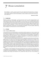
topics will also be described in an information transfer context (Figure 3.1 and also Figure 36.1 in
Chapter 36).
The fields describing relevant levels of information transfer and complexity are genomics →
transcriptomics → proteomics → metabolomics → bioenergetics or biochemical physiology →
molecular toxicology. All these areas of study explore different, yet linked, levels of organiza-
tion relative to biological information flow and complexity. Genomics explores the entire nuclear
DNA complement and variations within it.
1
Toxicogenomics specifically focuses on the influence
of toxicants on the nuclear DNA. The next level of the biochemical information flow emerges at
transcription. Transcription initiation occurs when RNA polymerase attaches to promoter regions
of DNA. Nucleotides are added according to the DNA base sequence to produce mRNA during the
elongation step of translation that ends with mRNA release. Transcriptomics attempts to describe
and explain the complement of mRNA transcripts and their abundances present in cells or tissues
under various conditions. Through translation, pools of various proteins are created in the cytoplasm.
Proteomics is the study of the full complement of these proteins, their relative abundances, changes,
and interactions. Finally, metabolomics attempts to explain the metabolite complement in cells or
tissues under various conditions, including toxicant exposure. Repeating an important theme in this
book, the greatest insight is gained by applying combinations of these approaches to a research
question.
1
Despite the focus here on nuclear DNA, mitochondrial DNA can also provide valuable information about contaminant
effects. Baker et al. (1999) quantified genetic damage in voles from the contaminated area surrounding the Chernobyl reactor
using a portion of the mitochondrial cytochrome b gene. They measured heteroplasmy (DNA sequence variation within an
individual) to suggest increased rates of somatic mutation in the liver of irradiated voles.
23
© 2008 by Taylor & Francis Group, LLC
Clements: “3357_c003” — 2007/11/9 — 12:42 — page 24 — #2
24 Ecotoxicology: A Comprehensive Treatment
DNA
RNA
Proteins
Metabolites
Energy currency,
structural and
storage molecules
By-products and
dysfunctional
molecules
Function
or
purpose
Associated
process
Metabolism
(anabolism
and catabolism)
Translation TranscriptionExcretion,
respiration,
detoxification,
and sequestration
Maintain
soma, control
aging
Maintain and
increase soma,
reproduction
Cellular information
processing
FIGURE 3.1 Hierarchical organization of biochemical effects discussed in this chapter.
The genome contains the instructions for growing and maintaining the soma. Although genomics
often focuses on consequences to the germ line, somatic risks are also created by toxicant-induced
changes to the genome. Carcinogenesis gives rise to the most obvious somatic risk (see Burdon
(1999) for a fuller treatment of this topic). Changes in the genome will be discussed below relative
to toxicant-induced modification of the DNA molecule.
Transcription and translation activities can provide evidence of response to a toxicant. As an
example, El-Alfy and Schlenk (1998) discovered that up-regulation of a monooxygenase in Japanese
medaka (Oryzias latipes) explained salinity-enhanced toxicity of aldicarb. In another study, differ-
ences in cytochrome P450 1A induction for chub (Leuciscus cephalus) populations with different
contaminant exposure histories was taken as evidence of pollutant-induced changes in population
genomics (Larno et al. 2001).
Shifts in metabolites can also suggest effects of, or responses to, toxicants. Kramer et al. (1992)
measured glycolysis and Krebs cycle metabolites in mosquitofish (Gambusia holbrooki) exposed to
mercury, finding decreased Krebs cycle flux during exposure. De Coen et al. (2001) noted increased
Krebs cycle activity during Daphnia magna exposure to lindane, suggesting that biochemical assays
be used to define the metabolic state of daphnids under stress.
Proteomics also has diverse applications in biochemical toxicology. Examples range from indu-
cible detoxification proteins to evidence of effects at higher levels of organization.Aspecific example
of evidence of potential effect at a higher level of biological organization is the abnormal induction of
the egg protein, vitellogenin, in male fish exposed to methoxychlor (Schlenk et al. 1997) or synthetic
estrogens (Schultz 2003). This induction will be discussed again in the following chapters in the
context of endocrine dysfunction.
Processes ensuing at higher levels of biological organization can manifest as shifts in biochemical
pools. Stressor-induced changes in bioenergetics can be detected with shifts in energy storage or
pools of high-energy molecules. Biochemical by-products can also be assessed in cells, tissues, and
physiological fluids. These types of biochemical shifts (e.g., shifts in heme biosynthesis) will also be
© 2008 by Taylor & Francis Group, LLC
Clements: “3357_c003” — 2007/11/9 — 12:42 — page 25 — #3
Biochemistry of Toxicants 25
discussed. The discussion of cellular, tissue, and bioenergetic effects detected with biochemical
qualities will be addressed again in chapters exploring these higher levels of biological organization
(i.e., Chapters 4–6).
3.2 DNA MODIFICATION
Damage to DNA occurs in several ways. It can result from strand breakage and subsequent imperfect
repair. Damage can also result from chemical bonding directly to the DNA or by some similar DNA
modification.
Although cancer is a paramount concern relative to somatic risk following toxicant-induced DNA
modification, some DNA changes to the germ line have population consequences, and in some cases,
these germ line-associated changes affect an exposed individual’s Darwinian fitness. The population
ecotoxicology section describes such changes and their consequences. As an example, men working
in certain conditions or occupations can have elevated risks of teratogenic effects in their children or
of their children developing cancer (Gardner et al. 1990, Stone 1992). In an even broader context, the
mutation accumulation theory proposes that the accumulation of genetic damage determines the rate
of aging for individuals (see Medvedev (1990) for details). Somatic longevity may be determined
by DNA modifications accrued during an individual’s life.
DNA can be damaged by contaminants or their metabolites that are free radicals or can facilitate
free radical
2
generation. Free radicals can break one or both strands of the DNA molecule, or can
oxidize bases in the DNA molecule. As an example of manifest breakage, Shugart (1996) noted
elevated levels of double-strand breaks in DNA of sunfish from contaminated reaches of East Poplar
Creek (Tennessee).As an example of base modification, Malins (1993) reported high concentrations
of the guanine product, 2,6-diamino-4-hydroxy-5-formaminidopyrimidine, in tumors of English sole
exposed to carcinogens in the field.
Contaminants or their metabolites can also bind covalently to DNAto form adducts. For example,
Ericson and Larsson (2000) found DNA adducts in perch caught below a Kraft pulp mill. As another
important example, metabolites of the carcinogen benzo[a]pyrene combined with guanine to form a
guanosine adduct.
Still other modes of DNA damage are possible. Mercury cross-links DNA with proteins. Some
metals bind to phosphate groups and heterocyclic bases of DNA. This changes the stability of the
molecule and increases the incidence of mismatched bases.
Damage, modification, and imperfect repair of protooncogenes or tumor suppressor genes can
initiate carcinogenesis (Burdon 1999). It can also accelerate the rate at which somatic mutations
accumulate, and in doing so, accelerate the rate of aging. Genomic damage changes cell functioning
and ultimately influences individual fitness.
3.3 DETOXIFICATION OF ORGANIC COMPOUNDS
A wide range of organic contaminants are transformed within organisms. The design behind such
transformations is to render the toxic chemical more amenable to elimination; however, this is
not always achieved without adverse consequences. The products of detoxification reactions can
sometimes be more toxic or reactive than the original compound. Such a transformation that makes
an inactive compound bioactive or an active compound more bioactive is called activation. In the
case of cancer-producing agents, the original compound is a procarcinogen and the cancer-causing
metabolite is called the carcinogen.
Detoxifying reactions are often classified as Phase I or II reactions. Phase I reactions produce a
more reactive, and sometimes more hydrophilic, metabolite from the original compound; the product
2
Free radicals are extremely reactive molecules possessing an unshared electron.
© 2008 by Taylor & Francis Group, LLC
Clements: “3357_c003” — 2007/11/9 — 12:42 — page 26 — #4
26 Ecotoxicology: A Comprehensive Treatment
is more amenable to further reaction and, in some cases, elimination. The reactive groups
−−
OH,
−−
NH
2
,
−−
SH, and
−−
COOH are added or made available by oxidation, hydrolysis, or reduction.
Products of a Phase I reaction can be eliminated directly, be subject to additional Phase I trans-
formations, or undergo Phase II transformations. Phase II reactions conjugate the compound or its
Phase I metabolite(s) with some compound such as acetate, cysteine, glucuronic acid, sulfate, gly-
cine, glutamine, or glutathione. The conjugate is more hydrophilic and readily eliminated than the
compound was before conjugation.
3.3.1 PHASE IREACTIONS
In Phase I, reactive groups are added or existing sites are made more readily available to further
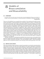
reactions. This can be illustrated with the metabolism of the dioxin benzo[a]pyrene (Figure 3.2).
The addition of oxygen by the microsomal mixed function oxidase system (MFO, also referred to
as the cytochrome P450 monooxygenase system) is the most prominent Phase I reaction. The cyto-
chrome P450 system is present in diverse species from bacteria to vertebrates, and functions in the
metabolism of endogenous (e.g., steroids and fatty acids) as well as xenobiotic compounds (Synder
2000). Associated Phase I oxidations involve two membrane-bound enzymes (cytochrome P450
isozymes and NADPH–cytochrome P450 reductase), NADPH, and molecular oxygen. The epoxida-
tions of benzo[a]pyrene to benzo[a]-4,5-oxide, benzo[a]-7,8-oxide, and benzo[a]-9,10-oxide shown
in Figure3.2 areachieved bythe MFO system. The MFOsystem isalso responsible for the conversion
of benzo[a]pyrene-7,8-dihydrodiol to benzo[a]pyrene-7,8-dihydrodiol-9,10-oxide.
Phase I enzymes also include epoxide hydrolases, esterases, and amidases that expose existing
functional groups on compounds (George 1994). For example, epoxide hydrolase is responsible for
Bay
region
K
Region
O
O
Benzo[a]pyrene-9,10-oxide
Benzo[a]pyrene
Benzo[a]pyrene-7,8-dihydrodiolBenzo[a]pyrene-7,8-oxide
Benzo[a]pyrene-7,8-dihydrodiol-9,10-oxideBenzo[a]pyrene-4,5-oxide
O
O
O
HO
HO
HO
HO
FIGURE 3.2 Phase I reactions for benzo[a]pyrene.
© 2008 by Taylor & Francis Group, LLC
Clements: “3357_c003” — 2007/11/9 — 12:42 — page 27 — #5
Biochemistry of Toxicants 27
the Phase I conversion of benzo[a]pyrene-7,8-oxide to benzo[a]pyrene-7,8-dihydrodiol, shown in
Figure 3.2. Epoxide hydrolase catalyzes the addition of water to MFO-generated epoxides. Other
enzymes such as alcohol and aldehyde dehydrogenases, aldehyde oxidases, and carbonyl reductase
generate products that are more rapidly eliminated than the original compound (George 1994,
Parkinson 1996). As an example, ethanol is oxidized to acetaldehyde by alcohol dehydrogenase.
This aldehyde is then oxidized by aldehyde dehydrogenase to acetic acid.
Type I reactions can also activate compounds to produce more poisonous or carcinogenic
ones (Figure 3.2). The epoxide formed at the K region of benzo[a]pyrene (e.g., the epoxide
in benzo[a]pyrene-4,5-oxide) and bay region dihydrodiols (e.g., benzo[a]pyrene-7,8-dihydrodiol)
of polycyclic aromatic hydrocarbons are potent carcinogens (Timbrell 2000). These products of
benzo[a]pyrene metabolism are strong electrophiles that bind to guanosine in the DNA molecule.
Formation of such adducts within protooncogenes can result in cancer. Another example of Phase I
activation is MFO-mediated epoxidation of the organochlorine pesticide aldrin to produce the more
toxic dieldrin (Chambers and Yarbrough 1976).
3.3.2 PHASE II (CONJUGATIVE)REACTIONS
In Phase II reactions, endogenous compounds are conjugated with contaminants or their metabolites
to detoxify them or to accelerate their elimination. Phase II conjugation can occur without any Phase I
reactions if the appropriate groups are already available. A compound is made more polar by binding
it to some amino acid, carbohydrate derivative, glutathione, or sulfate. However, Phase II reactions
can also involve methylation or acetylation that does not generally increase hydrophilicity.
Many Phase II reactions produce hydrophilic compounds readily eliminated from the indi-
vidual. Conjugates are commonly organic anions that are eliminated by glomerular filtration
and tubular transport in vertebrates (James 1987). Conjugation with glucuronic acid by UDP-
glucuronosyltransferases involves generation of a polar, hydrophilic glucuronide by combining the
compound with uridine diphosphate-glucuronic acid. As a relevant example, stimulated by con-
cern about birth control compounds released from sewage treatment plants into waterways, Schultz
(2003) studied the conjugation of the synthetic estrogen 17α-ethynylestradiol after its injection into
trout. Sulfate conjugation by sulfotransferases produces hydrophilic conjugates of polyaromatic
compounds, aliphatic alcohols, aromatic amines, and hydroxylamines. Xenobiotics with aromatic
or aliphatic hydroxyl groups are prone to such sulfation (James 1987). Amino acids may be con-
jugated to carboxylic acid or aromatic hydroxylamine groups of contaminants or their metabolites.
The amino acids most often involved are glycine, glutamine, and taurine (Jones 1987). Glutathione
(i.e., glycine–cysteine–glutamic acid) can be conjugated by glutathione S-transferases with a wide
array of electrophilic compounds. As examples, the benzo[a]-9,10-oxide and benzo[a]-4,5-oxides
shown in Figure 3.2 can undergo further Phase I transformations and the products of these reactions
conjugated with glutathione.
In contrast to the Phase II reactions just described, Phase II methylation and acetylation are
reactions thatdo notgenerally producemore hydrophilicproducts. The reader is directed to Parkinson
(1996) for more details about such reactions.
Box 3.1 There Is More to It Than Phase I and II Reactions
Our understanding ofreactions associated withxenobiotic conversion andelimination has grown
to include those outside the conventional Phase I and II reactions. The associated mechanisms
have been referred to as Phase III reactions (Zimniak et al. 1993). The ATP-dependent gluta-
thione S-conjugate export pump described byIshikawa (1992) facilitates a Phase III reactionthat
removes xenobiotic Phase II metabolites from the cell. Probably the best Phase III example is
the membrane-associated P-glycoprotein (P-gp) that acts as an energy-requiring efflux pump for
© 2008 by Taylor & Francis Group, LLC
Clements: “3357_c003” — 2007/11/9 — 12:42 — page 28 — #6
28 Ecotoxicology: A Comprehensive Treatment
xenobiotics and is described by Bard (2000) as the cell’s first line of defense. It also eliminates
metabolites from Phase I and II reactions from cells.
The P-gp mechanism for xenobiotic removal is similar to the multidrug resistance (MDR)
transporter protein discovered first in cancer cells that had become resistant to chemotherapeutic
agents. The cancer cell resistance results from reduced intracellular concentrations of these
chemotherapeutic agents due to the overexpression of an efficient ATP-dependent membrane-
bound pump, P-gp. This 170-kDa protein not only increases resistance to the original anticancer
drug, but also improves resistance to unrelated chemotherapy agents. The P-gp acts as a bar-
rier to xenobiotic absorption and accelerates their removal if they gained entry into the cell
(Abou-Donia et al. 2002). The mammalian P-gp is expressed at high levels in the kidney,
adrenal glands, liver, and lungs. Expression in mammalian brain capillary endothelial cells has
also been shown to reduce neurotoxicity of the pesticide ivermectin (Sckinkel et al. 1994).
The multixenobiotic resistance (MXR) mechanism is similar to MDR, involving a
membrane-associated transport P-gp that removes moderately hydrophobic, planar compounds
(Segner and Braunbeck 1998). Bard (2000) defines its substrates as “moderately hydrophobic,
amphipathic (i.e., somewhat soluble in both lipid and water), low molecular weight, planar
molecules with a basic nitrogen atom, cationic or neutral but never anionic, and natural
products.” P-gp can be induced during exposure to xenobiotics and has regulatory genes in com-
mon with the cytochrome P450 system. It has been found in mussel (Mytilus galloprovincialis)
cell membranes, leading Kurelec and Piv
ˇ
cevi
´
c (1991) to speculate that this mechanism could
account for the relatively high tolerance of these mussels to contaminants. The MXR gene was
also found recently in marine fish (Anoplarchus purpurescens) (Bard et al. 2002), Mytilus edulis
(Luedeking and Koehler 2004), and the Asiatic clam, Corbicula fluminea (Achard et al. 2004).
Their levels have been correlated with elevated concentrations of a variety of toxicants ranging
from crude oil (Hamdoun et al. 2002) to metals (Achard et al. 2004). Induction by metals
likely reflects the fact that protein-damaging chemicals induce several systems simultaneously,
including stress proteins, MXR, and cytochrome P450.
How does the P-gp work? A “flippase” model was proposed by Higgins and Gottesman
(1992) in which the xenobiotic binds to the P-gp at the inner surface of the cell membrane and
is “flipped” via an energy-requiring mechanism to the outside surface of the cell membrane.
The MXR’s presence in many taxonomic groups and its role in detoxification of many con-
taminants led Smital and Kurelec (1998) to define a new group of pollutants, that is, those that
modify the MXR response. In the laboratory, MXR can be readily inhibited with verapamil, so
there is potential for some environmental chemicals doing the same. A water-soluble fraction of
weathered crude oil, for example, appears to competitively inhibit MXR in larvae of the marine
worm, Urechis caupo (Hamdoun et al. 2002). Bard (2000) reviewed reports of such chemo-
sensitizers (Smital and Kurelec 1998), listing the following contaminants: pentchlorophenol,
2-acetylaminofluorene, diesel oil, and several pesticides (chlorbenside, sulfallate, and dacthal).
3.4 METAL DETOXIFICATION, REGULATION,
AND SEQUESTRATION
Predicting the consequences of metal exposure is complicated because metals may be essential or
nonessential. Very low concentrations of essential metals
3
can be as harmful as high concentrations
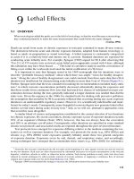
(Figure 3.3, upper panel). Nonessential metals display more conventional toxicity curves, showing
3
The essential metals are currently believed to be Co, Cr, Cu, Fe, Mn, Mo, Ni, Se, V, and Zn (Fraústo da Silva and
Williams 1991, Mertz 1981).
© 2008 by Taylor & Francis Group, LLC
Clements: “3357_c003” — 2007/11/9 — 12:42 — page 29 — #7
Biochemistry of Toxicants 29
Tox
icity
Metal concentration
Mortality (proportion dying)
Optimal
Deficiency
Toxicity
Sublethal
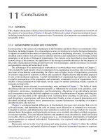
FIGURE 3.3 Mortality versus concentration for essential (upper panel) and nonessential (lower panel) metals.
A deficiency occurs if an essential metal is present below a certain concentration. This is not the case for a
nonessential metal. An essential metal will have an optimal range above and below which mortality begins to be
expressed. Increasing concentrations of the nonessential metal will increase the level of mortality experienced
in a group of exposed individuals. There might or might not be an apparent threshold concentration below which
no effect is expressed.
a sigmoidal increase in proportion of exposed individuals dying with an increase in metal concentra-
tion (Figure 3.3, lower panel). Essential metal deficiencies manifest in many ways other than death.
For example, insufficient intake of copper or zinc causes immunodeficiencies in mice (Beach et al.
1982, Prohaska and Lukasewycz 1981).
Understanding this dichotomy of essential and nonessential metal concentration–effect curves
can still be insufficient for sound prediction of metal effects. For example, chronic exposure to the
nonessential element cadmium can cause symptoms of zinc deficiency because cadmium displaces
zinc in metalloenzymes. Excessive amounts of nonessential tungsten can cause an apparent defi-
ciency of molybdenum, an essential and chemically similar element (Mertz 1981). Such an effect
would appear as a shift to the left for the curve shown in the upper panel of Figure 3.3 (x-axis
being the essential metal concentration). The bioactivity of some nonessential elements can also be
affected by another element. For example, mercury toxicity is lowered if sufficient concentrations of
selenium are also present. This would cause the curve in the lower panel of Figure 3.3 to shift to the
right.
Excess metals are dealt with in two ways, elimination or sequestration. Sequestration can involve
metal complexation with proteins or incorporation into granules. Sequestration in granules will
be discussed in the next chapter. Biomolecules involved in lessening metal intoxication will be
described here.
Metallothioneins are low-molecular-weight, cytosolic proteins that take up and facilitate trans-
port, sequestration, and excretion of metals such as cadmium, copper, silver, mercury, and zinc. They
© 2008 by Taylor & Francis Group, LLC
Clements: “3357_c003” — 2007/11/9 — 12:42 — page 30 — #8
30 Ecotoxicology: A Comprehensive Treatment
have high cysteine content, giving them the ability to form metal–thiolate clusters. Elevated metal
concentrations induce the production of metallothioneins to levels above those needed for normal
metal homeostasis. Metallothioneins bind metals, lowering the concentrations of metal available
to interact with sites of adverse action. Titers of metallothionein-coding mRNA or metallothionein
itself are often used as biomarkers of response to elevated metal concentrations.
Phytochelatin serves a similar protective role in plants. Phytochelatins are peptides of the form
(γ-glutamic acid–cysteine)
n
-glycine where n = 3, 5, 6, or 7 (Grill et al. 1985). Elevated concen-
trations of other phytochelatin-like peptides have recently been found in zinc-tolerant green algae
(Pawlik-Skowro
´
nska 2003).
3.5 STRESS PROTEINS AND PROTEOTOXICITY
The adverse effects of some agents result from protein damage (proteotoxicity). Indeed, this mode
of action is so pervasive that a general cellular stress response has evolved in most animal, plant,
or microbial species. Early studies of the stress-induced synthesis of protective proteins involved
the heat shock reaction—the organisms’ response to an abrupt change in temperature (Craig 1985).
Consequently, the proteins involved were first referred to as heat shock proteins. However, we
now know that a wide range of agents stimulate their production, including metals, metalloids,
ultraviolet (UV) radiation, and diverse organic compounds such as amino acid analogs, puromycin,
and ethanol (Hightower 1991, Sanders and Dyer 1994, Vedel and DePledge 1995). Because of their
induction by stressors other than heat, these proteins are now referred to as stress proteins. They
function to facilitate normal protein folding, protection of proteins under conditions that might lead
to denaturation, repair of denatured proteins, and movement of irreparably denatured protein to
lysosomes (Sanders and Dyer 1994).
4
Some stress proteins are present at basal levels but others are
present only after induction by some agent. Regardless of whether they were present under normal
conditions or induced by proteotoxic conditions, they collectively function to maintain homeostasis
by fostering essential protein levels, structure, and function.
The stress proteins are classified and named based on their molecular size. Stress70 and Stress90
are 70 and 90 kDa stress proteins, respectively. Smaller (60 kDa) stress proteins are called chaperons
owing to their role in mediating proper protein folding. Chaperons are abbreviated cpn60 (Di Giulio
et al. 1995). Stress70, Stress90, and cpn60 are present at basal levels that increase to reduce pro-
teotoxicity on appropriate induction. Another group of stress proteins (20–30 kDa) are the Low
Molecular Weight (LMW) stress proteins that are present only after induction.
Proteomic analysis of stress proteins is advocated by Sanders and Dyer (1994) for potentially
identifying agents responsible for adverse impact on species in the field. Their argument was based
on the observation that different chemicals induce different stress proteins to varying degrees.
Comparison of stress protein expression in field organisms to those of organisms exposed to each
candidate toxicant individually in the laboratory could provide causal insight. For example, Vedel
and DePledge (1995) measured Stress70 increase in crabs (Carcinus maenas) after laboratory copper
exposure. Currie and Tufts (1997) explored the combination of anoxia and heat stress on Stress70
induction in trout (Oncorhychus mykiss) red blood cells. Still other researchers focus on stress protein
genomics. Hightower (1991) made the novel suggestion that we could use the change in heat shock
protein genomes of various species to track the consequences of global warming. He hypothesized
that, as suggested by laboratory studies and field studies of desert species, the heat shock genes will
move in the direction of overexpression with adaptation to rapid warming.
4
Because our focus is chemical toxicology, other stress proteins will be ignored here. However, it should be mentioned
for the sake of completeness that glucose-regulated proteins (GRPs), metallothionein, hemeoxygenase, and the multidrug-
resistant p-glycoprotein are considered by many to be stress proteins (Di Giulio et al. 1995, Hightower 1991, Sander and
Dyer 1994).
© 2008 by Taylor & Francis Group, LLC
Clements: “3357_c003” — 2007/11/9 — 12:42 — page 31 — #9
Biochemistry of Toxicants 31
3.6 OXIDATIVE STRESS
Molecular oxygen is both benign and malign. On the one hand it provides enormous advantages and
on the other it imposes a universal toxicity. This toxicity is largely due to the intermediates of oxygen
reduction, that is, O
•
−
2
,H
2
O
2
, and OH
•
, and any organism that avails itself of the benefits of oxygen
does so at the cost of maintaining an elaborate system of defenses against these intermediates.
(Fridovich 1983)
A price was levied when much of the life on Earth took on the energetic advantage of using
molecular oxygen as a terminal electron acceptor for respiration. Very reactive, free oxyradicals
5
and
oxyradical-producing molecules suchashydrogen peroxide aregeneratedduring aerobic metabolism.
Oxyradicals oxidize lipids, proteins, and DNA, causing diverse effects ranging from membrane
damage to enzymedysfunction tocancer to accelerated aging. Consequently, organisms usingaerobic
respiration had to develop ways of coping with oxidative stress.
Oxidative stress is reduced in two ways. Antioxidant molecules are produced that react with
oxyradicals and enzymes are synthesized that consume oxyradicals or oxyradical-generating chem-
icals.Antioxidantsinclude catecholamines, glutathione, uric acid, andVitaminsA, C,andE. Enzymes
include superoxide dismutase, catalase, and glutathione peroxidase that catalyze the reactions shown
in Equations 3.1–3.3, respectively. (The unpaired electron in free radicals is designated as a dot by
convention. GSH and GSSG in these equations are reduced and oxidized glutathione, respectively.)
2O
•
−
2
+2H
+
→ H
2
O
2
+O
2
(3.1)
2H
2
O
2
→ 2H
2
O + O
2
(3.2)
2GSH +H
2
O
2
→ GSSG + 2H
2
O (3.3)
The removal of hydrogen peroxide, which is not itself an oxyradical, is crucial because it produces
the hydroxyl radical (OH
•
). This is accomplished through the Fenton reaction which, catalyzed by
a transition metal ion, generates OH
•
and OH
−
from H
2
O
2
(Equation 3.4). The transition metal ion
can be Cu(I), Cr(V), Fe(II), Mn(II), or Ni(II) (Gregus and Klaassen 1996).
H
2
O
2
+Fe
2+
→ Fe
3+
+HO
−
+HO
•
(3.4)
Why is this discussion relevant to environmental toxicants? Many organic chemicals become free
radicals during biochemical reactions or can generate oxyradicals. For example, paraquat reacting
within the MFO system becomes a charged free radical that reacts with molecular oxygen to produce
the superoxide anion, O
•
−
2
. After reacting with molecular oxygen, the paraquat becomes available
again to enter the same reactions, producing more superoxide anions each time it passes through
the redox cycle. Another example is carbon tetrachloride, which is converted to the trichloromethyl
radical (CCl
4
+ e
−
→ CCl
•
−
3
+ Cl
−
) during Phase I reactions (Slater 1984). As a final example,
enhanced oxidative damage at high metal concentrations occurs due to hydroxyl radical formation.
In such a case, more metal ion is available to catalyze the Fenton reaction and more oxyradicals are
formed as a consequence.
Responses to oxidative stress are used with field and laboratory exposures as evidence for xeno-
biotic hazard (Livingston et al. 1990, Winston and Di Giulio 1991). As an example, glutathione
and antioxidant enzymes shifted in mussels (M. galloprovincialis) transplanted from clean to metal-
contaminated conditions (Regoli and Principato 1995). Regoli (2000) later used the total oxyradical
scavenging capacity of mussels to indicate adverse effect of field exposure to metals.
5
A free radical is a charged or uncharged molecule or molecular fragment that has an unpaired electron (Slater 1984). An
oxyradical is a free radical in which the unpaired electron is associated with an oxygen atom.
© 2008 by Taylor & Francis Group, LLC
Clements: “3357_c003” — 2007/11/9 — 12:42 — page 32 — #10
32 Ecotoxicology: A Comprehensive Treatment
3.7 ENZYME DYSFUNCTION
Metals inhibit many types of enzymes that range in function from facilitating digestion (Chen et al.
2002) toheme synthesis (Dwyer et al. 1988). Eichhorn (1975) and, more extensively, Fraústo da Silva
and Williams (1991) provide details about metal binding to, and modifying the activity of, enzymes.
Ametal can displace another metal from an enzyme’s active site or otherwise interact with the enzyme
to change its secondary or tertiary structure. Metal ions can produce dysfunction by either increasing
or decreasing enzyme activity (Brown 1976, Eichhorn 1975).
Organic contaminants can also modify enzyme activity and, in so doing, modify an exposed
individual’s fitness. For example, brain cholinesterase activity was depressed for individuals of
several bird species found dying after organophosphorus or carbamate insecticide spraying (Hill
and Fleming 1982). More global examples exist such as the population consequences of DDT or
DDE inhibition of Ca–ATPase in the eggshell gland of birds. Its inhibition resulted in thin-shelled
eggs that broke before full development and hatching (e.g., Kolaja and Hinton 1979). Inhibition
of this one enzyme resulted in abrupt decreases in population size for osprey, Pandion haliaetus
(Ambrose 2001, Spitzer et al. 1978), bald eagle, Haliaeetus leucocephalus (Bowerman et al.
1995), falcon, Falco peregrinus (Ratcliffe 1967, 1970), and brown pelican, Pelecanus occidentalis
(Hall 1987).
3.8 HEME BIOSYNTHESIS INHIBITION
Porphyrin and heme synthesis (Figure 3.4) is central to producing hemoglobin, myoglobin, cyto-
chromes, tryptophan pyrrolase, catalase, and peroxidase.Although all cells produce heme, mammals
produce most heme in the liver and erythroid cells (Marks 1985). In the mitochondria, where
Porphobilinogen
Linear tetrapyrrole
δ-Aminolevulinic acid
Succinyl CoA + Glycine
δ-Aminolevulinic
acid
3 Porphobilinogen
Uroporphyrinogen III
Coproporphyrinogen III
Protoporphyrin IX
Heme
Protoporphyrinogen IX
FIGURE 3.4 Steps in heme synthesis.
© 2008 by Taylor & Francis Group, LLC
Clements: “3357_c003” — 2007/11/9 — 12:42 — page 33 — #11
Biochemistry of Toxicants 33
the tricarboxylic acid cycle generates ample succinyl CoA, succinyl CoA and glycine are con-
verted to δ-aminolevulinic acid by δ-aminolevulinic acid synthetase. The δ-aminolevulinic acid
then passes into the cytoplasm where two molecules of δ-aminolevulinic acid are then combined
by δ-aminolevulinic acid dehydratase to form porphobilinogen. Four molecules of porphobilino-
gen are then acted on by uroporphyrinogen I synthetase to produce linear tetrapyrrole. Still in
the cytoplasm, the linear tetrapyrrole is converted to uroporphyrinogen III by uroporphyrino-
gen III synthase and uroporphyrinogen III cosynthase. Uroporphyrinogen I synthase catalyses
the initial process which, if left on its own, would eventually produce uroporphyrinogen I, a
product with no known biochemical utility. However, uroporphyrinogen III cosynthase completes
the process to yield uroporphyrinogen III instead. Uroporphyinogen III is converted to copropor-
phyrinogen III by uroporphyrinogen decarboxylase. Intermediate products are also generated by
uroporphyrinogen decarboxylase including the seven, six, and five carboxy intermediates, hepta-
carboxyporphyrinogen, hexacarboxyporphyrinogen, and pentacarboxyporphyrinogen. Porphyrins
with 4–8 carboxyl groups tend to be generated in excess during heme synthesis and are excreted
in the urine (Wood et al. 1993). The remaining steps take place within the mitochondria.
Coproporphyrinogen III is converted to protoporphyrin IX by coproporphyrinogen oxidase and this
protoporphyrin IX is acted on by protoporphyrinogen IX oxidase and then ferrochelatase to produce
heme. The heme concentration in the mitochondria completes a feedback loop that regulates further
synthesis.
Porphyrin and heme synthesis can be influenced by a variety of factors, and deviations from
normal synthesis can have serious effects on an individual’s fitness. A human example is the genetic
disorder in porphyrin synthesis called acute intermittent porphyria. Acute intermittent porphyria
results from a uroporphyrinogen I synthetase deficiency that substantially diminishes this enzyme’s
activity. The disorder is diagnosed by urine analysis for excess porphobilinogen, the substrate for
this enzyme. Because of the strong influence of hormonal changes, the disorder does not usually
manifest in humans until puberty (Tschudy et al. 1975). It manifests in a range of intermittent effects
including abdominal pain, constipation, hypertension, psychosis, and even death from respiratory
paralysis (Becker and Kramer 1977, Goldberg 1959, Stein and Tschudy 1970). Barbiturates that
induce the early steps of heme synthesis or ethanol can trigger the adverse effects of this disorder
(Tschudy et al. 1975).
Inorganic toxicants also interfere with heme synthesis. Lead decreases heme production by
binding to a susceptible sulfhydrl group of aminolevulinic acid dehydrase (ALAD). An excess of
δ-aminolevulinic acid in urine and anemia are indicative of lead poisoning as a consequence. The
inhibition of ALAD has been developed as a biomarker for effects to fish from exposure to lead,
cadmium, and other metals (e.g., Dwyer et al. 1988, Johansson-Sjöbeck and Larsson 1978, 1979,
Marks 1985, Schmitt et al. 1993). Mercury impairs enzymes involved in heme synthesis and also
directly oxidizes reduced porphyrins (Wood et al. 1993) (Box 2.1). Prolonged imbibing of sodium
arsenate in water by rats depressed δ-aminolevulinic acid synthetase activity and modified the activity
of other enzymes involved in heme synthesis (Wood and Fowler 1978).
Organic chemicals can also interfere with heme synthesis. An outbreak of human porphyrias
was precipitated in Turkey during the 1950s when hexachlorobenzene fungicide-treated wheat
was consumed by several thousand Turks (Marks 1985). In addition to the skin lesions res-
ulting from this unintentional consumption of treated grain, the decrease in uroporphyrinogen
decarboxylase activity in afflicted individuals resulted in the accumulation of so much uropor-
phyrin that their urine was the color of dark red wine. The heme synthesis dysfunction appears
to result from the action of a Phase I metabolite of hexachlorobenzene. A Phase I metabolite of the
dioxin 2,3,7,8-tetrachlorodibenzo-p-dioxin (TCDD) also causes uroporohyrinogen decarboxylase
dysfunction. Other organic chemicals affecting heme synthesis include polychlorinated biphen-
yls (PCBs), polybrominated biphenyls (PBBs), and pesticides (diazinon, lindane, and heptachlor)
(Marks 1985).
© 2008 by Taylor & Francis Group, LLC
Clements: “3357_c003” — 2007/11/9 — 12:42 — page 34 — #12
34 Ecotoxicology: A Comprehensive Treatment
Box 3.2 Of Mice and Men (Dentists)
Perhaps the best demonstrations of metal effects on heme synthesis are provided by Wood and
his coworkers (i.e., Wood and Fowler 1978, Woods et al. 1993).
Prompted by concern about chronic sodium arsenate exposure in drinking water, Wood and
Fowler (1978) exposed rats and mice to arsenate, and examined the dose–response relation-
ships for heme synthesis. The influence of graded doses of arsenate on δ-aminolevulinic acid
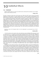
synthetase, uroporphyrinogen I synthase, and ferrochelatase activities are shown in Figure 3.5.
Notice that uroporphyrinogen I synthase activity increased slightly, but the activities of the other
two enzymes decreased. Concentrations of uroporphyrin and coproporphyrin also increased
with dose. Uroprophyrin concentrations in urine for mice exposed to 20, 40, and 85 µg/L doses
were 120%, 205%, and 910% of control concentrations, respectively. Similarly, coproporphyrin
concentrations were 104%, 142%, and 743% of control concentrations. Heme synthesis was
clearly influenced by arsenic exposure in drinking water.
Urinary porphyrins and mercury were measured in volunteer male dentists at the 1991
and 1992 American Dental Association meetings. Notionally, the dentists had been exposed to
mercury while working with the silver–mercury amalgam used for dental fillings. Urinary mer-
cury concentrations ranged from <0.5 to 556 µg/Lwith approximately 10% of screened dentists
having concentrations exceeding 20 µg/L. (The World Health Organization had recommended
an exposure limit of 25 µg/L in the urine.) Results were analyzed by splitting the dentists into
those with no detected urinary mercury (<0.5 µg/L) (n = 37) and those with ≥20 µg/L of
mercury in their urine (n = 56). The lower panel of Figure 3.5 shows the differences in mean
concentrations of urinary pentacarboxylporphyrin, precoproporphyrin, and coproporphyrin for
these two groups of dentists. All three were significantly higher in dentists with high exposures
(α = 0.05) but differences in concentrations of six- to seven-carboxyl porphyrins were not. The
authors concluded that these three porphyrins were excellent biomarkers for long-term mercury
exposure in humans.
FIGURE 3.5 (Upper panel) The change
in heme synthesis enzyme activities (relat-
ive to that of controls) for mice chronically
exposed to sodium arsenate. (Solid circle =
δ-aminolevulinic acid synthetase, shaded
circle = uroporphyrinogen I synthase, and
open circle = ferrochelatase activities.) (Data
from Table 2 of Wood and Fowler 1978.)
(Lower panel) The shift in porphyrins in dent-
ists exposed through their occupation to elev-
ated levels of elemental mercury. Mean con-
centrations of urinary pentacarboxylporphyrin
(solid squares), precoproporphyrin (shaded
squares), and coproporphyrin (open squares)
are shown for these two groups of dentists.
(Data from Table 3 of Wood et al. 1993.)
Activity (% of control)
70
80
90
100
110
120
130
20 µg/L
40 µg/L
85 µg/L
Porphyrin concentration (
µg/L )
No detected Hg
>20 µg Hg/L
75
50
25
10
5
0
© 2008 by Taylor & Francis Group, LLC
Clements: “3357_c003” — 2007/11/9 — 12:42 — page 35 — #13
Biochemistry of Toxicants 35
3.9 OXIDATIVE PHOSPHORYLATION INHIBITION
Some chemicals such as salicylic acid or pentachlorophenol act by uncoupling oxidative phos-
phorylation in the mitochondria.
6
Understandably, this leads to distinct physiological shifts such as
the lowered blood carbon dioxide levels and elevated blood pH (alkalosis) seen in humans overdosed
with aspirin (Timbrell 2000), or the significant increase in total oxygen consumption and gill ventila-
tion volume for trout overdosed with pentachlorophenol (McKim et al. 1987). A number of toxicants
act by this mode of action, notably substituted phenols such as 2,4-dinitrophenol, pentachlorophenol,
and 2,4,5-trichlorophenol. In a study by Penttinen and Kukkonen (1998), exposure to substituted
phenols predictably shifted the metabolic rate of exposed aquatic invertebrates.
Intoxications by substituted phenols are also described belowas cases of narcosis with the relative
toxicities of 2,4-dinitrophenol, pentachlorophenol, and 2,4,5-trichlorophenol being related to their
“effects on the energy-transducing membrane by uncoupling oxidative phosphorylation” (Penttinen
and Kukkonen 1998). Magnitude ofeffectisrelated to eachchemical’slipophilicity(i.e., propensity to
enter the membrane) and reactivity (i.e., ability to react at the appropriate receptor site). Similarly, in
a study of eight phenols that were narcotics and uncouplers of oxidative phosphorylation, lipophility
(log K
ow
) and acidity (pK
a
) were found to be important predictors of potency (Schüürmann et al.
1997).
3.10 NARCOSIS
Narcosis, including that brought about by many xenobiotics, results from a general and reversible
disruption of cell membrane functioning. There is a general depression of biological activity due
to toxicant interaction with membranes. The most familiar case of narcosis is that occurring with
anesthetic administration. The exact nature of the narcotic–membrane interaction seems to be incom-
pletely understood at the moment although changes in nerve cell membranes are clearly important in
higher animals. The protein-binding theory suggests that anesthetics (narcotics) act on ion channels
by directly binding to membrane proteins but the critical volume theory suggests that anesthetics
enter the membrane and modify its lipid bilayer (Abernethy et al. 1988). The critical volume the-
ory proposes that the toxicant accumulates in the lipid bilayer to such an extent that the membrane
swells, causing dysfunction. The toxicant molecule’s volume determines its capacity to swell the
membrane. Narcosis occurs when the membrane is swollen beyond a critical volume. In contrast,
the protein-binding theory suggests that the toxicant causes dysfunction by binding reversibly to
critical protein sites on the membrane. Franks and Lieb (1978) applied x-ray and neutron diffraction
techniques to find no change in the lipid bilayer of nerve cells, suggesting that anesthetic effect
did not involve lipid bilayer swelling. Later, they subjected a model protein, luciferase, to a wide
range of anesthetics and found that the anesthetics could modify the protein’s activity by binding
to specific receptors (Franks and Lieb 1984). The potency of an anesthetic in animal tests was also
highly correlated with its ability to inhibit luciferase. Franks and Lieb argued from this evidence that
anesthetic action likely results from competition with endogenous ligands for protein receptor sites.
They (Franks and Lieb 1985) also explained the cutoff phenomena of many anesthetic series with this
model system. A series of anesthetics appears to have increasing potency as lipophilicity increases,
but only to a certain cutoff point. Potency decreases quickly beyond that point. Although the strong
correlation with lipid solubility had provided support to the explanation of anesthetic cutoff point
on the basis of critical volume theory, they demonstrated with luciferase exposed to n-alcohols and
n-alkanes that the cutoff point was related to the anesthetic’s binding to a hydrophobic protein pocket
site of very specific dimensions. By analogy to the luciferase binding pocket, they suggested that
a similar situation occurs for membrane-associated proteins. The importance of protein binding in
6
Salicylic acid is produced from acetylsalicylic acid (aspirin) by a Phase I hydrolysis. It can then undergo conjugation
with glucuronic acid or glycine (Timbrell 2000).
© 2008 by Taylor & Francis Group, LLC
Clements: “3357_c003” — 2007/11/9 — 12:42 — page 36 — #14
36 Ecotoxicology: A Comprehensive Treatment
determining anesthetic potency was reinforced in another study using optical isomers of isoflurane.
These optimal isomers are equally soluble in lipids but have very different potencies and binding
capacities for ion channels of molluscan nerves (Franks and Lieb 1991). The remarkable work
of Franks and Lieb lends strong, but not yet definitive, support for the protein-binding theory of
narcosis.
Narcotics can be defined as “polar” (weak acids) and “nonpolar” (neutral or nonelectrolyte).
Many of the narcotics used in the Franks and Lieb studies were nonpolar, and lipophility was
adequate to predict trends in potency for them. Almost any nonelectrolyte organic compound that
can become associated with the cell membrane can express a nonspecific narcosis, but chemicals
commonly categorized as nonelectrolyte narcotics are ethers, alcohols, and chlorinated alkanes.
Other narcotics are weak acids. The most important of these polar narcotics have already been
discussed (i.e., the substituted phenols). Ionization also becomes important in predicting potency
for these narcotics because the unionized form of a compound is generally believed to be the most
capable of passage into lipid-rich membranes. McCarty et al. (1993) suggested that pK
a
and log K
ow
were important in predicting relative lethal effects of polar narcotics. (See Box 9.3 in Chapter 9 for
related details.) The concentration of an unionized narcotic in an exposure solution can be calculated
if the compound’s pK
a
and the medium’s pH are known. The Henderson–Hasselbach relationship
can be used to estimate the proportion of a weak acid that is unionized:
f
u
=
1
1 + 10
pH−pK
a
. (3.5)
The critical bodyresidue (CBR) approachis often appliedin dealingwithnarcotics. Theconcept is
simply thata narcotic’s actionis a direct function of the whole body dose at any moment. For example,
Penttinen and Kukkonen (1998) modeled effects of substituted phenols on aquatic invertebrates with
a threshold model of narcotic tissue concentration versus metabolic rate.
Before leaving the topic of polar narcotics, it is important to highlight a minor inconsist-
ency. Narcosis was described as a general phenomenon associated with cell membrane changes
(Section 3.9), but several substituted phenols act specifically on oxidative phosphorylation. This
specificity is inconsistent with the definition of narcosis as a nonspecific phenomenon. Although this
inconsistency does not impede understanding, it does cause confusion.
3.11 SUMMARY
Many, but not all, biochemical responses and consequences of toxicant exposure were discussed in
this brief chapter. Others will emerge in the next few chapters in discussions such as that addressing
cellular accumulation of degradation products from oxidative damage. Others such as the important
MXR transporter (Hamdoun et al. 2002) are relevant to discussions of contaminant uptake and
elimination. Together, they provide strong causal insights and sensitive biomarkers of contaminant
exposure or effect.
3.11.1 SUMMARY OF FOUNDATION CONCEPTS AND PARADIGMS
• The fields of study describing levels in the biological information hierarchy covered in this
chapter are the following: genomics → transcriptomics → proteomics → metabolomics
→ bioenergetics or biochemical physiology → molecular toxicology.
• Damage to DNA occurs by DNA strand breakage and subsequent imperfect repair, by
chemical bonding of a toxicant or its metabolite directly with the DNA, or by some
similar DNA modification such as DNA-protein cross-linking. Consequent effects to the
© 2008 by Taylor & Francis Group, LLC
Clements: “3357_c003” — 2007/11/9 — 12:42 — page 37 — #15
Biochemistry of Toxicants 37
soma include cancer and perhaps accelerated aging (i.e., the mutation accumulation theory
of aging).
• Many organic contaminants are subjectto transformationwithin organisms thatrenders the
toxic chemical more amenable to elimination. In some cases, the transformation products
can be more toxic or reactive than the original compound. A transformation in which an
inactive compound becomes bioactive or an active compound becomes more bioactive is
called activation.
• A series of Phase I and II reactions can occur, which render a toxicant more amenable
to elimination. Phase I reactions make compounds more reactive and sometimes more
hydrophilic. Reactive groups are added or existing sites are made more readily available
to further reactions. In Phase II (conjugative) reactions, endogenous compounds are con-
jugated with contaminants or their metabolites to accelerate their elimination. Phase II
conjugation can occur without any Phase I reactions if the appropriate groups are already
available.
• Toxic metals can bind with metallothioneins or phytochelatins to enhance transport,
sequestration, and elimination, or they can be incorporated into granules.
• Stress proteins lessen proteotoxicity of a wide range of stressors including metals,
metalloids, UV radiation, many organic compounds, and abrupt changes in temperature.
• Oxidative stress is reduced by the production of antioxidant molecules and by produc-
tion of enzymes that reduce the concentrations of free radicals or free radical generating
molecules.
• Organic and inorganic toxicants can also bind to enzymes, causing dysfunction.
• Heme synthesis is also sensitive to the action of organic and inorganic contaminants.
Shifts in porphyrin pools in body fluids such as urine can be a sensitive biomarker as
a consequence.
• Some toxicants (e.g., substituted phenols) act by uncoupling oxidative phosphorylation
in mitochondria.
• Narcosis, a result of a reversible disruption of cell membrane functioning, generally
depresses biological activity. Many toxicants act as narcotics. Two theories exist for
narcosis but current information supports the theory emphasizing action through narcotic
binding to membrane proteins and disruption of their functioning.
REFERENCES
Abernethy, S.G., Mackay, D., and McCarty, L.S., “Volume Fraction” correlation for narcosis in aquatic
organisms: The key role of partitioning, Environ. Toxicol. Chem., 7, 469–481, 1988.
Abou-Donia, M., Elmasry, E.M., and Abu-Qare, A.W., Metabolism and toxicokinetics of xenobiotics, In Hand-
book of Toxicology, 2nd ed., Derelanko, M.J. and Hollinger, M.A. (eds.), CRC Press, Boca Raton, FL,
2002, pp. 769–833.
Achard, M., Baudrimont, M., Boudou, A., and Bourineaud, J.P., Induction of a multixenobiotic resistance
protein (MXR) in the Asiatic clam Corbicula fluminea after heavy metal exposure, Aquat. Toxicol., 67,
347–357, 2004.
Ambrose P., Osprey revival from DDT complete in Chesapeake Bay, Mar. Pollut. Bull., 42, 388, 2001.
Baker, R.J., DeWoody, J.A., Wright, A.J., and Chesser, R.K., On the utility of heteroplasmy in genotoxicity
studies: An example from Chornobyl, Ecotoxicology, 8, 301–309, 1999.
Bard, S.M., Multixenobiotic resistance as a cellular defense mechanism in aquatic organisms, Aquat. Toxicol.,
48, 357–389, 2000.
Bard, S.M., Woodin, B.R., and Stegeman, J.J., Expression of P-glycoprotein and cytochrome P-450 1A in
intertidal fish (Anoplarchus purpurescens) exposed to environmental contaminants, Aquat. Toxicol.,
60, 17–32, 2002.
© 2008 by Taylor & Francis Group, LLC
Clements: “3357_c003” — 2007/11/9 — 12:42 — page 38 — #16
38 Ecotoxicology: A Comprehensive Treatment
Beach, R.S., Gershwin, M.E., and Hurley, L.S., Gestational zinc deprivation in mice: Persistence of
immunodeficiency for three generations, Science, 218, 469–471, 1982.
Becker, D.M. and Kramer, S., The neurological manifestations of porphyria: A review, Medicine, 56, 411–423,
1977.
Bowerman, W.W., Giesy, J.P., Best, D.Q., and Kramer, V.J., A review of factors affecting productivity of bald
eagles in the Great lakes regions: Implications for recovery, Environ. Health Perspect., 103(Suppl. 4),
51–59, 1995.
Brown, G.W., Jr., Effects of polluting substances on enzymes of aquatic organisms, J. Fish. Res. Board Can.,
33, 2018–2022, 1976.
Burdon, R.H., Genes and the Environment. Taylor & Francis Ltd., Philadelphia, PA, 1999.
Craig, E.A., The heat shock response, CRC Crit. Rev. Biochem., 18, 239–280, 1985.
Chambers, J.E. and Yarbrough, J.D., Xenobiotic biotransformation systems in fishes, Comp. Biochem.
Physiol. C, 55, 77–84, 1976.
Chen, Z., Mayer, L.M., Weston, D.O., Bock, M.J., and Jumars, P.A., Inhibition of digestive enzyme activit-
ies by copper in the guts of various benthic invertebrates, Environ. Toxicol. Chem., 21, 1243–1248,
2002.
Currie, S. and Tufts, B., Synthesis of stress protein 70 (Hsp70) in rainbow trout (Oncorhynchus mykiss) red
blood cells, J. Exp. Biol., 200, 607–614, 1997.
De Coen, W.M., Janssen, C.R., and Segner, H., The use of biomarkers in Daphnia magna toxicity test-
ing. V. In vivo alterations in the carbohydrate metabolism of Daphnia magna exposed to sublethal
concentrations of mercury and lindane, Ecotoxicol. Environ. Saf., 48, 223–234, 2001.
Di Giulio, R.T., Benson, W.H., Sanders, B.M., and Van Veld, P.A., Biochemical mechanisms: Metabolism,
adaptation, and toxicity, In Fundamentals of Aquatic Toxicology, 2nd ed., Rand, R.M. (ed.), Taylor &
Francis, Washington, D.C., 1995, pp. 523–561.
Dwyer, F.J., Schmitt, C.J., Finger, S.E., and Mehrle, P.M., Biochemical changes in longear sunfish, Lepomis
megalotis, associated with lead, cadmium and zinc from mine tailings, J. Fish Biol., 33, 307–317,
1988.
Eichhorn, G.L., Active sites of biological macromolecules and their interaction with heavy metals, In Ecological
Toxicology: Effects of Heavy Metal and Organohalogen Compounds, McIntyre, A.D. and Mills, C.F.
(eds.), Plenum Press, New York, 1975, pp. 123–142.
El-Alfy,A. and Schlenk, D., Potential mechanisms of the enhancement of aldicarb toxicity to Japanese medaka,
Oryzias latipes, at high salinity, Toxicol. Appl. Pharmacol., 152, 175–183, 1998.
Ericson, G. and Larsson, A., DNA adducts in perch (Perca fluviatilis) living in coastal water polluted with
bleached pulp mill effluents. Ecotoxicol. Environ. Saf., 46, 167–173, 2000.
Franks, N.P. and Lieb, W.R., Where do general anaesthetics act? Nature, 274, 339–342, 1978.
Franks, N.P. and Lieb, W.R., Do general anaesthetics act by competitive binding to specific receptors? Nature,
310, 599–601, 1984.
Franks, N.P. and Lieb, W.R., Mapping of general anaesthetic target sites provides a molecular basis for cutoff
effects, Nature, 316, 349–351, 1985.
Franks, N.P. and Lieb, W.R., Stereospecific effects of inhalational general anesthetic optical isomers on nerve
ion channels, Science, 254, 427–430, 1991.
Fraústo da Silva, J.J.R. and Williams, R.J.P., The Biological Chemistry of the Elements, Oxford University
Press, Oxford, UK, 1991.
Fridovich, I., Superoxide radical: An endogenous toxicant, Annu. Rev. Pharmacol. Toxicol., 23, 239–257,
1983.
Gardner, W.S., Snee, M.P., Hall, A.J., Powell, C.A., Downes, S., and Terrell, J.D., Results of case-control study
of leukaemia and lymphoma among young people near Sellafield nuclear plant in West Cumbria, BMJ,
300, 423–434, 1990.
George, S.G., Enzymology and molecular biology of Phase II xenobiotic-conjugating enzymes in fish, In
Aquatic Toxicology. Molecular, Biochemical and Cellular Perspectives, Malins, D.C. and Ostrander,
G.K. (eds.), CRC Press/Lewis Publishers, Boca Raton, FL, 1994, pp. 37–85.
Goldberg, A., Acute intermittent porphyria, Q. J. Med., 28, 183–209, 1959.
Gregus, Z. and Klaassen, C.D., Mechanisms of toxicity, In Casarett and Doull’s Toxicology. The
Basic Science of Poisons, 5th ed., Klaassen, C.D. (ed.), McGraw-Hill, New York, 1996,
pp. 35–74.
© 2008 by Taylor & Francis Group, LLC
Clements: “3357_c003” — 2007/11/9 — 12:42 — page 39 — #17
Biochemistry of Toxicants 39
Grill, E., Winnaker, E L., and Zenk, M.H., Phytochelatins: The principal heavy-metal complexing peptides in
higher plants, Science, 230, 674–676, 1985.
Hall, R.J., Impact of pesticides on bird populations, In Silent Spring Revisited, Marco, G.L.,
Hollingworth, R.M., and Durham, W. (eds.), American Chemical Society, Washington, D.C., 1987,
p. 214.
Hamdoun, A.M., Griffin, F.J., and Cherr, G.N., Tolerance to biodegraded crude oil in marine invertebrate
embryos and larvae is associated with expression of a multixenobiotic resistance transporter, Aquat.
Toxicol., 61, 127–140, 2002.
Hightower, L.E., Heat shock, stress proteins, chaperons, and proteotoxicity, Cell, 66, 191–197,
1991.
Hill, E.F. and Fleming, W.J., Anticholinesterase poisoning of birds: Field monitoring and diagnosis of acute
poisoning, Environ. Toxicol. Chem., 1, 27–38, 1982.
Ishitawa, T., The ATP-dependent glutathione S-conjugate export pump, Trends Biochem. Sci., 18, 164–166,
1992.
Jagoe, C.H., Responses at the tissue level: Quantitative methods in histopathology applied to ecotoxicology,
In Ecotoxicology. A Hierarchical Treatment, Newman, M.C. and Jagoe, C.H. (eds.), CRC Press/Lewis
Publishers, Boca Raton, FL, 1996, pp. 163–196.
James, M.O., Conjugation of organic pollutants in aquatic species, Environ. Health Perspect., 71, 97–103,
1987.
Johansson-Sjöbeck, M L. and Larsson, Å., The effect of cadmium on the hematology and on the activity
of δ-aminolevulinic acid dehydratase (ALA-D) in blood and hematopoietic tissues of the flounder,
Pleuronectes flesus L., Environ. Res., 17, 191–204, 1978.
Johansson-Sjöbeck, M L. and Larsson, Å., Effects of inorganic lead on delta-aminolevulinic acid dehydratase
activity and hematological variables in the rainbow trout, Salmo gairdnerii, Arch. Environ. Contam.
Toxicol., 8, 419–431, 1979.
Kolaja, G.L. and Hinton, D.E., DDT-induced reduction in eggshell thickness, weight, and calcium is accom-
panied by calcium ATPase inhibition, In Animals as Monitors of Environmental Pollutants, National
Academy of Sciences, Washington, D.C., 1979, pp. 309–318.
Kramer, V.J., Newman, M.C., and Ultsch, G.R., Changes in concentrations of glycolysis and Krebs cycle
metabolites inmosquitofish, Gambusiaholbrooki, induced by mercuric chloride and starvation, Environ.
Biol. Fishes, 34, 315–320, 1992.
Kurelec, B. and Piv
ˇ
cevi
´
c, B., Evidence for a multixenobiotic resistance mechanism in the mussel Mytilus
galloprovincialis, Aquat. Toxicol., 19, 291–302, 1991.
Larno, V., LaRoche, J., Launey, S., Flammarion, P., and DeVaux, A., Responses of chub (Leuciscus cephalus)
populations to chemical stress, assessed by genetic markers, DNA damage and cytochrome P4501A
induction, Ecotoxicology, 10, 145–158, 2001.
Livingston, D.R., Garcia Martinez, P., Michel, X., Narbonne, J.F., O’Hara, S., Ribera, D., and Winston, G.W.,
Oxyradical production as a pollution-mediated mechanism of toxicity in the common mussel, Mytilus
edulis L., and other molluscs, Funct. Ecol., 4, 415–424, 1990.
Lucier, G.W., Portier, C.J., and Gallo, M.A., Receptor mechanisms and dose-response models for the effects of
dioxins, Environ. Health Perspec., 101, 36–44, 1993.
Luedeking, A. and Koehler, A., Regulation of expression of multixenobiotic resistance (MXR)
genes by environmental factors in the bluw mussel Mytilus edulis, Aquat. Toxicol., 69, 1–10,
2004.
Malins, D.C., Identification of hydroxyl radical-induced lesions in DNA base structure: Biomarkers with a
putative link to cancer development, J. Toxicol. Environ. Health, 40, 247–261, 1993.
Marks, G.S., Exposure to toxic agents: The heme biosynthetic pathway and hemoproteins as indicator, Crit.
Rev. Toxicol., 15, 151–179, 1985.
McCarty, L.S., Mackay, D., Smith, A.D., Ozburn, G.W., and Dixon, D.G., Residue-based interpretation of
toxicity and bioconcentration QSARs from aquatic bioassays: Polar narcotic organics, Ecotoxicol.
Environ. Saf., 25, 253–270, 1993.
McKim, J.M., Schmieder, P.K., Carlson, R.W., Hunt, E.P., and Niemi, G.J., Use of respiratory-cardiovascular
responses of rainbow trout (Salmo gairdneri) in identifying acute toxicity syndromes in fish: Part 1.
Pentachlorophenol, 2,4-dinitrophenol, tricaine methanesulfonate and 1-octanol, Environ. Toxicol.
Chem., 6, 295–312, 1987.
© 2008 by Taylor & Francis Group, LLC
Clements: “3357_c003” — 2007/11/9 — 12:42 — page 40 — #18
40 Ecotoxicology: A Comprehensive Treatment
Medvedev, Z.A., An attempt at a rational classification of theories of ageing, Biol. Rev. Camp. Philos. Soc., 65,
375–398, 1990.
Mertz, W., The essential trace elements, Science, 213, 1332–1338, 1981.
Pawlik-Skowro
´
nska, B., When adapted to high zinc concentrations the periphytic green alga Stigeoclonium
tenue produces high amounts of novel phytochelatin-related peptides, Aquat. Toxicol., 62, 155–163,
2003.
Parkinson, A., Biotransformation of xenobiotics, In Casarett & Doull’s Toxicology. The Basic Science of
Poisons, 5th ed., Klaassen, C.D. (ed.), McGraw-Hill, New York, 1996, pp. 113–186.
Penttinen, O P. and Kukkonen, J., Chemical stress and metabolic rate in aquatic invertebrates: Threshold,
dose–response relationships, and mode of toxic action, Environ. Toxicol. Chem., 17, 883–890,
1998.
Prohaska, J.R. and Lukasewycz, O.A., Copper deficiency suppresses the immune response of mice, Science,
213, 599–561, 1981.
Ratcliffe, D.A., Decrease in eggshell weight in certain birds of prey, Nature, 215, 208–210,
1967.
Ratcliffe, D.A., Changes attributable to pesticides in egg breakage frequency and eggshell thickness in some
British birds, J. Appl. Ecol., 7, 67–107, 1970.
Regoli, F., Total oxyradical scavenging capacity (TOSC) in polluted and translocated mussels: A predictive
biomarker of oxidative stress, Aquat. Toxicol., 50, 351–361, 2000.
Regoli, F. and Principato, G., Glutathione, glutathione-dependent and antioxidant enzymes in mussel, Mytilus
galloprovincialis, exposed to metals under field and laboratory conditions: Implications for use of
biochemical biomarkers, Aquat. Toxicol., 31, 143–164, 1995.
Sander, B.M. and Dyer, S.D., Cellular stress response, Environ. Toxicol. Chem., 13, 1209–1210,
1994.
Schlenk, D., Stresser, D., McCants, J., Nimrod, A., and Benson, W., Influence of beta-naphthoflavone
and methoxychlor pretreatment on the biotransformation and estrogenic activity of methoxy-
chlor in channel catfish (Ictalurus punctatus), Toxicol. Appl. Pharmacol., 145, 349–356,
1997.
Schinkel, A.D., Smit, J.J., Tellingen, O., Beijnen, J.H., Wagenaar, E., van Deemter, L., Mol, C.A., et al.,
Disruption of the mouse mdr1a P-glycoprotein gene leads to a deficiency in the blood-brain barrier and
to increased sensitivity to drugs, Cell, 77, 491–502, 1994.
Schmitt, C.J., Wildhaber, M.L., Hunn, J.B., Nash, T., Tieger, M.N., and Steadman, B.L., Biomonitoring of
lead-contaminated Missouri streams with an assay for erythrocyte δ-aminolevulinic acid dehydratase
activity in fish blood, Arch. Environ. Contam. Toxicol., 25, 464–475, 1993.
Schultz, I., Environmental estrogens: Occurrence of ethynylestradiol and adverse effects on fish repro-
duction, In Fundamentals of Ecotoxicology, 2nd ed., Newman, M.C. and Unger, M.A. (eds.),
CRC Press/Lewis Publishers, Boca Raton, FL, 2003, pp. 156–160.
Schüürmann, G., Segner, H., and Jung, K., Multivariate mode-of-action analysis of acute toxicity of phenols,
Aquat. Toxicol., 38, 277–296, 1997.
Segner, H. and Braunbeck, T., Cellular response profile to chemical stress, In Ecotoxicology, Schüürmann, G.
and Braunbeck, T. (eds.), John Wiley & Sons, New York, 1998, pp. 521–569.
Shugart, L.R., Molecular markers to toxic agents, In Ecotoxicology. A Hierarchical Treatment, Newman, M.C.
and Jagoe, C.H. (eds.), CRC Press/Lewis Publishers, Boca Raton, FL, 1996, pp. 133–161.
Slater, T.F., Free-radical mechanisms in tissue injury, Biochem. J., 222, 1–15, 1984.
Smital, T. and Kurelee, B., The chemosensitizers of multixenobiotic resistance mechanisms in aquatic
invertebrates: A new class of pollutants, Mutat. Res., 399, 43–53, 1998.
Spitzer, P.R., Risebrough, R.W., Walker, W., Hernandez, R., Poole, A., Pulleston, D., and Nisbet., I.C.T.,
Productivity of ospreys in Connecticut-Long Island increases as DDE residues decline, Science, 202,
333–335, 1978.
Stein, J.A. and Tschudy, D.P., Acute intermittent porphyria: A clinical and biochemical study of 46 patients,
Medicine, 49, 1–16, 1970.
Stone, R., Can a father’s exposure lead to illness in his children? Science, 258, 31, 1992.
Timbrell, J., Principles of Biochemical Toxicology, Taylor & Francis, Philadelphia, PA, 2000.
Vedel, G.R. and DePladge, M.H., Stress-70 levels in the gills of Carcinus maenas exposed to copper, Mar.
Pollut. Bull., 31, 84–86, 1995.
© 2008 by Taylor & Francis Group, LLC
Clements: “3357_c003” — 2007/11/9 — 12:42 — page 41 — #19
Biochemistry of Toxicants 41
Winston, G.W. and Di Giulio, R.T., Prooxidant and antioxidant mechanisms in aquatic organisms, Aquat.
Toxicol., 19, 137–161, 1991.
Wood, J.S. and Fowler, B.A., Altered regulation of mammalian hepatic heme biosynthesis and urinary por-
phyrin excretion during prolonged exposure to sodium arsenate, Toxicol. Appl. Pharm., 43, 361–371,
1978.
Wood, J.S., Martin, M.D., Naleway, C.A., and Echeverria, D., Urinary porphyrin profiles as a biomarker of
mercury exposure: Studies on dentists withoccupational exposure tomercury vapor, J. Toxicol. Environ.
Health, 40, 235–246, 1993.
Zimniak, P., Awasthi, S., andAwasthi, Y.C., Phase III detoxification system. Trends Biochem. Sci., 18, 164–166,
1993.
© 2008 by Taylor & Francis Group, LLC