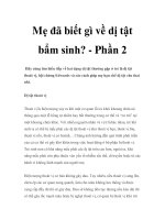
Dị tật bẩm sinh cột sống ở trẻ em doc
Bạn đang xem bản rút gọn của tài liệu. Xem và tải ngay bản đầy đủ của tài liệu tại đây (228.16 KB, 9 trang )
Congenital and
Developmental Deformities
of the Spine in Children With
Myelomeningocele
Abstract
The treatment of spinal deformities in children with myelomenin-
gocele poses a formidable task. Multiple medical comorbidities,
such as insensate skin and chronic urinary tract infection, make
care of the spine difficult. A thorough understanding of the natural
history of these deformities is mandatory for appropriate treatment
to be rendered. A team approach that includes physicians from
multiple specialties provides the best care for these patients. The
two most challenging problems are paralytic scoliosis and rigid
lumbar kyphosis. The precise indications for surgical intervention
are multifactorial, and the proposed benefits must be weighed
against the potential risks. Newer spinal constructs now allow for
fixation of the spine in areas previously difficult to instrument.
Complications appear to be decreasing with improved understand-
ing of the pathophysiology associated with myelomeningocele.
S
coliosis and kyphosis with sec-
ondary adaptive changes are
common in the patient with myelo-
meningocele. Developmental defor-
mities are acquired and are related to
the level of paralysis; congenital de-
formities result from malforma-
tions, such as hemivertebrae. Both
forms may exist concurrently.
From 1983 to 1990, the preva-
lence of neural tube defects (ie, my-
elomeningocele) in the United States
was 4.6 per 10,000.
1
However, with
the increased awareness of the im-
portance of folic acid consumption
during pregnancy, there has been a
decrease of between 72% and 100%
in the number of overall new neural
tube defects.
1
Nearly every patient
with myelomeningocele will de-
velop hydrocephalus, and approxi-
mately 50% of all patients will have
some degree of mental retardation.
The clinical experience of the au-
thors has shown that most of these
patients have motor levels at the
lumbosacral or sacral level. As the
motor level ascends the spine, the
prevalence of scoliosis and associ-
ated musculoskeletal anomalies in-
creases.
Prevalence of Spinal
Deformity
Multiple studies document the
so-called incidence of the various
types of spine problems in children
with myelomeningocele at different
ages.
2-7
Whether these numbers rep-
resent an actual incidence or a prev-
alence is unknown. Cobb measure-
ments also have been used to define
developmental scoliosis, the preva-
James T. Guille, MD
John F. Sarwark, MD
Henry H. Sherk, MD
S. Jay Kumar, MD
Dr. Guille is Orthopaedic Surgeon,
Shriners Hospital for Children,
Philadelphia, PA. Dr. Sarwark is
Chairman, Department of Orthopaedic
Surgery, The Children’s Memorial
Hospital, Chicago, IL, and Professor of
Orthopaedic Surgery, Northwestern
University Feinberg School of Medicine,
Chicago. Dr. Sherk is Professor,
Department of Orthopaedic Surgery,
Drexel University College of Medicine,
Philadelphia. Dr. Kumar is Director,
Spinal Dysfunction Clinic, Alfred I.
duPont Hospital for Children,
Wilmington, DE, and Clinical Professor
of Orthopaedic Surgery, Jefferson
Medical College of Thomas Jefferson
University, Philadelphia.
None of the following authors or the
departments with which they are
affiliated has received anything of value
from or owns stock in a commercial
company or institution related directly or
indirectly to the subject of this article:
Dr. Guille, Dr. Sarwark, Dr. Sherk, and
Dr. Kumar.
Reprint requests: Dr. Guille, Shriners
Hospital for Children, 3551 North Broad
Street, Philadelphia, PA 19140.
J Am Acad Orthop Surg 2006;14:294-
302
Copyright 2006 by the American
Academy of Orthopaedic Surgeons.
294 Journal of the American Academy of Orthopaedic Surgeons
lence of which ranges from 52% to
89% in this population. The preva-
lence of congenital scoliosis ranges
from 7% to 20%. More than 80% of
patients aged >10 years will have
scoliosis. Using rigid criteria, Trive-
di et al
2
recently defined develop-
mental scoliosis in this patient pop-
ulation as a Cobb magnitude >20°.
This figure was chosen because
curves of less than this magnitude
often were observed to improve or
even to resolve. The authors con-
cluded that when a scoliosis did not
develop by age 15 years, the child
would not develop a curve later in
life.
Associated Health
Issues
The global health concerns in these
children are numerous and may dra-
matically influence the care of the
spinal deformity. Common issues
include central nervous system in-
volvement, such as mental retarda-
tion, hydrocephalus requiring shunt-
ing, and tethering problems of the
brain and spinal cord. Insensate skin,
latex allergy, renal anomalies, bacte-
rial colonization of the urinary tract,
bowel and bladder incontinence, and
lower extremity malalignment are
other factors that often require eval-
uation and treatment. Ongoing care
and assessment are most effectively
done by a team approach. In addition
to the orthopaedic surgeon, mem-
bers of the team should include a pe-
diatrician, neurosurgeon, urologist,
physiatrist, orthotist, physical ther-
apist, and social worker.
Renal anomalies occur in 4% to
17% of patients with myelomenin-
gocele, with a higher association in
those with congenital vertebral
anomalies.
8
Aplasia or dysgenesis of
the kidneys is associated with tho-
racic and upper lumbar level defects;
anomalies of the ureters, especially
duplication, are associated with low-
er lumbar and sacral level defects.
Secondary changes, such as scarring,
may occur in the urinary tract as a
result of chronic or recurrent infec-
tion. Therefore, renal dysfunction is
common and requires routine mon-
itoring.
Latex allergy occurs in 18% to
40% of patients with myelomenin-
gocele and may be life-threatening.
9
The allergy is a type I immunoglob-
ulin E (IgE)-mediated response to
natural plant antigens in the latex
and is not an actual allergy to the la-
tex itself. Repeated exposure to latex
products (from urinary catheteriza-
tions, hospitalizations, or surgical
procedures) ultimately may sensi-
tize these patients to these antigens.
The value of an allergen-specific
IgE antibody test in detecting indi-
viduals with latex allergy is ques-
tionable; therefore, a latex-free pro-
tocol should be used for every
patient at all times.
Musculoskeletal
Evaluation
A complete history and baseline
physical examination by the special-
ty team should be done in every
child. Serial examinations are rec-
ommended every 4 to 6 months to
document changes in neurologic and
functional status. The extremities
are examined for range of motion,
muscle strength, and the presence of
skin ulcers or breakdown. Truncal
and sitting alignment are evaluated,
with decompensation and rotational
prominences noted. Wheelchair and
prosthetic modifications are made as
needed in consultation with the ap-
propriate skilled vendor and thera-
pists.
Baseline radiographs should be
done of the entire spine in the an-
teroposterior and lateral planes, with
notation made as to whether the ra-
diographs were made in the supine,
sitting, or standing position. Except
in low lumbar and sacral level pa-
tients who can stand independently,
radiographs preferably are done in
the sitting position. Evidence of
deformity, associated congenital
anomalies, and the level of the spi-
nal dysraphism should be recorded.
Serial radiographs should be per-
formed every 6 months to document
curve stability or progression. A
magnetic resonance imaging (MRI)
study of the brain and spinal canal
should be done within the first
2 years of life for baseline purposes;
studies should be repeated when
clinically indicated. Myelography is
reserved for patients in whom the
spinal deformity is sufficiently se-
vere to preclude adequate interpreta-
tion of MRI scans, in patients with
metal implants, and as part of the
evaluation of a child with a tethered
cord.
10
Anomalies of the central nervous
system include hydrocephalus, cere-
bellar malformations, hydromyelia,
syringomyelia, spinal cord anoma-
lies, and tethered cord. After birth
but before discharge, >90% of chil-
dren with myelomeningocele devel-
op hydrocephalus and a Chiari II
malformation following spinal/skin
closure. Hydromyelia and syringo-
myelia have been reported in ap-
proximately 50% of patients.
5,10
At-
rophy of the spinal cord is seen in
15% of patients, lipomas and der-
moids in 11% to 38%, and diastem-
atomyelia in 2% to 7%; any of these
anomalies may cause neurologic de-
terioration. Scoliosis associated with
hydromyelia or syringomyelia is typ-
ically an S-shaped curve in the tho-
racic or thoracolumbar region. No
good guidelines exist as to when and
how a syringomyelia becomes symp-
tomatic and when it should be
drained; evaluation of a syringomy-
elia requires the consultation of a
neurosurgeon. MRI is a mainstay in
detecting and monitoring these con-
ditions.
Signs of a tethered cord in the pa-
tient with myelomeningocele can
include deterioration in gait, increas-
ing spasticity, weakness, limb defor-
mities, back pain, changes in conti-
nence, and rapid increase in the
curve magnitude of scoliosis.
11,12
The curve, when present, is usually
in the thoracolumbar or lumbar re-
James T. Guille, MD, et al
Volume 14, Number 5, May 2006 295
gion and is associated with increased
lumbar lordosis. Some authors now
think that the signs of a tethered spi-
nal cord are a result of pressure on
the cord from repeated flexion and
extension of the spine at the site
causing focal flattening, rather than
from an actual tethering effect on
the ascent of the spinal cord.
11
Sar-
wark et al
12
found that in a select
group of children with L3 motor lev-
el or lower and no hydromyelia, re-
lease of a tethered cord resulted in a
58% chance of stabilization or im-
provement of curve magnitude.
Equivocal results also were seen in
patients with spasticity. Pierz et
al
11
found no improvement in curve
magnitude following detethering in
patients who presented with thorac-
ic neurologic levels or a curve >40°.
Developmental
Scoliosis
Frequently seen in young children,
developmental scoliosis primarily re-
sulting from paralysis typically is a
long, sweeping, C-shaped curve with
or without pelvic obliquity. The con-
vexity of the curve often is opposite
the side of the elevated pelvis. A dis-
location of the hip alone does not ap-
pear to be the cause of the scoliosis;
the curve develops from muscle im-
balance secondary to paralysis and is
commonly associated with kyphosis,
not lordosis. Often upper extremity
function is diverted because the
hands are used to support the trunk.
Many factors correlate with the
occurrence of developmental scolio-
sis. Clinical motor level is an impor-
tant predictor.
2,6
Trivedi et al
2
found
the prevalence of scoliosis to be
93%, 72%, 43%, and <1%, respec-
tively, in patients with thoracic, up-
per lumbar , lower lumbar, and sacral
motor levels. The level of the last in-
tact laminar arch (LILA) is another
important predictive factor in the
development of scoliosis.
4,7
Trivedi
et al
2
found the prevalence of scolio-
sis to be 89%, 44%, 12%, and 0%,
respectively, in patients with thorac-
ic, upper lumbar, lower lumbar, and
sacral LILAs. The LILA is not always
synonymous with the motor level.
Overall, it appears that the three
most important factors in predicting
the development of scoliosis are the
motor level, ambulatory status, and
LILA. To a lesser degree, hip
dislocation/subluxation and lower
extremity spasticity also are predic-
tive factors.
Muller et al
13
studied the progres-
sion of scoliosis in 64 patients. The
fastest progression was seen during
the early teenage years, although it
may occur earlier; the scoliosis typ-
ically stopped with the cessation of
growth. Progression was found to be
related to the size of the curve:
curves <20° progressed slowly,
whereas those >40° progressed more
quickly (approximately 13° per year).
Nonambulatory patients had a great-
er progression rate; however, no cor-
relation was made between the lev-
el of spinal defect and progression.
The authors concluded that all
curves should be observed closely
and treated before a magnitude of
40° is reached. Marchesi et al
14
also
found that scoliosis is a progres-
sive condition, especially in young-
er children, and that there was
less chance for progression when
the curve was detected after age
10 years.
In a child with a scoliosis <20°,
observation with radiographs every
4 to 6 months is suggested. When
the curve is >20°, use of a brace
should be considered. Because the
role of bracing in these patients is
controversial, its use is left to the
choice of the surgeon and his or her
experience. Although most agree
that bracing does not stop curve pro-
gression or completely eliminate the
need for spinal fusion, bracing may
slow the progression of a curve and
allow for further trunk growth be-
fore eventual spinal fusion.
Muller and Nordwall
15
reported
on the use of the Boston brace in the
management of scoliosis and found
that when treatment was instituted
early and before the curve reached
45°, the brace could arrest progres-
sion of the cur ve. These results,
however, have not been reported by
others. The brace we have recom-
mended is custom-molded and does
not interfere with pulmonary
function, lower extremity bracing,
self-catheterization, or sitting—a
challenge in some patients. Obesity
may be a relative contraindication.
The brace aids in sitting balance and
frees the hands for function. Patients
who use a brace need to have their
skin checked daily for areas of pres-
sure and breakdown, although a
custom-fitted brace usually avoids
these problems.
Surgical Indications and
Principles
Listing the absolute indications
for surgical intervention is difficult
because the long-term natural histo-
ry of untreated spinal deformity in
this population is relatively un-
known. Most agree, however, that
progressive scoliosis >50° that caus-
es sitting imbalance is an important
indication. McMaster
16
thought that
loss of function as an indication was
more important than the degree of
curvature. Ideally, spinal reconstruc-
tion would be done after most adult
sitting height is attained; however,
the surgeon is infrequently afforded
this optimal scenario.
Because it is common for sur-
geons to want to procrastinate in
treating these curves surgically, a di-
lemma arises when a younger child
presents with a large progressive
curve and sitting imbalance. The
ideal solution to this problem has
yet to be found. The use of a growing
rod system in these patients has
been reported. Medical comorbidi-
ties, such as shunt function, pulmo-
nary function, skin condition, and
urinary tract infection, require eval-
uation and treatment before surgery.
When poor tissue coverage is a con-
cern, consultation with a plastic sur-
geon is advised to consider the use of
preoperative tissue expanders.
Congenital and Developmental Deformities of the Spine in Children With Myelomeningocele
296 Journal of the American Academy of Orthopaedic Surgeons
Combined anterior and posterior
arthrodesis and instrumentation
provide the best chance to achieve a
durable fusion.
16-20
Anterior diskec-
tomy improves the preinstrumenta-
tion flexibility and correctability of
the curve, and anterior interbody fu-
sion increases the strength of the en-
tire fusion mass. The addition of al-
lograft bone may be necessary in
patients who have had prior bone
graft harvesting, in those with small
ilia, and in those requiring long fu-
sions. Bone graft substitutes and
growth factors may play an impor-
tant role in the future.
Anterior fusion and instrumenta-
tion alone is again being considered
for selected curves. Sponseller et
al
21
revisited this technique and had
good results with anterior fusion
alone only when the thoracolumbar
curve was <75°; when there were no
syrinx, no increased kyphosis, and
no compensatory curve >40°; and
when there was independent sitting
balance. Parsch et al
19
recommended
instrumented anterior and posterior
fusions, especially in patients with
thoracic level paralysis, to decrease
the rate of implantation failure and
to prevent postoperative loss of cor-
rection. In the series of Stella et al,
22
the best corrections were obtained in
patients who had instrumented an-
terior and posterior fusions.
The fusion should extend from
the upper thoracic vertebrae to the
sacrum in nonambulators and
should include all curves. Careful
consideration should be given to the
type of posterior incision and surgi-
cal approach. Although either a
transverse or triradiate incision of-
fers better exposure laterally, the
triradiate incision is associated with
a 40% rate of skin necrosis;
18
there-
fore, a single longitudinal straight in-
cision generally is preferred. Wide
flaps should be developed laterally to
aid in wound closure.
Instrumentation without spinal
fusion is not recommended in this
patient population; neither is fusion
without instrumentation, except for
congenital anomalies requiring in
situ fusions. Segmental posterior in-
strumentation provides a means of
curve correction and all but elimi-
nates postoperative immobilization.
Posterior spinal fusion with instru-
mentation alone has unacceptably
high rates of failure.
20,23
Instrumen-
tation of the dysraphic spine is diffi-
cult, and the surgeon must use skill
and experience in determining
surgical strategies and in choosing
the vertebral elements to the im-
plant. When laminae are present,
sublaminar wires or cables are
passed in the standard caudocepha-
lad fashion; when laminae are ab-
sent, drill holes may be made in the
vertebral bodies for anchor sites.
Pedicle screw fixation offers many
solutions; in this population, how-
ever, the pedicles often are small,
dysplastic, and maloriented. Rodgers
et al
24
have shown that pedicle screw
instrumentation allowed preserva-
tion and correction of lumbar lordo-
sis and that the anterior approach
possibly could be avoided. This tech-
nique also may allow the surgeon to
end the fusion above the sacrum,
which may be beneficial in select
ambulatory patients. Multihook sys-
tems are effective in the thoracic
spine in which the anatomy is more
nearly normal. Consideration should
be given to the use of titanium im-
plants if MRI studies of the region
will be needed later. Sacral and pel-
vic fixation generally is thought to be
mandatory in patients with fixed pel-
vic obliquity. However, Wild et al
25
reported spontaneous correction of
the pelvic obliquity following ante-
rior and posterior spinal fusion.
Use of the Galveston technique
may be challenging secondary to
small, dysplastic, osteoporotic ilia.
The Dunn-McCarthy or Warner-
Fackler techniques of sacral fixation
may be preferred in these pa-
tients.
26-29
Postoperatively, the pa-
tient should be mobilized as soon as
possible. When stable, rigid fixation
is achieved, postoperative immobi-
lization with a cast or brace is op-
tional and left to the surgeon’s dis-
cretion. Prolonged postoperative
immobilization is associated with
skin problems as well as fractures
from disuse osteoporosis.
Complications
Spinal surgery in this challenging
patient population is associated with
higher rates of complication.
18,23
Wound infection may occur in up to
half of these patients, as well as inci-
sional necrosis (commonly seen
when a triradiate incision is used).
However, Ward et al
18
found no long-
term disability from incisional skin
necrosis in their patients. Infection
rates have approached 43% and are
highest when surgery is performed
with concurrent urinary tract infec-
tion.
18
Preoperative urinary cultures
are mandatory, as is treatment with
antibiotics preoperatively and post-
operatively.
Foley catheters should be re-
moved as soon as the patient is med-
ically stable. Intravenous antibiotics
should be continued postoperatively
until discharge. The rate of neuro-
logic deficit is low but can be perma-
nent.
18,30
Cerebrospinal fluid leaks
may occur as a result of the surgical
dissection or tethering of the spinal
cord. Progression of the curve may
occur above and below the fusion
mass when selection of the fus-
ion levels is inappropriately short.
Pseudarthrosis occurs in up to 76%
of patients and is related to the sur-
gical approach, type and presence of
instrumentation, or use of a posteri-
or approach alone.
20,22
The pseudar-
throsis rate is 0% to 50% with an
isolated anterior arthrodesis, 26% to
76% with an isolated posterior ar-
throdesis, and 5% to 23% with a
combined anterior and posterior ar-
throdesis (Figure 1). Pseudarthrosis
secondary to implant failure has oc-
curred in up to 65% of cases.
16,18,30
Fractures of the extremities second-
ary to disuse osteoporosis from im-
mobilization are common. Shunt
malfunction may occur following
acute correction of large curves.
23
James T. Guille, MD, et al
Volume 14, Number 5, May 2006 297
In more than half of patients
in some series, reduced walking
ability in preoperative ambulators af-
ter surgery has been reported.
30,31
There may occasionally be an im-
provement in activities of daily liv-
ing (eg, sitting balance), but hip flex-
ion contractures may increase. A
greater potential for ambulation ex-
ists when the scoliosis is <40° and
pelvic obliquity is <25°. The eval-
uation of postoperative patient activ-
ity (and function) is multifactorial
and can be affected by older age, obe-
sity, neurologic level, central axis le-
sions, and motivation of the patient.
Improved pulmonary function has
been reported after anterior and pos-
terior spine fusion procedures.
32
Pa-
tients may have problems with self-
catheterization after spinal surgery;
however, i n these situations, modal-
ities such as mirrors with central
holes can be used by the patient. As
an alternative, urologic bladder di-
version procedures may be per-
formed. Skin sores may develop
when changes in sitting balance re-
distribute pressure on the skin.
Congenital Scoliosis
Congenital scoliosis in the patient
with myelomeningocele should be
treated using the same principles as
those used in otherwise normal chil-
dren. The natural history of this
anomaly is similar in children both
with and without a myelomeningo-
cele. In both clinical settings, a
strong association between the pres-
ence of congenitally dysplastic ver-
tebrae and renal anomalies exists.
Rigid Lumbar and
Thoracolumbar
Kyphosis
Banta and Hamada
33
found that 46 of
457 patients had developmental ky-
phosis, rigid congenital kyphosis, or
kyphoscoliosis that progressed an
average of 8°, 8.3°, and 6.8° per year,
respectively.
34
The prevalence of rig-
id kyphosis of the lumbar spine rang-
es from 8% to 15%, depending on
the series.
35-38
The curve may be ini-
tially large at birth, and progression
can range from 4° to 12° per year.
39
Mintz et al
35
reviewed 51 children
who had a rigid kyphosis at birth;
40 of these patients had a thoracic
level paralysis, and 9 of the remain-
ing 11 patients had grade 3 motor
strength in the quadriceps. Progres-
sion becomes more rapid after the
first year of life, when the child be-
gins to sit. The fixed compensatory
thoracic lordosis, so commonly seen
in older patients, is not present at
birth and progresses by approximate-
ly 2.5° per year.
36
Children with rigid lumbar ky-
phosis have a characteristic clinical
appearance: they sit on the posterior
aspect of the sacrum with a protu-
berant abdomen and kyphotic gib-
bus. Occasionally, an extension de-
formity of the cervical spine may
develop to balance the trunk. The
legs appear to be long because of
the flexed position of the pelvis,
and the lower ribs are splayed later-
ally. These children usually are
more severely neurologically in-
volved, have a higher prevalence of
hydrocephalus, and have a poorer
quality of life. Thoracolumbar ky-
phosis is characterized by a collaps-
ing C-shaped curve with its apex
found in the lower thoracic or lum-
bar region; it is supple early but can
become rigid.
Figure 1
Postoperative images of a patient with myelomeningocele scoliosis. A, Anteropos-
terior radiograph following anterior fusion with placement of interbody cages and
posterior fusion/instrumentation to the sacropelvis. Note restoration of coronal bal-
ance. B, Lateral radiograph demonstrating restoration of sagittal balance.
Congenital and Developmental Deformities of the Spine in Children With Myelomeningocele
298 Journal of the American Academy of Orthopaedic Surgeons
Atrophic or absent erector spinae
muscles allow the quadratus lumbo-
rum muscle to become a flexor of
the spine.
37
The erector spinae mus-
cles, when present and functioning,
act as flexors of the spine in their po-
sition anterior to the pedicles. Hy-
pertrophic psoas muscles also may
act as flexors of the spine, along with
the crura of the diaphragm. The dys-
plastic laminae and pedicles are di-
rected laterally, and the interverte-
bral articulations are absent or
rudimentary. With the development
of sitting, the increased moment
arm and physiologic load lead to a
progressive kyphotic deformity,
which continues until the vertebral
bodies become wedge-shaped ante-
riorly and the rib cage rests on the
pelvis.
The rationale for surgical treat-
ment of rigid lumbar kyphosis is
based on many functional factors,
but the absolute criteria remain ill-
defined. The defor mity is progres-
sive in all cases and is recalcitrant to
nonsurgical treatment. The abnor-
mal sitting posture often forces the
child to rely on the hands for sup-
port, thus diverting their use from
functional activities. Repeated epi-
sodes of skin breakdown occurring
over the apex of the kyphosis are dif-
ficult to prevent and create risk for
the patient. These two clinical sce-
narios—abnormal sitting and skin
breakdown—are perhaps the most
compelling reasons for surgical in-
tervention. Compression of the ab-
dominal contents from the kyphotic
deformity also has been suggested as
a theoretic concern, yet no study has
documented functional benefits re-
lated to this parameter following
kyphectomy. Families often worry
about shortened trunk height; how-
ever, performing multilevel corpec-
tomies and osteotomies may exacer-
bate this problem. Respiratory
compromise in untreated deformity
also is a concern. However, most of
these patients have low aerobic de-
mand, and adaptive changes (eg,
flared ribs, barrel-shaped chests) may
partially compensate. Mar tin et al
38
showed that, with wheelchair mod-
ifications, these children can do well
and may complain only of the cos-
metic deformity.
The most appropriate timing and
the optimal type of surgery are areas
of controversy. Bracing can be used
early to slow the progression of de-
formity, but a surgical intervention
is nearly always required. As the
child ages, the deformity becomes
more rigid, and a compensatory fixed
thoracic lordosis develops. Initial at-
tempts at correction involve resec-
tion or osteotomy of the apical ver-
tebrae, with little attention directed
to the proximal thoracic lordosis.
Proponents of neonatal kyphectomy
at the time of closure of the my-
elomeningocele report that the pro-
cedure is safe and provides good
initial correction.
40
Even though re-
currence of the kyphosis was com-
mon, the new deformity was less rig-
id and easier to address.
40
More
extensive fusion and instrumenta-
tion are required in the older child,
and complication rates are higher.
41
These children also require an ex-
tensive preoperative evaluation. An
area of interest has been determining
the course of the abdominal aorta
42
as well as the method of most effec-
tive evaluation (ie, aortography,
MRI, ultrasound, computed tomog-
raphy). All published studies noted
here have shown that the abdominal
aorta does not follow the path of the
kyphosis and is at little risk during
kyphectomy. Preoperative shunt
function should be tested. When cor-
dotomy is to be performed, the pro-
cedure should not be done at the
same level of the dura. This will al-
low the cerebrospinal fluid to circu-
late and avoids an increase in intra-
cranial pressure. Lalonde and
Jarvis
43
showed that cordotomy al-
lows for better correction, potential-
ly decreasing spasticity and poten-
tially positively affecting bladder
function. However, undertaking a
cordotomy increases surgical time
and the degree of blood loss.
For thoracolumbar kyphosis,
Lindseth and Stelzer
39
described re-
moval of the cancellous bone from
the vertebra above and the one be-
low the apical ver tebra, which can
be performed at any age. The poste-
rior elements are removed, and an
eggshell-type procedure is performed
without violating the end plates.
The apical vertebra is then pushed
forward, thus correcting the kypho-
sis. Fusion is done posteriorly only
so that continued anterior growth
may provide further correction of
the kyphosis. Fixation is with
tension-band wiring around the
pedicles in younger children and
with sublaminar wires, pedicle
screws, and rods in older children.
For rigid lumbar kyphosis, the pro-
cedures include kyphectomy as de-
scribed by Lindseth and Stelzer.
39
This entails resection or osteotomy
of the proximal portion of the apical
vertebra of the gibbus and of the dis-
tal segments of the adjacent lordosis,
with limited fusion and wire fixa-
tion.
44
In 39 patients (average follow-
up, 11.1 years), Lintner and Lind-
seth
44
reported that 34 had a partial
loss of cor rection, but only 2 patients
settled into a position worse than
their preoperative deformity. The
three remaining patients maintained
their correction. In the younger child,
the kyphectomy is followed by a lim-
ited fusion to preserve growth of the
adjacent vertebrae. In the older child,
kyphectomy should be accompanied
by more extensive fusion and instru-
mentation to the pelvis or sacrum
(Figure 2). The optimal instrumenta-
tion and distal fixation technique
have yet to be determined.
45-48
Sarwark
49
reported on the sub-
traction osteotomy of multiple ver-
tebral bodies at the apex, which cre-
ates lordosing osteotomies at each
level.
50
The vertebral body can be
entered and subtracted via the pedi-
cles with a curette, distal to proxi-
mal. A closing osteotomy is then
done posteriorly to obtain correc-
tion. This procedure is done in chil-
dren younger than age 5 years and is
James T. Guille, MD, et al
Volume 14, Number 5, May 2006 299
supplemented with full sagittal in-
strumentation from the midtho-
racic level to the sacrum. Excellent
correction and restoration of the
sagittal alignment can be obtained,
but long-term results after comple-
tion of growth are needed to observe
all related losses of correction (Fig-
ure 3). Reported advantages include
less blood loss, decreased morbidity,
no need for cordotomy, and contin-
ued growth because the end plates
are not violated.
Summary
Care of the child with myelomenin-
gocele who has a spinal deformity,
such as paralytic scoliosis or rigid
lumbar kyphosis, is challenging be-
cause of the presence of medical co-
morbidities, such as central nervous
system involvement, renal anoma-
lies, and potential latex allergy. Eval-
uation and management of these
children requires a team approach
with physicians from multiple spe-
cialties. Preoperative discussions
with the patient and family must ad-
dress their perceived as well as the
actual benefits of treatment.
51
Early
treatment, which may include com-
bined anterior and posterior arthro-
Figure 2
Rigid lumbar kyphosis in a 13-year-old
boy. A, Preoperative lateral radiograph
showing a 119° curve. B, Antero-
posterior radiograph showing minimal
deformity in the coronal plane.
C, Clinical photograph of the deformity.
Eighteen months postoperatively, there
is excellent correction in the antero-
posterior (D) and lateral (E) planes
following resection of the first and
second lumbar vertebrae and
instrumentation with Dunn-McCarthy
rods. (Case courtesy of B. Stephens
Richards, MD, Dallas, TX.)
Congenital and Developmental Deformities of the Spine in Children With Myelomeningocele
300 Journal of the American Academy of Orthopaedic Surgeons
desis and instrumentation, anterior
diskectomy, anterior interbody fu-
sion, or the addition of allograft
bone, is required to avoid the pro-
gression of curves to severe deformi-
ty that later may require extensive
measures. Besides using skill and ex-
perience during surgical procedures
of the spine, the surgeon must be fa-
miliar with and be prepared to use
different kinds of implants. Al-
though the surgical treatment of
these patients remains difficult and
is associated with higher complica-
tion rates, new implant designs,
careful attention to detail, and pre-
operative planning can yield suc-
cessful results with minimal associ-
ated problems and complications.
References
Citation numbers printed in bold
type indicate references published
within the past 5 years.
1. Oakley GP: Folic acid: Preventable
spina bifida, in Sarwark JF, Lubicky JP
(eds): Caring for the Child With Spina
Bifida. Rosemont, IL: American
Academy of Orthopaedic Surgeons,
2001, pp 19-28.
2. T rivedi J, Thomson JD, Slakey JB, Banta
JV, Jones PW: Clinical and radiographic
predictors of scoliosis in patients with
myelomeningocele. J Bone Joint Surg
Am 2002;84:1389-1394.
3. Raycroft JF, Curtis BH: Spinal curva-
ture in myelomeningocele: Natural
history and etiology. AAOS Sympo-
sium on myelomeningocele. 1972;
186-201.
4. Piggott H: The natural history of
scoliosis in myelodysplasia. J Bone
Joint Surg Br 1980;62:54-58.
5. Samuelsson L, Eklof O: Scoliosis in
myelomeningocele. Acta Orthop
Scand 1988;59:122-127.
6. Muller EB, Nordwall A: Prevalence of
scoliosis in children with myelome-
ningocele in western Sweden. Spine
1992;17:1097-1102.
7. Shurtleff DB, Goiney R, Gordon LH,
Livermore N: Myelodysplasia: The
natural history of kyphosis and
scoliosis. A preliminary report. Dev
Med Child Neurol Suppl 1976;37:
126-133.
8. Tori JA, Dickson JH: Association of
congenital anomalies of the spine and
kidneys. Clin Orthop Relat Res
1980;148:259-262.
9. Emans JB: Current concepts review:
Allergy to latex in patients who have
myelodysplasia. J Bone Joint Surg
Am 1992;74:1103-1109.
10. Beaty JH, Canale ST: Current con-
cepts review: Orthopaedic aspects of
myelomeningocele. J Bone Joint Surg
Am 1990;72:626-630.
11. Pierz K, Banta J, Thomson J, Gahm N,
Hartford J: The effect of tethered cord
release on scoliosis in myelomeningo-
cele. J Pediatr Orthop 2000;20:362-
365.
12. Sarwark JF, Weber DT, Gabrieli AP,
McLone DG, Dias L: Tethered cord
syndrome in low motor level children
with myelomeningocele. Pediatr
Neurosurg 1996;25:295-301.
13. Muller EB, Nordwall A, Oden A: Pro-
gression of scoliosis in children with
myelomeningocele. Spine 1994;19:
147-150.
Figure 3
A, Preoperative lateral radiograph of an 8-year-old patient with rigid kyphosis. B, Anteroposterior radiograph taken 2 years
postoperatively showing that there has been continued growth, as noted at the proximal instrumentation, following sagittal
reconstruction using the subtraction technique and long instrumentation. C, Postoperative lateral radiograph.
James T. Guille, MD, et al
Volume 14, Number 5, May 2006 301
14. Marchesi D, Rudeberg A, Aebi M: De-
velopment in conservatively treated
scoliosis in patients with myelomen-
ingocele (patients of the years 1964-
1977). Acta Orthop Belg 1991;57:
390-398.
15. Muller EB, Nordwall A: Brace treat-
ment of scoliosis in children with my-
elomeningocele. Spine 1994;19:151-
155.
16. McMaster MJ: Anterior and posterior
instrumentation and fusion of thora-
columbar scoliosis due to myelome-
ningocele. J Bone Joint Surg Br 1987;
69:20-25.
17. Banta JV: Combined anterior and pos-
terior fusion for spinal deformity in
myelomeningocele. Spine 1990;15:
946-952.
18. Ward WT, Wenger DR, Roach JW: Sur-
gical correction of myelomeningocele
scoliosis: A critical appraisal of vari-
ous spinal instrumentation systems.
J Pediatr Orthop 1989;9:262-268.
19. Parsch D, Geiger F, Brocai DR, Lang
RD, Carstens C: Surgical manage-
ment of paralytic scoliosis in my-
elomeningocele. J Pediatr Orthop B
2001;10:10-17.
20. Banit DM, Iwinski HJ, Talwalkar V,
Johnson M: Posterior spinal fusion in
paralytic scoliosis and myelomenin-
gocele. J Pediatr Orthop 2001;21:
117-125.
21. Sponseller PD, Young AT, Sarwark JF,
Lim R: Anterior only fusion for scoli-
osis in patients with myelomeningo-
cele. Clin Orthop Relat Res 1999;
364:117-124.
22. Stella G, Ascani E, Cervellati S, et al:
Surgical treatment of scoliosis as-
sociated with myelomeningocele.
Eur J Pediatr Surg 1998;8(suppl 1):22-
25.
23. Geiger F, Parsch D, Carstens C: Com-
plications of scoliosis surgery in chil-
dren with myelomeningocele. Eur
Spine J 1999;8:22-26.
24. Rodgers WB, Williams MS, Schwend
RM, Emans JB: Spinal deformity in
myelodysplasia: Correction with pos-
terior pedicle screw instrumentation.
Spine 1997;22:2435-2443.
25. Wild A, Haak H, Kumar M, Krauspe R:
Is sacral instrumentation mandatory
to address pelvic obliquity in neuro-
muscular thoracolumbar scoliosis
due to myelomeningocele? Spine
2001;26:E325-E329.
26. McCarthy RE, Dunn H, McCullough
FL: Luque fixation to the sacral ala us-
ing the Dunn-McCarthy modifica-
tion. Spine 1989;14:281-283.
27. McCall RE: Modified Luque instru-
mentation after myelomeningocele
kyphectomy. Spine 1998;23:1406-
1411.
28. McCarthy RE, Bruffett WL, Mc-
Cullough FL: S rod fixation to the
sacrum in patients with neuromuscu-
lar spinal deformities. Clin Orthop
Relat Res 1999;364:26-31.
29. Thomsen M, Lang RD, Carstens C:
Results of kyphectomy with the
technique of Warner and Fackler in
children with myelomeningocele.
J Pediatr Orthop B 2000;9:143-147.
30. Mazur J, Menelaus MB, Dickens DRV,
Doig WG: Efficacy of surgical man-
agement for scoliosis in myelomenin-
gocele: Correction of deformity and
alteration of functional status.
J Pediatr Orthop 1986;6:568-575.
31. Muller EB, Nordwall A, vonWendt L:
Influence of surgical treatment of
scoliosis in children with spina bifida
on ambulation and motoric skills.
Acta Paediatr 1992;81:173-176.
32. Carstens C, Paul K, Niethard FU, Pfeil
J: Effect of scoliosis surgery on pulmo-
nary function in patients with my-
elomeningocele. J Pediatr Orthop
1991;11:459-464.
33. Banta JV, Hamada JS: Natural history
of the kyphotic deformity in myelo-
meningocele. J Bone Joint Surg Am
1976;58:279.
34. Lindseth RE: Spine deformity in my-
elomeningocele. Instr Course Lect
1991;40:273-279.
35. Mintz LJ, Sarwark JF, Dias LS, Schafer
MF: The natural history of congenital
kyphosis in myelomeningocele: A re-
view of 51 children. Spine 1991;16:
S348-S350.
36. Doers T, Walker JL, van den Brink KD,
Stevens DB, Heavilon J: The progres-
sion of untreated lumbar kyphosis
and the compensatory thoracic lordo-
sis in myelomeningocele. Dev Med
Child Neurol 1997;39:326-330.
37. Carstens C, Koch H, Brocal DRC,
Niethard FU: Development of patho-
logical lumbar kyphosis in myelo-
meningocele. J Bone Joint Surg Br
1996;78:945-950.
38. Martin J Jr, Kumar SJ, Guille JT, Ger
D, Gibbs M: Congenital kyphosis in
myelomeningocele: Results follow-
ing operative and nonoperative treat-
ment. J Pediatr Orthop 1994;14:323-
328.
39. Lindseth RE, Stelzer L: Vertebral exci-
sion for kyphosis in children with my-
elomeningocele. J Bone Joint Surg
Am 1979;61:699-704.
40. Crawford AH, Strub WM, Lewis R, et
al: Neonatal kyphectomy in the pa-
tient with myelomeningocele. Spine
2003;28:260-266.
41. Heydemann JS, Gillespie R: Manage-
ment of myelomeningocele kyphosis
in the older child by kyphectomy and
segmental spinal instrumentation.
Spine 1987;12:37-41.
42. Loder RT, Shapiro P, Towbin R, Aron-
son DD: Aortic anatomy in children
with myelomeningocele and congen-
ital lumbar kyphosis. J Pediatr
Orthop 1991;11:31-35.
43. Lalonde F, Jarvis J: Congenital kypho-
sis in myelomeningocele. J Bone
Joint Surg Br 1999;81:245-249.
44. Lintner SA, Lindseth RE: Kyphotic de-
formity in patients who have a my-
elomeningocele. J Bone Joint Surg
Am 1994;76:1301-1307.
45. Warner WC, Fackler CD: Comparison
of two instrumentation techniques in
treatment of lumbar kyphosis in my-
elodysplasia. J Pediatr Orthop 1993;
13:704-708.
46. Torode I, Godette G: Surgical correc-
tion of congenital kyphosis in my-
elomeningocele. J Pediatr Orthop
1995;15:202-205.
47. McMaster MJ: The long-term results
of kyphectomy and spinal stabiliza-
tion in children with myelomeningo-
cele. Spine 1988;13:417-424.
48. Christofersen MR, Brooks AL: Exci-
sion and wire fixation of rigid my-
elomeningocele kyphosis. J Pediatr
Orthop 1985;5:691-696.
49. Sarwark JF: Kyphosis deformity in
myelomeningocele. Orthop Clin
North Am 1999;30:451-455.
50. Nolden MT, Sarwark JF, Vora A, Gray-
hack JJ: A kyphectomy technique
with reduced perioperative morbidity
for myelomeningocele kyphosis.
Spine 2002;27:1807-1813.
51. Wai EK, Young NL, Feldman BM, Bad-
ley EM, Wright JG: The relationship
between function, self-perception,
and spinal deformity: Implications for
treatment of scoliosis in children
with spina bifida. J Pediatr Or thop
2005;25:64-68.
Congenital and Developmental Deformities of the Spine in Children With Myelomeningocele
302 Journal of the American Academy of Orthopaedic Surgeons