Báo cáo sinh học: " Role of CD151, A tetraspanin, in porcine reproductive and respiratory syndrome virus infection" pdf
Bạn đang xem bản rút gọn của tài liệu. Xem và tải ngay bản đầy đủ của tài liệu tại đây (1.04 MB, 12 trang )
BioMed Central
Page 1 of 12
(page number not for citation purposes)
Virology Journal
Open Access
Research
Role of CD151, A tetraspanin, in porcine reproductive and
respiratory syndrome virus infection
Kumar Shanmukhappa
1
, Jeong-Ki Kim
2
and Sanjay Kapil*
3
Address:
1
Division of Pediatric Gastroenterology, Hepatology and Nutrition, Cincinnati Children's Hospital Medical Center, University of
Cincinnati. Cincinnati, OH 42229, USA,
2
Division of Virology, Department of Infectious Diseases, St. Jude Children's Research Hospital,
Memphis, TN 38105, USA and
3
Oklahoma Animal Disease Diagnostic Laboratory, Center for Veterinary Health Sciences, Oklahoma State
University, Stillwater, OK 74078, USA
Email: Kumar Shanmukhappa - ; Jeong-Ki Kim - ; Sanjay Kapil* -
* Corresponding author
Abstract
Background: Porcine reproductive and respiratory syndrome virus (PRRSV) is a RNA virus
causing respiratory and reproductive diseases in swine. The susceptibility for PRRSV varies
between the different breeds of swine. In cell culture, PRRSV virus can be propagated in primary
porcine alveolar macrophages and some African green monkey kidney cell lines, such as MARC-
145 cells. Previous studies have shown that 3' untranslated region (UTR) RNAs of the arteriviruses
play an important role in the replication of the virus through interactions with cellular proteins. To
better understand the differences in the replication capability of PRRSV in different cell lines, we
sought to identify the host cellular proteins interacting with PRRSV 3' UTR RNA. We constructed
a cDNA library of MARC-145 cell line in lambda ZAP Express vector and screened the library with
the positive sense 3' UTR RNA of PRRSV.
Results: We found that CD151, a host cellular protein, interacting with PRRSV 3' UTR RNA. The
specificity of the interaction between CD151 and PRRSV 3' UTR RNA was examined by gel shift
assay as well as North-Western hybridization. The transfection of CD151 expression clone into
BHK-21 rendered these cells susceptible to PRRSV infection, and the transfection of siRNA against
CD151 into MARC-145 significantly reduced the level of PRRSV infection. Also, anti-CD151
antibody treatment to MARC-145 completely blocked PRRSV infection.
Conclusion: Based on our results, we suggest that CD151 should cooperate in PRRSV infection
in vitro in MARC-145 and BHK-21 cells.
Background
Porcine reproductive and respiratory syndrome virus
(PRRSV) is the causative agent of viral disease in swine
that is endemic in swine producing regions throughout
the world resulting in severe economic losses in affected
areas. The disease is characterized by severe reproductive
failure in sows and gilts and respiratory distress in pigs of
all ages [1-3]. PRRSV is an enveloped virus containing sin-
gle-stranded positive-sense RNA as the genome. Its
genome is 14.5 kb in length and is composed of nine
open reading frames (ORFs; ORF 1a, ORF 1b, ORF 2a,
ORF 2b, ORF 3, ORF 4, ORF 5, ORF 6 and ORF 7) flanked
by 5' and 3' untranslated regions (UTRs) [4,5]. PRRSV
belongs to the family Arteriviridae, grouped together with
Published: 16 June 2007
Virology Journal 2007, 4:62 doi:10.1186/1743-422X-4-62
Received: 4 April 2007
Accepted: 16 June 2007
This article is available from: />© 2007 Shanmukhappa et al; licensee BioMed Central Ltd.
This is an Open Access article distributed under the terms of the Creative Commons Attribution License ( />),
which permits unrestricted use, distribution, and reproduction in any medium, provided the original work is properly cited.
Virology Journal 2007, 4:62 />Page 2 of 12
(page number not for citation purposes)
the Coronaviridae and Roniviridae in the order Nidovirales
[6-8]. Other members in the family Arteriviridae include
equine arteritis virus, lactate dehydrogenase-elevating
virus of mice, and simian hemorrhagic fever virus [9].
PRRSV has a restricted cell tropism in its host (pig). It pri-
marily infects alveolar macrophages although the virus
has been detected in macrophages of other tissues like
spleen, liver, peyers patches, thymus as well as microglial
cells, however peritoneal macrophages are refractory
[10,11]. Primary cultures of porcine alveolar macrophages
are routinely used for in vitro isolation of PRRSV. Other
established cell lines such as MA104 (a monkey kidney
cell line) or its derivatives MARC-145 and CL2621 cells
are commonly used for its in vitro propagation [2,12]. Sev-
eral candidate molecules have been identified to be the
receptors/co-receptors for PRRSV entry including heparin
sulfate and sialoadhesin [13-15]. Our laboratory has dem-
onstrated that PRRSV utilizes vimentin as a receptor in
MARC-145 cells [16]. During infection, PRRSV enters the
host cells by a receptor-mediated endocytosis through
interaction with its receptor(s) and/or co-receptor(s)
[17,18]. There are few other cell lines that supports bind-
ing of PRRSV but are not permissive to virus infection. Fol-
lowing receptor mediated endocytosis, PRRSV replication
proceeds by discontinuous transcription forming a 3'-cot-
erminal nested set of functionally monocistronic mRNA.
The common leader sequences in mRNA are joined to the
coding sequences by consensus intergenic sequences
through the junction sequence UCAACC. The interactions
between the leader sequence, the intergenic sequence, and
the body of RNA are regulated by cis- and trans-acting ele-
ments as well as host cellular factors [4,5,19,20]. In sev-
eral RNA viruses, the secondary or tertiary structures of 5'
and/or 3' UTRs have been reported to be critical for the
viral replication process. In this process, host cellular pro-
teins are thought to bind to 3' UTR of viral RNA [8,20-23].
For example, translation elongation factor 1 alpha was
found to bind to the 3' UTR RNAs of West Nile virus [24],
dengue virus [25], and tobacco mosaic virus [26]. In
corona viruses, Mitochondrial heat shock proteins (hsp
40, 60 and 70) were reported to bind to the 3' UTR RNA
of murine hepatitis virus in cooperation with mitochon-
drial aconitase [27,28]. Recently, glyceraldehyde-3-phos-
phate dehydrogenase was also reported to interact with
hepatitis A virus RNA [29]. These studies indicate that
host proteins interacting with 3' UTR RNA of viruses play
a very important role in viral infection. Previous studies in
our laboratory have identified at least 11 MARC-145 cel-
lular proteins that bind to the 3' UTR RNA of PRRSV
(Fahad and Kapil, unpublished data). We performed this
study with the aim of identifying these cellular proteins
interacting with 3' UTR RNA of PRRSV and to study their
role in viral infection.
In this study, we identified a PRRSV 3' UTR RNA-binding
protein, CD151, by RNA-ligand screening of a MARC-145
cell expression library. CD151 is a member of the tet-
raspanin superfamily, which has several cellular functions
that include cell signaling, cell activation and platelet
aggregation [30-33]. Transfection of CD151 rendered
BHK-21, a non-susceptible cell line, susceptible to PRRSV
infection. The transfection of siRNA against CD151 inhib-
ited PRRSV infection into MARC-145 cells. Additionally,
polyclonal anti-CD151 antibody (Ab) completely
blocked PRRSV infection into MARC-145 cells. These
results suggest that CD151 plays a critical role in PRRSV
infection in vitro.
Results
Identification of PRRSV 3' UTR RNA-binding clone
To identify the host cellular proteins binding to 3' UTR of
PRRSV, we constructed a MARC-145 cell line cDNA
library in our laboratory. The library had a titer of 10
8
plaque forming units/ml with an average insert size of 1–
4 kb (data not shown). The library was screened by North-
Western hybridization using α-
32
P-labeled 3' UTR RNA of
PRRSV. Approximately 6 × 10
6
plaques were screened, and
a single reacting clone was obtained by repeated plaque
purification and re-screening five times (data not shown).
In the last round of screening, a single plaque was iso-
lated, rescued and sequenced. The insert was identified as
CD151 by NCBI BLAST search. Figure 1 shows the align-
ment of the simian CD151 amino acid sequence
(Genebank accession number: AF275666
) with human,
bovine, murine, and porcine CD151 amino acid
sequences. The simian CD151 amino acid sequence has
95%, 92%, 89% and 83% identity with human, bovine,
murine and porcine CD151 amino acid sequences respec-
tively.
In vitro binding activity of simian CD151 to PRRSV 3' UTR
RNA
North-Western hybridization was performed to demon-
strate the interaction between CD151 protein and PRRSV
3' UTR RNA. MARC-145 and BHK-21 cells were trans-
fected with CD151 plasmid isolated from the cDNA
library screened and the protein was immunoprecipitated
with anti-CD151 Ab. Then, the immunocomplex was
electrophoresed by SDS-PAGE, and the RNA-binding
activity was detected by North-Western hybridization
using α-
32
P-labeled PRRSV 3' UTR RNA probe. Figure 2A
(1) demonstrates the RNA-binding activity of the CD151
protein in CD151-transfected MARC-145 {Fig. 2A (1),
lane 2} or BHK-21 cell lysates {Fig. 2A (1), lane 3}. The
endogenous CD151 also has PRRSV 3' UTR RNA-binding
activity (untransfected MARC-145 cell lysates Fig. 2A, lane
4). However the untransfected BHK-21 cells did not dem-
onstrate any RNA binding activity as these cells lack
CD151 protein.
Virology Journal 2007, 4:62 />Page 3 of 12
(page number not for citation purposes)
Since simian CD151 was expressed as a lac Z fusion pro-
tein, simian CD151-transfected or untransfected cell
lysates were also immunoprecipitated with anti-β-galac-
tosidase MAb. Fig. 2A (2) shows PRRSV 3' UTR RNA-bind-
ing activity of the immunocomplex immunoprecipitated
with anti-β-galactosidase MAb from simian CD151-trans-
fected BHK-21 {Fig. 2A (2), lane 3} or MARC-145 cell
lysates; {Fig. 2A (2), lane 4}. However, the immunocom-
plex immunoprecipitated with anti-β-galactosidase MAb
from untransfected BHK-21 {Fig. 2A (2), lane 2} or
MARC-145 cell lysates {Fig. 2A (2), lane 5} did not show
PRRSV 3' UTR RNA-binding activity.
To directly demonstrate the interaction between CD151
and PRRSV 3' UTR RNA, we performed gel shift assay {Fig
2B lane 1}. Upon addition of the cold, unlabelled RNA,
we found that the interaction was inhibited {lane 3} nor
did the PRRSV RNA interact with CD151 antibody {lane
2}. These results indicate that CD151 interacts specifically
with PRRSV 3' UTR RNA.
In vivo binding activity of simian CD151 to PRRSV 3' UTR
RNA
After demonstrating that CD151 protein interacts with
PRRSV 3' UTR RNA in vitro, we wanted to determine if the
interaction also occurs in vivo. It has been demonstrated
earlier that UV cross-linking strengthens and preserve
RNA-protein complexes that also withstands immunpre-
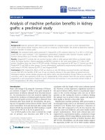
RNA-binding activity of CD151 in vitro and in vivoFigure 2
RNA-binding activity of CD151 in vitro and in vivo. (A)
In vito RNA-binding activity of CD151 was demonstrated by
Immunoprecipitation/North-Western blot analysis. BHK-21
and MARC-145 cells were transfected with pBK-CMV plas-
mid expressing CD 151 as a β-galactosidase fusion protein.
The cell lysates were immunoprecipitated with anti-CD151
MAb (A1) and anti-β-galactosidase MAb (A2). In A1, lane1,
MARC-145 cytoplasmic protein lysate (without immunopre-
cipitation); lanes 2, transfected MARC-145; lane 3, trans-
fected BHK-21; lane 4, untransfected MARC-145; lane 5,
untransfected BHK-21. In A (2), lane1, MARC-145 cytoplas-
mic protein lysate (without immunoprecipitation); lane 2,
untransfected BHK-21; lane 3, transfected BHK-21; lane 4,
transfected MARC-145; lane 5, untransfected MARC-145.
FIG 2B, gel shift assay demonstrating the interaction of
CD151 protein with the PRRSV 3'UTR RNA. MARC cell
lysate was immunoprecipitated with CD151 antibody (lanes
1&3) and the complex was incubated radiolabelled PRRSV 3'
UTR RNA. Addition of unlabelled RNA (lane 3) prevented
the formation of complex, while the radiolabelled RNA did
not interact with the CD151 antibody (lane 3). FIG 2C, In
vivo RNA-binding activity of CD151 was demonstrated by
immunoprecipitation/RT-PCR assay (149 bp amplicon).
PRRSV-infected or uninfected MARC-145 cell lysates were
immunoprecipitated with anti-CD151 MAb or a negative
control MAb (wasp, Cotesia folepis MAb), and RT-PCR was
performed using PRRSV 3' UTR RNA-specific primers for
RNAs extracted from the immunocomplexes. M, 123 bp lad-
der; lane 1, negative PCR control; lane 2, PRRSV-uninfected/
CD151 MAb-immunoprecipitated; lane 3, PRRSV-infected/
wasp MAb-immunoprecipitated; lane 4, PRRSV-infected/
CD151 MAb-immunoprecipitated (without UV cross-link-
ing); lane 5, PRRSV-infected/CD151 MAb-immunoprecipi-
tated (UV cross-linked for 15 min); lane 6, PRRSV-infected/
CD151 MAb-immunoprecipitated (UV cross-linked for 30
min); lane 7, PRRSV-infected/CD151 MAb-immunoprecipi-
tated (UV cross-linked for 45 min).
Alignment of CD151 amino acid sequencesFigure 1
Alignment of CD151 amino acid sequences. Simian
CD151 amino acid sequence was generated from the cDNA
sequence. The amino acid sequence was aligned with human,
bovine, murine and porcine CD151 amino acid sequences.
Dots represent similarity of amino acid residues. Genbank
accession number is AF 275666 [Genbank: AF275666
].
Virology Journal 2007, 4:62 />Page 4 of 12
(page number not for citation purposes)
cipitation [34-38]. MARC-145 cells were infected with
PRRSV, and after UV cross-linking, the cytoplasmic pro-
teins were isolated and immunoprecipitated with anti-
CD151 MAb. Then, RNA was isolated from the immuno-
complex, and RT-PCR was performed using PRRSV 3' UTR
RNA-specific primers. PRRSV 3' UTR was detected in the
immunocomplex demonstrating that the CD151 protein
interacts in vivo with PRRSV 3' UTR. {Fig. 2C, lane 4–7}.
However, PRRSV 3' UTR RNA was neither detected in the
immunocomplex from uninfected MARC-145 cells using
anti-CD151 MAb {Fig. 2C, lane 2} nor detected in the
immunocomplex from PRRSV-infected MARC-145 cells
using the isotype control MAb against wasp protein
Cotesia folepis. {Fig. 2C, lane 3}. These results clearly
demonstrate that CD151 protein interacts with 3' UTR
RNA of PRRSV.
Correlation between CD151 expression and susceptibility
to PRRSV infection
To determine the possible relationship between the pres-
ence of CD151 and susceptibility to PRRSV infection, we
screened various PRRSV susceptible and non-susceptible
cell lines using RT-PCR for CD151. As shown in Fig. 3A, a
105 bp amplicon of CD151 was amplified in MARC-145
{Fig. 3A, lane 4}, ST {Fig. 3A, lane 7}, MA-104 {Fig. 3A,
lane 8}, ST-K {Fig. 3A, lane 9}, Vero {Fig. 3A, lane 10},
CL-2621 {Fig. 3A, lane 11}, COS-7 {Fig. 3A, lane 12},
and simian CD151-transfected BHK-21 cells {Fig. 3A,
lane 13}. However, the 105 bp amplicon of CD151 was
not amplified in HRT {Fig. 3A, lane 3}, MDBK {Fig. 3A,
lane 5} and BHK-21 cells {Fig. 3A, lane 6}. MARC-145,
MA-104, CL-2621 and Vero cells are known to be suscep-
tible to PRRSV infection, while BHK-21 cells are known to
be non-susceptible [19,20]. We also performed Western
blot analysis using anti-CD151 MAb to determine the
presence of CD151 in some of the PRRSV-susceptible and
-non-susceptible cell lines. As shown in Figure 3B, CD151
was detected in susceptible cell lines, MARC-145 {Fig. 3B,
lane 1} and Vero {Fig. 3B, lane 3}, while CD151 was not
detected in a non-susceptible cell line, BHK-21 {Fig. 3B,
lane 2}. Additionally, we also found the expression of
CD151 protein by flow cytometric analysis in MARC-145
and BHK-21 cells. CD151 protein was expressed on the
surface of MARC 145 cells but not on surface of BHK-21
cells {Fig 3C}.
Transfection of non-susceptible cell line (BHK-21) with
CD151 confers susceptibility to PRRSV
The PRRSV non-susceptible cell line, BHK-21 was trans-
fected with the pBK-CMV plasmid containing CD151
gene and then was infected with PRRSV. Immunohisto-
chemical staining was performed to detect the presence of
PRRSV in simian CD151-transfected BHK-21 cells using
SR-30, a MAb against PRRSV nucleocapsid protein. As
shown in Fig. 4, CD151-transfected BHK-21 cells could be
Detection of the presence of CD151 by RT-PCR and Western blot
Figure 3
Detection of the presence of CD151 by RT-PCR and West-
ern blot. Correlation between CD151 expression and susceptibil-
ity to PRRSV infection was demonstrated by RT-PCR and Western
blot analysis. (A) RT-PCR showing the amplification of 105 bp
amplicon with CD151-specific primers was performed for RNAs
isolated from PRRSV-susceptible and -non-susceptible cell lines. M,
123 bp ladder; lane 1, negative RT control; lane 2, negative PCR
control; lane 3, HRT; lane 4, MARC-145; lane 5, MDBK; lane 6,
BHK-21; lane 7, ST; lane 8, MA-104; lane 9, ST-K; lane 10, Vero;
lane 11, CL-2621; lane 12, COS; lane 13, CD151-transfected BHK-
21. (B) Western blot analysis using anti-CD151 MAb was per-
formed for cell lysates from PRRSV-susceptible and -non suscepti-
ble cell lines. Lane 1, MARC-145; lane 2, BHK-21; lane 3, Vero. (C)
Flow cytometric analysis using polyclonal anti-CD151 Ab was per-
formed for MARC-145 (C (1)) and BHK-21 (C (2)) cell lines. An
isotype-matched control is represented by the dotted lines.
Virology Journal 2007, 4:62 />Page 5 of 12
(page number not for citation purposes)
infected with PRRSV {Fig. 4A}, while untransfected BHK-
21 cells could not be infected with PRRSV {Fig. 4B}.
Where as the BHK-21 cells transfected with control plas-
mid (CMV driven β-gal protein) did not confer suscepti-
bility to PRRSV infection (data not shown). These results
indicate that CD151 should be one of the susceptibility
factors to PRRSV infection.
Interaction between CD151 and PRRSV proteins
The interaction between CD151 and PRRSV proteins and
CD151 was investigated by (co-) immunoprecipitation.
The infected MARC-145 cells were immunoprecipitated
with anti-CD151 MAb, and the presence of PRRSV pro-
teins in the immunocomplex was examined by PRRSV
hyperimmune serum, followed by detection with the ECL
system. The co-immunoprecipitation was also performed
by immunoprecipitating with PRRSV hyperimmune
serum, and the presence of CD151 in the immunocom-
plex was examined by anti-CD151 MAb. Virus overlay
protein binding assay (VOPBA) was performed to investi-
gate if there is any direct interaction between PRRSV pro-
teins and CD151 as described ([39]. However, any direct
interactions between the CD151 and PRRSV proteins were
not detected (data not shown).
Effect of CD151-overexpression on PRRSV infection levels
To address the effect of CD151-overexpression on PRRSV
infection, MARC cells were examined with respect to the
effect on infectivity level. Both CD151-transfected and
untransfected MARC-145 cells were infected with equal
amounts of plaque-purified PRRSV. The cells were
allowed to grow for one complete replication cycle (18
hr), and the infectivity levels of PRRSV in both simian
CD151-transfected and -untransfected MARC-145 cells
were measured by plaque assay. Additionally, simian
CD151-transfected BHK-21 cells were also examined. As
shown in Fig. 5, there was approximately a 100-fold
increase in the amount of virus in the simian CD151-
transfected MARC-145 cells overexpressing CD151 {Fig.
5, column 1} as compared to untransfected MARC-145
cells {Fig. 5, column 2}. The simian CD151-transfected
BHK-21 cells also allowed for PRRSV replication at a
higher level than untransfected MARC-145 cells {Fig. 5,
column 3}.
Effect of siRNA against CD 151
To study the effect of suppression of CD151 expression on
PRRSV replication, the transfection of siRNA against
CD151 was performed with MARC-145 cells. Figure 6A
shows the effect of the transfection of siRNA against
CD151 on CD151 expression. The expression level of
CD151 was reduced (36% to 19%) by the transfection of
siRNA against CD151 {Fig. 6A (2)}, even though the
expression level of CD151 in the mock-transfected MARC-
145 cells was not high {Fig. 6A (1)}. Figure 6B shows the
effect of the transfection of siRNA against CD151 on
Effect of CD151-overexpression on PRRSV infectionFigure 5
Effect of CD151-overexpression on PRRSV infection.
The effect of CD151-overexpression on PRRSV infection
was demonstrated by virus burst assay. To induce CD151-
overexpression, the simian CD151 expressing clone was
transfected into MARC-145 cells. Column 1, CD151-trans-
fected/PRRSV-infected MARC-145; column 2, β-galactosi-
dase-transfected/PRRSV-infected MARC-145; column 3,
CD151-transfected/PRRSV-infected BHK-21; column 4,
CD151-untransfected/PRRSV-infected BHK-21; column 5,
CD151-transfected/PRRSV-uninfected MARC.
Transfection of simian CD151 into BHK-21 cellsFigure 4
Transfection of simian CD151 into BHK-21 cells. To
detect the presence of PRRSV in simian CD151-transfected
BHK-21 cells, immunohistochemical staining was performed
using SR-30, a MAb against PRRSV nucleocapsid protein. (A)
Simian CD151-untransfected BHK-21 cells, and (B) Simian
CD151-transfected BHK-21 cells. The presence of PRRSV is
shown by DAB substrate in brown color.
Virology Journal 2007, 4:62 />Page 6 of 12
(page number not for citation purposes)
PRRSV infection. PRRSV infection was significantly
reduced (50% reduction as determined by fluorescent
staining) by the transfection of siRNA against CD151
{Fig. 6B (2)}.
Blocking activity of anti-CD151 Ab on PRRSV infection
into MARC-145 cells
To investigate the effect of polyclonal anti-CD151 Ab on
PRRSV infection into MARC-145 cells, a checkerboard
titration assay was performed. As shown in Table 1, poly-
clonal anti-CD151 Ab blocked PRRSV infection in a dose-
dependent manner. Even at the highest concentration of
the virus (10
-1
-dilution), polyclonal anti-CD151 Ab com-
pletely blocked PRRSV infection. However, a negative
control Ab, anti-β-galactosidase MAb, did not block
PRRSV infection (data not shown). Figure 7 shows the
complete blocking activity of polyclonal anti-CD151 Ab
on PRRSV infection by immunofluorescence antibody
assay.
Discussion
Viruses are obligate intracellular parasites, which use host
cellular factors and energy supplies for replication. In sev-
eral RNA viruses, the interaction between 5' and/or 3' UTR
RNA and host cell proteins was already reported to play an
important role in virus replication mechanisms, such as
the transcription, translation, orientation and transport of
viral RNA [23,40].
In this study we were able to demonstrate for the first time
that CD151 protein binds to 3' UTR RNA of PRRSV. Inter-
action between CD151 and RNA of PRRSV is specific (Gel
shift assay) and interaction also occurs in vivo (detection
of PRRSV RNA in immunoprecipitation). Another impor-
tant observation of our study is that CD151 confers
PRRSV susceptibility to BHK-21 cells. Previously it has
been shown that BHK-21 cells are non-susceptible to
Effect of siRNA against CD151 on PRRSV infectionFigure 6
Effect of siRNA against CD151 on PRRSV infection.
(A) To examine the effect of siRNA against CD151 on
PRRSV infection, siRNA was transfected into MARC-145
cells. The suppression of the cell surface expression of
CD151 by the transfection of siRNA was shown by flow
cytometric analysis for the untransfected MARC-145 cells
(A1) and the transfected MARC-145 cells (A2). An isotype-
matched control is represented by the dotted lines. (B) The
effect of siRNA on PRRSV infection was shown by immun-
ofluorescence antibody assay using FITC-conjugated SDOW-
17, a MAb against PRRSV nucleocapsid protein for the
untransfected MARC-145 cells (B 1) and the transfected
MARC-145 cells (B 2).
Table 1: Checkerboard titration assay for measuring the blocking activity of anti-CD151 Ab
Virus dilution (1:9-diluted) ↓ 10
-1
CCC0012.5 3 33 33
10
-2
Ccc0012.5 3 33 33
10
-3
Cc c 0 0 0.5 1 2 2 2 22
10
-4
ccc 0 0 0 0 0.5 1 1 11
10
-5
c c c 0 0 0 0 0 0.5 0.5 0.5 0.5
10
-6
ccc 0 0 0 0 0 0 0 00
10
-7
ccc 0 0 0 0 0 0 0 00
No virus Ccc000 0 0 00 00
Ab dilution(1:1-diluted) → No Ab
20
-1
40
-1
80
-1
160
-1
320
-1
640
-1
1280
-1
2560
-1
5120
-1
MARC-145 cells were cultured with polyclonal anti-CD151 Ab and/or PRRSV in a 96-well tissue culture plate. Polyclonal anti-CD151 Ab was
1:1-serially diluted from 20
-1
-dilution, and the PRRSV preparation was 1:9-serially diluted from 10
-1
-dilution. At 2 days post infection,
immunofluorescence microscopy analysis was performed. The cells were stained with FITC-conjugated SDOW-17, a MAb against PRRSV
nucleocapsid protein. The cells were examined by fluorescent microscopy. C means the cytopathic effect of Ab, and the numbers mean the
intensity of fluorescence (0 means no fluorescence detected, and 3 means the highest intensity of fluorescence)
Virology Journal 2007, 4:62 />Page 7 of 12
(page number not for citation purposes)
PRRSV infection. However these cells when transfected
with either PRRSV RNA or infectious cDNA clones, it
results in productive infection of PRRSV without spread-
ing to neighbouring cells [19]. The major factor that is
lacking in BHK-21 cells that prevent the infection seems to
be in entry. Since CD151 is a transmembrane protein, we
reasoned that it might function as the entry molecule and
performed (co-) immunoprecipitation experiments to
determine if there is direct interaction between CD151
and the PRRSV protein. We could not detect any direct
interaction between them using (co-) immunoprecipita-
tion and virus overlay protein binding assay (data not
shown). Our results are in agreement with role of another
tetraspanin molecule CD9 that has been shown to render
MDBK cells susceptible to infection by a canine distemper
virus (CDV) and predicted that this molecule serves as the
entry molecule. However, they also could not demon-
strate any direct interaction between CD9 and CDV pro-
teins [40]. Therefore we cannot completely rule out the
possibility of interaction between the CD151 and PRRSV
proteins leading to helping of virus entry into BHK-21
cells.
CD151 is a 29-kDa transmembrane glycoprotein with an
N-glycosylation site and several palmitoylation sites
[41,42]. CD151 is a member of the tetraspanin super-
family, alternately known as the transmembrane 4 super-
family, which is characterized by the presence of four
highly conserved hydrophobic transmembrane domains.
CD151 was initially identified as a human platelet surface
glycoprotein (platelet endothelial tetraspan antigen-3;
PETA-3) by a monoclonal antibody inducing platelet
aggregation [43]. CD151 was also independently cloned
as SF-HT-activated gene 1 (SFA-1), which was up-regu-
lated in human T cells by transformation with human T-
cell-leukemia virus type 1 [44]. We found that CD151
protein is highly conserved across the species examined
with high homology between human and simian species
and our results are in agreement with previous report [45].
In this study, we examined the expression of CD151 in
several cell lines to determine if it is the susceptibility fac-
tor in PRRSV infection. CD151 was expressed in all sus-
ceptible cell lines namely, MA-104, MARC-145, COS-7
and Vero cells, which are derived from African green mon-
key kidney. However, CD151 was not expressed in BHK-
21 and MDBK cells, which are derived from kidneys of the
other species. CD151 has a wide cell and tissue distribu-
tion, including platelets, megakaryocytes, activated T lym-
phocytes, dendritic cells, Schwann cells, epithelial cells,
endothelial cells, and muscle cells [43,44,46]. In account
of our novel observation of RNA binding activity of
CD151, we looked for RNA binding domains on CD151
protein by bioinformatic analysis, we could not find any
known RNA binding activity but there were some motifs
in second extracellular domain which could be potential
RNA binding sites. Current experiments are underway to
identify potential RNA binding motifs.
Evidence presented in this study definitely points that
CD151 confers susceptibility to PRRSV infection. It is evi-
dent when transfection of a CD151 expressing clone into
MARC-145 cells increased the susceptibility of MARC cells
to PRRSV. Conversely, decreased expression of CD151 by
using siRNA also inhibited the susceptibility of MARC-
145 cells to PRRSV infection. Furthermore, the antibody
against CD151 completely inhibited PRRSV infection of
MARC-145 cells. These results indicate that CD151 plays
very important role in PRRSV infection of MARC-145
cells. To this end, only direct interaction between CD151
and PRRSV is that of RNA-protein interaction. How can
CD151, a transmembarane protein, by virtue of its bind-
ing to PRRSV RNA help in virus infection? PRRSV and
other arteriviruses, enter into host cells by receptor-medi-
ated endocytosis. CD151, by virtue of its expression on
the plasma membranes and in intracellular vesicles, like
endosomes [33,46], interacts with PRRSV in cooperation
with other molecules [13-18]. Even though we could not
directly demonstrate the interaction between CD151 and
PRRSV protein, we cannot rule if there is any direct inter-
action between them. Another example of tetraspan mol-
ecule promoting viral entry is CD82 and CD81 molecules
in case of HTLV-1 virus [47-49], however in this case,
binding of CD81 to viral glycoprotein E2 does not corre-
late with permissiveness of cells to virus infection. This
implies that other cellular factors are required for viral
Effect of anti-CD151 Ab on PRRSV infectionFigure 7
Effect of anti-CD151 Ab on PRRSV infection. To
examine the effect of anti-CD151 Ab on PRRSV infection,
immunofluorescence antibody assay was performed. MARC-
145 cells were incubated with polyclonal anti-CD151 Ab (A)
or PBS (B) and infected with PRRSV. At 2 days post infection,
the presence of PRRSV in the cells was detected by FITC-
conjugated SDOW-17, a MAb against PRRSV nucleocapsid
protein.
Virology Journal 2007, 4:62 />Page 8 of 12
(page number not for citation purposes)
infection [47-49]. During endocytosis, lowering of pH in
the endosome results in fusion event between viral enve-
lope and endosome [18] possibly involving CD151.
Another role of CD151 by virtue of RNA binding ability is
possibly in localization of ribonucleoprotein complexes
to the site of viral replication [21,41] that has been dem-
onstrated to promote viral replication.
Conclusion
Based on our results, we propose that CD151 is one of the
key molecule in facilitating PRRSV infection. To our
knowledge, it is the first demonstration of the interaction
between PRRSV 3' UTR RNA and a host cell protein,
CD151.
Methods
Cell lines and virus
African green monkey kidney cell lines (MARC-145, COS-
7, Vero, CL-2621 and MA-104), a baby hamster kidney
cell line (BHK-21), a bovine kidney cell line (MDBK), a
swine testis cell line (ST) and a human rectal tumor cell
line (HRT) were used in the study. These cell lines
obtained from ATCC were already available in our labora-
tory. The cell lines were grown in Eagle's minimum essen-
tial medium (MEM; Life Technologies, Inc., Gaithersburg,
MD) supplemented with 10% fetal bovine serum (FBS;
Hyclone, Logan, UT). The ATCC VR-2332 strain of PRRSV
was used in the study. The virus was propagated in MARC-
145 cells.
Construction of MARC-145 cDNA library
The cDNA library from MARC-145 cells was constructed
in our laboratory using a λ ZAP Express cDNA synthesis
kit (Stratagene, La Jolla, CA) by following manufacturer's
instructions. Briefly, total cellular RNA from MARC-145
cells was extracted according to the Chomczynski and Sac-
chi method [34]. The mRNA was purified from total cellu-
lar RNA using an oligo (dT) cellulose column (Stratagene,
La Jolla, CA), and then 5 µg of mRNA was converted to
cDNA. The cDNA was then directionally cloned in the λ
ZAP Express vector. The cDNA library was packaged using
the ZAP Express cDNA Gigapack III Gold cloning kit
(Stratagene, La Jolla, CA).
Cloning of PRRSV 3' UTR RNA and RNA probe
preparation
PRRSV 3' UTR was amplified by RT-PCR using forward 5'-
CCCCATTTTCCTCTA
GCGACTG-3' and reverse 5'-CGGCCGCATGGT-
TCTCGCCAAT-3' primers (regions corresponding to
15,386 to 15,846 bp of the PRRSV VR-2332) and then
cloned into the pCR II vector (Invitrogen, Carlsbad, CA).
α-
32
P-labeled 3' UTR RNA transcript was prepared by in
vitro transcription using a T7 RNA synthesis kit,
Riboscribe™ (Epicentre Technologies, Madison, WI) by
following the manufacturer's instructions. The probe was
purified either by Quick Spin™ columns (Boehringer Man-
nheim, Indianapolis, IN) for North-Western blotting or
by acrylamide gel electrophoresis [35] method of purifica-
tion for gel mobility shift assay.
North-Western screening of MARC-145 cDNA library
The MARC-145 cDNA library was screened using PRRSV 3'
UTR RNA by North-Western hybridization described [36].
In all the rounds of the screening, protein expression was
induced using nitrocellulose membranes impregnated
with 10 mM IPTG for 2 hr. The nitrocellulose membranes
were denatured in 6 M guanidinium hydrochloride for 30
min, followed by sequential renaturation every 10 min
with equal changes of single-binding (SB) buffer (15 mM
HEPES [pH 7.9], 50 mM KCl, 0.01% [vol/vol] Nonidet P-
40, 0.1% [wt/vol] Ficoll 400-DL, 0.1% [wt/vol] PVP-40,
0.1 mM MnCl
2,
0.1 mM ZnCl
2,
0.1 mM EDTA and 0.5 mM
DTT) for 1 hr. Hybridization was performed in SB buffer
containing the α-
32
P-labeled PRRSV 3' UTR RNA probe at
500,000 cpm/ml in presence of 10 µg/ml of yeast tRNA
and 100 µg/ml of denatured sheared salmon sperm DNA
overnight. The blots were washed with SB buffer for 1.5
hr, and RNA-binding activity was detected by autoradiog-
raphy. The corresponding positive plaques were cored,
eluted and then rescued using the ZAP Express cDNA
Gigapack III Gold cloning kit (Stratagene, La Jolla, CA).
Sequencing was performed at the Iowa State University
Sequencing Facility in Ames, IA.
Transfection of CD151 clone
BHK-21 and MARC-145 cells were transfected with pBK-
CMV plasmid containing CD151 gene using Lipo-
fectamine™ reagent (Life Technologies, Inc., Gaithersburg,
MD) by following manufacturer's instructions. For tran-
sient transfection, the cells were tested for protein expres-
sion 24 hrs after transfection. For stable transfection,
media was changed to selection medium containing G418
sulfate (Omega Scientific, Inc., Tarzana, CA) in growth
medium (1 mg/ml for BHK-21 cells and 0.7 mg/ml for
MARC-145 cells). After selection, the cells were main-
tained in the presence of G418 sulfate at 0.5 mg/ml for
BHK-21 cells and 0.35 mg/ml for MARC-145 cells. The
expression of CD151 was measured by immunoprecipita-
tion followed by North-Western hybridization.
Immunoprecipitation/North-Western hybridization
CD151 protein was immunoprocipitated using anti-
CD151 antibody and the RNA binding activity was
detected by North-Western hybridization. BHK-21 or
MARC-145 cells were transfected with CD151 as
described above. The transfected cells were lysed in 1 ml
of single detergent lysis buffer (50 mM Tris-HCl [pH8.0],
150 mM NaCl, Phenylmethylsulfonyl fluoride 100 µg/ml
Virology Journal 2007, 4:62 />Page 9 of 12
(page number not for citation purposes)
and 1% [vol/vol] Nonidet-P40). Proteins were quantified
using Bradford method based Bio-Rad assay (Bio-Rad
Laboratory Inc., Hercules, CA). To 500 µg of cell lysate, 1
mg/ml of anti-CD 151 MAb (BD Biosciences, Franklin
Lakes, NJ) or anti-β-galactosidase MAb (Boehringer Man-
nheim, Indianapolis, IN) was added and rocked overnight
at 4°C. The immunocomplexes were precipitated on ice
for 2 h with the addition of 40 µl of protein A-sepharose
beads (Sigma, St. Louis, MO) and then centrifuged at
4,000 × g for 10 min. The pellets were washed once in cold
Tris saline azide (TSA) buffer (0.05 M Tris-HCl [pH 8.0];
0.15 M NaCl; 0.025% NaN
3
) containing 1% Triton X-100
and 1% SDS. The second wash was done in cold TSA
buffer alone, followed by two washes in 10 mM Tris-HCl
[pH 7.5] containing 1 mM EDTA. The pellet was sus-
pended in 20 µl of SDS-loading buffer and electrophore-
sized by SDS-PAGE. The proteins were transferred onto a
nitrocellulose membrane, and North-Western hybridiza-
tion was performed as described above.
Gel mobility shift assay
To determine the specificity of interaction between
CD151 protein and the PRRSV 3' UTR RNA, we performed
gel mobility shift assay as described [25] with slight mod-
ifications. 500 µg of MARC cell lysate was immunoprecip-
itated with anti-CD151 MAb as described above. After
washing the immunocomplexes, the immunoprecipitate
was resuspended in 50 µl of incubation buffer (50 mM
HEPES [pH7.4], 0.1 mM DTT, 40 mM MgCl
2
, 0.5 mM
EDTA, 20 mM Spermidine, 1.5 mM ATP, 10 mM GTP)
along with 4 µg of yeast tRNA and incubated for 10 min
at 4°C. Labeled RNA (500,000 cpm) was added and incu-
bated further for 15 min. For competition experiments,
unlabelled RNA (3 fold excess) was included in the pre-
incubation prior to addition of labeled RNA.
In vivo cross-linking and reverse transcription (RT)-PCR
assay
To investigate in vivo interaction between CD151 and
PRRSV 3' UTR RNA, In vivo cross-linking followed by
immunoprecipitation and then RT-PCR was performed as
described with slight modifications [37,38] ([39]. MARC-
145 cells were infected with PRRSV at 37°C for 1 hr. The
cells were washed 3 times in PBS and twice in MEM, and
replaced with MEM supplemented with 1% FBS. At 18 hr
postinfection, the cells were washed twice in PBS and cov-
ered in PBS. Irradiation was performed on ice in a UV
cross-linker (Fisher Scientific, Pittsburgh, PA) at a distance
of 10 cm from the 300 λ light-source for 0, 15, 30 and 45
min. PBS was removed, and the cells were lysed by adding
ice cold RIPA lysis buffer (20 mM Tris-HCl [pH8.0], 150
mM NaCl, 1% Nonidet P-40, 1% SDS and 0.5% deoxy-
cholic acid) supplemented with 20 U of DNase and 20 U
of RNasin inhibitors (Life Technologies, Inc., Gaithers-
burg, MD). Immunoprecipitation was performed using
anti-CD151 MAb as described above, except that RNase
inhibitor (20 U) was added in all incubations. Immuno-
precipitate was treated with Proteinase K (4 µg/ml) at
37°C for 15 min, and RNA was extracted as described pre-
viously [34]. To determine the presence of PRRSV 3' UTR
RNA, RT-PCR was performed as described below. To
detect PRRSV 3' UTR RNA bound to the immunocomplex
in In vivo cross-linking and RT-PCR assay, RT-PCR was per-
formed using the GeneAmp EZ rTth RNA PCR kit (Roche
Molecular System, Inc., Branchburg, NJ) with PRRSV 3'
UTR RNA-specific primers; 5'-TGGGCTGGCATTCTT-
GAGGC-3' (forward) and 5'-TTCGGGCCGCATGGT-
TCTCGC-3' (reverse) that cover 15,262 bp to 15,410 bp
regions of PRRSV VR-2332 strain. Reverse transcription
was performed at 42°C for 45 min, 95°C for 10 min and
5°C for 5 min. Standard PCR was done at 95°C for 2 min,
95°C for 30 s, 55°C for 30 s, 72°C for 60 s for 25 cycles
and 72°C for 30 min. To demonstrate the correlation
between CD151 presence and susceptibility to PRRSV
infection, RT-PCR was carried out using CD151 specific
primers 5'-CCTACCTGGCCACAGCCTAC-3' (forward)
and 5'-ACAGGCGCAGCAGGTTCCGA-3' (reverse) that
amplifies 167 bp to 277 bp region of CD151. RNA was
isolated from PRRSV-susceptible and non-susceptible cell
lines as described previously [34]. Reverse transcription
reaction was performed at 42°C for 45 min, 95°C for 10
min and 5°C for 5 min. Standard PCR was done at 95°C
for 2 min, 95°C for 30 s, 55°C for 30 s, 72°C for 15 s for
25 cycles and 72°C for 30 min. The PCR products were
detected by agarose gel electrophoresis.
Western blot analysis
To examine the presence of CD151 in MARC-145, BHK-
21 and Vero cells, Western blot analysis was performed.
MARC-145, BHK-21 and Vero cytoplasmic proteins were
electrophoresed by SDS-PAGE and transferred onto a
nitrocellulose membrane. After blocking in 5% skim-milk
in PBS, the membrane was stained with anti-CD151 MAb
at room temperature for 1 hr, followed by staining with
the peroxidase-conjugated horse anti-mouse IgG (H+L)
(Vector Laboratories, Inc., Burlingame, CA) at room tem-
perature for 45 min. The proteins were detected by the
enhanced chemiluminescence (ECL) system (Amersham
Biosciences, Piscataway, NJ) by following manufacturer's
instructions.
Flow cytometric analysis
To investigate the cell surface expression of CD151 and
quantify CD151 protein in MARC-145 and BHK-21 cells,
flow cytometry was performed. After trypsinization, cells
(5 × 10
5
total) were washed twice in staining solution
(0.1% bovine serum albumin [BSA] in PBS) and blocked
in 3% BSA in staining solution on ice for 10 min, and then
incubated with polyclonal goat anti-CD151 Ab (Santa
Cruz Biotechnology, Inc., Santa Cruz, CA) on ice for 30
Virology Journal 2007, 4:62 />Page 10 of 12
(page number not for citation purposes)
min. After washing twice in staining solution, cells were
incubated with rabbit anti-goat FITC conjugated second-
ary Ab (Bethyl Laboratories, Montgomery, TX) on ice for
30 min. Cells were resuspended in 1% paraformalehyde
in PBS after washing twice in staining solution. Flow cyto-
metric analysis was performed on a FACSCalibur (BD Bio-
sciences, San Jose, CA). In transfection experiment
involving siRNA against CD151, the siRNA-transfected
MARC-145 cells were stained as described above.
Immunohistochemistry
To determine if the CD151-transfected BHK-21 cells
become susceptible to PRRSV infection, immunohisto-
chemical staining was performed using a MAb against
PRRSV nucleocapsid protein. The cells were cultured in a
24 well plate and infected with PRRSV. At 24 hr post infec-
tion, the cells were fixed in 75% acetone in PBS at 4°C for
10 min and stained with SR-30 (Rural Technologies, Inc.,
Brookings, SD), a MAb against PRRSV nucleocapsid pro-
tein at 37°C for 1 h, followed by staining with a bioti-
nylated anti-mouse IgG (Vector Labs, Burlingame, CA) at
RT for 30 min. Finally, the avidin-biotin-enzyme complex
(Vector Labs, Burlingame, CA) was added. The presence of
PRRSV in the cells was detected by the addition of DAB
substrate (Vector Labs, Burlingame, CA). The cells were
counterstained with Gill's-1 hematoxylin and examined
by light microscopy.
Immunoprecipitation/co-immunoprecipitation
To examine the interaction between CD151 and PRRSV
proteins, immunoprecipitation was performed. MARC-
145 cells were infected with PRRSV, and the cell lysate was
prepared in single detergent lysis buffer 2 days post infec-
tion. The PRRSV-infected MARC-145 cell lysate was
immunoprecipitated with anti-CD151 MAb as described
above. The immunocomplex was electrophoresized by
SDS-PAGE and transferred onto a nitrocellulose mem-
brane. After blocking in 5% skim-milk in PBS, the mem-
brane was stained with PRRSV hyper immune serum at
room temperature for 1 hr, followed by staining with the
peroxidase-conjugated secondary Ab (goat anti-porcine
IgG [H+L]; ICN Biomedicals, Inc., Aurora, OH) at room
temperature for 1 hr. The presence of PRRSV proteins was
determined by the addition of TMB membrane peroxidase
substrate (one component) (KPL, Inc., Gaithersburg,
MD). Also, the PRRSV-infected MARC-145 cell lysate was
co-immunoprecipitated with PRRSV hyper immune
serum. The immunocomplex was electrophoresed by
SDS-PAGE and transferred onto a nitrocellulose mem-
brane. After blocking in 5% skim-milk in PBS, the mem-
brane was stained with anti-CD151 MAb, followed by
staining with the peroxidase-conjugated secondary Ab
(horse anti-mouse IgG [H+L]). The presence of CD151
bound to PRRSV proteins was determined by the addition
of TMB membrane peroxidase substrate (one compo-
nent).
Virus replication assay
To investigate the effect of CD151-overexpression in
MARC-145 cells, a virus replication assay was performed.
The simian CD151-transfected MARC-145 cells were
infected with PRRSV at 37°C for 1 hr, washed twice in
MEM, and then overlaid with MEM supplemented with
1% FBS. At 18 hr postinfection, the cells were lysed by
freezing and thawing, and cell debris was removed by cen-
trifugation. The amount of virus in the supernatant was
titrated by plaque assay using MARC-145 cells. In plaque
assay, the supernatant was initially diluted 1:10 and in 10-
fold dilutions thereafter, and used for infection to MARC-
145 cells. After infection, the cells were washed twice in
MEM and overlaid with MEM containing 1% FBS and 1%
agar. After incubation at 37°C for 24 h, plaques were vis-
ualized by staining with 0.01% neutral red.
Transfection of siRNA against CD151
Silencer™ pre-designed siRNA against CD151 (Ambion,
Austin, TX) was used for transfection. The sequence of the
siRNA strands was as follows: 5'-GUUGGAGACC
UUCAUCCAGTT-3' (sense) and 5'-CUGGAUGAAG-
GUCUCCAACTT-3' (antisense). The transfection of the
siRNA was performed with DharmaFECT™ reagent (Dhar-
macon, Lafayette, CO) by following the manufacturer's
instructions. MARC-145 cells were cultured overnight in a
96- or 6-well tissue culture plates. The siRNA (10 – 100
nM) was complexed with DharmaFECT™ reagent by incu-
bating together at room temperature for 20 min. After
removing the cell culture supernatant, the complex was
added. After incubation for 3 days, the cells were infected
with PRRSV. At 3 days post-infection, flow cytometric
analysis and immunofluorescence antibody assay were
performed. Flow cytometric analysis was performed as
described above. For immunofluorescence antibody
assay, the siRNA-transfected MARC-145 cells were fixed
with 80% acetone in PBS and stained with FITC-conju-
gated SDOW-17 (Rural Technologies, Inc., Brookings,
SD), a MAb against PRRSV nucleocapsid protein. The cells
were examined by fluorescence microscopy for PRRSV.
Checkerboard titration assay for measuring blocking
activity of anti-CD151 Ab
To examine the blocking activity of anti-CD151 Ab,
checkerboard titration assay was performed. MARC-145
cells were cultured overnight in a 96-well tissue culture
plate (1 × 10
5
cells/well). The cells were incubated with
PRRSV, which were pre-incubated with polyclonal anti-
CD151 Ab (Santa Cruz Biotechnology, Inc., Santa Cruz,
CA) or anti-β-galactosidase MAb (Boehringer Mannheim,
Indianapolis, IN). The antibodies were prepared as serial
Virology Journal 2007, 4:62 />Page 11 of 12
(page number not for citation purposes)
two-fold dilutions starting with a 1:20 dilution, and the
PRRSV preparation was initially diluted 1:10 and in 10-
fold dilutions thereafter. At 2 days postinfection, the cells
were fixed with cold 80% acetone at 4°C for 10 min and
then incubated at 37°C for 30 min with FITC-conjugated
SDOW-17, a MAb against PRRSV nucleocapsid protein.
After being washed twice in PBS, the cells were examined
by fluorescence microscopy.
Competing interests
The author(s) declare that they have no competing inter-
ests.
Authors' contributions
KS designed and carried out the experiment and drafted
the manuscript.
JKK designed and carried out the experiment and drafted
the manuscript.
SK designed and carried out the experiment and drafted
the manuscript.
All authors read and approved the final manuscript
Acknowledgements
Contribution No 00-414-J from the Kansas Agricultural Experiment Sta-
tion, Manhattan, KS 66503. This project was supported by USDA Health
Funds (NC 229 Project). This work was conducted at Louise C. Averill
Research Laboratory, Department of Diagnostic Medicine/Pathobiology,
College of Veterinary Medicine, Kansas State University, Manhattan, KS
66506
We thank Teresa Yeary for excellent editorial assistance.
References
1. Bautista EM, Goyal SM, Collins JE: Serologic survey for Lelystad
and VR-2332 strains of porcine respiratory and reproductive
syndrome (PRRS) virus in US swine herds. J Vet Diagn Invest
1993, 5:612-614.
2. Bautista EM, Goyal SM, Yoon IJ, Joo HS, Collins JE: Comparison of
porcine alveolar macrophages and CL 2621 for the detection
of porcine reproductive and respiratory syndrome (PRRS)
virus and anti-PRRS antibody. J Vet Diagn Invest 1993, 5:163-165.
3. Goyal SM: Porcine reproductive and respiratory syndrome. J
Vet Diagn Invest 1993, 5:656-664.
4. Meulenberg JJ, de Meijer EJ, Moormann RJ: Subgenomic RNAs of
Lelystad virus contain a conserved leader-body junction
sequence. J Gen Virol 1993, 74(Pt 8):1697-1701.
5. Meulenberg JJ, Hulst MM, de Meijer EJ, Moonen PL, den Besten A, de
Kluyver EP, Wensvoort G, Moormann RJ: Lelystad virus, the caus-
ative agent of porcine epidemic abortion and respiratory
syndrome (PEARS), is related to LDV and EAV. Virology 1993,
192:62-72.
6. Cavanagh D: Nidovirales: a new order comprising Coronaviri-
dae and Arteriviridae. Arch Virol 1997, 142:629-633.
7. Lai MM, Cavanagh D: The molecular biology of coronaviruses.
Adv Virus Res 1997, 48:1-100.
8. Cao XQ, Liu TY, Nakhasi HL: The cis-acting 3'-element of
rubella virus RNA has DNA promoter activity. Gene 1992,
114:251-256.
9. Plagemann PG, Moennig V: Lactate dehydrogenase-elevating
virus, equine arteritis virus, and simian hemorrhagic fever
virus: a new group of positive-strand RNA viruses. Adv Virus
Res 1992, 41:99-192.
10. Larochelle R, Mardassi H, Dea S, Magar R: Detection of porcine
reproductive and respiratory syndrome virus in cell cultures
and formalin-fixed tissues by in situ hybridization using a dig-
oxigenin-labeled probe. J Vet Diagn Invest 1996, 8:3-10.
11. Sur JH, Cooper VL, Galeota JA, Hesse RA, Doster AR, Osorio FA: In
vivo detection of porcine reproductive and respiratory syn-
drome virus RNA by in situ hybridization at different times
postinfection. J Clin Microbiol 1996, 34:2280-2286.
12. Kim HS, Kwang J, Yoon IJ, Joo HS, Frey ML: Enhanced replication
of porcine reproductive and respiratory syndrome (PRRS)
virus in a homogeneous subpopulation of MA-104 cell line.
Arch Virol 1993, 133:477-483.
13. Delputte PL, Vanderheijden N, Nauwynck HJ, Pensaert MB: Involve-
ment of the matrix protein in attachment of porcine repro-
ductive and respiratory syndrome virus to a heparinlike
receptor on porcine alveolar macrophages. J Virol 2002,
76:4312-4320.
14. Vanderheijden N, Delputte P, Nauwynck H, Pensaert M: Effects of
heparin on the entry of porcine reproductive and respiratory
syndrome virus into alveolar macrophages. Adv Exp Med Biol
2001, 494:683-689.
15. Vanderheijden N, Delputte PL, Favoreel HW, Vandekerckhove J, Van
Damme J, van Woensel PA, Nauwynck HJ: Involvement of siaload-
hesin in entry of porcine reproductive and respiratory syn-
drome virus into porcine alveolar macrophages. J Virol 2003,
77:8207-8215.
16. Kim JK, Fahad AM, Shanmukhappa K, Kapil S: Defining the cellular
target(s) of porcine reproductive and respiratory syndrome
virus blocking monoclonal antibody 7G10. J Virol 2006,
80:689-696.
17. Kreutz LC: Cellular membrane factors are the major deter-
minants of porcine reproductive and respiratory syndrome
virus tropism. Virus Res 1998, 53:121-128.
18. Kreutz LC, Ackermann MR: Porcine reproductive and respira-
tory syndrome virus enters cells through a low pH-depend-
ent endocytic pathway. Virus Res 1996, 42:137-147.
19. Meulenberg JJ, Bos-de Ruijter JN, van de Graaf R, Wensvoort G,
Moormann RJ: Infectious transcripts from cloned genome-
length cDNA of porcine reproductive and respiratory syn-
drome virus. J Virol 1998, 72:380-387.
20. Meulenberg JJ, Bos-de Ruijter JN, Wensvoort G, Moormann RJ: An
infectious cDNA clone of porcine reproductive and respira-
tory syndrome virus. Adv Exp Med Biol 1998, 440:199-206.
21. Liu Q, Yu W, Leibowitz JL: A specific host cellular protein bind-
ing element near the 3' end of mouse hepatitis virus genomic
RNA. Virology 1997, 232:74-85.
22. Nakhasi HL, Singh NK, Pogue GP, Cao XQ, Rouault TA: Identifica-
tion and characterization of host factor interactions with cis-
acting elements of rubella virus RNA. Arch Virol Suppl 1994,
9:255-267.
23. Yu W, Leibowitz JL: A conserved motif at the 3' end of mouse
hepatitis virus genomic RNA required for host protein bind-
ing and viral RNA replication. Virology 1995, 214:128-138.
24. Blackwell JL, Brinton MA: Translation elongation factor-1 alpha
interacts with the 3' stem-loop region of West Nile virus
genomic RNA. J Virol 1997, 71:6433-6444.
25. De Nova-Ocampo M, Villegas-Sepulveda N, del Angel RM: Transla-
tion elongation factor-1alpha, La, and PTB interact with the
3' untranslated region of dengue 4 virus RNA. Virology 2002,
295:337-347.
26. Zeenko VV, Ryabova LA, Spirin AS, Rothnie HM, Hess D, Browning
KS, Hohn T: Eukaryotic elongation factor 1A interacts with
the upstream pseudoknot domain in the 3' untranslated
region of tobacco mosaic virus RNA. J Virol 2002, 76:5678-5691.
27. Nanda SK, Johnson RF, Liu Q, Leibowitz JL: Mitochondrial HSP70,
HSP40, and HSP60 bind to the 3' untranslated region of the
Murine hepatitis virus genome. Arch Virol 2004, 149:93-111.
28. Nanda SK, Leibowitz JL: Mitochondrial aconitase binds to the 3'
untranslated region of the mouse hepatitis virus genome. J
Virol 2001, 75:3352-3362.
29. Dollenmaier G, Weitz M: Interaction of glyceraldehyde-3-phos-
phate dehydrogenase with secondary and tertiary RNA
structural elements of the hepatitis A virus 3' translated and
non-translated regions. J Gen Virol 2003, 84:403-414.
Publish with BioMed Central and every
scientist can read your work free of charge
"BioMed Central will be the most significant development for
disseminating the results of biomedical researc h in our lifetime."
Sir Paul Nurse, Cancer Research UK
Your research papers will be:
available free of charge to the entire biomedical community
peer reviewed and published immediately upon acceptance
cited in PubMed and archived on PubMed Central
yours — you keep the copyright
Submit your manuscript here:
/>BioMedcentral
Virology Journal 2007, 4:62 />Page 12 of 12
(page number not for citation purposes)
30. Fitter S, Sincock PM, Jolliffe CN, Ashman LK: Transmembrane 4
superfamily protein CD151 (PETA-3) associates with beta 1
and alpha IIb beta 3 integrins in haemopoietic cell lines and
modulates cell-cell adhesion. Biochem J 1999, 338(Pt 1):61-70.
31. Hasegawa H, Nomura T, Kishimoto K, Yanagisawa K, Fujita S: SFA-
1/PETA-3 (CD151), a member of the transmembrane 4
superfamily, associates preferentially with alpha 5 beta 1
integrin and regulates adhesion of human T cell leukemia
virus type 1-infected T cells to fibronectin. J Immunol 1998,
161:3087-3095.
32. Roberts JJ, Rodgers SE, Drury J, Ashman LK, Lloyd JV: Platelet acti-
vation induced by a murine monoclonal antibody directed
against a novel tetra-span antigen. Br J Haematol 1995,
89:853-860.
33. Sincock PM, Fitter S, Parton RG, Berndt MC, Gamble JR, Ashman LK:
PETA-3/CD151, a member of the transmembrane 4 super-
family, is localised to the plasma membrane and endocytic
system of endothelial cells, associates with multiple integrins
and modulates cell function. J Cell Sci 1999, 112(Pt 6):833-844.
34. Chomczynski P, Sacchi N: Single-step method of RNA isolation
by acid guanidinium thiocyanate-phenol-chloroform extrac-
tion. Anal Biochem 1987, 162:156-159.
35. Maxam AM, Gilbert W: A new method for sequencing DNA.
Proc Natl Acad Sci USA 1977, 74:560-564.
36. Sagesser R, Martinez E, Tsagris M, Tabler M: Detection and isola-
tion of RNA-binding proteins by RNA-ligand screening of a
cDNA expression library. Nucleic Acids Res 1997, 25:3816-3822.
37. Buckanovich RJ, Darnell RB: The neuronal RNA binding protein
Nova-1 recognizes specific RNA targets in vitro and in vivo.
Mol Cell Biol 1997, 17:3194-3201.
38. Shen ZJ, Esnault S, Malter JS: The peptidyl-prolyl isomerase Pin1
regulates the stability of granulocyte-macrophage colony-
stimulating factor mRNA in activated eosinophils. Nat Immu-
nol 2005, 6:1280-1287.
39. Ule J, Jensen KB, Ruggiu M, Mele A, Ule A, Darnell RB: CLIP identi-
fies Nova-regulated RNA networks in the brain. Science 2003,
302:1212-1215.
40. Loffler S, Lottspeich F, Lanza F, Azorsa DO, ter Meulen V, Schneider-
Schaulies J: CD9, a tetraspan transmembrane protein, renders
cells susceptible to canine distemper virus. J Virol 1997,
71:42-49.
41. Lai MM: Cellular factors in the transcription and replication of
viral RNA genomes: a parallel to DNA-dependent RNA tran-
scription. Virology 1998, 244:1-12.
42. Charrin S, Manie S, Oualid M, Billard M, Boucheix C, Rubinstein E:
Differential stability of tetraspanin/tetraspanin interactions:
role of palmitoylation. FEBS Lett 2002, 516:139-144.
43. Fitter S, Tetaz TJ, Berndt MC, Ashman LK: Molecular cloning of
cDNA encoding a novel platelet-endothelial cell tetra-span
antigen, PETA-3. Blood 1995, 86:1348-1355.
44. Hasegawa H, Utsunomiya Y, Kishimoto K, Yanagisawa K, Fujita S:
SFA-1, a novel cellular gene induced by human T-cell leuke-
mia virus type 1, is a member of the transmembrane 4
superfamily. J Virol 1996, 70:3258-3263.
45. Hasegawa H, Watanabe H, Nomura T, Utsunomiya Y, Yanagisawa K,
Fujita S: Molecular cloning and expression of mouse homo-
logue of SFA-1/PETA-3 (CD151), a member of the trans-
membrane 4 superfamily. Biochim Biophys Acta 1997,
1353:125-130.
46. Sincock PM, Mayrhofer G, Ashman LK: Localization of the trans-
membrane 4 superfamily (TM4SF) member PETA-3
(CD151) in normal human tissues: comparison with CD9,
CD63, and alpha5beta1 integrin. J Histochem Cytochem 1997,
45:515-525.
47. Imai T, Fukudome K, Takagi S, Nagira M, Furuse M, Fukuhara N,
Nishimura M, Hinuma Y, Yoshie O: C33 antigen recognized by
monoclonal antibodies inhibitory to human T cell leukemia
virus type 1-induced syncytium formation is a member of a
new family of transmembrane proteins including CD9,
CD37, CD53, and CD63. J Immunol 1992, 149:2879-2886.
48. Nagira M, Imai T, Ishikawa I, Uwabe KI, Yoshie O: Mouse homo-
logue of C33 antigen (CD82), a member of the transmem-
brane 4 superfamily: complementary DNA, genomic
structure, and expression. Cell Immunol 1994, 157:144-157.
49. Nagira M, Sato A, Miki S, Imai T, Yoshie O: Enhanced HIV-1 repli-
cation by chemokines constitutively expressed in secondary
lymphoid tissues. Virology 1999, 264:422-426.