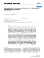
Báo cáo sinh học: " Characterization of vaccinia virus A12L protein proteolysis and its participation in virus assembly" pptx
Bạn đang xem bản rút gọn của tài liệu. Xem và tải ngay bản đầy đủ của tài liệu tại đây (1.4 MB, 12 trang )
BioMed Central
Page 1 of 12
(page number not for citation purposes)
Virology Journal
Open Access
Research
Characterization of vaccinia virus A12L protein proteolysis and its
participation in virus assembly
Su Jung Yang*
Address: Department of Microbiology, Oregon State University, Corvallis, Oregon 97331-3804, USA
Email: Su Jung Yang* -
* Corresponding author
Abstract
Vaccinia virus (VV) undergoes a proteolytic processing to evolve from immature virus particles into
intracellular mature virus particles. Most of structural core protein precursors such as p4a, p4b,
and p25K are assembled into previrions and then proteolytically processed to yield core proteins,
4a, 4b, and 25 K, which become components of a mature virus particle. These structural
rearrangements take place at a conserved cleavage motif, Ala-Gly-X (where X is any amino acid)
and catalyzed by a VV encoded proteinase, the I7L gene product. The VV A12L gene product, a 25
kDa protein synthesized at late times during infection is cleaved at an N-terminal AG/A site,
resulting in a 17 kDa cleavage product. However, due to the distinct characteristics of A12L
proteolysis such as the localization of both the A12L full-length protein and its cleavage product in
mature virions and two putative cleavage sites (Ala-Gly-Lys) located at internal and C-terminal
region of A12L ORF, it was of interest to examine the A12L proteolysis for better understanding
of regulation and function of VV proteolysis. Here, we attempted to examine the in vivo A12L
processing by: determining the kinetics of the A12L proteolysis, the responsible viral protease, and
the function of the A12L protein and its cleavage events. Surprisingly, the A12L precursor was
cleaved into multiple peptides not only at an N-terminal AG/A but also at both an N- and a C-
terminus. Despite the involvement of I7L proteinase for A12L proteolysis, its incomplete
processing with slow kinetics and additional cleavages not at the two AG/K sites demonstrate
unique regulation of VV proteolysis. An immunoprecipitation experiment in concert with N-
terminal sequencing analyses and mass spectrometry led to the identification of VV core and
membrane proteins, which may be associated with the A12L protein and suggested possible
involvement of A12L protein and its cleavage products in multiple stages in virus morphogenesis.
Background
Vaccinia virus (VV), the prototype member of the Poxviri-
dae family has a large double-stranded DNA genome. Rep-
lication and viral assembly occur entirely in the cytoplasm
of host cells, in particular, in areas referred as viroplasms
or virosomes. Virus assembly initiates at virosomes sur-
rounded by crescent membranes, which subsequently
engulf granular materials forming spherical-shaped parti-
cles named immature virions (IV). The IVs transform into
brick-shaped structures referred to as intracellular mature
virions (IMV) where viral DNAs become condensed and
packaged in an electron dense area and are covered by a
viral envelope membrane. A portion of IMVs is
enwrapped by a membrane cisternae derived from the
Published: 1 August 2007
Virology Journal 2007, 4:78 doi:10.1186/1743-422X-4-78
Received: 25 June 2007
Accepted: 1 August 2007
This article is available from: />© 2007 Yang; licensee BioMed Central Ltd.
This is an Open Access article distributed under the terms of the Creative Commons Attribution License ( />),
which permits unrestricted use, distribution, and reproduction in any medium, provided the original work is properly cited.
Virology Journal 2007, 4:78 />Page 2 of 12
(page number not for citation purposes)
trans-Golgi network and results in the formation of intra-
cellular enveloped virus (IEV), which then becomes fused
with the plasma membrane. If the IEVs remain associated
with the cells, they are referred to as cell-associated envel-
oped virus (CEV), or if the IEVs bud through the plasma
membrane spreading outside of the cells, they are consid-
ered extracellular enveloped virus (EEV).
Despite intensive study of VV morphogenesis, the mecha-
nism required for the transformation of IV to IMV still
remains poorly understood. The complex morphological
development during the transition initiates with success-
ful DNA replication, concatermer resolution [1,2] and
condensation/packaging of the viral genome in IV parti-
cles [3]. This is followed by encapsidation of a transcrip-
tion complex, formation of a defined core, and
reorganization of virion membranes [4]. In order to com-
plete this morphogenic transformation, VV undergoes a
various post-translational modifications such as proteo-
lytic processing of VV structural proteins, which contrib-
utes to proper virus morphogenic development and
acquisition of viral infectivity.
The cleavage processing of VV structural precursor pro-
teins are well studied. The cleavage reactions take place
after the second Gly residue of an Ala-Gly-X (AG/X) con-
served motif, as indicated in Figure 1. Most precursor pro-
teins show acidic upstream and basic downstream charge
differential across the cleavage site, which are usually
located within the N-terminal 60 amino acid residues and
catalyzed by I7L, a cysteine proteinase [5]. As an example,
p4b (A3L) and p25K (L4R) are synthesized at a late stage
in the virus life cycle with molecular weights of 66 kDa
and 28 kDa, and are proteolytically processed at an N-ter-
minal AG/A site to yield a 60 kDa peptide, 4b and a 25
kDa cleavage product, 25 K respectively [6]. P4a, however,
a 102 kDa precursor protein undergoes cleavage events at
two different AG/X motifs: an AG/S and an AG/T located
at amino acids 619 and 697 [7,8]. Proteolysis at the AG/S
and the AG/T sites leads to the release of a 62 kDa (4a)
and a 23 kDa C-terminal peptide. Cleavage at the N-termi-
nal AG/A site in A17L processes a 23 kDa full-length pre-
cursor protein (p21K) into a 21 kDa peptide (21 K) and
additional cleavage at the C-terminal AG/N site is cata-
lyzed by the I7L core protein proteinase [9]. G7L also uti-
lizes two distinct motifs, AG/F and AG/L. Mutagenesis
studies have demonstrated that both of these sites are
essential for the production of infectious virus [10].
Although a partial cleavage was observed at an AG/S motif
in the p25K ORF with an larger molecular weight of 25K,
referred as 25K' (Fig. 1), the tripeptides such as AG/L and
AG/N located in the N-terminus of p4b and p4a ORF do
not serve as reaction sites. These alternate sites, however,
do appear to be utilized for the proteolysis of G7L and
A17L. Thus, it is of interest to note that the presence of the
consensus cleavage motif is not sufficient enough to
induce VV proteolysis. Rather, the proteins destined for
VV AG/X cleavages are 1) late gene products, 2) catalyzed
by I7L proteinase, and 3) incorporated within the core of
assembling virions. These represent the characteristics of
VV morphogenic proteolysis, which requires a contextu-
ally constrained regulation.
The VV A12L protein is synthesized at a late stage with an
apparent molecular weight of 25 kDa (p17K) and is pro-
teolytically processed at an N-terminal AG/A site yielding
a 17 kDa polypeptide (17 K) similar to p4b and p25K.
However, unlike the core protein precursors, of which
only the processed forms, 4b and 25 K, are localized to the
mature virion, both p17K and 17 K are observed in the
core of mature virus, indicating distinct regulation/func-
tion of VV proteolysis [11]. In addition, A12L contains
two other AG/K sites in the internal region and C-termi-
nus of A12L open reading frame (ORF), of which utiliza-
tion for VV cleavage events has not been reported. Thus,
the research on A12L proteolytic processing may contrib-
ute to the discovery of requirements to initiate and regu-
late viral cleavage processing other than the consensus of
cleavage residues, identification of novel AG/X cleavage
motif, and elucidation of more detailed function of VV
proteolysis in the morphogenic transition. Here, we
attempted to characterize the proteolytic processing of the
A12L protein through determination of the kinetics, the
sites selected for the cleavage reactions, and identification
of the responsible protease. We also sought to demon-
strate possible A12L associations with other VV proteins,
Vaccinia virus morphogenic proteolysisFigure 1
Vaccinia virus morphogenic proteolysis. VV has six
structural precursor proteins, which undergo morphogenic
proteolysis. The consensus motif is not enough to induce VV
proteolysis. From left to right, the figure shows the name of
gene products, their cleavage motif (italic: not utilized, under-
lined: not determined), the localization of cleavage product,
and the responsible proteinase.
Virology Journal 2007, 4:78 />Page 3 of 12
(page number not for citation purposes)
providing a clue to the biological function of the A12L
proteolysis in virus assembly.
Results
Multiple cleavage products of A12L protein in vivo
Previous work by Whitehead and Hruby [11] demon-
strated that both the A12L precursor, p17K, and the AG/A
cleavage product, 17 K, were present in the core of assem-
bling virions. To determine if any other A12L-derived pro-
tein species were evident within the cytoplasm of VV-
infected cells, cytoplasmic extracts were prepared and sub-
jected to immunoblot analysis using A12L antisera (anti-
A12L) directed against the entire A12L protein. Surpris-
ingly, not only were the 25 kDa (p17K) and the 17 kDa
(17 K) proteins detected, but also five other peptides with
apparent molecular weights of 21, 18, 15, 13 and 11 kDa
were observed (Fig. 2A). Pre-immune sera of A12L did not
cross-react with any of these peptides, suggesting that all
of the proteins are indeed A12L-derived products (data
not shown).
In order to determine if VV proteolysis produces a number
of A12L-derived peptides, we compared the pattern of
A12L maturation processing in the presence and absence
of rifampicin (Rif). Rifampicin, an antibiotic, is known to
reversibly block the assembly of VV by disrupting the viral
membrane biogenesis and arresting maturational events
of the structural core proteins, such as p4a and p4b [12].
Thus, rifampicin has been used to determine the relation-
ship of VV precursor proteins and cleavage products. VV-
infected cells were incubated with rifampicin at various
concentrations from 100 to 400 μg/ml for 24 hours (Fig.
2B). Using p4b as a positive control, we were able to show
the suppressed cleavage at concentrations of 100~200 μg/
ml of rifampicin, while proteolysis was observed only in
the absence of rifampicin. Drug concentrations of more
than 200 μg/ml inhibited the expression of both precursor
proteins, p4b and p17K. Similar to p4b processing, p17K
was expressed in the presence and absence of rifampicin,
whereas the smaller peptides were produced only in the
absence of the drug, indicating that p17K is processed into
multiple peptides by VV proteolytic processing. Next, we
performed a rifampicin-reversibility experiment to con-
firm that the A12L proteolytic processing is regulated by
rifampicin (Fig. 2C). The hypothesis that the rifampicin-
arrested proteolysis of A12L would be re-initiated by the
removal of the drug was proposed from the previous core
protein processing experiments. Infected cells were treated
with rifampicin at 5 hpi to allow sufficient A12L precur-
sors to be expressed, and incubated for the next 14 hours
to suppress VV proteolysis. Rifampicin-induced suppres-
sion of VV cleavage processing resulted in no production
of the A12L-derived peptides (Fig. 2C, lane 4). The
removal of rifampicin, however, displayed the A12L-
derived multiple cleavage products whereas the continu-
ous presence of rifampicin completely suppressed the pro-
teolysis of A12L (Fig. 2C, lane 5 and 6), indicating a
rifampicin-regulated A12L proteolysis. In order to rule out
the possibility of protein degradation, all the cell lysates
were resuspended in PBS with a protein inhibitor cocktail
tablet and the same amount of proteins were loaded for
the immunoblot analysis. Thus, it is concluded that the
A12L protein is proteolytically processed into six peptides,
including 17 K, in a similar morphogenesis-associated
manner to other VV core proteins.
Kinetic analysis of A12L
For the kinetic analysis of A12L protein processing, cell
extracts were prepared at various times post infection and
equal amounts of the cell lysates were loaded for the
immunoblot analysis (Fig. 3A). The 25 kDa precursor of
A12L (p17K) was first detected at 5 hours post infection
Multiple cleavage products of A12L proteinFigure 2
Multiple cleavage products of A12L protein. A. BSC-40
cells were infected by VV WR and harvested at 24 hpi. Mock:
cells alone, WR: VV WR-infected cell extracts. B. In order to
determine whether A12L undergoes proteolysis, BSC-40
cells were infected with VV WR for 24 hours and incubated
with rifampicin at concentrations of 0, 100, 150, 200, 300,
and 400 μg/ml from left to right. As a positive control of drug
induced-inhibition of VV proteolysis, p4b processing was
demonstrated. C. Rifampicin-reversibility experiment. Cells
were infected with VV and treated with rifampicin (150 μg/
ml) at 5 hpi. The rifampicin was replaced with new infection
media with and without the drug at 19 hpi to determine the
effects of the drug on A12L protein processing for 12 hours.
Mock (lane1): cells alone, Rif- (lane 2): rifampicin-free cell
extracts harvested at 5 hpi, Rif- (lane 3): rifampicin-free cell
extracts harvested at 19 hpi, Rif+ (lane 4): rifampicin-treated
cell extracts harvested at 19hpi, Rif+/- (lane 5): rifampicin
treated cells at 5 hpi and placed with new media without the
drug at 19 hpi, Rif+/+ (lane 6): rifampicin treated cells at 5 hpi
and replaced new media containing rifampicin at 19 hpi. Both
Rif+/- and Rif+/+ were harvested at 31 hpi.
Virology Journal 2007, 4:78 />Page 4 of 12
(page number not for citation purposes)
(hpi), demonstrating that the A12L protein is a late gene
product. Over time the amount of the 25 kDa species
accumulated throughout from 5 to 24 hpi. The 18, 15, 13,
and 11 kDa bands were first detected at 8 hpi and accumu-
lated from 8 to 24 hpi whereas the 21 and 17 kDa pep-
tides began to appear at 12 till 24 hpi. Although the A12L
full-length protein is being expressed at 5 hpi, its process-
ing appears to be initiated at 8 hpi and reaches a steady-
state at 12 to 24 hpi. This is albeit slow compared to the
processing of other core proteins, which are completed
within 4 to 6 hpi [7]. The slow kinetics of the A12L cleav-
age event may be attributed to the possibilities of either
inefficient processing or different regulation of the A12L
proteolysis from other major core precursors. Moreover,
the total numbers of cleavage products imply other possi-
ble cleavage reactions, occurring not only at the AG/A site,
but also at other residues such as the two AGK sites.
To examine further characteristics of A12L processing, a
pulse-chase labeling experiment was conducted in concert
with immunoprecipitation (Fig. 3B). Using cells alone as
a negative control, we were able to demonstrate that the
full-length A12L protein was chased into four peptides
with apparent molecular weights of 25, 21, 17, and 11
kDa. P17K remained relatively faint while the 21, 17, and
11 kDa species became more evident after 19 hours of
chase. The absence of these four peptides in the
rifampicin-treated cells confirmed that all of these pep-
tides are cleavage products. Importantly, the precursor
remained after the chase suggests that the cleavage reac-
tion of the A12L protein did not proceed to completion.
Rather, the proteolysis of A12L was halted when a steady-
state mixture of intermediates was obtained. This could be
explained by the fact that the full-length protein by itself
may be required for assembling of mature virions or once
the quantitative requirement of the intermediate and final
peptides is met, the A12L proteolytic processing may be
arrested.
Predicted characterization of A12L proteolysis
Due to the multiple cleavage products, their molecular
sizes, and the slow kinetics, it was of interest to determine
the cryptic proteolysis events at AG/K sites. The sequences
of A12L proteins encoded by several representative
orthopoxviruses show a highly conserved alignment
(>95% identity), indicating that A12L may be essential for
virus replication. Moreover, both the N-terminal AG/A,
and the two AG/K motifs are conserved, suggesting that
these motifs are possibly required for maintaining protein
function and performing the cleavage reaction properly.
As an attempt to identify the cleavage motifs, we consid-
ered the possible schematic cleavage products by utilizing
different combinations of all three AG/X sites. The relative
position of the three AG/X motifs within the A12L ORF is
shown in Figure 4. The molecular sizes of the predicted
cleavage products and their calculated isoelectric points
(pI's) for both complete and incomplete processing of the
A12L precursor are also indicated. If all three sites were
utilized and the processing proceeds to completion, four
small proteins with molecular weights of 6.5, 6, 4.4, and
3.6 kDa would be produced. On the other hand, single
site utilization would produce only one or two major frag-
ments with molecular weights of 15, 12.4, 8, and 16 kDa.
Thus, the total six A12L cleavage products and their
molecular sizes from 11 to 21 kDa suggest that A12L pro-
teolysis may partially take place at all of the AG/X sites,
and some peptides are subject to following cleavage reac-
tions. However, due to the discrepancy observed between
a predicted and an apparent molecular weight of A12L
Kinetic analysis and pulse chase of A12L proteinFigure 3
Kinetic analysis and pulse chase of A12L protein. A.
To determine kinetic analysis of proteolytic processing of
A12L, BSC-40 cells were infected with VV WR synchro-
nously and harvested at different time courses as indicated
above each lane. A 25 kDa protein corresponds to the A12L
precursor (p17K), while smaller peptides with the molecular
weights from 21 to 11 kDa are suspected to be the A12L
cleavage products. B. Immunoprecipitation of pulse-chase
labeled VV-infected cell extracts. Infected cells were labeled
with [
35
S]-methionine for an hour at 5 hpi and chased with
100× non-radioactive methionine/cysteine. Each pulse (P)
and chase (C) of cells alone (Mock), rifampicin-treated (Rif+),
and WR infected cell extracts (WR) were analyzed.
Virology Journal 2007, 4:78 />Page 5 of 12
(page number not for citation purposes)
full-length protein, it was hard to figure out the AG/X serv-
ing residues for the proteolysis.
Of note, for the three major core protein precursors, p4a,
p4b, and p25K, the portion of the protein that is removed
by proteolysis is acidic (pI's of 4.04, 4.08, and 3.26,
respectively). Among the potential A12L fragments, only
the 6 kDa (pI 5.9) and the 3.6 kDa (pI 4.8) have similar
characteristics. Since the 6 kDa protein is not detected
after 17 K production, the 3.6 kDa peptide might be
designed to be cleaved off. This implies that the AG/K res-
idues may serve as a cleavage motif for A12L fragmenta-
tion.
AG/A utilization and C-terminal proteolysis
In order to demonstrate the utilization of each AG/X site
in the A12L ORF, we constructed A12L expression plas-
mids, which contained AG/A and AG/K site mutations
into ID/I and ID/R, respectively (Fig. 5A). In addition, a
FLAG epitope was attached at the C-terminus of the A12L
ORF to discriminate the mutated plasmid expression from
the wild-type endogenous protein processing. To examine
the capability of a single site as a cleavage residue, differ-
ent combinations of two sites were chosen as follows; N-
terminal AG/A and middle AG/K site-directed mutations
(SD1&2), N-terminal AG/A and C-terminal AG/K site-
directed mutations (SD1&3), and middle and C-terminal
AG/K site-directed mutations (SD2&3). Under the
assumption that each AG/X site is being utilized, there
would be peptides corresponding to the sizes of 15, 8, and
4 kDa, resulting from N-terminal AG/A, middle AG/K and
C-terminal AG/K cleavages respectively. Although all of
the A12L constructs with double mutations demonstrated
the full-length proteins, only the SD2&3 plasmid showed
the signals corresponding to a 17 K. This result directly
demonstrated a cleavage event only at the AG/A site with-
out the utilization of AG/K residues. Similarly, N-terminal
fragments produced by each cleavage at C-terminal AG/K
(SD1&2), middle AG/K (SD1&3), and N-terminal AG/A
(SD2&3) would be 16, 12.5, and 6 kDa in size, respec-
tively (Fig. 5B). None of the A12L mutant constructs con-
jugated with a FLAG epitope at the N-terminus displayed
a 17 kDa AG/A cleavage product due to the loss of N-ter-
minal signal. Instead, the N-terminal AG/A site mutated
A12L constructs such as SD1&2 and SD1&3 introduced a
21 kDa peptide (Fig. 5B, arrow), which is attributed to
possible proteolysis between C-terminal AG/K and the
end of C-terminus. The absence of a 21 kDa signal in
intact A12L with a FLAG at the N-terminus (pA12L-FN)
may be explained by the complete AG/A site cleavage
prior to the C-terminal processing while the absence of a
FLAG signal by the SD2&3 plasmid transfection is possi-
bly due to degradation of N-terminal residues as previ-
ously observed.
Here, we were able to report only the AG/A site selection
as an active cleavage residue, ruling out the possibility of
AG/K site utilization. Instead, possible proteolysis was
observed to take place at the C-terminus, yielding a 21
kDa species. These were confirmed by the transient exper-
iments of single site mutated A12L with FLAG tag at C-
and N-terminus (data not shown). Only the AG/A site
mutated A12L with a FLAG tag conjugated at the C-termi-
nus failed to demonstrate a 17 K while the same site
mutated A12L plasmid with a FLAG tag appended at N-
terminus displayed a 21 kDa peptide. In addition, we were
not able to detect the other A12L cleavage products in this
transient experiment. Possible reasons are that cleavage
events, which occur near the C- or N-terminus would
result in the degradation of FLAG-tagged small peptides,
or the FLAG epitope interrupts protein folding, allowing
only partial cleavage. More likely, the cleavage reactions
occur in a cascade. If proteolysis takes place first at an AG/
A site, followed by another cleavage in close proximity to
the C-terminus, a FLAG epitope at either end of A12L ORF
would not detect any further cleavage products.
AG/A site cleavage by I7L, the VV proteinase
Since its maturation showed similar characteristics as
p25K and p4b, whose cleavages are driven by the VV I7L
cysteine proteinase, it was likely that A12L might be
another substrate of I7L. By taking advantage of a temper-
ature-sensitive mutant virus of I7L, named Dts-8 [13], we
were able to compare the processing of transiently
The predicted molecular weights and pI's of potential A12L cleavage productsFigure 4
The predicted molecular weights and pI's of potential
A12L cleavage products. The schematic cleavage prod-
ucts at each AG/X site were drawn with the molecular
weights of 6, 6.5, 3.6, and 4.4 kDa. Utilizing single and double
AG/X sites, proteolytic processing of A12L were predicted
as follows: cleavage at the middle AG/K site would only pro-
duce a 12 kDa and a 8 kDa peptide, while cleavages at the C-
terminus AG/K site and the N-terminus AG/A site only
would introduce a 16 kDa and a 15 kDa product (bottom),
respectively. The utilization of both AG/A and N-terminal
AG/K site would generate a 10 kDa peptide.
Virology Journal 2007, 4:78 />Page 6 of 12
(page number not for citation purposes)
Proteolysis of A12LFigure 5
Proteolysis of A12L. In order to characterize the proteolytic processing of A12L, we examined the utilization of each AG/X
site and determined the responsible proteinase for the processing. A. The A12L ORF with double AG/X site mutations were
placed into pRB21 and appended with a C-terminal FLAG epitope (FC). The N-terminal AG/A site and internal AG/K site
mutations, the N-terminal AG/A and C-terminal AG/K site mutations, and the internal and C-terminal AG/K site mutations
were indicated as SD 1&2, SD 1&3, and SD 2&3, respectively. Each transient expression would result in 4, 8, and 15 kDa cleav-
age product by cleavages at the C-terminal and internal AG/K residues, and N-terminal AG/A site. B. All of the plasmids con-
tained the same mutations as described above except a FLAG epitope in the N-terminus (FN) of A12L ORF. Ara-C refers to
the cells transfected with pA12L-FN in the presence of cytosine arabinoside (Ara-C, 40 μg/mL) as an inhibitor of VV late gene
expression. The FLAG tag at the N-terminus of each mutant plasmid would represent the products of 16, 12, and 6 kDa pep-
tides resulted from utilization of the C-terminal, internal AG/K, and N-terminal AG/A site. pA12L-FN: A12L intact ORF under
an early/late synthetic promoter. An Arrow indicates a cleavage product near N-terminus. C. BSC-40 cells were transfected
with a plasmid containing a FLAG epitope at C-terminus of A12L ORF (pA12L-FC) and infected with WR or Dts-8 (I7L tem-
perature-sensitive mutant virus). Having WR-infected cells as a positive control, Dts-8 infection at the permissive (31°C) and
non-permissive (39°C) temperatures showed I7L participation in A12L cleavage event. pRB21: vector plasmid containing an
early/late synthetic promoter. pI7L: plasmid born I7L in pRB21. D. To determine another cleavage reaction at N-terminus as
indicated with arrow at Fig. 5C, the pA12L-FC and pA12L-FN were transfected into BSC-40 cells and infected with VV WR
and Dts-8 at an MOI of 5 PFU/cell. Both infections were incubated at permissive temperature.
Virology Journal 2007, 4:78 />Page 7 of 12
(page number not for citation purposes)
expressed A12L protein with a FLAG epitope at its C-ter-
minus (pA12L-FC, Fig. 5C). While the full-length protein
and 17 K species were observed at the permissive temper-
ature (31°C), the 17 K species were absent at the non-per-
missive temperature (39°C), suggesting that I7L is the
protease responsible for the AG/A cleavage of A12L. This
result was confirmed by a rescue experiment using plas-
mid borne I7L (pI7L), which permitted p17K to be proc-
essed into 17 K at the non-permissive temperature. Using
as a plasmid vector alone, pRB21 as a negative control, we
did not see any signal under the permissive and non-per-
missive temperatures, indicating the signals are FLAG-spe-
cific. Consequently, we concluded that the I7L protease is
responsible for an AG/A site cleavage reaction. However,
it has not been determined whether I7L protein partici-
pates in the production of the peptides other than a 17 K.
Priority of N-terminal cleavage of A12L
The transient expression of the A12L with a FLAG epitope
and pI7L showed not only a 17 K but also some faint sig-
nal at the approximate molecular weight of 21 kDa (Fig.
5C, arrow). In order to determine if a 21 kDa species is not
Dts-8 virus specific or non-specific FLAG signal but
another cleavage product of A12L protein, we repeated the
transient experiment of pA12L-FC and pA12L-FN, fol-
lowed by WR and Dts-8 infection at the permissive tem-
perature. As shown in Figure 5D, both WR and Dts-8
infection demonstrated a 17 K and a 21 kDa species in the
expression of pA12L-FC. However, none of the cleavage
products appeared in the expression of pA12L-FN. This
indicates that a 21 kDa peptide is not non-specific FLAG
signal but an A12L fragment processed near N-terminal
end. The relatively weak intensity of 21 kDa species sug-
gests that it might exist as an intermediate cleavage pep-
tide rather than a final product. Taken together with the
fact that a FLAG tag at an N-terminus of A12L did not
show any band, A12L proteolysis events are expected to
occur at an N-terminus and then followed by a C-terminal
proteolysis.
Intracellular localization of A12L and its cleavage products
Since an N-terminal AG/A cleavage is observed in the
A12L protein, it was hypothesized that the removal of N-
terminal residues might be required for the proper locali-
zation of A12L-derived peptides. Other core proteins such
as p25K (L4R) have been shown to be cleaved at an N-ter-
minal AG/A site like A12L protein. Failure of this cleavage
in p25K resulted in impaired intraviral localization and
loss of packaging into virions. [14] This is commonly
observed among different viruses, which express polypep-
tides and localize their cleavage products into different
subcellular locations. Thus, we attempted to determine
whether the AG/A cleavage of A12L results in different
intracellular localization of the cleavage products from
the precursor. The infected cell lysates were fractionated
by differential centrifugation to yield a nuclear pellet frac-
tion (NP), a particulate cytosolic fraction (PC), which
includes whole virions and membraneous components,
and a soluble cytosolic fraction (SC). As a control, the sub-
cellular localization of the L1R gene product was exam-
ined (Fig. 6). The L1R gene product, a VV membrane
protein, is known to be located in the nucleic and the
membraneous fraction but not in the soluble cytosolic
fraction [15]. The distribution of L1R demonstrated the
differential centrifugation was conducted properly. Both
A12L full-length protein and its cleaved peptides were
localized to not only nuclear pellet fractions but also sol-
uble/particulate cytosolic fractions of the total lysates.
Identification of A12L-derived peptidesFigure 7
Identification of A12L-derived peptides. BSC-40 cells
were infected with WR at an MOI of 5 PFU/cell, of which cell
lysates were subjected to immunoprecipitation analyses with
anti-A12L. The immunoprecipitates were resolved in 12%
gel, transferred to PVDF membrane, followed by Coomassie
staining. The four bands in molecular weights of 20, 15, 13,
and 11 kDa were cut out and sent for N-terminal sequencing.
The sequence data we obtained from N-terminal sequencing
is represented in the table below. Arrows indicate the pep-
tides, which are N-terminally blocked or not enough protein
to analyze the amino acid sequences.
Subcellular localization of A12L proteinFigure 6
Subcellular localization of A12L protein. BSC-40 cells
were infected with WR at an MOI of 10 PFU/cell and the cell
extracts were separated by differential centrifugations. TCE:
total cell extracts, NP: nuclear pellet fraction, PC: particulate
cytosolic fraction, SC: soluble cytosolic fraction. Right and
left panels show the localization of A12L and L1R proteins.
Virology Journal 2007, 4:78 />Page 8 of 12
(page number not for citation purposes)
This implies that the cleavage at an AG/A site in the A12L
ORF does not lead to different subcellular localization of
the cleavage products. Rather, the full-length proteins dis-
tributed all around the cytoplasm undergo proteolytic
processing, generating multiple peptides, which are not
re-located into the virion-containing fraction. It is an
indicative of the unique characteristics of A12L proteoly-
sis not subjected to the contextual processing, which refers
to as a cleavage reaction occurred within the context of
assembling mature virions [16].
Possible association of A12L with a variety of VV proteins
In order to identify the cleavage residues of the A12L-
derived peptides, immunoprecipitation of A12L was per-
formed and resolved on 12% NuPAGE Bis-Tris gel electro-
phoresis. Figure 7 shows the PVDF membrane, which
A12L immunoprecipitates were transferred onto and
stained with Commassie R-250. Five bands were detected
with approximate molecular weights of 21, 17, 15, 13,
and 11 kDa. Surprisingly, only one of the four peptides
corresponding to 11 kDa turned out to be A12L, which
was cleaved at an N-terminal AG/A site. In contrast, the
~21 kDa peptide was identified as an A17L gene product,
a virion membrane protein while the 13 kDa peptide
matched with the A14L protein. The sequence of the 21
kDa peptide represents a cleavage product (21 K) of the 23
kDa full-length A17L protein (p21K), being generated by
the removal of the N-terminal 16 amino acids. The cleav-
age product of A17L, a 21 K is previously reported to inter-
act with the gene product of A14L, a phosphorylated
membrane protein and induce the initial sequence of
events of VV membrane formation [17,18]. Although we
were able to obtain sequence of each of the three peptides,
some of them were mixed with other protein sequences
and not enough protein of the 17 and 15 kDa (as indi-
cated with arrows at Fig. 7) was obtained for N-terminal
sequencing analysis. Thus, to identify other cleavage resi-
dues and determine more clearly which viral proteins
A12L protein incorporates with, we loaded the A12L
immunoprecipitates on 2-dimensional (2D) PAGE gel for
better resolution, analyzed them through N-terminal
sequencing analysis and mass-spectrometry for acquisi-
tion of protein sequences.
Compared to a negative control, mock (Fig. 8) and anti-
body of A12L alone (data not shown), A12L specific pep-
tides were separated into six different sizes; 37, 28, 25, 23,
15, 13, and 11 kDa. Through the N-terminal sequencing
analysis (Fig. 8 bottom panel), we identified a 13 kDa
peptide as an A12L gene product, which contains the
amino acids (aa) of 57 to 66 residues and a 11 kDa pep-
tide as a F17R gene product with amino acid sequences
from 11 to 19 residues, which were mixed with the same
sequences as the 13 kDa A12L peptide. Due to N-terminal
blockage of the other peptides, we employed mass spec-
trometry to identify the proteins. As a result, a variety of
different VV proteins with sequence coverage from 12 to
55% were obtained, which is above the minimum cover-
age (5%) for protein identification. The A12L-immuno-
precipitates with the molecular weights of 37, 28, 25, 23,
15, 13, and 11 kDa turned out to be a gene product of
A4L, L4R, A12L (full-length), A10L, A27L, A12L (cleaved
at AG/A) and F17R, respectively (Fig. 8). It is interesting to
report that the A12L immunoprecipitates turned out to be
VV core (A4L, A10L, L4R, and F17R) and membrane
(A17L, A14L, A27L) proteins. The gene product of A4L, a
39 kDa core protein, associates with a 60 kDa cleavage
product (4a) of A10L, and stimulates proper progression
of IV to IMV [19,20] as two other core proteins, L4R and
Possible association of A12L with other VV proteinsFigure 8
Possible association of A12L with other VV proteins.
The anti-A12L immunoprecipitates were absorbed in IPG
strips for two dimensional gel eletrophoresis (2D-gel), which
were stained with Coomassie R-250. The distinguished spots
were cut out and sent for either N-terminal sequencing or
MS analyses (LC-ESI-Q-TOF MS). The upper panel shows the
immunoprecipitates of the cells alone (Mock) while the bot-
tom panel is WR-infected cell lysates (WR) immunoprecipi-
tated with A12L antibody. Arrowheads are the A12L-derived
peptides distinguished from mock (upper panel) and antibody
alone (data not shown). The table underneath the 2D gel
stains shows the summary of the total results from both anal-
yses.
Virology Journal 2007, 4:78 />Page 9 of 12
(page number not for citation purposes)
F17R, are participated in correct viral genome packaging,
which is an essential step for assembling mature virions.
On the other hand, A27L, a 15 kDa VV envelope protein
also incorporates with A17L just like A14L, and responsi-
ble for envelopment of IMV particles [17,21,18]. There-
fore, the A12L protein with these viral associates may
imply its possible participations in different stages during
VV morphogenic transitions.
Discussion
Investigation of the proteolytic maturation of the VV A12L
core protein yielded several unexpected results. It is most
interesting that proteolytic processing of the VV A12L pro-
tein produces a mixture of products and does not proceed
to completion, as do the other VV core proteins. There are
two hypotheses to consider for this phenomenon. First,
perhaps some of the multiple cleavages are "accidental",
occurring due to a quirk of having cryptic AG/X sites
within the precursor. This assumption appears unlikely
since all of the sites are well conserved with the orthopox-
viruses and the viruses have had ample time to remove the
sites by mutation if cleavage was deleterious. Further-
more, other core protein precursors have cryptic cleavage
sites, (AG/S in p25K, and AG/N in p4a) which are either
not recognized or do not interfere with the reaction pro-
ceeding to completion. Second, a more intriguing possi-
bility is that the incomplete processing of the A12L
precursor is required to produce multiple protein species,
some of which might have different functions. Certainly
for other viruses such as poliovirus, partially cleaved pep-
tides are known to have different functions from the fully
maturated products [22]. In addition, the A12L proteoly-
sis not in context with assembly of mature virions suggests
that both of A12L precursor and cleavage fragments may
play dual roles as structural components of mature virion
and as non-structural proteins.
In contrast to the presence of multiple cleavage products
in vivo, only AG/A site cleavage is reported here, catalyzed
by the I7L VV core protein proteinase. Despite no observa-
tion of cleavage at the putative AG/K residues, it cannot be
ruled out that the AG/K sites may become recognizable by
the proteinase after the first cleavage. In consideration of
the fact that the A12L proteolysis takes place at an N-ter-
minus in advance to a C-terminal cleavage, it is more con-
vincing to speculate that the A12L cleavage is regulated in
order, so that a blockage of cleavage reaction may inhibit
subsequent cleavage processing by forming an improper
structure, which is not fully accessible to the proteinase.
The proteolysis at both ends of A12L ORF, however, raises
another possibility of cleavage reactions at a new motif
other than the AG/X sites in concert with involvement of
another proteinase. Given this atypical behavior, it is of
interest to determine the essentiality of the A12L protein
in viral replication. Therefore, a conditional A12L mutant
virus may need to be designed and used to address the role
of A12L as well as how important each AG/X site is to the
function of A12L.
The identification of the numbers of viral proteins immu-
noprecipitated with A12L antibody is contradictory to the
fact that A12L precursor proteins are processed into the
multiple peptides. This result could be explained by cross-
reactivity of A12L antibody. Considering the rifampicin-
regulated A12L cleavage processing, it would be likely that
the antibody of A12L precipitates virus-encoded late gene
products. However, the parallel immunoprecipitation
with A17L and F17R antibodies, followed by immunoblot
analyses with A12L antibody demonstrated positive signal
of A12L from each A17L and F17R immunoprecipitate,
(see Additional file 1). This confirms the A12L associa-
tions with A17L and F17R proteins and supports the pos-
sible association of A12L with A14L and A27L proteins. In
case of F17R, the precipitated A12L fragment by F17R
antibody has previously demonstrated (personal commu-
nication). Thus, it is more likely that A12L may have asso-
ciations with other viral membrane and core proteins,
ruling out the non-specific cross reactivity of A12L anti-
body. To confirm the association of A12L with the other
proteins and determine their biological function, each
associate needs to be more characterized.
Recent studies of early morphogenic processing events
have provided the participation of the membrane proteins
such as A17L, A14L and A27L in early development of IV
particles as well as IEV particles, recruiting nascent viral
membranes to the viral foci, inducing their stable attach-
ment to the surfaces of viral factories, and developing
envelopment of IEV particles [23]. Unlike these membra-
nous proteins, the association of A4L with A10L plays a
role in the correct assembly of nucleoprotein complex and
organization of IV content with the membranes while
F17R (a DNA-binding phosphoprotein), and L4R (a
DNA-binding protein) are proposed to work for the cor-
rect viral genome packaging and efficient transcription
[20,24-26]. These participations of the A12L-associated
proteins throughout the progression of IV to IMV and IEV
particles suggest that the A12L may also be involved in
multiple stages of virus morphogenesis.
Conclusion
In conclusion, we were able to demonstrate that A12L
undergoes unique proteolysis, which occurs multiple
times in order, utilizing both AG/A site and new cleavage
residue other than the AG/X motif, not in context of
assembling virions, and shows the possible association
with various VV proteins. These characteristics imply more
extensive participations of VV proteolytic maturation
processing not limited to viral morphogenesis. Further
investigation on A12L proteolysis and biological function
Virology Journal 2007, 4:78 />Page 10 of 12
(page number not for citation purposes)
of each A12L cleavage product will elucidate more details
of regulation and function of VV proteolysis.
Methods
Cell cultures
VV WR (Western Reserve strain) was grown on confluent
monolayers of BSC-40 cells maintained in Eagle's mini-
mal essential medium (EMEM, Invitrogen) supplemented
with 10% fetal calf serum (FCS, Invitrogen), 2 mM
glutamine (Invitrogen), and 10 mM gentamicin sulfate
(Invitrogen) at 37°C in a 95% humidified atmosphere
containing 5% CO
2
. For infection of WR, BSC-40 cells
were maintained in infection media (EMEM) supple-
mented with 5% FCS, 2 mM glutamine, and 10 mM gen-
tamicin sulfate and were infected at a multiplicity of
infection (MOI) as indicated. Infected cells were harvested
by centrifugation at 750 × g for 10 min., and resuspended
in phosphate buffered saline solution (PBS), which con-
tained a protease inhibitor mix tablet (Roche), followed
by three cycles of freezing and thawing to lyse the cells.
After a post nuclear spin at 350 × g at 4°C, cell extracts
were subjected to immunoblot or immunoprecipitation
analyses.
Rifampicin-reversibility experiment
Rifampicin stock solution (10 mg/ml, Sigma-Aldrich) was
prepared in 100% Dimethyl sulfoxide (DMSO) and
diluted out with dH
2
O for various concentrations. BSC-40
cells were synchronously infected with VV WR at an MOI
of 5 plaque forming units (PFU)/cell and then treated
with rifampicin (150 μg/ml). The treatment with
rifampicin was performed at 5 hpi for the rifampicin-
reversibility experiment. In order to compare the pattern
of proteolysis in the absence and presence of the drug, the
VV infected cell extracts were harvested when the drug was
added and removed. After the removal of rifampicin, new
infection media with and without the drug was replaced.
Infected cell pellets were re-suspended in PBS, subjected
to three cycles of freezing and thawing, and clarified by
low speed centrifugation. Immunoblot analysis was per-
formed on 12% NuPAGE Bis-Tris gels (Invitrogen). Anti-
body of A12L was generated by bacterial expression of
A12L full-length protein, which was fused with an N-ter-
minal 7× His tag and affinity purified over a Ni-NTA-aga-
rose column [11].
Kinetics of A12L processing
Confluent BSC-40 cells were synchronously infected with
VV WR at an MOI of 10 PFU/cell. The infected cells were
harvested at various time points after infection (5, 8, 12,
and 24 hpi) and resuspended in protease inhibitor-con-
taining PBS, followed by a post-nuclear spin as previously
described. The same amount of each sample was resolved
on a 12% NuPAGE Bis-Tris gel (Invitrogen) prior to
immunoblot analysis with A12L antisera and pre-
immune serum was used as a control (data not shown).
Pulse chase
Confluent monolayers of BSC-40 cells were synchro-
nously infected with VV WR at an MOI of 10 PFU/cell. At
5 hpi, [
35
S]-methionine (10 μCi/mL, EasyTag EXPRE
35
S
protein labeling mixture, Perkin Elmer Life Science) was
added to the infection medium. After 1 hour, the radioac-
tive medium was replaced with the medium containing
100× non-radioactive methionine/cysteine and chased for
19 hours. The infected cell extracts were used for immuno-
precipitation and analyzed by electrophoresis on a 12%
NuPAGE Bis-Tris gel. The gel was dried and exposed to a
film for 72 hours.
Immunoprecipitation
Protein A-Sepharose beads (Amersham) were prepared
according to manufacturer's instructions. Infected cell
extracts were lysed and diluted with Radioimmunoprecip-
itation buffer (RIPA buffer: 50 mM Tris [pH7.4], 1 mM
NP-40, 150 mM NaCl, 1 mM EDTA, 0.25% sodium deox-
ycholate and protease inhibitor cocktail tablets) and pre-
cleared for an hour-incubation with re-hydrated beads at
4°C. After a short spin, the supernatant was transferred to
a fresh tube and incubated with A12L antibody overnight
at 4°C with shaking. Fresh beads were added and incu-
bated for 2–3 hours at the same temperature. The beads
were collected by a short centrifugation at 14,000 × g for
40 sec., followed by three cycles of washing with 50%
PBS/RIPA buffer and the final re-suspension in 4× sample
buffer. After 5 min. of boiling, the samples were analyzed
by gel electrophoresis on a 12% NuPAGE Bis-Tris gel.
Plasmid construction and transfection
To determine the cleavage residues for A12L protein cleav-
age processing, three possible AG/X sites (AG/A and two
AG/Ks) were changed into IDI and IDR, respectively by
Quickchange site-directed mutagenesis kit (Stratagene).
The open reading frame (ORF) of both the wild-type A12L
(pA12L) and the mutated A12L genes were placed into the
pRB21 plasmid [27], which has a VV early/late synthetic
promoter. Primers for the site mutations were designed as
follows: site-directed mutation 1 (SD1) for the first AG/A
mutation at the residues 55–57, 5'-CTT AAT TCT CAA
ACA GAT GTG ACT ATC GAC ATC
TGT GAT ACA AAA
TCA AAG AGT TCA-3', site-directed mutation 2 (SD2) for
the middle AGK site mutation at the residues 119–121
into IDR, 5'-CAG ATT GTC CAA GCT GTT ACT AAT ATC
GAC CGC ATA GTT TAT GGT ACC GTC AGA GAC-3', and
site-directed mutation (SD3) for the C-terminal AGK site
mutation at the residues 153–155 into IDR, 5'-CTT CTA
GGT ATC GAC TCA GTT AAT ATC GAC CGC
AAG AAA
CCA TCT AAA AAG ATG CCT-3'. Underlined characters
indicate the mutation sites. SD1&2, SD1&3, and SD2&3
Virology Journal 2007, 4:78 />Page 11 of 12
(page number not for citation purposes)
are double site mutations generated by using each combi-
nation of the primers. In addition, a FLAG-epitope was
appended to the C-terminus (FC) and N-terminus of each
ORF (FN) to discriminate the transient expression from
an endogenous protein processing.
For transfection of the plasmids into BSC-40 cells, infec-
tion media of EMEM was placed in new eppendorf tubes
and mixed with 2 to 10 μg of DNA and 30 μl of a transfec-
tion reagent (DMRIE-C, Invitrogen). The mixture was vor-
texed, placed at room temperature for 20 min. and loaded
on 6-well plates of ~100% confluent BSC-40 cells. Each
infection of VV WR or Dts-8 (IHD-J derived I7L-termpera-
ture sensitive mutant virus, kindly provided by Dr. Rich
Condit) was performed with an MOI as indicated. To
determine the responsible protease for A12L proteolysis,
we have used pA12L-FC under Dts-8 infection and com-
pared the cleavage pattern at permissive (31°C) and non-
permissive (39°C) temperatures. For rescue experiment of
I7L proteinase activity, we constructed I7L plasmid in the
control of an early/late synthetic promoter as described
[13].
Two dimensional gel electrophoresis (2D gel
eletrophoresis)
Monolayers of BSC-40 cells in 100 mm plates were
infected with VV WR at an MOI of 10 PFU/cell and har-
vested at 24 hpi for the immunoprecipitation with anti-
A12L as described above. The beads after the final spin
were resuspended with 180 μl of rehydration buffer (9 M
Urea, 4% CHAPS, 50 mM DTT, 2% ampholyte, and
Bromophenol blue) for an hour at room temperature with
shaking. After a short spin, the rehydration solution was
applied into the strip tray where 11 cm IPG Readystrips
with a pH range of 3–10 (BioRad) were positioned over-
night. The IPG strips were transferred to a Protean IEF tray
(BioRad), which was placed to the Protean IEF cell for iso-
electro-focusing. For the second dimensional (2D) gel
electrophoresis, the IPG strips were treated with sample
preparation buffer (0.0625 M Tris [pH 6.8], 5% β-mercap-
toethanol, and 2% SDS), followed by treatment with
Equilibration buffer (EB) I and II, which contained 200
mg of DTT and 250 mg of Iodoacetamide respectively in
10 mL of EB (6 M Urea, 2% SDS, 0.05 M Tris [pH 8.8], and
20% glycerol). Then, the IPG strips were rinsed with 1×
Running buffer and loaded on precast Criterion gels (Bio-
Rad) for separation on the basis of molecular weight. The
gels were either stained with Coomassie R-250 solution
(0.1% Coomassie R-250, 40% MeOH, and 1% Acetic acid
[HoAC]) or transferred to PVDF membrane, followed by
the Coommassie stain R-250.
Mass spectrometry of the A12L-immunoprecipitated
peptides
The BSC-40 cell extracts infected with VV WR at an MOI of
5 PFU/cell were subjected to immunoprecipitation with
anti-A12L as described above. The immunoprecipitates of
A12L protein were resolved on 2D gel, followed by stain-
ing with Coomassie R-250 and de-staining until protein
bands could be easily visualized. Protein bands of interest
were excised in as small of piece of gel as possible. The gel
slices were then dehydrated with acetonitrile (AcN) and
re-hydrated with 50 mM ammonium bicarbonate. This
procedure was repeated and the final dehydration was
dried under a vacuum. To each tube 10–40 μL of 1 μg/μL
Promega trypsin in 10 mM Tris-HCl, pH = 8.0 was added.
After the enzyme solution was fully absorbed, the excess
trypsin solution was removed and replaced with 40 μL of
10 mM Tris-HCl, pH = 8.0. Each sample was incubated at
37°C for 12–16 hours. The peptides were then extracted
from the gel by vortexing with 40–80 μL of 80% AcN/5%
TFA. The extraction fluid was placed in a new tube and
concentrated to 10–15 μL. The tryptic peptides were
injected onto an HPLC system with a C
18
column system
(Jupiter, 0.2 × 10 mm, 300 Å) followed by liquid chroma-
tography electrospray ionization quadrupole ion trap
(LC-ESI-QIT) mass spectrometry (Finnigan LCQ). HPLC
was performed with a gradient from 90% Buffer A (0.1%
TFA in water) to 90% Buffer B (0.01% TFA and 5% water
in acetonitrile) over 80 min [28]. The LC-ESI-QIT MS data
was converted into Sequest DTA files and searched with
the Mascot program. Mascot (Matrix Science, London,
UK) software was used for the protein identification. The
uninterpreted tandem mass spectral data were searched
against the MSDB database, a composite, non-identical
protein sequence database built from a number of pri-
mary source databases (Matrix Science).
Differential centrifugation for subcellular fractionation
Confluent BSC-40 cells were infected with VV WR at an
MOI of 10 PFU/cell and harvested as described. From 1
mL of total cell lysates, 100 μl was used as total cell
extracts while the rest of the lysate was centrifuged at 700
× g for 10 min. to pellet the nuclei. Subsequent centrifuga-
tion at 20,000 × g for 30 min of the supernatant separated
the soluble cytosolic fraction from the insoluble cytosolic
fraction. Each pellet of nuclei and insoluble fraction was
resuspended in 900 μl of PBS [15].
Abbreviations
VV: Vaccinia virus; IV: Immature virus; IMV: Intracellular
mature virus; IEV: Intracellular enveloped virus; WR: VV
Western Reserve strain; SD: Site-directed mutagenesis;
MOI: Multiplicity of infection; Hpi: Hours post infection.
Publish with BioMed Central and every
scientist can read your work free of charge
"BioMed Central will be the most significant development for
disseminating the results of biomedical researc h in our lifetime."
Sir Paul Nurse, Cancer Research UK
Your research papers will be:
available free of charge to the entire biomedical community
peer reviewed and published immediately upon acceptance
cited in PubMed and archived on PubMed Central
yours — you keep the copyright
Submit your manuscript here:
/>BioMedcentral
Virology Journal 2007, 4:78 />Page 12 of 12
(page number not for citation purposes)
Competing interests
The author(s) declare that they have no competing inter-
ests.
Additional material
Acknowledgements
This work was supported by NIH research grant number, AI-060106. We
also would like to appreciate Neil Bersani, who initiated this study and Dr.
Dennis E. Hruby at Oregon State University, who gave scientific guidance.
Dr. Mike Reddy at University of Wisconsin provided F17R antibody and Dr.
Rich Condit at University of Florida provided Dts-8, temperature sensitive
mutant virus.
References
1. DeLange AM: Identification of temperature sensitive mutants
of vaccinia virus that are defective in conversion of concate-
meric replicative intermdediates to the mature linear DNA
genome. J Virol 1989, 63:2437-2444.
2. Merchlinsky M, Moss B: Resolution of vaccinia virus DNA con-
catemer junctions requires late-gene expression. J Virol 1989,
63:1595-1603.
3. Morgan C: Vaccinia virus reexamined: development and
release. Virology 1976, 73:43-58.
4. Zhang Y, Ahn BY, Moss B: Targeting of a multicomponent tran-
scription apparatus into assembling vaccinia virus particles
requires RAP94, an RNA polymerase-associated protein. J
Virol 1994, 68:1360-1370.
5. Byrd CM, Bolken TC, Hruby DE: The vaccinia virus I7L gene
product is the core protein proteinase. J Virol 2002,
76:8973-8936.
6. VanSlyke JK, Franke CA, Hruby DE: Proteolytic maturation of
vaccinia virus core proteins: identification of a conserved
motif at the N-termini of the 4b and 25 K virion proteins. J
Gen Virol 1991, 72:411-416.
7. VanSlyke JK, Whitehead SS, Willson EM, Hruby DE: The multiple
proteolytic maturation pathway utilized by vaccinia virus
p4a protein: A degenerative conserved cleavage motif within
core proteins. Virology 1991, 183:467-478.
8. Whitehead SS, Bersani NA, Hruby DE: Physical and molecular
genetic analysis of the multistep proteolytic maturation
pathway utilized by vaccinia virus p4a protein. J Gen Virol 1995,
76:717-721.
9. Ansarah-Sobrinho C, Moss B: Role of the I7L protein in proteo-
lytic processing of vaccinia virus membrane and core com-
ponents. J Virol 2004, 78:6335-6343.
10. Mercer J, Traktman P: Genetic and cell biological characteriza-
tion of the vaccinia virus A30 and G7 phosphoproteins.
J Virol
2005, 79:7146-7161.
11. Whitehead SS, Hruby DE: Differential utilization of a conserved
motif for the proteolytic maturation of vaccinia virus core
proteins. Virology 1994, 200:154-161.
12. Katz E, Moss B: Formation of a vaccinia structural polypeptide
from a higher molecular weight precursor:Inhibition by
rifampicin. Proc Natl Sci USA 1970, 66:677-684.
13. Moerdyk MJ, Byrd CM, Hruby DE: Analysis of vaccinia virus tem-
perature-sensitive I7L mutants reveals two potential func-
tional domains. Virol J 2006, 3:64.
14. Lee P, Hruby DE: Analysis of the role of the amino-terminal
peptide of vaccinia virus structural protein precursors during
proteolytic processing. Virology 1995, 207:229-233.
15. Grosenbach DW, Hansen SG, Hruby DE: Identification and anal-
ysis of vaccinia virus palmitylproteins. Virology 2000,
275:193-206.
16. Byrd CM, Hruby DE: Vaccinia virus proteolysis-a review. Rev
Med Virol 2006, 16:187-202.
17. Rodriguez D, Risco C, Rodriguez JR, Carrascosa JL, Esteban M:
Inducible expression of the vaccinia virus A17L gene pro-
vides a synchronized system to monitor sorting of viral pro-
teins during morphogenesis. J Virol 1996, 70:7641-7653.
18. Rodriguez JR, Risco C, Carrascosa JL, Esteban M, Rodriguez D: Char-
acterization of early stages in vaccinia virus membrane bio-
genesis:implications of the 21-kilodalton protein and a newly
identified 15-kilodalton envelope protein. J Virol 1997,
71:1821-1833.
19. Williams O, Wolffe EJ, Weisberg AS, Merchlinsky M: Vaccinia virus
WR gene A5L is required for morphogenesis of mature viri-
ons. J Virol 1999, 73:4590-4599.
20. Risco C, Rodriguez JR, Demkowicz W, Heljasvaara R, Carrascosa JL,
Esteban M, Rodriguez D: The vaccinia virus 39-kDa protein
forms a stable complex with the p4a/4a major core protein
early in morphogenesis. Virology
1999, 265:375-386.
21. Rodriguez D, Rodriguez JR, Esteban M: The vaccinia virus 14-kilo-
dalton fusion protein forms a stable complex with the proc-
essed protein encoded by the vaccinia virus A17L gene. J Virol
1993, 67:3435-3440.
22. Hellen CUT, Wimmer E: Maturation of poliovirus capsid pro-
teins. Virology 1992, 187:391-397.
23. Traktman P, Liu K, DeMasi J, Rollins R, Jesty S, Unger B: Elucidating
the essential role of the A14L phosphoprotein in vaccinia
virus morphogenesis: construction and characterization of a
tetracycline-inducible recombinant. J Virol 2000, 74:3682-3695.
24. Heljasvaara R, Rodriguez D, Risco C, Carrascosa JL, M Esteban, Rod-
riguez JR: The major core protein p4a (A10L gene) of vaccinia
virus is essential for correct assembly of viral DNA into the
nucleoprotein complex to form immature viral particles. J
Virol 2001, 75:5778-5795.
25. Wilcock D, Smith GL: Vaccinia virus core protein VP8 is
required for virus infectivity, but not for core protein
processing or for INV and EEV formation. Virol 1994,
202:294-304.
26. Zhang Y, Moss B: Vaccinia virus morphogenesis is interrupted
when expression of the gene encoding an 11-kilodalton phos-
phorylated protein is prevented by the Escherichia coli lac
repressor. J Virol 1991, 65:6101-6110.
27. Blasco R, Moss B: Selection of recombinant vaccinia viruses on
the basis of plaque formation. Gene 1995, 158:157-162.
28. Yoder JD, Chen TS, Gagnier CR, Vemulapalli S, Maier CS, Hruby DE:
Pox proteomics: mass spectrometry analysis and identifica-
tion of vaccinia virion proteins. Virol J 2006, 3:10.
Additional file 1
Parallel immunoprecipitation of each A17L and F17R antiserum followed
by A12L antibody immunoblot analyses. The immunoprecipitates (IP) of
A17L and F17R antibody were analyzed with immunoblot assay (IB) with
each antibody of A17L, F17R, and A12L.
Click here for file
[ />422X-4-78-S1.jpeg]