Báo cáo sinh học: " Kaposi''''s sarcoma associated herpesvirus G-protein coupled receptor activation of cyclooxygenase-2 in vascular endothelial ce" pptx
Bạn đang xem bản rút gọn của tài liệu. Xem và tải ngay bản đầy đủ của tài liệu tại đây (573.57 KB, 9 trang )
BioMed Central
Page 1 of 9
(page number not for citation purposes)
Virology Journal
Open Access
Research
Kaposi's sarcoma associated herpesvirus G-protein coupled
receptor activation of cyclooxygenase-2 in vascular endothelial cells
Bryan D Shelby
1,3
, Heather L LaMarca
1,3
, Harris E McFerrin
1,3
,
Anne B Nelson
1
, Joseph A Lasky
2
, Gang Sun
4
, Leslie Myatt
4
,
Margaret K Offermann
5
, Cindy A Morris
1,3
and Deborah E Sullivan*
1,3
Address:
1
Department of Microbiology and Immunology, Tulane University Health Sciences Center, New Orleans, LA, 70112, USA,
2
Department
of Medicine, Tulane University Health Sciences Center, New Orleans, LA, 70112, USA,
3
Interdisciplinary Program in Molecular and Cellular
Biology, Tulane University Health Sciences Center, New Orleans, LA, 70112, USA,
4
Department of Obstetrics and Gynecology, University of
Cincinnati College of Medicine, Cincinnati, OH 45267, USA and
5
Department of Hematology, Winship Cancer Institute, Emory University,
Atlanta, Georgia 30322, USA
Email: Bryan D Shelby - ; Heather L LaMarca - ; Harris E McFerrin - ;
Anne B Nelson - ; Joseph A Lasky - ; Gang Sun - ;
Leslie Myatt - ; Margaret K Offermann - ; Cindy A Morris - ;
Deborah E Sullivan* -
* Corresponding author
Abstract
Background: Kaposi's sarcoma associated herpesvirus (KSHV) is the etiologic agent of Kaposi's
sarcoma (KS), a highly vascularized neoplasm characterized by endothelial-derived spindle-shaped
tumor cells. KSHV-infected microvascular endothelial cells demonstrate increased cyclooxygenase-
2 (COX-2) expression and KS lesions have high levels of prostaglandin E
2
(PGE
2
), a short-lived
eicosanoid dependent on cyclooxygenase activity that has been linked to pathogenesis of other
neoplasias. To determine whether increased COX-2 expression and PGE
2
production is mediated
by the angiogenic and tumorigenic KSHV-encoded G-protein coupled receptor (vGPCR), we
developed a recombinant retrovirus to express vGPCR in Human Umbilical Vascular Endothelial
Cells (HUVEC).
Results: In the present study, we show that vGPCR-expressing HUVEC exhibit a spindle-like
morphology that is characteristic of KS endothelial cells and demonstrate selective induction of
PGE
2
and COX-2. By treating vGPCR-expressing HUVEC with selective and non-selective COX
inhibitors, we show that vGPCR-induced PGE
2
production is dependent on the expression of
COX-2 but not COX-1.
Conclusion: Taken together, these results demonstrate that vGPCR induces expression of COX-
2 and PGE
2
that may mediate the paracrine effects of this key viral protein in KS pathogenesis.
Background
Kaposi's Sarcoma (KS) is a multi-cellular, highly vascular-
ized neoplasm that is primarily composed of lymphoid,
epithelial, and endothelial cells. The appearance of spin-
dle-shaped cells, believed to be of endothelial origin, is a
hallmark of KS lesions [1]. Kaposi's sarcoma associated
Published: 14 September 2007
Virology Journal 2007, 4:87 doi:10.1186/1743-422X-4-87
Received: 26 July 2007
Accepted: 14 September 2007
This article is available from: />© 2007 Shelby et al; licensee BioMed Central Ltd.
This is an Open Access article distributed under the terms of the Creative Commons Attribution License ( />),
which permits unrestricted use, distribution, and reproduction in any medium, provided the original work is properly cited.
Virology Journal 2007, 4:87 />Page 2 of 9
(page number not for citation purposes)
herpesvirus (KSHV), also known as Human herpesvirus-8
(HHV-8), is the etiologic agent of both KS [2] and primary
effusion lymphoma (PEL) [3] and is associated with mul-
ticentric castleman's disease (MCD) [4]. In KS, KSHV is
found in spindle cells at all stages of the disease [1].
Changes in endothelial gene expression resulting from
KSHV encoded gene products could provide insights into
pathogenesis of KS.
KSHV has two distinct replication cycles, lytic and latent.
During the lytic replication phase, infected cells express
nearly all KSHV genes, including those genes required for
viral DNA replication, virus packaging, and host immune
response modulation to produce new virus. Latent KSHV
replication is characterized by the expression of a small
subset of KSHV genes that maintain infection and mediate
evasion from host immune detection. Unlike lytic phase
replication, viral DNA replication during latent phase is
coupled to host cell replication and the latently infected
cell does not produce new virus [5]. Most KSHV infected
cells within KS lesions (>85%) persist in a latent state of
replication [6,7].
The KSHV G-protein-coupled receptor (vGPCR) is a con-
stitutively active lytic phase protein with significant
homology to the human interleukin-8 (IL-8) receptor and
has angiogenic and tumorigenic properties [8,9]. Trans-
fection of vGPCR into endothelial and epithelial cells acti-
vates multiple transcription factors and signaling
molecules including nuclear factor kappa B (NF-κB),
extracellular signal regulated kinase 1/2 (ERK 1/2), p38
mitogen activated protein kinase (p38), nuclear factor of
activated T cells (NFAT), c-Jun N-terminal kinase/stress-
activated protein kinase (JNK/SAPK), and protein kinase
C/activator protein-1 (PKC/AP-1), all of which regulate
COX-2 expression [8,10-15]. The KSHV vGPCR also
induces the expression of paracrine factors [8,12].
Previous studies indicate that KS lesions have increased
prostaglandin E
2
(PGE
2
) production [16] and KSHV-
infected human adult dermal microvascular endothelial
cells have increased cyclooxygenase-2 (COX-2) expression
[17,18]. COX-2 catalyzes the conversion of arachidonic
acid to Prostaglandin H
2
(PGH
2
), a precursor for prostag-
landins including PGE
2
that are synthesized by cell-type
specific prostaglandin synthases [19-21]. Human umbili-
cal vascular endothelial cells (HUVEC) express two COX
isoforms, COX-1 and COX-2, where COX-1 is ubiqui-
tously expressed and COX-2 expression can be induced by
inflammatory agents such as lipopolysaccharide (LPS)
[22], and Interleukin-1-β (IL-1-β) [23], as well as
mitogenic stimuli [24]. The mechanisms by which KSHV
modulates expression of COX-2 and PGE
2
in endothelial
cells have yet to be associated with specific KSHV gene
products.
Since vGPCR induces transcription factors known to acti-
vate COX-2 expression, we tested the hypothesis that
COX-2 may be a downstream target of vGPCR intracellu-
lar signaling. Here, we demonstrate that vGPCR induces
synthesis of COX-2 in HUVEC that in turn leads to PGE
2
expression that may participate in KS pathogenesis. To our
knowledge, the experiments described within this report
provide the first association between a specific KSHV pro-
tein and COX-2-mediated prostaglandin production in
endothelial cells.
Results
vGPCR induces a morphological change in primary
endothelial cells
To stably express the KSHV vGPCR in primary HUVEC,
cells were infected with a moloney murine leukemia virus-
based BABE recombinant retrovirus (BABE-vGPCR)
expressing vGPCR upstream of the coding sequence for
green fluorescent protein (GFP) so that the bicistronic
transcript mRNA should lead to the dual expression of
vGPCR and GFP (Figure 1). At 72 hours post-infection,
BABE-vGPCR-infected HUVEC exhibited a spindle cell-
like morphology that mimicked the endothelial-derived
KS spindle cells that populate KS lesions consistent with
previous reports [12,25] (Fig. 2). Conversely, infection of
HUVEC with the control retrovirus, BABE, failed to induce
a spindle cell-like morphology; rather, these cultures
maintained a cobblestone-like appearance, which is con-
sistent with the morphology of uninfected HUVEC. The
infection efficiency for both control and vGPCR-express-
ing viruses was approximately 60%, as determined by
quantifying GFP-positive cells using fluorescence micros-
copy.
vGPCR induces expression of COX-2 but not COX-1 in
primary endothelial cells
Most cell types, including HUVEC, express two COX iso-
forms, COX-1 and COX-2, which perform the same func-
tion of converting arachidonic acid to PGH
2
. Sharma-
Wailia et al recently demonstrated that KSHV infection of
human adult dermal microvascular endothelial (HMVEC-
d) cells and human foreskin fibroblasts (HFF) induced the
expression of COX-2 but not COX-1 [17]. To determine
whether vGPCR, alone, may regulate the expression of
COX isoform expression, protein lysates of BABE or BABE-
vGPCR-infected HUVEC were analyzed for COX-1 and
COX-2 expression by western blot at 24-hour intervals
post-infection. HUVEC infected with BABE-vGPCR dis-
played a time-dependent increase in COX-2 expression
that began at 24 hours post-infection and remained ele-
vated through 72 hours (Fig. 3). COX-2 expression in
BABE-infected-HUVEC was identical to that in uninfected
HUVEC at each time point, providing evidence that the
COX-2 expression in BABE-infected-HUVEC cultures does
not reflect a reduction in COX-2 expression by the retrovi-
Virology Journal 2007, 4:87 />Page 3 of 9
(page number not for citation purposes)
ral control. KSHV vGPCR did not increase COX-1 expres-
sion at any time point analyzed in the experiment. Taken
together, these results demonstrate that vGPCR induces
expression of COX-2 but not COX-1 in primary HUVEC in
a time-dependent manner.
vGPCR induces COX-2 transcription
Since COX-2 expression is transcriptionally regulated in
most cases, we asked whether vGPCR activated COX-2
transcription. To assess vGPCR-mediated COX-2 pro-
moter activity, a vGPCR expression vector (pcDNA3-
vGPCR) was co-transfected along with a human COX-2
promoter-luciferase construct into HeLa cells. Expression
of vGPCR transactivated the COX-2 promoter in a dose-
dependent manner, leading to increased luciferase activity
that was 4, 5, or 6 times greater than that from cells
cotransfected with the COX-2 promoter luciferase con-
struct and pcDNA3 lacking vGPCR (Fig. 4A). The pcDNA3
vector alone displayed minimal COX-2 promoter activity
at each amount analyzed in these experiments. Activation
of the cellular promoter should lead to increased mRNA
synthesis and this was the case. Total RNA isolated from
BABE and BABE-vGPCR transduced HUVEC were ana-
lyzed by quantitative real-time RT-PCR using COX-2 spe-
cific primers. As expected, COX-2 mRNA levels in vGPCR-
expressing HUVEC were 3.2 and 4-fold higher than BABE-
infected HUVEC at 16 and 24 hours post infection, respec-
tively (Fig. 4B). Collectively, these results suggest that
KSHV vGPCR induces COX-2 transcription through the
activation of the COX-2 promoter.
vGPCR-expression leads to increased PGE
2
synthesis
PGE
2
synthesis is downstream of the cyclooxygenase con-
version of arachidonic acid to PGH
2
. To determine
whether the vGPCR-induced increase in COX-2 led to an
increase in PGE
2
synthesis, conditioned medium from
BABE and BABE-vGPCR-transduced HUVEC was assayed
to directly quantify secreted PGE
2
(Fig. 5A). At 24 hours
post-infection, PGE
2
production from BABE-vGPCR-
transduced HUVEC was two times greater than that from
BABE-tranduced (control) HUVEC. PGE
2
secretion from
BABE-vGPCR-transduced HUVEC increased to over 20
times greater at 48 hours post-infection with the concen-
tration of PGE
2
exceeding 1.5 ng/ml in conditioned
medium from BABE-vGPCR-transduced HUVEC. The
uninfected HUVEC used in these experiments secreted
low levels of PGE
2
(50 pg/ml or less per culture) and infec-
tion with BABE did not increase PGE
2
secretion above that
detected from uninfected HUVEC (data not shown). Thus,
KSHV vGPCR significantly increased PGE
2
secretion in a
time-dependent manner when expressed in primary
HUVEC and, since vGPCR induces COX-2 but not COX-1
expression (Fig. 2), the increased PGE
2
secretion was likely
to result from increased COX-2 expression.
vGPCR-induced PGE
2
secretion requires COX-2
To confirm the role of COX-2 in vGPCR-induced PGE
2
secretion, SC-560 (COX-1 selective inhibitor), NS-398
(COX-2 selective inhibitor), or Indomethacin (Non-selec-
tive COX inhibitor) was added to vGPCR-expressing
HUVEC at 24 hours post-infection for a 24-hour treat-
ment. The concentrations of each COX inhibitor used
were based upon the published IC
50
values: SC-560
(COX-1, 9 nM; COX-2, 6 μM), NS-398 (COX-1, 100 μM;
COX-2, 0.1 μM), and Indomethacin (COX-1, 2 μM; COX-
2, 20 μM) [26-28]. BABE-vGPCR-transduced HUVEC
treated with either NS-398 or with indomethacin demon-
strated a dose-dependent decrease in PGE
2
secretion. In
contrast, treatment with the selective COX-1 inhibitor SC-
560 had no significant effect on PGE
2
secretion of vGPCR-
expressing HUVEC (Fig. 5B). These results demonstrate
that the increased production of PGE
2
in vGPCR-express-
ing HUVEC is dependent on vGPCR-induced COX-2
activity.
Discussion
KS lesions are multi-cellular, highly vascularized neo-
plasms that express high levels of growth and inflamma-
tory factors. There is increasing evidence that vGPCR plays
a key role in KS development but the mechanisms
involved are not fully understood. Aberrant induction of
COX-2 and up-regulation of the prostaglandin cascade is
believed to pay a significant role in carcinogenesis [29].
The data presented in this study demonstrate that KSHV
vGPCR-induces COX-2 mRNA and protein expression in
primary vein endothelial cells, yet has no effect on COX-1
expression. We further showed in transient expression
assays, vGPCR activates COX-2 promoter-directed gene
expression. The vGPCR-induced COX-2 expression results
in a 20-fold increase in PGE
2
that is significantly reduced
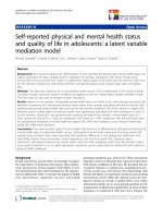
Schematic for the retroviral expression vectorFigure 1
Schematic for the retroviral expression vector. The
KSHV vGPCR is expressed as part of a bicistronic RNA
upstream of an internal ribosomal entry site (IRES)-regulated
GFP reporter cassette in the BABE retroviral plasmid [34].
The proviral segment of the BABE retroviral plasmid con-
tains long terminal repeats (LTRs) that participate in proviral
replication, an encapsidation sequence (ψ), and a selectable
puromycin marker. This diagram is not drawn to scale.
Virology Journal 2007, 4:87 />Page 4 of 9
(page number not for citation purposes)
in the presence of a COX-2 specific inhibitor. These results
demonstrate a mechanism by which a lytic phase protein,
vGPCR, encoded by KSHV may exert paracrine effects
through COX-2 dependent prostaglandin induction.
Over-expression of COX-2 has emerged as a prominent
feature of virtually every form of cancer [29]. The role of
COX-2 as a critical mediator of cancer progression is sup-
ported by numerous studies showing that the induction of
constitutive COX-2 expression and the resulting biosyn-
thesis of PGE
2
are sufficient to stimulate all of the key fea-
tures of carcinogenesis including mutagenesis,
mitogenesis, angiogenesis, metastasis, inhibition of apop-
tosis and immunosuppression [29]. KS lesions are charac-
terized by increased levels of PGE
2
and cyclic AMP
phosphodiesterase compared to those within the sur-
rounding tissues [16] suggesting that induction of COX-2
may be an important mediator of KS. Importantly,
Sharma-Walia et al recently demonstrated that infection
with KSHV induces robust COX-2, but not COX-1, expres-
sion in HMVEC and HFF and increased their secretion of
PGE
2
[17]. They further showed that although viral bind-
ing and entry induced moderate levels of COX-2, viral
gene expression significantly increased COX-2 induction.
To date, no specific KSHV gene products have been asso-
ciated with the observed COX-2 increase. Our studies pro-
vide evidence that KSHV vGPCR induces COX-2
expression in primary vascular endothelial cells and may
be responsible for the COX-2 and PGE
2
increases observed
following infection in microvascular endothelial cells and
in KS clinical specimens, respectively.
Emerging evidence points to vGPCR expression as essen-
tial for KS development. It is the only KSHV gene that
when expressed in the vascular endothelium of mice is
able to produce vascular tumors [30] and transgenic mice
that express vGPCR under either a ubiquitous (SV40) pro-
moter or a T cell-specific (CD2) promoter also develop
dermal angioproliferative lesions that closely resemble
those seen in KS [31,32]. Moreover, siRNA-mediated sup-
pression of vGPCR in mice expressing the entire KSHV
genome was sufficient to block VEGF secretion and tum-
origenesis, leading to significant retardation in tumor
growth [33]. Whether induction of COX-2 leads to the
angiogenic and tumorigenic effects of vGPCR is currently
under investigation.
Increased expression of PGE
2
due to vGPCR expression
may have a role in KSHV replication as well. The study by
Sharma-Walia et al suggests that COX-2 and PGE
2
play
roles in facilitating latent viral gene expression and the
establishment and maintenance of latency [17]. This data
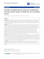
vGPCR-expressing HUVEC mimic spindle cellsFigure 2
vGPCR-expressing HUVEC mimic spindle cells. HUVEC were infected with BABE or BABE-vGPCR retroviruses and
grown for 72 hours. GFP-positive vGPCR expressing HUVEC exhibit a spindle cell-like morphology that resemble the spindle
cells found in KS lesions. GFP expression ca. 60%. Images taken at 400× magnification.
Virology Journal 2007, 4:87 />Page 5 of 9
(page number not for citation purposes)
together with results presented here could explain how
vGPCR, a lytic gene normally expressed only in cells des-
tined for lysis, might induce a tumor. Even though only a
small percentage of KS cells express vGPCR within KS
lesions, vGPCR induced COX-2 expression and conse-
quent PGE
2
secretion could initiate tumorigenesis and
promote viral latency through paracrine mechanisms.
There are no published reports describing a clinical link
between COX-2 and KS, however, we feel that this
hypothesis warrants further investigation.
COX inhibitors such as aspirin inhibit COX activity by
blocking the conversion of arachidonic acid to PGH
2
by
competing with free arachidonic acid for the cyclooxygen-
ase active sites. The COX-1 and COX-2 pharmacological
inhibitors NS-398, SC-560, and Indomethacin used in
this study abrogate cyclooxygenase activity by a similar
mechanism. The use of COX-2 inhibitors would provide a
viable therapeutic strategy to abrogate PGE
2
secretion
since prostaglandin E synthase (PGES) cannot synthesize
PGE
2
without PGH
2
and there are no direct inhibitors of
PGES currently available. Given the correlation of COX-2
in other cancer models, and evidence that regular intake
of a COX-2 inhibitor reduces cancer risk [29], future inves-
tigations into the mechanisms of KSHV induced COX-2
expression and prostaglandin activity may lead to new
treatments for KS patients.
Methods
Reagents
NS-398 was purchased from Cayman Chemical, Ann
Arbor, MI and sodium butyrate (NaB) was purchased
from Sigma, St. Louis, MO.
Cell culture
Pooled HUVEC (Cambrex BioScience, Walkersville, MD)
were cultured on 0.2% gelatin coated plates in medium
199 (M-199) (Invitrogen, Carlsbad, CA) supplemented
with 20% fetal bovine serum (FBS), 2 mM L-glutamine, 2
mM penicillin-streptomycin, and 1% endothelial cell
growth supplement (ECGS) (BD Biosciences, Bedford,
MA). Phoenix GP retroviral packaging cells (ATCC, Man-
assas, VA) were grown in Dulbecco's modified essential
medium (DMEM, Invitrogen) supplemented with 10%
FBS, 2 mM L-glutamine, and 2 mM penicillin-streptomy-
cin. HeLa cells (ATCC) were grown in minimal essential
media supplemented with 10% FBS, 2 mM L-glutamine, 2
mM penicillin-streptomycin, non-essential amino acids,
and sodium pyruvate. All cells were grown at 37°C with
5% CO
2
.
Plasmids
To construct vGPCR-expressing plasmids, KSHV vGPCR
cDNA was cut from MIGR-ORF74 (generous gift from
Marvin Reitz, Institute of Human Virology, Baltimore,
MD) by BglII and EcoRI digestion [12]. The digested
vGPCR fragment was separated by gel electrophoresis,
purified using QIAquick PCR Purification Kit (Qiagen,
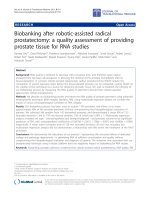
vGPCR induces COX-2 expressionFigure 3
vGPCR induces COX-2 expression. HUVEC were infected at time 0 with either BABE or BABE-vGPCR. Whole cell
lysates were prepared at the indicated time points and analyzed by western blot for the indicated protein. BABE-vGPCR-
infected HUVEC demonstrate increased COX-2 expression beginning at 24 hour post-infection (p.i.), while neither BABE nor
BABE-vGPCR-transduced HUVEC express COX-1. 50 ng of ovine COX-1 and COX-2 electrophoretic standards (S) served as
positive controls. β-actin expression served as a loading control. Results are representative of three independent experiments.
Virology Journal 2007, 4:87 />Page 6 of 9
(page number not for citation purposes)
Valencia, CA) and ligated into either BamHI and EcoRI
digested pBABE-green fluorescent protein (GFP) retroviral
plasmid (generous gift from Andrew Rice, Baylor Univer-
sity, Houston, TX) [34] or BamHI and EcoRI digested
pcDNA3 (Invitrogen) using T4 DNA Ligase (Invitrogen).
pBABE-GFP and pBABE-GFP-vGPCR were propagated in
Escherichia coli strain STBL2 (Invitrogen), whereas
pcDNA3-vGPCR, pcDNA3, and plasmids containing the
full-length COX-2 promoter upstream of luciferase (gen-
erous gift of Nicholas Bazan, Louisiana State University
Health Sciences Center, New Orleans, LA) or vesicular sto-
matitis virus G envelope protein (pVSV-G, generous gift of
Gary Nolan, Stanford University, Palo Alto, CA) were
propagated in Escherichia coli strain DH5α (Invitrogen).
All plasmids were harvested using QIAfilter Plasmid Maxi
Kit (Qiagen) according to the manufacturer's instructions
and quantified by spectrophotometry.
Retrovirus Production and Infection
BABE(VSV-G) pseudotypes were produced by transfecting
Phoenix GP cells with an equal amount of either pBABE-
GFP or pBABE-GFP-vGPCR and pVSV-G using calcium
phosphate precipitation as previously described [35]. The
medium was removed from each culture at 15 hours post-
transfection, cells were washed twice with phosphate-
buffered saline (PBS), and DMEM supplemented with 5
mM NaB was added. Fourteen hours later, the cells were
washed twice with PBS and the medium was changed to
M-199 supplemented with 10% FBS. At 48 hours post-
transfection, the virus-containing medium was collected,
centrifuged at 200 x g for 10 minutes to remove debris,
passed through a 0.45-μm filter, aliquoted and stored at -
80°C. Virus titers were quantified by GFP expression in
retrovirus-transduced NIH3T3 cells (ATCC) using fluores-
cent microscopy. HUVEC between passages 4 and 6 were
plated 24 hour prior to infection at a confluence of 40%.
At the time of infection, the medium was replaced with
BABE or BABE-vGPCR retrovirus-containing medium
diluted in M-199 (20% FBS) with ECGS to an MOI of 0.5.
The virus-containing medium was removed 24 hour post-
infection and fresh M-199 (20% FBS) with ECGS was
added every 24 hours for the remainder of each experi-
ment.
Western Blot Analysis
Whole cell extracts were harvested in RIPA Buffer (50 mM
Tris-HCl pH 7.5, 1% Nonidet P-40, 150 mM NaCl, 0.5%
sodium deoxycholate, 0.1% SDS, 1 mM phenylmethylsul-
fonylflouride, 1 mM sodium orthovanadate, 1 mM
sodium flouride, and 10 μg/ml aprotinin), rotated for 30
min at 4°C, and centrifuged at 14,000 rpm for 15 min.
Clarified protein was quantified by Bradford assay
(Sigma). 45 μg of protein from each sample was separated
by SDS-PAGE, transferred to a PVDF membrane, and
incubated overnight with COX-1 or COX-2 monoclonal
antibody (Cayman Chemicals, Ann Arbor, MI) diluted
1:1000 in 5% non-fat milk/0.1% Tween-TBS. After incu-
bation with an anti-mouse secondary antibody conju-
gated with horseradish peroxidase (1:5000 dilution,
Sigma), the immunocomplexes were visualized by
enhanced chemiluminescence (Amersham Biosciences,
Buckinghamshire, England). β-actin expression was meas-
ured as a loading control using an anti-β-actin rabbit pol-
yclonal antibody (1:1000) (Sigma) and detected by a
vGPCR induces COX-2 mRNA expressionFigure 4
vGPCR induces COX-2 mRNA expression. A) A COX-
2 promoter-luciferase plasmid (5 μg) was co-transfected with
pcDNA3 or pcDNA3-vGPCR at the indicated concentra-
tions. These results demonstrate a dose-dependent increase
in COX-2 promoter activity in vGPCR-expressing HeLa at
48 hours (* = p < 0.001). Graph and SEM are representative
of three independent experiments. B) RNA was collected
from BABE and BABE-vGPCR-infected HUVEC at the indi-
cated time points and analyzed by quantitative real-time RT-
PCR for COX-2 mRNA expression. The graph demonstrates
a time-dependent increase in COX-2 expression in vGPCR-
expressing HUVEC and represents the mean of 3 independ-
ent infections each measured in triplicate (*= p < 0.001).
Virology Journal 2007, 4:87 />Page 7 of 9
(page number not for citation purposes)
1:5000 dilution of horseradish peroxidase conjugated
donkey anti-rabbit antibody (Amersham Biosciences).
PGE
2
Quantification
Conditioned medium from BABE and BABE-vGPCR
transduced HUVEC was centrifuged at 14,000 rpm for 15
min at 4°C and supernatants assayed immediately using
a monoclonal PGE
2
EIA kit (Cayman, Ann Arbor, MI)
according to the manufacturer's protocol.
Luciferase Assay
HeLa cells were co-transfected in duplicate with 5 μg of
full-length COX-2 promoter luciferase plasmid and
increasing amounts of either pcDNA3 or pcDNA3-vGPCR
using calcium phosphate precipitation as previously
described [35]. Medium was removed 18 hours post-
transfection and replaced with fresh culture medium. At
48 hours post-transfection, medium from each well was
replaced with Luciferase Lysis Buffer (Promega, Madison,
WI) and incubated at -80°C for 10 min. Each sample was
thawed to room temperature, scraped, and centrifuged at
14,000 rpm for 15 min at 4°C. Clarified protein for each
sample was quantified by Bradford assay (Sigma) accord-
ing to the manufacturer's protocol. Luciferase Reporter
Buffer (Promega) was added to each sample and relative
luciferase activity was measured in triplicate using a lumi-
nometer (Lumat LB9507, Berthold).
Real-Time Reverse Transcriptase PCR
Total cellular RNA was isolated using RNeasy Total RNA
Kit (Qiagen) according to the manufacturer's instructions.
DNA was eliminated from all samples using Turbo-DNase
I (Ambion, Austin, TX). The RNA from each sample was
quantified by spectrophotometry. RNA (250 ng) from
each sample was converted to cDNA using iScript Reverse
Transcriptase (RT) (Bio-Rad, Hercules, CA) following the
manufacturer's protocol. Two microliters of cDNA was
amplified in 20 μl reactions containing primers at 250 nM
in iQ SYBR Green Supermix (Bio-Rad). PCR was per-
formed for 40 cycles consisting of 95°C for 15 sec and
60°C for 45 sec using an iCycler iQ Real Time Detection
System (Bio-Rad) using primers specific for COX-2 mRNA
[36] and human riboprotein 36B4 [37]. Dilution curves
showed that PCR efficiency was 96–100% for all primer
sets used. All samples were run in triplicate on the same
plate for each primer set. Negative controls, such as cDNA
reactions without reverse transcriptase or RNA, and PCR
mixtures lacking cDNA were included in each PCR to
detect possible contaminants. Following amplification,
specificity of the reaction was confirmed by melt curve
analysis. Relative quantitation was determined using the
comparative C
T
method with data normalized to 36B4
mRNA and calibrated to the average ΔC
T
of untreated con-
trols.
Statistical analysis
Data are presented as the means +/- standard error of the
means (SEM). Data from vGPCR-expressing groups were
compared to control groups and significant differences
were determined by one-way analysis of variance
(ANOVA) followed by Tukey's post hoc t-test (GraphPad
Prism Home, San Diego, CA).
vGPCR induced PGE
2
secretion is dependent on COX-2Figure 5
vGPCR induced PGE
2
secretion is dependent on
COX-2. A) HUVEC were infected with BABE or BABE-
vGPCR at time 0 and analyzed at the indicated time points
for the amount of PGE
2
in conditioned medium using an EIA
kit. The vGPCR-expressing HUVEC conditioned media dem-
onstrates a time-dependent increase in PGE
2
secretion (* = p
< 0.001). Graph and SEM is representative of three inde-
pendent experiments. B) At 24 hours post-infection, BABE-
vGPCR infected HUVEC were treated with the non-selective
COX inhibitor indomethacin (Indo), the selective COX-2
inhibitor (NS-398), or the selective COX-1 inhibitor SC-560
and PGE
2
in the conditioned media was quantified at 48
hours post-infection by EIA. PGE
2
secretion is reduced in a
dose-dependent manner in Indo and NS-398 treated BABE-
vGPCR infected HUVEC. The COX-1 selective inhibitor SC-
560 had a minimal effect on PGE
2
secreted from vGPCR
expressing HUVEC. Graph indicates fold induction in PGE
2
secretion over BABE-HUVEC and is representative of three
independent experiments.
Virology Journal 2007, 4:87 />Page 8 of 9
(page number not for citation purposes)
Competing interests
The author(s) declare that they have no competing inter-
ests.
Authors' contributions
BDS participated in experimental design, implementa-
tion, interpretation of results and drafting the maunscript.
HLL helped with real-time PCR analyses. HEM helped
with retroviral production. ABN performed western blot
analyses. JAL participated in experimental design. GS and
LM provided expertise in real-time PCR analyses. MKO
participated in experimental design and data interpreta-
tion. CAM participated in experimental design, data inter-
pretation and manuscript preparation. DES participated
in experimental design, data interpretation and manu-
script preparation. All authors read and approved the final
manuscript.
Acknowledgements
We thank Marvin Reitz, University of Maryland-Baltimore for the MIGR and
MIGR-ORF retroviral plasmids, Andy Rice, Baylor University for the BABE
retroviral plasmid, Gary Nolan, Stanford University for the VSV-G plasmid,
and Nicholas Bazan, Louisiana State University for the full-length COX-2
promoter luciferase plasmid. We also thank MaryBeth Ferris for excellent
technical assistance in confirmation of specific experiments. This study was
supported in part by NIH/NICHD045768 (CAM), NIH/NHLBI HL083480
(JAL) and a grant from the Louisiana Cancer Research Consortium (DES).
References
1. Antman K, Chang Y: Kaposi's sarcoma. N Engl J Med 2000,
342(14):1027-1038.
2. Chang Y, Cesarman E, Pessin MS, Lee F, Culpepper J, Knowles DM,
Moore PS: Identification of herpesvirus-like DNA sequences
in AIDS-associated Kaposi's sarcoma. Science 1994,
266(5192):1865-1869.
3. Cesarman E, Chang Y, Moore PS, Said JW, Knowles DM: Kaposi's
sarcoma-associated herpesvirus-like DNA sequences in
AIDS-related body-cavity-based lymphomas. N Engl J Med
1995, 332(18):1186-1191.
4. Soulier J, Grollet L, Oksenhendler E, Cacoub P, Cazals-Hatem D, Bab-
inet P, d'Agay MF, Clauvel JP, Raphael M, Degos L, Sigaux F: Kaposi's
sarcoma-associated herpesvirus-like DNA sequences in mul-
ticentric Castleman's disease. Blood 1995, 86(4):1276-1280.
5. Ganem D: Kaposi's Sarcoma-associated Herpesvirus. In Fields
Virology Volume 2. Fifth edition. Edited by: Knipe DM, Howley PM. Phil-
adelphia , Lippincott Williams and Wilkins; 2007:2847-2888.
6. Zhong W, Wang H, Herndier B, Ganem D: Restricted expression
of Kaposi sarcoma-associated herpesvirus (human herpesvi-
rus 8) genes in Kaposi sarcoma. Proc Natl Acad Sci U S A 1996,
93(13):6641-6646.
7. Staskus KA, Zhong W, Gebhard K, Herndier B, Wang H, Renne R,
Beneke J, Pudney J, Anderson DJ, Ganem D, Haase AT: Kaposi's sar-
coma-associated herpesvirus gene expression in endothelial
(spindle) tumor cells. J Virol 1997, 71(1):715-719.
8. Bais C, Santomasso B, Coso O, Arvanitakis L, Raaka EG, Gutkind JS,
Asch AS, Cesarman E, Gershengorn MC, Mesri EA: G-protein-cou-
pled receptor of Kaposi's sarcoma-associated herpesvirus is
a viral oncogene and angiogenesis activator. Nature 1998,
391(6662):86-89.
9. Guo HG, Browning P, Nicholas J, Hayward GS, Tschachler E, Jiang
YW, Sadowska M, Raffeld M, Colombini S, Gallo RC, Reitz MS Jr.:
Characterization of a chemokine receptor-related gene in
human herpesvirus 8 and its expression in Kaposi's sarcoma.
Virology 1997, 228(2):371-378.
10. Arvanitakis L, Geras-Raaka E, Varma A, Gershengorn MC, Cesarman
E: Human herpesvirus KSHV encodes a constitutively active
G-protein-coupled receptor linked to cell proliferation.
Nature 1997, 385(6614):347-350.
11. Cannon M, Philpott NJ, Cesarman E: The Kaposi's sarcoma-asso-
ciated herpesvirus G protein-coupled receptor has broad sig-
naling effects in primary effusion lymphoma cells. J Virol 2003,
77(1):57-67.
12. Pati S, Cavrois M, Guo HG, Foulke JS Jr., Kim J, Feldman RA, Reitz M:
Activation of NF-kappaB by the human herpesvirus 8 chem-
okine receptor ORF74: evidence for a paracrine model of
Kaposi's sarcoma pathogenesis. J Virol 2001, 75(18):8660-8673.
13. Pati S, Foulke JS, Barabitskaya O, Kim J, Nair BC, Hone D, Smart J,
Feldman RA, Reitz M: Human herpesvirus 8-encoded vGPCR
activates nuclear factor of activated T cells and collaborates
with human immunodeficiency virus type 1 Tat. J Virol 2003,
77(10):5759-5773.
14. Smit MJ, Verzijl D, Casarosa P, Navis M, Timmerman H, Leurs R:
Kaposi's Sarcoma-Associated Herpesvirus-Encoded G Pro-
tein-Coupled Receptor ORF74 Constitutively Activates p44/
p42 MAPK and Akt via Gi and Phospholipase C-Dependent
Signaling Pathways. J Virol 2002, 76(4):1744-1752.
15. Sodhi A, Montaner S, Patel V, Bais C, Zohar M, Mesri EA, Gutkind JS:
The Kaposi's sarcoma-associated herpes virus G protein-
coupled receptor up-regulates vascular endothelial growth
factor expression and secretion through mitogen-activated
protein kinase and p38 pathways acting on hypoxia-inducible
factor 1alpha. Cancer Res 2000, 60(17):4873-4880.
16. Ambrus JL, Stoll HL, Klein EA, Karakousis CP, Stadler S: Increased
Prostaglandin E2 and cAMP Phosphodiesterase levels in
Kaposi's Sarcoma - A virus against host defense. Res Commun
Chem Pathol Pharmacol 1992, 78(2):249-252.
17. Sharma-Walia N, Raghu H, Sadagopan S, Sivakumar R, Veettil MV,
Naranatt PP, Smith MM, Chandran B: Cyclooxygenase 2 induced
by Kaposi's sarcoma-associated herpesvirus early during in
vitro infection of target cells plays a role in the maintenance
of latent viral gene expression. J Virol 2006, 80(13):6534-6552.
18. Naranatt PP, Krishman HH, Svojanovsky SR, Bloomer C, Mathur S,
Chandran B: Hose gene induction and transcriptional repro-
gramming in Kaposi's Sarcoma-Associated Herpesvirus
(KSHV/HHV-8)-infected endothelia, fibroblasts, and B cells:
Insights into modulation events early during infection. Cancer
Res 2004, 64(January):72-84.
19. Vane JR, Bakhle YS, Botting RM:
Cyclooxygenases 1 and 2. Annu
Rev Pharmacol Toxicol 1998, 38:97-120.
20. Fosslien E: Molecular Pathology of cyclooxygenase-2 in neo-
plasia. Ann Clin Lab Sci 2000, 30:3-21.
21. DuBois RN, Abramson SB, Crofford L, Gupta RA, Simon LS, Van De
Putte LB, Lipsky PE: Cyclooxygenase in biology and disease.
FASEB 1998, 12:1063-1073.
22. Akarasereenont P, Chotewuttakorn S, Techatraisak K, Thaworn A:
The effects of COX-metabolites on cyclooxygenase-2 induc-
tion in LPS-treated endothelial cells. J Med Assoc Thai 2001, 84
Suppl 3:S696-709.
23. Akarasereenont P, Techatrisak K, Chotewuttakorn S, Thaworn A:
The induction of cyclooxygenase-2 in IL-1beta-treated
endothelial cells is inhibited by prostaglandin E2 through
cAMP. Mediators Inflamm 1999, 8(6):287-294.
24. Smith WL, DeWitt DL, Garavito RM: Cyclooxygenases: struc-
tural, cellular, and molecular biology. Annu Rev Biochem 2000,
69:145-182.
25. Bais C, Van Geelen A, Eroles P, Mutlu A, Chiozzini C, Dias S, Silver-
stein RL, Rafii S, Mesri EA: Kaposi's sarcoma associated herpes-
virus G protein-coupled receptor immortalizes human
endothelial cells by activation of the VEGF receptor-2/KDR.
Cancer Cell 2003, 3(2):131-143.
26. Barnett J, Chow J, Ives D, Chiou M, Mackenzie R, Osen E, Nguyen B,
Tsing S, Bach C, Freire J: Purification, characterization, and
selective inhibition of human prostaglandin G/H synthase 1
and 2 expressed in the baculovirus system. Biochim Biophys Acta
1994, 1209(1):130-139.
27. Gierse JK, Hauser SD, Creely DP, Koboldt C, Rangwala SH, Isakson
PC, Seibert K: Expression and selective inhibition of the con-
stitutive and inducible forms of human cyclo-oxygenase. Bio-
chem J 1995, 305 (Pt 2):479-484.
28. Smith CJ, Zhang Y, Koboldt CM, Muhammad J, Zweifel BS, Shaffer A,
Talley JJ, Masferrer JL, Seibert K, Isakson PC: Pharmalogical anal-
Publish with BioMed Central and every
scientist can read your work free of charge
"BioMed Central will be the most significant development for
disseminating the results of biomedical research in our lifetime."
Sir Paul Nurse, Cancer Research UK
Your research papers will be:
available free of charge to the entire biomedical community
peer reviewed and published immediately upon acceptance
cited in PubMed and archived on PubMed Central
yours — you keep the copyright
Submit your manuscript here:
/>BioMedcentral
Virology Journal 2007, 4:87 />Page 9 of 9
(page number not for citation purposes)
ysis of cyclooxygenase-1 in inflammation. Proc Natl Acad Sci U
S A 1998, 95:13313-13318.
29. Harris RE: Cyclooxygenase-2 (COX-2) and the Inflammogen-
esis of Cancer. In Inflammation in the Pathogenesis of Chronic Diseases
Volume 42. Edited by: Harris RE. Netherlands , Springer ; 2007:93-126.
30. Montaner S, Sodhi A, Molinolo A, Bugge TH, Sawai ET, He Y, Li Y, Ray
PE, Gutkind JS: Endothelial infection with KSHV genes in vivo
reveals that vGPCR initiates Kaposi's sarcomagenesis and
can promote the tunorigenic potential of viral latent genes.
Cancer Cell 2003, 3(January 2003):23-36.
31. Guo HG, Sadowska M, Reid W, Tschachler E, Hayward G, Reitz M:
Kaposi's Sarcoma-Like Tumors in a Human Herpesvirus 8
ORF74 Transgenic Mouse. J Virol 2003, 77(4):2631-2639.
32. Yang TY, Chen SC, Leach MW, Manfra D, Homey B, Wiekowski M,
Sullivan L, Jenh CH, Narula SK, Chensue SW, Lira SA: Transgenic
expression of the chemokine receptor encoded by human
herpesvirus 8 induces an angioproliferative disease resem-
bling Kaposi's sarcoma. J Exp Med 2000, 191(3):445-454.
33. Mutlu AD, Cavallin LE, Vincent L, Chiozzini C, Eroles P, Duran EM,
Asgari Z, Hooper AT, La Perle KM, Hilsher C, Gao SJ, Dittmer DP,
Rafii S, Mesri EA: In vivo-restricted and reversible malignancy
induced by human herpesvirus-8 KSHV: a cell and animal
model of virally induced Kaposi's sarcoma. Cancer Cell 2007,
11(3):245-258.
34. Foskett SM, Ghose R, Tang DN, Lewis DE, Rice AP: Antiapoptotic
function of Cdk9 (TAK/P-TEFb) in U937 promonocytic cells.
J Virol 2001, 75(3):1220-1228.
35. Sambrook J, Fritsch EF, Maniatis T: Molecular Cloning A Labora-
tory Manual. Volume 3. 2nd edition. Plainview , Cold Spring Harbor
Laboratory Press; 1989.
36. Sun K, Ma R, Cui X, Campos B, Webster R, Brockman D, Myatt L:
Glucocorticoids induce cytosolic phospholipase A2 and pros-
taglandin H synthase type 2 but not microsomal prostaglan-
din E synthase (PGES) and cytosolic PGES expression in
cultured primary human amnion cells. J Clin Endocrinol Metab
2003, 88(11):5564-5571.
37. Simpson DA, Feeney S, Boyle C, Stitt AW: Retinal VEGF mRNA
measured by SYBR green I fluorescence: A versatile
approach to quantitative PCR. Mol Vis 2000, 6:178-183.