Báo cáo sinh học: " Imperfect DNA mirror repeats in the gag gene of HIV-1 (HXB2) identify key functional domains and coincide with protein structural elements in each of the mature proteins" doc
Bạn đang xem bản rút gọn của tài liệu. Xem và tải ngay bản đầy đủ của tài liệu tại đây (879.94 KB, 13 trang )
BioMed Central
Page 1 of 13
(page number not for citation purposes)
Virology Journal
Open Access
Research
Imperfect DNA mirror repeats in the gag gene of HIV-1 (HXB2)
identify key functional domains and coincide with protein structural
elements in each of the mature proteins
DorothyMLang
Address: School of Contemporary Sciences, University of Abertay-Dundee, Bell Street, Dundee DD1 1HG, Scotland, UK
Email: Dorothy M Lang -
Abstract
Background: A DNA mirror repeat is a sequence segment delimited on the basis of its containing
a center of symmetry on a single strand, e.g. 5'-GCATGGTACG-3'. It is most frequently described
in association with a functionally significant site in a genomic sequence, and its occurrence is
regarded as noteworthy, if not unusual. However, imperfect mirror repeats (IMRs) having ≥ 50%
symmetry are common in the protein coding DNA of monomeric proteins and their distribution
has been found to coincide with protein structural elements – helices, β sheets and turns. In this
study, the distribution of IMRs is evaluated in a polyprotein – to determine whether IMRs may be
related to the position or order of protein cleavage or other hierarchal aspects of protein function.
The gag gene of HIV-1 [GenBank:K03455
] was selected for the study because its protein motifs and
structural components are well documented.
Results: There is a highly specific relationship between IMRs and structural and functional aspects
of the Gag polyprotein. The five longest IMRs in the polyprotein translate a key functional segment
in each of the five cleavage products. Throughout the protein, IMRs coincide with functionally
significant segments of the protein. A detailed annotation of the protein, which combines structural,
functional and IMR data illustrates these associations. There is a significant statistical correlation
between the ends of IMRs and the ends of PSEs in each of the mature proteins. Weakly symmetric
IMRs (≥ 33%) are related to cleavage positions and processes.
Conclusion: The frequency and distribution of IMRs in HIV-1 Gag indicates that DNA symmetry
is a fundamental property of protein coding DNA and that different levels of symmetry are
associated with different functional aspects of the gene and its protein. The interaction between
IMRs and protein structure and function is precise and interwoven over the entire length of the
polyprotein. The distribution of IMRs and their relationship to structural and functional motifs in
the protein that they translate, suggest that DNA-driven processes, including the selection of
mirror repeats, may be a constraining factor in molecular evolution.
Background
A DNA mirror repeat is a sequence segment delimited on
the basis of its containing a center of symmetry on a single
strand and identical terminal nucleotides. For example, in
the sequence below, TACACG is the mirror image of GCA-
CAT.
Published: 26 October 2007
Virology Journal 2007, 4:113 doi:10.1186/1743-422X-4-113
Received: 28 September 2007
Accepted: 26 October 2007
This article is available from: />© 2007 Lang; licensee BioMed Central Ltd.
This is an Open Access article distributed under the terms of the Creative Commons Attribution License ( />),
which permits unrestricted use, distribution, and reproduction in any medium, provided the original work is properly cited.
Virology Journal 2007, 4:113 />Page 2 of 13
(page number not for citation purposes)
< >
5'- T A C A C G G C A C A T -3'
3'- A T G T G C C G T G T A -5'
Imperfect DNA mirror repeats (IMRs) are less than 100%
symmetrical.
The identification of mirror repeats is highly dependent
on how they are defined. One method is to identify all
mirror repeats within a sequence by systematically evalu-
ating the symmetry of each string within in it. This
method identifies relatively long (or maximal) symmetric
strings (mIMRs). Using symmetry criteria of ≥ 50% and
discounting strings completely contained within other
strings, the longest mIMRs in TnsA were found to coincide
with key structural domains [1].
Another type of mirror repeat is identified by progres-
sively evaluating, from the start to the end of a sequence,
symmetric sub-strings bounded by reverse dinucleotides
(rdIMRs). These are generally shorter than and often con-
tained within mIMRs. Lang [1] found statistically signifi-
cant correlations for the coincidence of the ends of rdIMRs
and the ends of protein structural elements – helices, β-
sheets and turns – in 17 monomeric proteins. In TnsA (E.
coli), 88% of the known or potential functional motifs
occur within rdIMRs and the longest mIMRs translate key
functional and/or structural sequences of the protein.
In this study, the distribution of IMRs is evaluated in a
gene that translates a polyprotein. The specific goals were
to determine whether IMRs span the entire polyprotein, to
identify the relationship of IMRs in the precursor to IMRs
in the mature cleavage products and to assess the relation-
ship between IMRs and protein functional and structural
motifs. The HIV-1 gag sequence used for this analysis is
HXB2_LAI_IIIB_BRU [Genbank: K03455
], the most com-
monly used reference sequence for the HIV-1 genome [2].
The gag gene of HIV-1 is about twice as long as TnsA, and
translates the following proteins (in the order of their
occurrence within the sequence): matrix (MA), capsid
(CA), p2 (SP1), nucleocapsid (NC), and either (a) p1
(SP2) and p6 or (b) GagTF. CA is about the same length
as TnsA. The cleavage positions for each of the mature pro-
teins of Gag (HXB2) are summarized in Table 1.
Gag proteins are the structural components of the HIV-1
virus and cleavage of the Gag polyprotein into several
mature proteins is essential to replication. Near the C-ter-
minal of Gag (at the NC-p1 cleavage site), the protein
becomes polycistronic. The ribosome "slips" within the
DNA motif "tttttt", once in every 20
th
Gag transcription
and the resulting transcript is GagTF-Pol. At maturation,
the Pol segment is cleaved into enzymatic proteins. Gag
and Gag-Pol are cleaved differentially and in stages. This
process is summarized in Table 2.
In order to facilitate the comparison of multiple types of
data within the context of the protein, a comprehensive
annotation of complete Gag sequence was made (Addi-
tional file 1) that combines experimentally determined
functional and structural motifs, and the sequence posi-
tions of IMRs found in this study.
Results
The five longest mIMRs in gag that are ≥ 50% symmetric
each translate an essential protein motif in a different
cleavage product, indicating that the association between
mIMR length and function may be related to selection in
both the polyprotein and its cleaved products. Most IMRs
translate distinct, functionally significant protein motifs.
At symmetry ≥ 50% there are significant statistical correla-
tions between the ends of both mIMRs and rdIMRs, and
the ends of protein structural elements (PSEs). Several
mIMRs that are ≥33% symmetric start or stop at cleavage
positions.
The DNA and amino acid sequence positions of the long-
est L1 mIMRs are listed in Table 3. The designation L1
means that it is the longest IMR for a unique span of the
Table 1: Nucleotide and amino acid sequences adjacent to cleavage sites in Gag (HXB2) [2]
Segment DNA Amino Acid
nt start stop start stop
gag thru slip 1296 1-atgggtgcg gctaat-1296 1-MGARAS ERQAN-432
matrix 396 1-atgggtgcg aattac-0396 1-MGARAS VSQNY-132
capsid 693 397-cctata gttttg-1089 133-PIVQN KARVL-363
p2 42 1090-gctgaa ataatg-1131 364-AEAMS SATIM-377
p7 nucleocapsid 162 1132-atgcag gctaat-1296 378-MQRGN ERQAN-432
p1 start is slip 48 1297-ttttta aatttt-1344 433-FLGKI RPGNF-448
p6 159 1345-cttcag caataa-1503 449-LQSRP DPSSQ$-501
gag-pol TF 165 1299-tttagg aacttc-1463 433-FREDL VSFNF-488
Virology Journal 2007, 4:113 />Page 3 of 13
(page number not for citation purposes)
DNA sequence. MIMRs are identified by evaluating the
symmetry of every possible sub-string of a DNA sequence,
then nesting them sequentially, beginning at the 5' end.
The span of the first IMR is designated L1; all shorter IMRs
within the span are designated progressively higher levels
(L2, L3, etc.) based on whether they are completely con-
tained within another IMR. The next L1 IMR ends down-
stream from the end of the preceding IMR; it may begin
within a preceding IMR or downstream from it. For the
remainder of this article, all references to IMRs refer to L1
IMRs. Each (L1) mIMR is assigned an ID number based on
rank by length, and is preceded by a hash mark (e.g. #1-
gag). The position of some mIMRs differ by only a few
amino acids, so it is possible to simplify the data by dis-
counting mIMRs that substantially overlap. Table 4 sum-
marizes this simplification and illustrates that although
mIMRs occur throughout most of the Gag protein each
span is associated with distinct structural or functional
domains.
MIMRs were found separately for the Gag polyprotein and
each of the cleavage products. It was anticipated that the
mIMRs for Gag CDS would be different than those for the
components, but they were not except that there are two
mIMRs in the NC that only attain L1 status when NC is
evaluated separately (not as part of gag). The distribution
of mIMRs in Gag indicates that most of the largest mIMRs
do not span sequences that will be cleaved into separate
proteins. The single exception is E419 E454 (#3-gag),
which spans NC-p1, and terminates at the p1-p6 cleavage
site; this is the segment that is differentially cleaved in Gag
and Gag-Pol.
Table 5 lists the DNA and amino acid sequence positions
of the longest rdIMRs. RdIMRs are identified by sequen-
tially evaluating, from 5' to 3', the symmetry of each sub-
string delineated by each dinucleotide and the next
downstream reverse dinucleotide. They are nested by the
same process described for mIMRs. Most of the protein
segments translated by rdIMRs coincide with experimen-
tally determined structural or functional motifs of the pro-
tein.
MIMRs and rdIMRs vary in distribution, beyond that
which would occur due to the differences in their lengths.
MIMRs occur throughout most of gag, as a series of over-
lapping, or nearly overlapping spans; within many
mIMRs, there are one or two spatially separated rdIMRs.
MIMRs are, however, noticeably absent in some segments
Table 3: mIMRs in gag that are ≥50% symmetrical
Rank m-IMR ID protein length DNA positions protein positions overlaps
1 #1-gag MA 95 0270-aa ca-0364 091-RI DT-122
2 #2-gag CA 87 0742-gg tg-0828 248-GW RM-276
3 #3-gag NC-p1-p6 85 1256-aa aa-1340 419-EG GN-447
#4-gag MA 82 0266-at ca-0347 089-HQ AQ-116 ~#1-gag
#5-gag CA 81 0758-at ta-0838 253-NP PT-280 ~#2-gag
4 #6-gag NC 81 1171-aa ga-1251 391-KC CG-417
5#7-gagp2801065-ac ca-1144 356-PG GN-382
#8-gag CA 77 0764-ct cc-0840 255-PP PT-280 ~#2-gag
#9-gag gag 77 1100-tg gt-1176 367-MS KC-392 ~#7-gag
6 #10-gag CA 76 0812-at ta-0887 271-NK DY-296
7 #11-gag CA 75 0920-ag ga-0994 307-EQ KT-332
#12-gag MA 74 0299-ct ac-0372 100-AL GH-124 ~#1-gag
#13-gag MA 71 0303-ag ca-0373 102-DK HS-125 ~#1-gag
8 #14-gag CA 69 0985-ga ag-1053 329-DC QG-351
9 #15-gag CA 64 0543-ca aa-0606 181-PQ LK-202
#16-gag CA 64 0810-aa aa-0873 271-NK KE-291 ~#10-gag
10 #17-gag p6 64 1362-gc ag-1425 455-PT QK-475
#18-gag MA 63 0265-ca ac-0327 089-HQ QN-109 ~#1-gag
11 #1-NC NC 59 1209-aa aa-1267 404-NC QM-423
12 #2-NC NC 47 1153-aa ca-1199 385-NQ GH-400
ID numbers for each mIMR (e.g. #1-gag) are based on rank by length (#1 being the longest). MIMRs terminated by reverse dinucleotides are bold.
Table 2: Gag and Gag-Pol are differentially cleaved at
maturation
Gag stage 1 MA-CA-p2\1/NC-p1-p6
Gag-Pol stage 1 MA-CA-p2\1/NC-GagTF-pol
Gag stage 2 MA\2/CA-p2\1/NC-p1\2/p6
Gag-Pol stage 2 MA\2/CA-p2\1/NC\2/GagTF-Pol
Gag stage 3 MA\2/CA\3/p2\1/NC\3/p1\2/p6
Gag-Pol stage 3 MA\2/CA\3/p2\1/NC\2/PR\3/RT\3/RNase\3/IN
GagTF-Pol results from a frame shift at the end of NC. In Gag, p1 is
not cleaved from NC until stage 3. GagTF is cleaved from NC at stage
2 [3-7].
Virology Journal 2007, 4:113 />Page 4 of 13
(page number not for citation purposes)
of gag; in these segments, e.g. M1 R91 (MA) and
P133 G248 (CA), rdIMRs form a nearly continuous
series, end-to-end. The sequence spans in MA and CA that
do not contain mIMRs are illustrated in Figure 1. These
regions are both highly reactive and mobile (detailed in
the legend).
Figures 2A and 2B illustrate the protein translation of the
two largest mIMRs in gag – the largest helix in MA (2A)
and CA (2B) and the adjacent turns essential to the tertiary
structure. The PDB structure used for this illustration –
1L6N – is of the immature Gag protein; the structure of
MA and CA is not substantially different in the mature
proteins, except that the long loop between them is cut
and refolded [8]. The MA-H5 helix is distinct from the
other matrix components, and in the mature protein
projects directly into the center of the virion [13]; the MA-
H5 helix may also contain a nuclear localization signal
[11]. The CA-H7 helix stabilizes interface 1 (planar strips)
of the viral core [14].
Figures 2C and 2D illustrate the three largest rdIMRs in
MA and CA. The protein translation of $3-gag spans a
nuclear localization signal; $6-gag and $10-gag are essen-
tial to structural transformation at maturation [15]. The
protein translation of $16-gag spans a region that refolds
to create a CA-CA interface essential to assemble the core
[16]; $18-gag spans the MA-CA cleavage site; $22-gag
translates part of the loop on the surface of the virion core
and interacts with CypA [12].
Figure 3 illustrates the two largest mIMRs in the nucleo-
capsid. The largest (Fig. 3A) spans the entire region con-
necting the two Cys-His boxes. The second largest (Fig.
3B) spans the EF1α binding site and first Cys-His box. The
largest rdIMRs in the NC overlap (Fig. 3C), and a Zn ion
is bound within the region translated by the overlap. The
Cys-His boxes are zinc finger binding domains which ena-
ble NC to bind to nucleic acids, and the Zn ion increases
the affinity of NC for nucleic acids; NC also has unwind-
ing properties, resembling a DNA topoimerase [17].
The coincidence of the ends of IMRs and PSEs was tested
for several gene segments – MA-CA-p2-NC, MA, CA and
NC segments – using Fisher's exact test (FET) [20]. The
Kabsch and Sander [21] secondary structure prediction
was used with the 1L6N tertiary structure (PDB) and sta-
tistically significant values were found for the MA-CA-p2-
NC, CA and NC segments; PROMOTIF secondary struc-
ture annotation was used for MA. These results are sum-
marized in Table 6.
The mIMRs included in the test are all ≥58 nt and often
span more than a single protein structural element. The
rdIMRs included in the test are all ≥15 nt. Both mIMRs
and rdIMRs begin and end at various positions within
codons and therefore, the composition of the two nucle-
otides at each end (which delimit the rdIMRs) are unlikely
to be strongly influenced by preferences related to second-
ary structure composition or codon preference. More than
50% of the mIMRs are terminated by reverse dinucle-
otides.
For almost all measurements of coincidence, the ends of
IMRs and PSEs were statistically significant over a range of
3 nt, similar to the span found in TnsA. The position at
which the coincidence is maximal is listed in Table 6. The
coincidence of IMR and PSE at position 0 indicates that
the span of a PSE exactly coincides with the span of an
IMR. When the position is negative, the IMR begins
Table 4: Simplification of Table 3 by removal of slightly overlapping mIMRs
Rank mIMR prot len DNA positions AA positions Structure or function
1 #1-gag MA 95 0270-aa ca-0364 091-RI DT-122 MA-H5 related to viral entry
2 #2-gag CA 87 0742-gg tg-0828 248-GW RM-276 CA-H7 longest constituent of viral core
3 #3-gag p1 85 1256-aa aa-1340 419-EG GN-447 end NC, p1 to p1-p6 cleavage site
4 #6-gag NC 81 1171-aa ga-1251 391-KC CG-417 1st cys-his box; EF1α binding
5 #7-gag p2 80 1065-ac ca-1144 356-PG GN-382 p2, critical to budding
6 #10-gag CA 76 0812-at ta-0887 271-NK DY-296 major homology region
7 #11-gag CA 75 0920-ag ga-0994 307-EQ KT-332 endocytosis signal 1; CA-H9 helix
8 #14-gag CA 69 0985-ga ag-1053 329-DC QG-351 endocytosis signal 2; CA-H10 helix
9 #15-gag CA 64 0543-ca aa-0606 181-PQ LK-202 CA-H3-H4 helices, part of viral core
10 #17-gag NC 64 1362-gc ag-1425 455-PT QK-475 L-domain (budding); Tsg101
455-PT QK-475 docking; ubiquitin-gag conjugate
11 #1-NC NC 59 1209-aa aa-1267 404-NC QM-423 2cd cys-his box; end NC
12 #2-NC NC 47 1153-aa ca-1199 385-NQ GH-400 EF1α binding
MIMRs that begin and end within two amino acids of a larger mIMR have been removed. Although the distribution of mIMRs is nearly continuous
throughout gag, the functional and/or structural association of each is discrete, as indicated by the structure-function notation in the right hand
column of this table, which is described in greater detail in Additional File 1.
Virology Journal 2007, 4:113 />Page 5 of 13
(page number not for citation purposes)
slightly upstream of the start of the PSE; when the posi-
tion is positive, the IMR begins slightly downstream. The
difference is indicated as a nucleotide position, however,
so in the protein the equivalent distance is 1–2 amino
acids, which is similar to the variability of different struc-
ture prediction methods.
Table 5: rdIMRs in gag ranked by length
Beg end rd-IMRs nt prot AA structure or function
1215 ca ac 1261 $1-gag 47 NC R406 H421 primer annealing; Cys-His box
767 tc ct 803 $2-gag 37 CA I256 L268 N-terminal CA-H7 helix
73 gg gg 108 $3-gag 36 MA G025 W036 nuclear localization signal 1(NLS1)
1245 at ta 1279 $4-gag 35 NC C416 T427 Zn finger motifs. 2cd cys-his box
267 tc ct 300 $5-gag 34 MA Q090 A100 I92 V95 affect struct orientation of MA-H5
168 ct tc 200 $6-gag 33 MA C057 S067 MA H3 helix, C57S prevents particle fmtn
68 ca ac 97 $7-gag 30 MA P023 H033 basic residues target, bind Gag to PM
1379 aa aa 1408 $8-gag 30 p6 E460 T470 possible association with ubiquitin
184 gg gg 212 $9-gag 29 MA G062 G071 C-terminal MA-H3 helix
198 at ta 226 $10-gag 29 MA P066 R076 essential to structural transformation
246 ag ga 274 $11-gag 29 MA A083 I092 mutations retarget assembly
232 tt tt 259 $12-gag 28 MA L078 C087 MA-H4 central to 3D structure
1091 ct tc 1118 $13-gag 28 - A364 S373 cleavage site, most of p2
618 tg gt 644 $14-gag 27 CA E207 V215 C-terminal CA-H4 helix, CypA interaction
1074 ta at 1100 $15-gag 27 - K359 M367 cleavage CA-p2
490 tt tt 515 $16-gag 26 CA F164 F172 folds against MHR
1108 ag ga 1131 $17-gag 24 - V370 M378 cleavage site p2-p7, phosphorylation
385 ag ga 409 $18-gag 25 - S129 N137 spans cleavage site MA-CA
894 cc cc 918 $19-gag 25 CA R299 A306 end MHR
964 tt tt 988 $20-gag 25 CA L322 C330 interacts with LysRS
1027 ct tc 1051 $21-gag 25 CA L343 Q351 interacts with LysRS
650 ca ac 673 $22-gag 24 CA P217 P225 CypA interaction; surface of virion core
1043 ca ac 1066 $23-gag 24 CA T348 P356 interacts with LysRS
590 cc cc 612 $24-gag 23 CA A197 T204 N-terminal CA-H4 helix
931 ca ac 953 $25-gag 23 CA Q311 T318 N-terminal CA-H9 helix, LysRS interaction
1275 tt tt 1297 $26-gag 23 NC C426 F433 cleavage site p7-p1
12 ag ga 33 $27-gag 22 MA A005 G011 links myristoylation and calmodulin-binding
712 gg gg 733 $28-gag 22 CA G238 E245 links CA-H5 and -H6 helices
1109 ta at 1130 $1-p2 22 V370 M377 cleavage site p2-p7, phosphorylation
1 at ta 21 $1-MA 21 M001 V007 minimum signal required for myristoylation
135 ag ga 155 $2-MA 21 V046 S052 tether btwn MA at viral membrane and CA
150 gt tg 170 $3-MA 21 L051 C057 L50A-L51A prevents particle formation
109 gc cg 128 $4-MA 20 A037 R043 binds HIV to plasma membrane, calmodulin
327 ca ac 346 $5-MA 20 K110 Q116 nuclear localization signal 2
45 at ta 63 $6-MA 19 W016 L021 mutations retarget particle fmtn to Golgi
166 gg gg 184 $7-MA 19 G056 G062 G56E, C57D, C57S, I60E elim replication
95 aa aa 112 $8-MA 18 K032 S038 binds HIV to plasma membrane, calmodulin
139 aa aa 156 $9-MA 18 N047 E052 ?
320 ag ga 337 $10-MA 18 E107 K113 nuclear localization signal 2
22 tt tt 38 $11-MA 17 L008 L013 ?
367 gg gg 382 $12-MA 16 G123 V128 start labile structure near cleavage site
565 aa aa 585 $1-CA 21 N189 Q195 connects CA-H3 and CA-H4 helices
647 at ta 667 $2-CA 21 H216 I223 CypA interaction; surface of virion core
676 gg gg 696 $3-CA 21 G226 R232 CypA interaction; surface of virion core
947 gg gg 967 $4-CA 21 W316 V323 CA H9 helix, endocytosis signal
1039 at ta 1059 $5-CA 21 M347 V353 interacts with LysRS
832 ag ga 851 $6-CA 20 S278 D284 necessary for formation of dimer interface
910 ct tc 929 $7-CA 20 L304 S310 spans CA-H8-H9 helices
927 tt tt 946 $8-CA 20 S310 W316 N-terminal, LysRS interaction site
1306 aa aa 1325 $1-p1 20 K436 K442 start is slip site, p1 protein
1319 cc cc 1334 $2-p1 16 S440 P445 middle, p1 protein
The rank of each rdIMR within the entire gag gene was determined first, then rank within each mature protein. Multiple rdIMRs of the same length
were ordered by sequence position.
Virology Journal 2007, 4:113 />Page 6 of 13
(page number not for citation purposes)
Differences in the position of maximum coincidence
between the segments occur for several reasons. The meas-
urement includes coincidences over the entire range of the
sequence, and the position of maximum coincidence
would be expected to be somewhat different for each pro-
tein due to differences in secondary and tertiary structure.
The values, however, are consistent; the largest segment –
MA-CA-p2-NC – has a maximum coincidence at position
5 (for rdIMR ≥16 nt), which is central to positions 3, -2
and 7, which are maximal for MA, CA and NC, respec-
tively.
The coincidence of IMRs with PSEs may be enhanced by
the greater than expected numbers of them in the Gag
polyprotein. The following formula predicts the expected
number of occurrences.
P(t) predicted number of occurrences of mIMRs in the
sequence
P(o) probability of the occurrence of a mirror repeat in a
random sequence consisting of 4 nucleotides present in
approximately equal amounts
P(e) probability of the ends of a segment matching, for
mIMRs, P(e) = 1/4
P(m) probability of number of matches required for sym-
metry
l number of potential matches (1/2 total sequence length,
odd values disregarded)
m number of matches required for symmetry
P(o) = P(e) * P(m)
P(m) = (l!/((m!(l-m)!) * (1/4)
m
* (3/4)
l-m
In gag, 18 L1 mIMRs were identified that were ≥ 63 nt.
Therefore, as a generalization, this length will be evalu-
ated. Since we are only concerned that one side of the seg-
ment matches the other, l = 30 and m = 14.
P(m) = (30!/(14! * 14!)) * (1/4)
14
* (3/4)
16
P(m) = 0.005430
Adding the criteria that the ends must match,
P(o) = 0.001357
The length of gag is 1500 nt, from which is subtracted the
required length for the match (62), resulting in 1438
potential sites ≥ 63 nt.
P(t) = P(o) * 1438 = 1.95
This value indicates that it is likely that at least two mIMRs
≥ 63 nt will occur by chance. Since each possible site of an
mIMR is included to obtain this estimate, it should be
compared with the total number if mIMRs ≥ 63 nt that
were identified (= 49), not just L1 mIMRs (= 18). There-
fore, the observed frequency (49) is 25-fold greater than
the expected frequency (2).
A similar process for rdIMRs can be made, with the only
change of P(e) = (1/4)*(1/4), to reflect the reverse dinu-
cleotide criteria delimiter. The estimate will be for rdIMRs
≥20 nt, the length summarized in Table 5.
P(m) = (l!/((m!(l-m)!) * (1/4)
m
* (3/4)
l-m
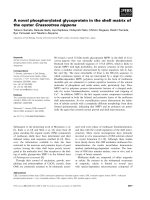
The distribution of mIMRs in the immature Gag protein [NCBI:1L6N, [8]]Figure 1
The distribution of mIMRs in the immature Gag pro-
tein [NCBI:1L6N, [8]]. MIMRs that are ≥ 50% symmetric
are noticeably absent from some segments of the protein.
These regions are characterized by a series of rdIMRs,
arranged end-to-end (illustrated in black). The spans lacking
mIMRs are highly reactive and mobile. The A3 C87 region of
matrix undergoes structural transformation at several stages
of the virion life cycle, and contains basic residues that target
Gag to the plasma membrane [9], a calmodulin-binding motif
[10] and a nuclear localization signal [11]. The T204 E245
region of capsid includes the exposed loop on the virion core
[8, 12], and the CypA binding site [12].
Capsid protein
Matrix protein
T204
E245
A3
C87
MA-H5
CA-H1
CA-H8
Virology Journal 2007, 4:113 />Page 7 of 13
(page number not for citation purposes)
P(m) = (8!/(3! * 5!)) * (1/4)
3
* (3/4)
5
= 0.2076
P(o) = P(e) * P(m) = (1/16) * 0.2076 = 0.01280
P(t) = P(o) * (1500-19) = 19.2
The observed frequency for rdIMRs ≥20 nt is 53, approxi-
mately 2.5 the predicted number.
Both mIMRs and rdIMRs occur at greater than expected
numbers, although the greater than expected number of
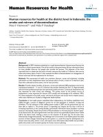
The longest IMRs coincide with key protein functional motifsFigure 2
The longest IMRs coincide with key protein functional motifs. Figures 2A and 2B [NCBI:1L6N [8]] illustrate the
two longest mIMRs in the Gag polyprotein – #1-gag in matrix and #2-gag in capsid. These mIMRs translate the MA H5 and CA
H7 helices which (in the illustrated structure) are approximately parallel to each other at a pitch of about 45°. Both are essen-
tial to the structure and function of each protein. Figure 2C illustrates the largest rdIMRs in matrix and Figure 2D the largest
rdIMRs in capsid, that do not coincide with mIMRs.
G25
W36
C57
S67
R76
G248
M276
A.
#1-gag mIMR R91 T122 MA H5 helix
B.
#2-gag mIMR G248 M276 CA H7 helix
$3-gag rdIMR G25 W36
nuclear localization
$6-gag rdIMR C57 S67 trimerization
$10-gag rdIMR P66 R76 maturation
F164
F172
S129
N137
P217
P225
$16-gag rdIMR F164 F172 viral core compo nent
$18-gag rdIMR S129 N137 MA-CA cleavage site
$22-gag rdIMR P217 P225
CypA
binding
C. D.
T122
R91
Virology Journal 2007, 4:113 />Page 8 of 13
(page number not for citation purposes)
mIMRs is much greater than for rdIMRs. These values
demonstrate that it is unlikely that the multiple occur-
rences of mIMRs ≥63 nt occur by chance. It is also unlikely
that chance occurrences will be at positions that are highly
significant to the function of the protein.
The affect of modifying symmetry criteria on IMR identity
was examined for both lower and higher levels of symme-
try. No evidence of a relationship between mIMRs and
protein cleavage sites for the entire Gag polyprotein was
found at levels of symmetry ≥50%. Table 7 summarizes L1
mIMRs that are ≥33% symmetrical. Using the formula
described previously, less than one (0.1128) mIMRs that
is 704 nt in length and ≥33% symmetric is expected
within the gag sequence of 1500 nt; in contrast, five are
observed and there are an additional 237 that are longer
than 705 nt, indicating that mirror symmetry pervades the
gene. About half of the L1 mIMRs translate protein seg-
ments that would end at or near cleavage sites, and one
mIMR coincides with the start of CA and the end of p6.
MIMRs that are not associated with cleavage sites begin
and end at functionally related domains.
The region M1 K32 encompasses the start of four mIMRs
(≥33% symmetrical) and is the region that targets Gag to
the cell membrane [22]. Two of these mIMRs terminate
within capsid D235 E260 which is a region of small heli-
ces and loops adjacent to the CypA binding site that is
probably essential to disassembling the core upon infec-
tion [14]; these mIMRs, then, begin at sequences that
localize Gag to the cell membrane – a process essential to
core formation – and end at sequences that dissolve the
virion core (upon infection). Similarly, E12 N271 begins
within the membrane localization domain, and ends at
CA-H7, the largest component of the structural core,
which stabilizes its constituent planar strips [14]. The
fourth mIMR, R15 Q379, begins within the membrane
localization region and terminates one amino acid down-
stream from the p2-NC cleavage site; cleavage at p2-NC is
the initial step in the Gag cleavage sequence [3]. MIMR
E52 K410 begins at positions essential to particle forma-
tion, trimerization and virus assembly, and terminates
immediately upstream of the second Cys-His box (zinc
finger) which is essential to packaging. Several mIMRs
begin within the region L101 D121, which includes most
of the MA-H5; this helix projects away from the plasma
membrane, directly into the center of the virion [23] and
deleterious deletions within it have been found to block
viral entry [13]. MIMRs that begin at the MA-H5 helix ter-
minate at the NC-p1 cleavage site and the end of Gag-Pol
TF and p6. The association of weakly symmetrical mIMRs
with cleavage sites in the polyprotein and functionally
related protein motifs suggests that different levels of IMR
symmetry may be related to different functional aspects of
the translated protein.
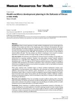
The largest mIMR in the nucleocapsid spans the two Cys-His boxes [NCBI:1F6U [18]]Figure 3
The largest mIMR in the nucleocapsid spans the two
Cys-His boxes [NCBI:1F6U [18]]. Figure 3A illustrates
the largest mIMR in the nucleocapsid – #6-gag. This mIMR
spans both zinc knuckles and the spacer between them. Each
of the next largest mIMRs in the NC, translates one of the
Cys-His boxes. Figure 3B illustrates the first Cys-His box.
Figure C (same polar orientation as A and B, but rotated)
illustrates the two longest rdIMRs in Gag that occur in the
nucleocapsid – $1-gag and $4-gag – which overlap; within the
overlap region (in purple) two amino acids bind the zinc ion
[19].
G417
K391
#6-gag K 391 G417
#2-NC N385 H400
$1-gag R406 H421
$4-gag C416 T427
A.
B.
C.
T427
R406
C416
Q422
H400
N385
N432
N432
N432
Virology Journal 2007, 4:113 />Page 9 of 13
(page number not for citation purposes)
At higher criteria for symmetry (≥66%), the sequence
positions of mIMRs and rdIMRs are nearly the same.
These results are summarized in Table 8. At this level of
symmetry the distribution of rdIMRs and mIMRs are
nearly identical.
Table 7: MIMRs ≥ 33% begin and end at cleavage sites (bold) and sites that have related functions in the translated protein
DNA mIMR len protein protein function
begin end begin end
1-atg tga-704 704 begin MA, M-1 start MA
end CA, D-235 CypA binding site – CA uncoating exposed loop virion core surface
9-gag gag-778 770 begin MA, R-4 min myristoylation signal
end CA, E-260 H7, largest component viral core
35-aat taa-811 777 begin MA, E-12 AP-3 binding
calmodulin binding
plasma membrane binding
end CA, N-271 H7, largest component viral core
43-cga tgc-1135 1093 begin MA, R-15 AP-3 binding
calmodulin binding
plasma membrane binding
end p2, Q-379 p2-NC cleavage site +1AA stage 1 Gag & Gag-Pol
153-aga gga-1228 1076 begin MA, E-52 -2AA essential to trimerization & virus assembly
end NC, K-410 motif crucial to NC-RT binding
302-tag gct-1293 992 begin MA, L-101 start, H5 helix related to viral entry
end NC, A-431 NC-p1 cleavage Gag
NC-GagTF cleavage Gag-Pol
337-aaa taa-1459 1123 begin MA, K-113 nuclear localization signal
end p6, F-487 end GagTF
360-tga aat-1501 1142 begin MA, D-121 end, H5 helix related to viral entry
end p6, $-501 end Gag (end p6)
400-ata taa-1503 1104 begin MA, I-134 MA-CA cleavage + 1AA Gag & Gag-Pol
end p6, $-501 end Gag (end p6)
Table 6: Both mIMRs and rdIMRs coincide with PSEs in each mature protein and the polyprotein
DNA
segment
MIMRs mIMRs terminated by reverse dinucleotides rdIMRs
N* max FET N* max FET N* length max FET
p-value p-value p-value
MA-CA-p2-NC 4337 -7 0.0513 2141 -7 0.0190 2529 all 4 0.0163
MA-CA-p2-NC 1267 ≥ 16 nt 5 0.0526
MA-CA-p2 3907 -8 0.0084 2045 -7 0.0085 2196 all none
MA-CA-p2 1302 ≥ 15 nt -5 0.0356
MA – K&S 1463 -8 0.0088 746 -8 0.0034 none
MA – promotif none
MA – promotif 502 ≥ 15 nt 3 0.0154
CA 2364 7 0.0103 1312 6 0.0409 1354 all 4 0.0757
CA 637 ≥ 16 nt -2 0.0019
NC 421 -2 0.0004 144 -1 0.0110 334 all 1 0.0019
NC 149 ≥ 16 nt 7 0.0422
NC 114 ≥ 19 nt 7 0.0027
The coincidence of IMRs and PSEs was tested for each of the sequentially cleaved segments, and found to be valid for all of them. For most
segments, the correlation is improved when short IMRs below the essential value are removed, indicating that the coincidence is related to
sequence segments longer than 15 nt.
Virology Journal 2007, 4:113 />Page 10 of 13
(page number not for citation purposes)
Discussion
In this study, IMRs were found occur in gag in greater than
expected numbers, and in a hierarchal order in which
multiple shorter IMRs occur within the span of a longer
IMR. The longest IMRs coincide with protein functional
motifs that are highly significant to the gene. Some
mIMRs and rdIMRs overlap, and others are uniquely posi-
tioned in the gene.
Because there are so many IMRs, the question arises
whether the coincidence of IMRs and functional motifs
occurs by chance. This possibility is further complicated
by the uncertainty of the boundaries of functional motifs,
which becomes apparent in the detailed annotation in the
Additional File 1.
Functional motifs have been determined primarily
through the study of engineered mutants. However, a
slightly different experimental design seems to have fre-
quently led to the identifcation of a slightly different func-
tional motif. Additionally, there is the possibility that a
motif may not be complete. Therefore it is unlikely that a
probability for the coincidence of IMRs with functional
motifs can be computed. However, when IMRs are identi-
Table 8: mIMRs and rdIMRs that are ≥66% symmetric
mIMR DNA mIMR AA Protein function rdIMR AA
len begin end begin end begin end
37 1158 1194 386 398 1st cys-his box
37 1314 1350 438 450 p1-p6 cleavage site F449 L450 440 445
36 1087 1122 362 374 p2 helix, most of p2 A364 M377 364 373
36 1349 1384 450 461 L domain P455 P459 448 456
30 74 103 25 34 residues essential to binding to cell membrane 25 36
28 1302 1329 434 443 most of p1 F433 F448 436 442
27 17 43 6 14 myristoylation 8 13
26 562 587 187 196 bridges CA H3 and CA H4 helices 189 195
25 322 346 107 115 part of MA H5 helix T97 A120 107 113
25 478 502 159 167 bridges CA H1 and CA H2 helices
25 734 758 245 253 bridges CA H6 helix and downstream B-sheet
25 930 954 310 318 CA H9 helix, endocytosis signal T311 Q324 310 318
25 1022 1046 341 349 CA H11 helix L343 C350 343 351
25 1404 1428 468 476 T471A mutation leads to incomplete separation from host cell membrane
24 356 379 119 126 labile structure at end of MA 123 128
24 838 861 279 287 required for dimer interface 278 284
24 1146 1169 382 390 g helix, near start p7
23 772 794 257 265 CA H7, potential NEC cleavage site 256 268
23 1189 1211 396 404 1st cys-his box
22 1253 1274 418 425 2cd cys-his box 416 427
21 1 21 0 7 minimum signal required for myristoylation 0 7
21 268 288 89 96 loop with highly variable charge btwn MA H4-H5 90 100
21 361 381 120 127 near end of MA 123 128
21 1238 1258 413 419 2cd cys-his box C412 H421 406 421
21 1274 1294 425 431 ends at p7-p1 cleavage site N432 F433 426 433
20 990 1009 330 336 CA H10 helix, endocytosis signal P328 L337 426 433
19 63 81 21 27 basic residues essential to binding P23 H33 23 33
19 159 177 53 59 MA H3 helix, mutations affect virus assembly 51 57
19 192 210 64 70 MA H3 helix, mutations affect virus assembly 62 71
19 305 323 102 108 part of MA H5 helix, potential PDZ domain binding
19 956 974 319 325 CA H9 helix, endocytosis signal Q311 Q324 316 323
19 1060 1078 353 359 G-rich segment at end CA
19 1427 1445 476 482 end of Gag-PolTF 476 481
18 468 485 156 162 CA H1 helix
18 541 558 180 186 CA H3 helix, D184 essential to mature capsid
18 886 903 295 301 MHR 299 306
18 919 936 306 312 deletion causes major defect in particle formation 304 310
18 956 973 319 324 CA H9 helix, endocytosis signal T311 Q324 316 323
18 1389 1406 463 469 ubiquitin-gag conjugates found L449 Q500 460 470
18 1461 1478 487 493 vpr packaging L489 F493 487 493
Increased stringency for symmetry results in substantial overlap of mIMRs and rdIMRs. Many of the mIMRs listed in this table are relatively short
and therefore do not appear in Tables 3, 4 or 5.
Virology Journal 2007, 4:113 />Page 11 of 13
(page number not for citation purposes)
fied, solely on the basis of length, the longest of them
coincide with key functional motifs in the protein. The
relationship between length and significance first
becomes apparent in the polyprotein, but persists inde-
pendently in each of the mature proteins.
It is less problematic to identify the position of protein
structural elements, although, again, differences in exper-
imental design may result in slightly different boundaries
for helices, turns and β-sheets (see Additional File 1). In
this study, the ends of rdIMRs were found to coincide with
the ends of protein structural elements over a range of
about three nucleotides, a result consistent with a previ-
ous study of monomeric proteins. In HIV-1 Gag, this
property is also found in mIMRs, and reverse dinucleotide
pairs terminate 55% of the longest mIMRs in Gag. This
feature may be related to the structural nature of Gag pro-
teins, a premise that would also be consistent with the
absence of mIMRs in highly mobile segments of MA and
CA.
IMRs at low levels of symmetry begin and/or end at cleav-
age positions in the protein. IMRs having higher levels of
symmetry coincide with PSEs and significant functional
motifs in the protein. The highest levels of symmetry
delineate essential functional sites in the protein. Analysis
of the distribution of IMRs in the Gag polyprotein indi-
cates that the gene sequence exhibits a high degree of reg-
ularity, is stabilized by multiple levels of mirror
symmetry, and consists of sequence segments that are spe-
cifically associated with functional attributes of the pro-
tein segments that they translate.
Conclusion
Key structural and functional features of each protein are
almost always translations of IMRs. The distribution, by
length, of the segments that translate the most significant
motifs in each protein over the span of the polypeptide
indicates that the polypeptide is the functional unit of
organization for DNA motifs. The five longest mIMRs in
gag that are ≥ 50% symmetric each translate the most sig-
nificant protein motif in a different cleavage product.
Various thresholds for DNA symmetry differentiate func-
tional and structural properties of the polyprotein that is
translated. MIMRs that are ≥33% symmetric start or stop
at cleavage positions, and positions that are functionally
related in the mature proteins. IMRs that are ≥50% sym-
metric coincide with most of the functional motifs in the
mature proteins. At ≥ 66% symmetry, the distribution of
mIMRs and rdIMRs overlap and most of these motifs are
related to structural features.
The frequency and distribution of IMRs in HIV-1 Gag
indicates that DNA symmetry is a fundamental property
of protein coding DNA and that different levels of symme-
try are associated with different functional aspects of gene
and protein. The interaction between DNA and protein
structure and function is precise and interwoven over the
entire length of the protein. The distribution of mIMRs
and rdIMRs and their relationship to structural and func-
tional motifs in the protein that they translate, suggest
that DNA-driven processes, including selection for mirror
repeats, may be a constraining factor in molecular evolu-
tion.
Methods
Sequence analysis
The HIV-1 gag sequence used for this analysis is
HXB2_LAI_IIIB_BRU [GenBank:K03455
], the most com-
monly used reference sequence for the HIV-1 genome [2].
All numbering in this paper refer to positions from the
start of gag, unless stated otherwise.
Determination of mIMRs and rdIMRs
The mIMRs and rdIMRs were determined for the differen-
tial cleavage products of HXB2 Gag: the polyprotein, the
segments at the first cleavage – MA-CA-p2 and NC-p1-p6
– and MA, CA, NC, p6 and spacer proteins p2 and p1.
MIMRs were evaluated at symmetry criteria of ≥ 33%,
45%, 50%, 55% and 66%; rdIMRs were evaluated at
≥50% and ≥66%.
Evaluation of the coincidence of IMRs with PSEs
The coincidence of rdIMRs with PSEs was evaluated for
the entire polyprotein and separately for each of its cleav-
age products. Because a high number of sub-strings might
contribute to a false positive for the correlation of the ends
of PSEs and IMRs, the number of IMRs was reduced by
sequentially eliminating shorter lengths of IMRs, and test-
ing whether the Fisher's exact test (FET) remained signifi-
cant. The length of IMRs that have a positive FET
correlation when all shorter IMRs are removed is identi-
fied as the "essential value"; this value was determined for
each cleavage product.
The p6 region was not included in the rdIMR-PSE analysis
because its tertiary structure has not been determined.
Detailed annotation of Gag combined IMRs and
functional and structural data
The sequence motifs of experimentally determined func-
tional and structural data, and the sequence positions of
the translations of mIMRs and rdIMRs were summarized
and compared. Observed and expected frequencies of
mIMRs and rdIMRs were determined. The largest IMRs
were mapped to 3D structures from the NCBI Structure
Database [19].
Virology Journal 2007, 4:113 />Page 12 of 13
(page number not for citation purposes)
Abbreviations
IMR: imperfect mirror repeat
mIMR: maximal imperfect mirror repeat
rdIMR: reverse dinucleotide imperfect mirror repeat
MA: matrix
CA: capsid
SP1: p2
NC: nucleocapsid
PSE: protein structural element
L1: refers to largest IMR for a particular sequence span
FET: Fisher's exact test
PDB: Protein Data Bank
Competing interests
The author(s) declare that they have no competing inter-
ests.
Authors' contributions
DML performed all computer-based analysis. DML wrote
the manuscript and approved its final copy.
Additional material
Acknowledgements
Dr. John Palfreyman read the manuscript and made many helpful sugges-
tions. Doug MacLean provided technical support. The support of the Uni-
versity of Abertay-Dundee made the work possible. All are deeply
appreciated.
References
1. Lang DM: Imperfect DNA mirror repeats in E. coli TnsA and
other protein-coding DNA. Biosystems 2005, 81(3):183-207.
2. Korber BT, Foley BT, Kuiken CL, Pillai SK, Sodroski G: Numbering
Positions in HIV Relative to HXB2CG. In Human Retroviruses
and AIDS 1998: A Compilation and Analysis of Nucleic Acid and Amino Acid
Sequences Edited by: Korber B, Kuiken CL, Foley B, Hahn B,
McCutchan F, Mellors JW, Sodroski J. Theoretical Biology and Bio-
physics Group, Los Alamos National Laboratory, Los Alamos, NM.
3. Wiegers K, Rutter G, Kottler H, Tessmer U, Hohenberg H, Krauss-
lich HG: Sequential steps in human immunodeficiency virus
particle maturation revealed by alterations of individual Gag
polyprotein cleavage sites. J Virol 1998, 72(4):2846-54.
4. Pettit SC, Moody MD, Wehbie RS, Kaplan AH, Nantermet PV, Klein
CA, Swanstrom R: Free in PMC The p2 domain of human
immunodeficiency virus type 1 Gag regulates sequential pro-
teolytic processing and is required to produce fully infectious
virions. J Virol 1994, 68(12):8017-27.
5. Swanstrom RA, Wills JW: Synthesis, assembly and processing of
viral proteins. In Retroviruses Edited by: Coffin JM, Hughes SH, Var-
mus HE. Cold Spring Harbor Laboratory Press; 1997:263-334.
6. Freed EO: HIV-1 gag proteins: diverse functions in the virus
life cycle. Virology 1998, 251(1):1-15.
7. Shehu-Xhilaga M, Kraeusslich HG, Pettit S, Swanstrom R, Lee JY, Mar-
shall JA, Crowe SM, Mak J: Proteolytic processing of the p2/
nucleocapsid cleavage site is critical for human immunodefi-
ciency virus type 1 RNA dimer maturation. J Virol 2001,
75(19):9156-64.
8. Tang C, Ndassa Y, Summers MF: Structure of the N-terminal
283-residue fragment of the immature HIV-1 Gag polypro-
tein. Nat Struct Biol 2002, 9(7):537-43.
9. Yuan X, Yu X, Lee TH, Essex M: Mutations in the N-terminal
region of human immunodeficiency virus type 1 matrix pro-
tein block intracellular transport of the Gag precursor. J Virol
1993, 67(11):6387-94.
10. Radding W, Williams JP, McKenna MA, Tummala R, Hunter E, Tytler
EM, McDonald JM: Calmodulin and HIV type 1: interactions
with Gag and Gag products. AIDS Res Hum Retroviruses 2000,
16(15):1519-25.
11. Bukrinsky MI, Haggerty S, Dempsey MP, Sharova N, Adzhubel A, Spitz
L, Lewis P, Goldfarb D, Emerman M, Stevenson M: A nuclear local-
ization signal within HIV-1 matrix protein that governs infec-
tion of non-dividing cells. Nature 1993, 365(6447):666-9.
12. Luban J: Absconding with the chaperone: essential cyclophilin-
Gag interaction in HIV-1 virions. Cell 1996, 87(7):1157-1159.
13. Hill CP, Worthylake D, Bancroft DP, Christensen AM, Sundquist WI:
Crystal structures of the trimeric human immunodeficiency
virus type 1 matrix protein: implications for membrane asso-
ciation and assembly. Proc Natl Acad Sci USA 1996,
93(7):3099-104.
14. Gamble TR, Vajdos FF, Yoo S, Worthylake DK, Houseweart M, Sun-
dquist WI, Hill CP: Crystal structure of human cyclophilin A
bound to the amino-terminal domain of HIV-1 capsid. Cell
1996, 87(7):1285-94.
15. Massiah MA, Worthylake D, Christensen AM, Sundquist WI, Hill CP,
Summers MF: Comparison of the NMR and X-ray structures of
Additional file 1
Functional, structural and IMR motifs in Gag (HXB2). This table
compares experimentally determined structural and functional positions of
the Gag sequence with IMRs. The Gag sequence has a grey background.
Annotations based on experimental evidence occur above the sequence;
those that are translated by IMRs are bolded. The secondary structure of
the sequence (its PDB file indicated to the right) is below the sequence (H
= helix, B = residue in isolated beta bridge, E = extended beta strand, G =
310 helix, T = hydrogen bonded turn, S = bend). Below the structural
information are the protein translations of DNA-IMRs identified in this
study; to the right are this author's interpretation of the relationship
between the indicated IMR and the known function indicated above the
sequence. The IMR number indicates its rank, according to length. A
hatch mark (#) indicates an mIMR; a dollar sign ($) indicates an rdIMR.
Sequences that are protein translations of mIMRs are in bold letters. In
order to simply the descriptions of function or structure for each motif, the
earliest publication is referenced; if subsequent findings for the motif sub-
stantially altered interpretation, the motif is repeated with the new refer-
ence. References for this file are available in additional file 2.
Click here for file
[ />422X-4-113-S1.pdf]
Additional file 2
References for Additional file 1, not listed in main manuscript. Refer-
ences cited solely in Additional file 1 are listed in this document.
Click here for file
[ />422X-4-113-S2.doc]
Publish with BioMed Central and every
scientist can read your work free of charge
"BioMed Central will be the most significant development for
disseminating the results of biomedical research in our lifetime."
Sir Paul Nurse, Cancer Research UK
Your research papers will be:
available free of charge to the entire biomedical community
peer reviewed and published immediately upon acceptance
cited in PubMed and archived on PubMed Central
yours — you keep the copyright
Submit your manuscript here:
/>BioMedcentral
Virology Journal 2007, 4:113 />Page 13 of 13
(page number not for citation purposes)
the HIV-1 matrix protein: evidence for conformational
changes during viral assembly. Protein Sci 1996, 5(12):2391-8.
16. von Schwedler UK, Stemmler TL, Klishko VY, Li S, Albertine KH,
Davis DR, Sundquist WI: Proteolytic refolding of the HIV-1 cap-
sid protein amino-terminus facilitates viral core assembly.
EMBO J 1998, 17(6):1555-68.
17. Priel E, Aflalo E, Seri I, Henderson LE, Arthur LO, Aboud M, Segal S,
Blair DG: DNA binding properties of the zinc-bound and zinc-
free HIV nucleocapsid protein: supercoiled DNA unwinding
and DNA-protein cleavable complex formation. FEBS Lett
1995, 362(1):59-64.
18. Amarasinghe GK, De Guzman RN, Turner RB, Chancellor KJ, Wu
ZR, Summers MF: NMR structure of the HIV-1 nucleocapsid
protein bound to stem-loop SL2 of the psi-RNA packaging
signal. Implications for genome recognition. J Mol Biol 2000,
301(2):491-511.
19. Omichinski JG, Clore GM, Sakaguchi K, Appella E, Gronenborn AM:
Structural characterization of a 39-residue synthetic peptide
containing the two zinc binding domains from the HIV-1 p7
nucleocapsid protein by CD and NMR spectroscopy. FEBS Lett
1991, 292(1–2):25-30.
20. Langsrud O: Fisher's exact test. [ />fisher.htm].
21. Kabsch W, Sander C: Dictionary of protein secondary struc-
ture: pattern recognition of hydrogen-bonded and geometri-
cal features. Biopolymers 1983, 22(12):2577-637.
22. Zhou W, Parent LJ, Wills JW, Resh MD: Identification of a mem-
brane-binding domain within the amino-terminal region of
human immunodeficiency virus type 1 Gag protein which
interacts with acidic phospholipids. J Virol 1994, 68(4):2556-69.
23. NCBI Structure Database [ />ture/mmdb/mmdbsrv.cgi?Dopt=s&uid=19925]