Báo cáo sinh học: " Rotavirus nonstructural protein 1 antagonizes innate immune response by interacting with retinoic acid inducible gene I" pdf
Bạn đang xem bản rút gọn của tài liệu. Xem và tải ngay bản đầy đủ của tài liệu tại đây (1.19 MB, 20 trang )
This Provisional PDF corresponds to the article as it appeared upon acceptance. Fully formatted
PDF and full text (HTML) versions will be made available soon.
Rotavirus nonstructural protein 1 antagonizes innate immune response by
interacting with retinoic acid inducible gene I
Virology Journal 2011, 8:526 doi:10.1186/1743-422X-8-526
Lan Qin ()
Lili Ren ()
Zhuo Zhou ()
Xiaobo Lei ()
Lan Chen ()
Qinghua Xue ()
Xinlei Liu ()
Jianwei Wang ()
Tao Hung ()
ISSN 1743-422X
Article type Research
Submission date 23 August 2011
Acceptance date 8 December 2011
Publication date 8 December 2011
Article URL />This peer-reviewed article was published immediately upon acceptance. It can be downloaded,
printed and distributed freely for any purposes (see copyright notice below).
Articles in Virology Journal are listed in PubMed and archived at PubMed Central.
For information about publishing your research in Virology Journal or any BioMed Central journal, go
to
/>For information about other BioMed Central publications go to
/>Virology Journal
© 2011 Qin et al. ; licensee BioMed Central Ltd.
This is an open access article distributed under the terms of the Creative Commons Attribution License ( />which permits unrestricted use, distribution, and reproduction in any medium, provided the original work is properly cited.
Rotavirus nonstructural protein 1 antagonizes
innate immune response by interacting with
retinoic acid inducible gene I
ArticleCategory :
Research
ArticleHistory :
Received: 23-Aug-2011; Accepted: 16-Nov-2011
ArticleCopyright
:
© 2011 Qin et al; licensee BioMed Central Ltd. This is an Open Access
article distributed under the terms of the Creative Commons Attribution
License ( which permits
unrestricted use, distribution, and reproduction in any medium, provided
the original work is properly cited.
Lan Qin,
Aff1
Email:
Lili Ren,
Aff1
Email:
Zhuo Zhou,
Aff1
Email:
Xiaobo Lei,
Aff1
Email:
Lan Chen,
Aff1
Email:
Qinghua Xue,
Aff1
Email:
Xinlei Liu,
Aff1
Email:
Jianwei Wang,
Aff1
Corresponding Affiliation: Aff1
Phone: +86-10-67828516
Fax: +86-10-67828516
Email:
Tao Hung,
Aff1
Email:
Aff1
State Key Laboratory of Molecular Virology and Genetic Engineering,
Institute of Pathogen Biology, Peking Union Medical College &
Chinese Academy of Medical Sciences,
# 9 Dong Dan San Tiao,
Dongcheng District, Beijing 100730, P. R. China
Abstract
Background
The nonstructural protein 1 (NSP1) of rotavirus has been reported to block interferon (IFN)
signaling by mediating proteasome-dependent degradation of IFN-regulatory factors (IRFs) and
(or) the β-transducin repeat containing protein (β-TrCP). However, in addition to these targets,
NSP1 may subvert innate immune responses via other mechanisms.
Results
The NSP1 of rotavirus OSU strain as well as the IRF3 binding domain truncated NSP1 of
rotavirus SA11 strain are unable to degrade IRFs, but can still inhibit host IFN response,
indicating that NSP1 may target alternative host factor(s) other than IRFs. Overexpression of
NSP1 can block IFN-β promoter activation induced by the retinoic acid inducible gene I (RIG-I),
but does not inhibit IFN-β activation induced by the mitochondrial antiviral-signaling protein
(MAVS), indicating that NSP1 may target RIG-I. Immunoprecipitation experiments show that
NSP1 interacts with RIG-I independent of IRF3 binding domain. In addition, NSP1 induces
down-regulation of RIG-I in a proteasome-independent way.
Conclusions
Our findings demonstrate that inhibition of RIG-I mediated type I IFN responses by NSP1 may
contribute to the immune evasion of rotavirus.
Keywords
Rotavirus, Nonstructural protein 1, Interferon, Retinoic acid inducible gene I
Background
Rotavirus is a major cause of acute diarrhea in children under 5 years old, leading to
approximately 600,000 annual deaths in the world [1]. Although two live vaccines, an attenuated
human rotavirus strain (Rotarix™) and a pentavalent human-bovine reassortant (Rotateq™),
have been demonstrated to protect recipients from rotavirus infection effectively and safely in
clinical trials and have been licensed in several countries, the protective mechanisms of rotavirus
vaccines and the pathogenic mechanisms of rotavirus are not fully understood [2,3]. A better
understanding of the pathogenic mechanisms of rotavirus infection, especially how rotaviruses
subvert and evade host antiviral responses are essential for identifying novel strategies to
develop antiviral reagents and new vaccines.
The type I interferon (IFN) mediated immune response constitutes the first line of host defense
against virus infection [4]. Host cells respond to viral infection by producing IFNs, which further
trigger the expression of a variety of genes involved in antiviral responses through the Janus
Kinase/Signal Transducer and Activator of Transcription (JAK/STAT) pathway [5]. IFNs also
stimulate downstream immune events, leading to the activation of specific immune cells
involved in adaptive immune responses [6,7]. To counteract antiviral responses induced by IFN-
α/β, most viruses have evolved viral products to suppress the IFN-mediated signaling pathways
[8]. For example, NS1 of influenza virus, NS1/NS2 of respiratory syncytial virus (RSV), VP35
of Ebola virus, E6 protein of human papilloma virus (HPV), and 3C of enterovirus 71 suppress
IFN induction by inhibiting IFN signaling pathways [9-14].
Rotaviruses, members of the Reoviridae family, are non-enveloped icosahedra viruses containing
11 segments of a double stranded RNA (dsRNA) genome within a triple-layered particle. The
rotavirus genome encodes six structural proteins (VPs) and six nonstructural proteins (NSPs).
The structural proteins (VP1-4, VP6-7) form the virion. The NSPs (NSP1-6) function in dsRNA
replication, transcription and translation of viral mRNA, and maturation of viral particles [1].
Rotavirus NSP1, a 55-kDa RNA binding protein, is the product of the rotavirus gene 5. It has
been shown that the interaction between NSP1 and host signaling proteins is essential for
rotaviruses to subvert innate immune responses. NSP1 inhibits innate immune signaling by the
following mechanisms. First, NSP1 induces proteasome-dependent degradation of the interferon
transcription factors (IRF3, IRF7, and IRF5) to inhibit the IFN response [15-17]. Second, NSP1
inhibits nuclear factor-κB (NF-κB) activation by inducing proteasome-dependent degradation of
β-transducin repeat containing protein (β-TrCP) and subsequent IFN-β gene transcription [18].
Third, rotavirus efficiently antagonizes cellular antivirus responses by preventing the nuclear
accumulation of STAT1, STAT2, and NF-κB [19].
NSP1 is the least conserved protein among rotavirus strains [20]. The effect of NSP1 on innate
immunity appears rotavirus strain-specific [21]. Investigations on the NSP1 proteins of different
rotavirus strains have shown that some degrade IRFs, some degrade β-TrCP, and some target
both [21]. For instance, the porcine OSU strain NSP1 cannot induce IRF3 degradation, but it
induces the degradation of β-TrCP [21]. We hypothesize that, aside from IRFs and β-TrCP,
NSP1 might target other cellular substrates involved in antiviral signaling pathways.
In this study, we investigated whether NSP1 targets other proteins involved in IFN response. We
found that NSP1 can inhibit virus-induced activation of IFN-β promoter independent of IRF3
degradation. Furthermore, we show that retinoic acid inducible gene I (RIG-I)-mediated
induction of IFN-β is inhibited by NSP1. Our study also revealed that NSP1 interacts with RIG-I
and mediates RIG-I down-regulation in a proteasome-independent way. Thus, RIG-I may be an
additional target that is antagonized by rotavirus NSP1.
Results
Rotavirus NSP1 inhibits IFN-β promoter activation independent of IRF3
degradation
Previous studies have shown that the NSP1 protein of the simian rotavirus SA11 strain subverts
host innate immune response by inducing degradation of IRF family proteins [16,17]. NSP1
interacts with IRF3 through its C terminal IRF3 binding domain [15,17]. However, research on
the porcine rotavirus OSU demonstrated that OSU NSP1 bound weakly to IRF3 and did not
cause IRF3 degradation. This observation suggested the possibility of alternative targets for
NSP1 in counteracting antiviral responses.
To investigate whether NSP1 targets other proteins involved in IFN response, we tested whether
NSP1 could inhibit virus-induced IFN-β promoter activation in an IRF3 degradation-independent
way. For this purpose, we made NSP1 constructs expressing wild type OSU NSP1 and an IRF3
binding domain truncated SA11 NSP1 (NSP1∆IRF3BD) (Fig. 1A), and then tested the ability of
these constructs to mediate IRF3 degradation in 293FT cells. We found that unlike SA11 NSP1,
both OSU NSP1 and SA11 NSP1∆IRF3BD were unable to induce the degradation of IRF3
(Fig. 1B). We then evaluated whether OSU NSP1 and SA11 NSP1∆IRF3BD could inhibit virus
induced IFN-β promoter activity by transfecting 293FT cells with an IFN-β luciferase reporter
plasmid along with the OSU NSP1 or SA11 NSP1∆IRF3BD construct. After transfection, cells
were stimulated with Sendai virus and were then lysed for luciferase assays. Our results indicate
that OSU NSP1 and SA11 NSP1 significantly suppress the promoter activity of IFN-β in a dose-
dependent manner (Fig. 1C and D). Although a little weaker than wild type NSP1, SA11
NSP1∆IRF3BD still inhibited IFN-β promoter activity in a dose-dependent manner (Fig. 1E).
These results suggest the existence of an alternative target for NSP1-mediated IFN pathway
inhibition other than IRF3.
NSP1 inhibits RIG-I mediated IFN-β promoter activation
To investigate further the potential host target of NSP1, we tested the inhibition effects of NSP1
on IFN-β promoter activation induced by several key innate immune signaling proteins upstream
of IRF3, including RIG-I, melanoma differentiation-associated gene 5 (MDA5), and the
mitochondrial antiviral-signaling protein (MAVS, also known as IPS-1/VISA/Cardif). Notably,
RIG-I-mediated IFN-β activity was strongly inhibited by OSU NSP1 and SA11 NSP1∆IRF3BD
in a dose-dependent manner (Fig. 2A and D); whereas, MDA5, another helicase for RNA virus
recognition other than RIG-I, and MAVS, the downstream adaptor molecule for RIG-I, were
fully competent to induce IFN activity in the presence of OSU NSP1 and SA11 NSP1∆IRF3BD
(Fig. 2B, C, E and F). Taken together, these findings indicate that RIG-I could be a potential
target for NSP1.
NSP1 interacts with RIG-I
Subsequently, we examined whether NSP1 can interact with RIG-I. We cotransfected the OSU
NSP1 and RIG-I constructs into 293FT cells and performed immunoprecipitation analysis using
antibodies specific to Myc (RIG-I). Our findings show that OSU NSP1 coprecipitates with RIG-I
by anti-Myc antibody (Fig. 3A). We further tested the potential interaction between SA11 NSP1
and RIG-I, and immunoprecipitation analysis suggested that SA11 NSP1 could also interact with
RIG-I (Fig. 3B).
Given that OSU NSP1 is unable to degrade IRF3, unlike SA11 NSP1, and that both OSU NSP1
and SA11 NSP1 can interact with RIG-I, we questioned if IRF3 binding domain is involved in
NSP1-RIG-I interaction. SA11 NSP1∆IRF3BD and RIG-I were cotransfected into 293FT cells,
and immunoprecipitation analysis was performed using antibodies specific to the Myc (RIG-I).
We found that SA11 NSP1∆IRF3BD can interact with RIG-I (Fig. 3C), indicating that N-
terminal of SA11 NSP1 protein is involved in NSP1-RIG-I interaction, whereas the IRF3 binding
domain of NSP1 may not be involved.
Collectively, these data suggest that NSP1 protein of different rotavirus strains may be associated
with RIG-I. Whether or not this interaction between NSP1 and RIG-I is due to direct binding or
through a protein complex remains to be elucidated.
RIG-I is down-regulated at protein level by NSP1 but is proteasome-independent
Previous studies have demonstrated that NSP1 could inhibit the IFN-mediated antiviral response
through inducing proteasome-dependent degradation of the interferon transcription factors
(IRF3, IRF7, and IRF5) [8-10]. Because we found NSP1 could bind to RIG-I, we further
investigated whether NSP1 could mediate RIG-I down regulation. To address these possibilities,
293FT cells were co-transfected with OSU NSP1 or SA11 NSP1 along with RIG-I. Lysates were
prepared at 48 h post transfection and were analyzed by Western blot. We found that an
increased expression of OSU NSP1 or SA11 NSP1 correlated well with a decreased expression
of RIG (Fig. 4A and B), suggesting that NSP1 may induce the down-regulation of RIG-I.
To detect if down-regulation of RIG-I protein is related to inhibition of RNA transcription, RIG-I
was co-expressed with NSP1, and the mRNA level of RIG-I was monitored by RT-PCR at
different time points post transfection. As shown in Fig. 4C, the transcription of RIG-I was not
reduced when NSP1 was co-expressed, indicating that the reduction of RIG-I protein levels is
not a result of transcription inhibition. Further, we detected the levels of RIG-I protein in
rotavirus infected cells. Of more physiological relevance, our results showed that protein levels
of RIG-I were decreased upon rotavirus infection, whereas the mRNA level was not attenuated
(Fig. 4D and E). These findings indicate that the down-regulation of RIG-I by NSP1 is achieved
at the protein level.
As it appears that NSP1 induces the proteasome-mediated degradation of targeted proteins [16-
18], we tested whether NSP1 can mediate RIG-I down-regulation by proteasome. However,
proteasome inhibition assays revealed that the proteasome inhibitor MG132 could not inhibit
NSP1 induced RIG-I down-regulation (Fig. 4F and G), indicating that proteasome-dependent
proteolysis may not be involved in this process.
Discussion
Previous studies revealed that rotavirus NSP1 may antagonize IFN-β signaling to support
rotavirus replication [22,23]. Studies on the NSP1 proteins of several RV strains have shown that
the degradation targets could be IRFs or/and β-TrCP [15-18,21]. In the present study, we found
NSP1 protein of rotavirus OSU strain inhibits virus-induced activation of IFN-β promoter
independent of IRF3 degradation (Fig. 1), in agreement with previous reports [18,20]. In
addition, we observed that the truncated NSP1 of the SA11 strain lacking the IRF3 binding
domain (SA11 NSP1∆IRF3BD) reserve its ability to inhibit virus-induced IFN-β promoter
activity (Fig. 1), indicating that other targets may exist by which NSP1 inhibits the IFN response.
RIG-I is an intracellular molecule that responds to viral nucleic acids and activates downstream
signaling involved in innate immune responses. RIG-1 induces several members of type I
interferon (IFN) family, which consist of the most important effectors of the innate antiviral
immune system [24]. Accumulating evidence demonstrates that RIG-I is a key component in
antiviral immune responses [25]. The production of IFN was abrogated in conventional dendritic
cells (cDCs) from RIG-I
−/−
mice infected with Newcastle disease virus, Sendai virus, and
vesicular stomatitis virus (VSV) [26], indicating that RIG-I plays a pivotal role in sensing RNA
virus infections. Recently, some experiments assessed the importance of RIG-I in rotavirus
infection. Rotavirus infection-induced IFN-β secretion and interferon stimulated response
element (ISRE) activation were impaired by silencing RIG-I, but not by silencing toll-like
receptor 3 (TLR3) or protein kinase R (PKR). Furthermore, rotavirus replication was increased
in RIG-I depleted cells [27], indicating that RIG-I may be critical for combating rotavirus
infection [28,29].
It is not surprising that viruses have evolved a variety of mechanisms to counteract RIG-I-
mediated signaling. Some viruses, such as the Ebola virus, encode proteins that bind dsRNA to
prevent their detection by RIG-I and MDA-5 [30]. Some other viruses, such as rhinovirus,
echovirus, and encephalomyocarditis virus, encode proteases that cleave RIG-I and attenuate
RIG-I mediated IFN signaling [31,32]. Recently, our lab reported that the 3C protein of
Enterovirus 71 suppresses RIG-I signaling by disrupting the RIG-I-MAVS complex [13]. Here
we demonstrated that the non-structural protein of rotavirus, NSP1, could interact with RIG-I.
Furthermore, NSP1 could promote RIG-I down-regulation in a proteasome-independent way,
thus attenuating a RIG-I mediated immune response. These results suggest that RIG-I could be a
novel host factor antagonized by rotavirus NSP1.
Some reports demonstrate that rotavirus NSP1 is an E3 ubiquitin ligase [33,34]. NSP1 may
induce the proteasome-mediated degradation of targeted proteins [16-18]. This degradation
raises the possibility that NSP1 mediated RIG-I down-regulation may be proteasome-dependent.
However, here we show that the proteasome inhibitor MG132 did not block NSP1-induced RIG-
I down-regulation (Fig. 4). The underlying mechanism for this phenomenon is unknown. Further
experiments, such as investigating if lysosome or caspase inhibitors could block the NSP1-
induced RIG-I down-regulation, will provide insights into the detailed mechanisms of NSP1-host
interactions.
Based on our findings and those of earlier studies by others [15-18,21,27-29], we can present an
updated schematic diagram to show the proposed mechanism by which rotavirus NSP1 subverts
host innate immunity (Fig. 5). Rotaviruses were detected by RIG-I and/or MDA-5 when they
enter host cells. Then RIG-I and/or MDA5 associate with MAVS/IPS-1, and antiviral signals are
propagated to IRF3 and NF-κB. Rotavirus NSP1 mediates proteasomal degradation of IRF3 and
IRF7 to inhibit the antiviral response and IFN-β secretion [16,17]. It is possible that NSP1 of
some rotavirus strains mediates proteasomal degradation of β-TrCP, leading to inhibition of NF-
κB [18]. It is also possible that NSP1 mediates the down-regulation of RIG-I to subvert host
innate immune response.
Conclusions
This study demonstrated that the NSP1 protein of rotavirus may interact with RIG-I and mediate
down-regulation of RIG-I, thus interfering with the RIG-I-mediated signaling pathway. This
activity is unrelated to its IRF3 binding ability. Our findings provide insight into the
pathogenesis of rotavirus infection and new knowledge in the path to find novel targets for anti-
viral therapies.
Methods
Cells and viruses
Human embryo kidney cells 293FT (Invitrogen, Carlsbad, CA) were cultured in Dulbecco’s
modified Eagle’s medium (DMEM; Invitrogen) supplemented with 10% fetal bovine serum
(FBS; HyClone, Logan, UT), 100U/ml penicillin, and 100 µg/ml streptomycin at 37°C in a 5%
CO
2
humidified atmosphere. African green monkey kidney MA104 cells (ATCC, Manassas,
VA) were maintained in DMEM supplemented with 5% FBS, 100U/ml penicillin, and 100 µg/ml
streptomycin. Rotavirus SA11 strain (CDC, USA) was propagated and titrated in MA104 cells.
Sendai virus (SeV) was kindly provided by Professor Zhendong Zhao (Institute of Pathogen
Biology, Chinese Academy of Medical Sciences, China).
Plasmids
The full-length NSP1 open reading frame (ORF) of the rotavirus strain OSU (GenBank
accession number U08432) was artificially synthesized and inserted into the entry vector pUC57
and the sequence was verified by Sangon Biotech (Shanghai, China). The OSU NSP1 cDNA was
then transferred into the destination vector pEGFP-C1 (Clontech, Mountain View, CA). A vector
containing the NSP1 ORF of the rotavirus SA11 strain (GenBank accession number AF290883)
was prepared by PCR amplification of a plasmid that contains the SA11 gene 5 cDNA. The PCR
product was digested with applicable restriction enzymes and inserted into the entry vector
pCDNAII (Invitrogen) and then the insert was transferred into pEGFP-C1. NSP1 cDNA
fragments encoding truncated forms of the IRF3 binding domain at C-terminal residues (SA11
NSP1∆IRF3 BD) were cloned by PCR with the designed forward and reverse primers. The PCR
products were ligated into the sites of vector pEGFP-C1. Expression constructs for retinoic acid
inducible gene I (RIG-I), melanoma differentiation-associated gene 5 (MDA5), and
mitochondrial antiviral-signaling protein (MAVS) were kindly provided by Dr. Bin He
(University of Illinois at Chicago, USA). The IRF3 expressing plasmid pCMV6-XL4-IRF3 was a
product from Origene Technologies (Rockville, MD). The pRL-SV40 and pGL3-IFN-β-Luc
plasmids were generous gifts from Professor Zhendong Zhao.
Transfection
293FT cells were cultured to approximately 70–80% confluence in 24-well plates, 10 cm dishes
or 6-well plates and transfected with indicated plasmids using Lipofectamine 2000 (Invitrogen).
After 24 or 48 h, cells were harvested and lysed in RIPA buffer [150 mM NaCl, 25 mM Tris (pH
7.4), 1% NP-40, 0.25% sodium deoxycholate, proteinase inhibitor cocktail tablets (Roche,
Indianapolis, IN), and 1 mM EDTA]. In experiments examining the effect of the proteasome
inhibitor MG132 (Calbiochem, Darmstadt, Germany), the culture medium was removed at
different time points post-transfection (p.t.) and replaced with fresh medium containing 25 µM
MG132. Cells were harvested at 36 or 48 h p.t. by lysis in RIPA buffer. For RIG-I mRNA
detection, total cellular RNA was extracted using Trizol reagent (Invitrogen) at different time
points after transfection.
Rotavirus infection
The rotavirus SA11 strain was activated by incubation with acetylated trypsin (10 µg/ml) for
30 min at 37°C prior to infection. Approximately 2.5 × 10
5
MA104 cells in 6-well plates were
infected at a multiplicity of infection (MOI) of 0.1 with activated rotavirus SA11. Virus was
added to the cells for adsorption for 45 min at 37°C and then washed with media to remove
unbound virus. At different time points post-infection (p.i.), cells were lysed in RIPA buffer and
assayed for protein content by immunoblot analysis. Total cellular RNA was extracted
simultaneously.
Luciferase reporter assay
For the luciferase reporter assays, 293FT cells were cultured in 24-well plates at a cell density of
1.5 × 10
5
cells per well. Twelve hours later, cells were transfected with a control plasmid or
plasmids expressing RIG-I, MDA5, or MAVS, and OSU NSP1 or SA11 NSP1∆IRF3 BD along
with pGL3-IFN-β-Luc and pRL-SV40 using Lipofectamine 2000. The total amount of DNA was
kept consistent for all constructs by adding vector plasmids without inserts. Each sample was
collected in triplicate, and each experiment was performed three times. At 48 h after transfection,
cells were harvested, and cell lysates were used to determine luciferase activities using the Dual
Luciferase Reporter Assay Kit (Promega, Madison, WI). Luciferase activity was normalized
using Renilla luciferase as an internal control, and the fold induction of luciferase activity above
control was calculated.
Immunoprecipitation
Cells were collected at 48 h after cotransfection and then lysed with 25 mM Tris–HCl buffer (pH
7.4) containing 150 mM NaCl, 1% NP-40, 0.25% sodium deoxycholate, and proteinase inhibitor
cocktail (Roche, Indianapolis, IN). Cell lysates were incubated overnight at 4°C with monoclonal
antibodies against Myc (Sigma, St. Louis, MO) in the presence of protein A/G agarose beads
(Santa Cruz Biotechnology, Santa Cruz, CA). Immunocomplexes captured on the affinity gel or
protein A/G agarose beads were extensively washed with lysis buffer and eluted with SDS
loading buffer by boiling for 5 min. Then the samples were subjected to SDS-PAGE and
Western blot analysis.
Reverse transcription-polymerase chain reaction (RT-PCR)
RNA samples were treated with DNase I (Pierce, Rockford, IL), and reverse transcription was
carried out using a Superscript cDNA Synthesis Kit (Invitrogen) according to the manufacturer’s
instructions. cDNA samples were subjected to PCR amplification and electrophoresis to detect
RIG-I and NSP1 expression. Specific primers for RIG-I (RIG-I-F-5’-
CTCCCGGCACAGAAGTGT-3′ RIG-I-R-5′-CCTCTGCCTCTGGTTTGG-3′), SA11-NSP1
(NSP1-F-5′-CATCTAATCACCCAGGCAATG-3 ′NSP1-R-5′-TCACGAATCCGCCAATCA-
3′), SA11-VP6 (VP6-F-5′-GACCAGTCTTTCCACCAGG-3′ VP6-R-5′-
GCCACTGTAAATATGCGTTG-3′) and GAPDH (F-5′-GAAGGTGAAGGTCGGAGTC′-3′ R-
5′-GAAGATGGTGATGGGATTTC-3′) were used.
Western blot analysis
Cells were pelleted by centrifugation and lysed in RIPA buffer. Aliquots of cell lysates were
resolved on 12% SDS-PAGE and transferred to a nitrocellulose membrane (Pall, Port
Washington, NY). The membranes were blocked with 5% nonfat milk and then probed with anti-
GFP monoclonal antibody (Sigma), anti-Myc monoclonal antibody (Sigma), anti-IRF3
monoclonal antibody (Santa Cruz Biotechnology), anti-RIG-I monoclonal antibody (Santa Cruz
Biotechnology), anti-rotavirus VP6 monoclonal antibody (prepared in our laboratory) and anti-β-
actin monoclonal antibody (Sigma) primary antibodies at 4°C overnight, respectively. This was
followed by incubation with the corresponding IRD Fluor 800-labeled IgG or IRD Fluor 680-
labeled IgG secondary antibody (Li-Cor, Lincoln, NE). After washing, the membranes were
scanned using an Odyssey Infrared Imaging System (Li-Cor) at a wavelength of 700–800 nm and
analyzed with Odyssey software. The molecular sizes of the developed proteins were determined
by comparison with prestained protein markers (Fermentas, Maryland, CA).
List of abbreviations
NSP1: nonstructural protein 1, IFN: interferon, IRF: IFN-regulatory factor, β-TrCP: β-transducin
repeat containing protein, RIG-I: retinoic acid inducible gene I, MAVS: mitochondrial antiviral-
signaling protein, JAK: Janus Kinase, STAT: Signal Transducer and Activator of
Transcription, RSV: respiratory syncytial virus, HPV: human papilloma virus, dsRNA: double
stranded RNA, NF-κB: nuclear factor-κB, NSP1∆IRF3BD: an IRF3 binding domain truncated
NSP1, MDA5: melanoma differentiation-associated gene 5, cDCs: conventional dendritic
cells, VSV: vesicular stomatitis virus, ISRE: interferon stimulated response element, TLR3: toll-
like receptor 3, PKR: protein kinase R, SeV: Sendai virus, ORF: open reading frame, MOI:
multiplicity of infection, RT-PCR: reverse transcription-polymerase chain reaction
Competing interests
The authors declare that they have no competing interests.
Authors’ contributions
LQ, LR, ZZ, XL, LC, QX and ZS carried out the experiments. LQ, LR, ZZ, JW and TH designed
the research, wrote the manuscript and reviewed the drafts. All authors read and approved the
final manuscript.
Acknowledgements
We thank Drs. Bin He and Zhendong Zhao for providing the reagents and for their helpful
discussions. This research is supported in part by the Nature Science Foundation of China (No.
30700715).
References
1. Estes MKK: A. Z. in fields virology. Edited by D. M. Knipe et al.: Lippincott Williams &
Wilkins/Wolters Kluwer, Philadelphia; 2006:1917–1974.
2. Ruiz-Palacios GM, Perez-Schael I, Velazquez FR, Abate H, Breuer T, Clemens SC, Cheuvart
B, Espinoza F, Gillard P, Innis BL, et al: Safety and efficacy of an attenuated vaccine against
severe rotavirus gastroenteritis. N Engl J Med 2006, 354:11–22.
3. Vesikari T, Matson DO, Dennehy P, Van Damme P, Santosham M, Rodriguez Z, Dallas MJ,
Heyse JF, Goveia MG, Black SB, et al: Safety and efficacy of a pentavalent human-bovine
(WC3) reassortant rotavirus vaccine. N Engl J Med 2006, 354:23–33.
4. Garcia-Sastre A, Biron CA: Type 1 interferons and the virus-host relationship: a lesson in
detente . Science 2006, 312:879–882.
5. Stark GR, Kerr IM, Williams BR, Silverman RH, Schreiber RD: How cells respond to
interferons. Annu Rev Biochem 1998, 67:227–264.
6. Tough DF: Type I interferon as a link between innate and adaptive immunity through
dendritic cell stimulation. Leuk Lymphoma 2004, 45:257–264.
7. Theofilopoulos AN, Baccala R, Beutler B, Kono DH: Type I interferons (alpha/beta) in
immunity and autoimmunity. Annu Rev Immunol 2005, 23:307–336.
8. Haller O, Kochs G, Weber F: The interferon response circuit: induction and suppression
by pathogenic viruses. Virology 2006, 344:119–130.
9. Wolff T, Ludwig S: Influenza viruses control the vertebrate type I interferon system:
factors, mechanisms, and consequences. J Interferon Cytokine Res 2009, 29:549–557.
10. Swedan S, Musiyenko A, Barik S: Respiratory syncytial virus nonstructural proteins
decrease levels of multiple members of the cellular interferon pathways. J Virol 2009,
83:9682–9693.
11. Spann KM, Tran KC, Collins PL: Effects of nonstructural proteins NS1 and NS2 of
human respiratory syncytial virus on interferon regulatory factor 3, NF-kappaB, and
proinflammatory cytokines. J Virol 2005, 79:5353–5362.
12. Feng Z, Cerveny M, Yan Z, He B: The VP35 protein of Ebola virus inhibits the antiviral
effect mediated by double-stranded RNA-dependent protein kinase PKR. J Virol 2007,
81:182–192.
13. Lei X, Liu X, Ma Y, Sun Z, Yang Y, Jin Q, He B, Wang J: The 3C protein of enterovirus
71 inhibits retinoid acid-inducible gene I-mediated interferon regulatory factor 3 activation
and type I interferon responses. J Virol 2010, 84:8051–8061.
14. Lei X, Sun Z, Liu X, Jin Q, He B, Wang J: Cleavage of the Adaptor Protein TRIF by
Enterovirus 71 3C Inhibits Antiviral Responses Mediated by Toll-Like Receptor 3. J Virol
2011, 85:8811–8818.
15. Graff JW, Mitzel DN, Weisend CM, Flenniken ML, Hardy ME: Interferon regulatory
factor 3 is a cellular partner of rotavirus NSP1. J Virol 2002, 76:9545–9550.
16. Barro M, Patton JT: Rotavirus nonstructural protein 1 subverts innate immune response
by inducing degradation of IFN regulatory factor 3. Proc Natl Acad Sci U S A 2005,
102:4114–4119.
17. Barro M, Patton JT: Rotavirus NSP1 inhibits expression of type I interferon by
antagonizing the function of interferon regulatory factors IRF3, IRF5, and IRF7. J Virol
2007, 81:4473–4481.
18. Graff JW, Ettayebi K, Hardy ME: Rotavirus NSP1 inhibits NFkappaB activation by
inducing proteasome-dependent degradation of beta-TrCP: a novel mechanism of IFN
antagonism. PLoS Pathog 2009, 5:e1000280.
19. Holloway G, Truong TT, Coulson BS: Rotavirus antagonizes cellular antiviral responses
by inhibiting the nuclear accumulation of STAT1, STAT2, and NF-kappaB. J Virol 2009,
83:4942–4951.
20. Hua J, Mansell EA, Patton JT: Comparative analysis of the rotavirus NS53 gene:
conservation of basic and cysteine-rich regions in the protein and possible stem-loop
structures in the RNA. Virology 1993, 196:372–378.
21. Arnold MM, Patton JT: Diversity of interferon antagonist activities mediated by NSP1
proteins of different rotavirus strains. J Virol 2011, 85:1970–1979.
22. Sherry B: Rotavirus and reovirus modulation of the interferon response. J Interferon
Cytokine Res 2009, 29:559–567.
23. Liu K, Yang X, Wu Y, Li J: Rotavirus strategies to evade host antiviral innate
immunity. Immunol Lett 2009, 127:13–18.
24. Wilkins C, Gale MJ: Recognition of viruses by cytoplasmic sensors. Curr Opin Immunol
2010, 22:41–47.
25. Kawai T, Akira S: Innate immune recognition of viral infection. Nat Immunol 2006,
7:131–137.
26. Kato H, Sato S, Yoneyama M, Yamamoto M, Uematsu S, Matsui K, Tsujimura T, Takeda
K, Fujita T, Takeuchi O, Akira S: Cell type-specific involvement of RIG-I in antiviral
response. Immunity 2005, 23:19–28.
27. Hirata Y, Broquet AH, Menchen L, Kagnoff MF: Activation of innate immune defense
mechanisms by signaling through RIG-I/IPS-1 in intestinal epithelial cells. J Immunol 2007,
179:5425–5432.
28. Broquet AH, Hirata Y, McAllister CS, Kagnoff MF: RIG-I/MDA5/MAVS are required to
signal a protective IFN response in rotavirus-infected intestinal epithelium. J Immunol 2011,
186:1618–1626.
29. Sen A, Pruijssers AJ, Dermody TS, Garcia-Sastre A, Greenberg HB: The early interferon
response to rotavirus is regulated by PKR and depends on MAVS/IPS-1, RIG-I, MDA-5,
and IRF3. J Virol 2011, 85:3717–3732.
30. Haasnoot J, de Vries W, Geutjes EJ, Prins M, de Haan P, Berkhout B: The Ebola virus
VP35 protein is a suppressor of RNA silencing. PLoS Pathog 2007, 3:e86.
31. Barral PM, Sarkar D, Fisher PB, Racaniello VR: RIG-I is cleaved during picornavirus
infection. Virology 2009, 391:171–176.
32. Papon L, Oteiza A, Imaizumi T, Kato H, Brocchi E, Lawson TG, Akira S, Mechti N: The
viral RNA recognition sensor RIG-I is degraded during encephalomyocarditis virus
(EMCV) infection. Virology 2009, 393:311–318.
33. Graff JW, Ewen J, Ettayebi K, Hardy ME: Zinc-binding domain of rotavirus NSP1 is
required for proteasome-dependent degradation of IRF3 and autoregulatory NSP1
stability. J Gen Virol 2007, 88:613–620.
34. Lan Q, Xiao-bo L, Li-li R, Jian-wei W, Tao H: Study on the ubiquitln Iigase activity of
rotavlrns NSPI protein, vol. 24; 2010:451–454.
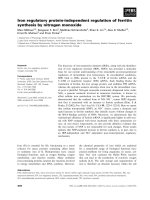
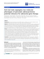
Figure 1 Rotavirus NSP1 inhibits IFN-β promoter activation independent of IRF3
degradation. (A) Scheme of full-length (wt) NSP1 structures of rotavirus OSU and SA11 strains
and C-truncated (∆IRF3 binding domain) NSP1 mutant of rotavirus SA11. (B) Western blot
analysis for degradation of IRF3 by OSU NSP1, SA11 NSP1 and NSP1∆IRF3BD (SA11).
293FT cells were transfected with pCMV-IRF3, pEGFP-OSU NSP1 or pEGFP-SA11 NSP1 or
pEGFP-NSP1∆IRF3BD (SA11) plasmids and cell extracts were assayed 48 h post-transfection
for the expression of IRF3. Immunoblots were probed with anti-IRF3 monoclonal antibody (top
panel). β-actin was used as a loading control (bottom panel). (C, D, E) Inhibition of virus-
induced IFN-β promoter activation by SA11 NSP1 (C), OSU NSP1 (D) or NSP1∆IRF3BD
(SA11) (E). 293FT cells were transfected with pGL3-IFN-β-Luc, pRL-SV40, and increasing
amounts of SA11 NSP1, OSU NSP1 or NSP1∆IRF3BD (SA11) expression plasmids. Cells were
infected with Sendai virus for 24 h and assayed for luciferase activities. Data are expressed as
folds of activation with standard deviations among triplicate samples
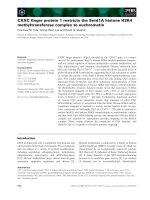
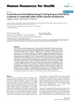
Figure 2 Rotavirus NSP1 inhibits RIG-I mediated IFN-β promoter activation. 293FT cells
were transfected with pGL3-IFN-β-Luc along with plasmids encoding RIG-I (A and D), MDA5
(B and F), MAVS (C and E) and OSU NSP1 or SA11 NSP1∆IRF3BD. pRL-SV40 was included
as a control. At 36 h after transfection, the luciferase activities were measured as described in the
Methods section. Results are expressed as folds of activation with standard deviations among
triplicate samples
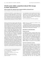
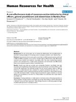
Figure 3 Analysis of interaction between NSP1 and RIG-I. (A) Immunoprecipitation analysis
for the association between OSU NSP1 and RIG-I. 293FT cells were transfected with plasmids
encoding full-length Myc-RIG-I, GFP, and GFP-OSU NSP1. At 48 h after transfection, cell
lysates were immunoprecipitated (IP) with antibodies against Myc, and detected by SDS-PAGE.
Immunoprecipitates and aliquots of cell lysates were then assayed by Western blot (WB)
analysis. (B) SA11 NSP1 interaction with RIG-I. 293FT cells were transfected with plasmids
encoding Myc-RIG-I, GFP-SA11 NSP1, and GFP separately. Immunoprecipitation and Western
blot analysis were performed as described for panel A. (C) Interaction between NSP1∆IRF3BD
of rotavirus SA11 and RIG-I. Transfection, immunoprecipitation and Western blot analysis were
performed as described above
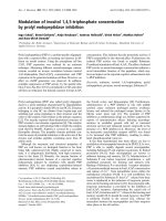
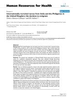
Figure 4 RIG-I is down-regulated by NSP1 at the protein level but is proteasome-
independent. (A, B) Western blot analysis of RIG-I down-regulation by NSP1. 293FT cells
were transfected with increased amount of pEGFP-OSU NSP1 (A) or pEGFP-SA11 NSP1 (B)
and plasmid encoding Myc-RIG-I. Cell extracts were prepared 48 h post-transfection.
Immunoblots were probed with anti-Myc monoclonal antibody to detect RIG-I (top panel). β-
actin was used as a loading control (bottom panel). (C) Transcription level of RIG-I at different
time points after transfection. 293FT cells were co-transfected with SA11-NSP1 and RIG-I
plasmids. At different time points after transfection, total RNA extracted from cells was
subjected to RT-PCR amplification and electrophoresis for RIG-I, NSP1 and GAPDH (inner
control) mRNAs. (D, E) RIG-I is degraded in rotavirus infected cells. MA104 cells were infected
with rotavirus SA11 at a m.o.i. of 0.1. Cell extracts were prepared at 0, 4, 8, 12, 24 and 36 h
post-infection (p.i). RIG-I protein levels at each time point p.i. were determined by Western blot
analyses using an anti-RIG-I antibody. The viral protein VP6 was used as an indicator for
rotavirus infection. β-actin was used as a loading control. RIG-I, NSP1, VP6 and GAPDH
mRNAs were also checked in parallel for evaluating the transcription level (E). (F, G) Effects of
a proteasome inhibitor on NSP1 mediated RIG-I down-regulation. 293FT cells were transfected
with Myc-RIG-I, pEGFP-OSU NSP1 (F) or pEGFP-SA11 NSP1 (G). The cells were treated with
the proteasome inhibitor MG132 or an equivalent volume of DMSO as described in Methods.
Lysates were prepared 36–48 h post-transfection. Immunoblots were probed with anti-Myc to
detect myc-tagged RIG-I
Figure 5 Schematic diagram of the proposed mechanisms by which rotavirus NSP1
subverts host innate immunity. Shown are the targets of NSP1 involved in host innate immune
signaling. NSP1, nonstructural protein 1 of rotavirus; RIG-I, retinoic acid inducible gene
I;MDA5, melanoma differentiation-associated gene 5;MAVS, mitochondrial antiviral-signaling
protein; IPS-1, IFNβ-promoter stimulator 1; IFN, interferon; IRF, interferon transcription factor;
Ub, ubiquitin
Figure 1
Figure 2
Figure 3
Figure 4
Figure 5