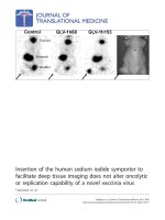
Báo cáo sinh học: " Insertion of the human sodium iodide symporter to facilitate deep tissue imaging does not alter oncolytic or replication capability of a novel vaccinia virus" pptx
Bạn đang xem bản rút gọn của tài liệu. Xem và tải ngay bản đầy đủ của tài liệu tại đây (3.05 MB, 14 trang )
Insertion of the human sodium iodide symporter to
facilitate deep tissue imaging does not alter oncolytic
or replication capability of a novel vaccinia virus
Haddad et al.
Haddad et al. Journal of Translational Medicine 2011, 9:36
(31 March 2011)
RESEARC H Open Access
Insertion of the human sodium iodide symporter
to facilitate deep tissue imaging does not alter
oncolytic or replication capability of a novel
vaccinia virus
Dana Haddad
1,2†
, Nanhai G Chen
3†
, Qian Zhang
3
, Chun-Hao Chen
2
, Yong A Yu
3
, Lorena Gonzalez
2
,
Susanne G Carpenter
2
, Joshua Carson
2
, Joyce Au
2
, Arjun Mittra
2
, Mithat Gonen
2
, Pat B Zanzonico
4
, Yuman Fong
2*
and Aladar A Szalay
1,3,5*
Abstract
Introduction: Oncolytic viruses show promise for treating cancer. However, to assess therapeutic efficacy and
potential toxicity, a noninvasive imaging modality is needed. This study aimed to determine if insertion of the
human sodium iodide symporter (hNIS) cDNA as a marker for non-invasive imaging of virotherapy alters the
replication and oncolytic capability of a novel vaccinia virus, GLV-1h153.
Methods: GLV-1h153 was modified from parental vaccinia virus GLV-1h68 to carry hNIS via homologous
recombination. GLV-1h153 was tested against human pancreatic cancer cell line PANC-1 for replication via viral plaque
assays and flow cytometry. Expression and transportation of hNIS in infected cells was evaluated using Westernblot and
immunofluorescence. Intracellular uptake of radioiodide was assessed using radiouptake assays. Viral cytotoxicity and
tumor regression of treated PANC-1tumor xenografts in nude mice was also determined. Finally, tumor radiouptake in
xenografts was assessed via positron emission tomography (PET) utilizing carrier-free
124
I radiotracer.
Results: GLV-1h153 in fected, replicated within, and killed PANC-1 cells as efficiently as GLV-1h68. GLV-1h153
provided dose-dependent levels of hNIS expression in infected cells. Immunofluorescence detected transport of the
protein to the cell membrane prior to cell lysis, enhancing hNIS-specific radiouptake (P < 0.001). In vivo, GLV-1h153
was as safe and effective as GLV-1h68 in regressing pancreatic cancer xenografts (P < 0.001). Finally, intratumoral
injection of GLV-1h153 facilitated imaging of virus replication in tumors via
124
I-PET.
Conclusion: Insertion of the hNIS gene does not hinder replication or oncolytic capability of GLV-1h153, rendering
this novel virus a promising new candidate for the noninvasive imaging and tracking of oncolytic viral therapy.
Introduction
Oncolytic viral therapies have shown such success in
preclinical testing as a novel cancer treatment modality
that several phase I and II trials are already underway.
Oncolytic vaccinia virus (VACV) strains have been of
particular interest due to several advantages. VACV’s
large 192-kb genome enables a large amount of foreign
DNA to be incorporated without reducing the replica-
tion efficiency of the virus, which has been shown not
to be the case with some other viruses such as adeno-
viruses [1]. It has fast and efficient replication, and cyto-
plasmic replication of the virus lessens the chance of
recombination or integration of viral DNA into cells.
Perhaps most importantly, its safety profile after its use
as a live vaccine in the World Health Organization’s
smallpox vaccination program makes it particularly
attractive as an oncolytic agent and gene vector [2].
* Correspondence: ;
† Contributed equally
1
Department of Biochemistry, University of Wuerzburg, Wuerzburg, D-97074,
Germany
2
Department of Surgery, Memorial Sloan-Kettering Cancer Center, New York,
NY 10065, USA
Full list of author information is available at the end of the article
Haddad et al. Journal of Translational Medicine 2011, 9:36
/>© 2011 Haddad et al; licensee BioMed Central Ltd. This is an Open Access article distributed under the terms of the Creative Commons
Attribution License ( which permits unrestricted use, distribution, and reproduction in
any medium, provided the origin al work is properly cited.
Currently, biopsy is the gold standard for monitoring
the therapeutic effect s of viral oncolysis [3-5] . This may
be feasible in preclinical or early clinical trials, however,
a noninvasive method facilitating ongoing monitoring of
therapy is needed for human studies. The tracking of
viral delivery could give clinicians the abil ity to correlate
efficacy and therapy and monitor potential viral toxicity.
Furthermore, a more sensitive and specific diagnostic
technique to detect tumor origin and, more importantly,
presence of metastases may be possible [3].
Here, we report on the construction and testing of a
VACV carrying the human sodium iodide symporter
(hNIS) as a marker gene for non-invasive tracking of
virusbyimaging.ThisviruswasderivedfromVACV
GLV-1h68, which has already been shown to be a simul-
taneously diagnostic and therapeutic agent in several
human tumor models including breast tumors [6],
mesothelioma [7], canine breast tumors [8], pancreatic
cancers [9], anaplastic thyroid cancers [10,11], mela-
noma [12], and squamous cell carcinoma [13].
Materials and methods
Virus and cell culture
African green monkey kidney fibroblast CV-1 cells and
human pancreatic ductal carcinoma PANC-1 cells were
purchased from American Type Culture Collection
(ATCC) (Manassas, VA) and were grown in Dulbecco’ s
modified Eagle’s medium (DMEM) supplemented with 1%
antibiotic-antimycotic solution (Mediatech, Inc., Herndon,
VA) and 10% fetal bovine serum (FBS) (Mediatech, Inc.) at
37°C under 5% CO
2
. Rat thyroid PCCL3 cells were a kind
gift from the lab of Dr. James Fagin at MSKCC and were
maintained in Coon’s modified medium (Sigma, St. Louis,
MO), 5% calf serum, 2 mM glutamine, 1% penicillin/strep-
tomycin, 10 mM NaHCO3, a nd 6H hormone (1 mU/ml
bovine TSH, 10 ug/ml bovine insulin, 10 nM hydrocorti-
sone, 5 ug/ml transferrin, 10 ng/ml somatostatin, and
2 ng/ml L-glycyl-histidyl-lysine) at 37°C under 5% CO
2
.
GLV-1h68 was derived from VACV LIVP, as described
previously [6].
Construction of hNIS transfer vector
The hNIS cDNA was amplified by polymerase chain
reaction (PCR) using human cDNA clone TC1240 97
(SLC5A5) from OriGene as the template with primers
hNIS-5 (5’-GTCGAC(Sal I) CACCATGGAGGCCGTG-
GAGACCGG-3’ )andhNIS-3(5’-TTAATTAA(Pac I)
TCAGAGGTTTGTCTCCTGCTGGTCTCGA-3’ ). The
PCR product was gel purified, and cloned into the pCR-
Blunt II-TOPO vector using Zero Blunt TOPO PCR
Cloning Kit (Invitrogen, Carlsbad, California). The
resulting construct pCRII-hNIS-1 was sequenced, and
found to contain an extra 33-bp segment in the middle
of the coding sequence, representing an alternative
splicing product for hNIS. To remove this extra 33-bp
segment, two additional primers were designed to flank
the segment, and used in the next set of PCR. In the
next round of reactions, hNIS-5 paired with hNIS-a3
(5’ -GAGGCATGTACTGGTCTGGGGCAGAGATGC-
3’ ), and hNIS-a5 (5’-CCCAGACCAGTACATGCCTCT
GCTGGTGCTG-3’) paired with hNIS-3 were used in
separate PCRs, both with pCRII-hNIS-1 as the template.
The respective PCR products were then mixed and used
as the templates in one reaction with hNIS-5 and hNIS-
3 as the primer pair. The final PCR product was again
cloned into the pCR-Blunt II-TOPO vector as pCRII-
hNISa-2, confirmed by sequencing to be ident ical to the
SLC5A5 sequence in GenBank (accession number
NM_000453). The hNIS cDNA was then released from
pCRII-hNIS-1 with Sal I and Pac I, and subcloned into
HA-SE-RLN-7 with the same cuts by replacing RLN
cDNA. The resulting construct HA-SE-hNIS-1 were
confirmed by sequencing a nd used for insertion of PE-
hNIS into the HA locus of GLV-1h68.
Generation of hNIS-expressing VACV
CV-1 cells were infected with GLV-1h68 at a multiplicity
of infection (MOI) of 0.1 for 1 hour, then transfected
using Fugene (Roche, Indianapolis, IN) with the hNIS
transfer vector. T wo days post infection, infected/ trans-
fected cells were ha rvested and the recombinant viruses
select ed and plaque purified as described previously [14].
The genotype of hNIS-expressing VACV GLV-1h153
was verified by PCR and sequencing. Also, expression
of GFP and b-galactosidase was confirmed by fluorescence
microscopy and 5-bromo-4-chloro-3-indolyl-b-D-galacto-
pyranoside (X-gal, Stratagene, La Jolla, CA), respectively,
and lack of expression of gusA was confirmed by
5-bromo-4-chloro-3-indolyl-b-D-glucuronic acid (X-GlcA,
Research Product International Corp., Mt. Prospect, IL).
Viral growth curves
PANC-1 cells were seeded onto 6-well plates at 5 × 10
5
cells per well. After 24 hours in culture, cells were
infected with either GLV-1h153 or GLV-1h68 at an
MOIof0.01or1.0.Cellswereincubatedat37°Cfor
1 hour with brief agitation every 30 minutes to allow
infection to occur. The infection m edium was then
removed, and cells were incubated in fresh growth med-
ium until cell harvest at 1, 24, 48, and 72 hours post
infection. Viral particles from the infected cells were
released by 3 freeze-thaw cycles, and the titers deter-
mined as (PFU/10
6
) in duplicate by p laque assay in
CV-1 cell monolayers.
Flow cytometry
Cells were seeded on 6-well plates at 5 × 10
5
cells per
well. Wells were then infected at MOIs of 0, 0.01, and
Haddad et al. Journal of Translational Medicine 2011, 9:36
/>Page 3 of 14
1.0, and cells then harvested at 6, 12, 24, 48, 72, and
96 hours postinfection by trypsinizing and washing with
phosphate-buff ered saline (PBS). For the second experi-
ments, cells were seeded on 6-well plates at 5 × 10
5
cells per well. Wells were then infected a t MOIs of 0,
0.01,0.1,0.5,1.0,2.0,and5,andwereharvestedinthe
same manner at 24 hours after infection. GFP expres-
sion was analyzed via a Becton-Dickinson FACScan Plus
cytometer (Becton-Dickinson,SanJose,CA).Analysis
was performed using CellQuest software (Becton-
Dickinson).
hNIS mRNA analysis via microarray
To evaluate the level of hNIS mRNA production in
infected cells, cells were plated at 5 × 10
5
cells per well
and infected with GLV-1h153 at an MOI of 5.0. Six and
24 hours postinfection, 3 samples of each time point
were harvested and lysis performed directly using
RNeasy mini kit protocol (Qiagen Inc., Valencia, CA).
The mRNA samples were measured by spectrophot-
ometer for proof of purity and hybridized to HG-U133A
cDNA microarray chips (Affymetrix Inc, Santa Clara,
CA) by the genomic core laborato ry at Memorial Sloan-
Kettering Cancer Center (MSKCC). The chip images
were scanned and processed to CEL files using the stan-
dard GCOS analysis suite (Affymetrix Inc). The CEL
fileswerethennormalizedandprocessedtosignal
intensities using the gcRMA algorithm from the Biocon-
ductor library for the R statistical programming system.
All subsequent analysis was done on the log (base 2)
transformed data. To find differentially expressed genes
a moderated t-test was used as implemented in the Bio-
conductor LIMMA package. To control for multiple
testing the False Discovery Rate (FDR) method was used
with a cutoff of 0.05.
hNIS protein analysis via Western blot
To confirm whether the hNIS protein was being
expressed in infected cells, cells were plated at 5 × 10
5
per well and infected with GLV-1h153 at various MOIs
of virus, harvested at 24 hours, and suspended with
SDS-PAGE and 0.5-m DDT reagent. After sonication,
30 ug of the protein samples were loaded on 10% Bis-
Tris-HCl buffered polyacrylamide gels using the Bio-rad
system (Bio-rad laboratories, San Francisco, CA). Fol-
lowing gel electrophoresis for 1 hour, proteins were
transferred to nitrocellulose membranes using electro-
blotting. Membranes were then preincubated for 1 hour
in 5% low fat dried milk in TBS-T (20 mm Tris, 137
mm NaCl, and 0.1% Tween-20) to block nonspecific
binding sites. Membranes were incubated with a purified
mouseantibodyagainsthNIS at 1:100 dilution (Abcam
Inc., Cambridge, MA) and incubated for 12 hour at
+4°C. After washing with TBS-T, secondary antibody
(horseradish peroxidase-conjugated g oat antimouse IgG
(Santa Cruz, Santa Cruz, California) was applied for
1 hour at room temperature at a 1:5,000 dilution. Perox-
idase-bound protein bands were visualized using
enhanced chemiluminescence Western blotting detec-
tion reagents (Amersham, Arlington Heights, IL) at
room temperature for approximately 1 minute and
using Kodak BIOMAX MR films for exposure. Normal
human thyroid lysate was used as a positive control, and
cells treated with GLV-1h68 and PBS were used as
negative controls.
Immunofluorescence
PANC-1 cells grown in a 12-well plate at 1 × 10
6
were
mock-infected with GLV-1h68 or infected with GLV-
1h153 at an MOI of 1.0. Twenty-four hours after infec-
tion the cells were fixed with 3.7% paraformaldehyde,
permeabilized with methanol, blocked with PBS contain-
ing BSA, and incubated with a mouse anti-hNIS mono-
clonal antibody (Abcam Inc., Cambridge, MA) at a
dilution of 1:100, followed by incubation with a second-
ary red fluorochrome-conjugated goat antimouse anti-
body (Invitrogen) at a dilution of 1:100. Pictures were
taken using a Nikon inverted fluorescence microscope.
In vitro radiouptake assay
Radio-uptake in cells infected with GLV-1h153 was com-
pared to rat thyroid cell line PCCL3 endogenously
expressing NIS and to cells infected with parental virus
GLV-1h68. Cells were plated at 5 × 10
5
per well in 6-well
plates. Twenty-four hours after infection with MOIs of
0.01, 0.10, and 1.0, cells we re treated w ith 0.5 μCi of
either carrier-free
131
Ior
131
I w ith 1 mM of sodium per-
chlo rate (NaC lO4), a competitive inhibitor of hN IS, for a
60-minute incubation period. Media was supplemented
with 10 μM of sodium iodide (NaI). Iodide uptake was
terminated by removing the medium and washing cells
twice with PBS. Finally, cells were solubilized in lysis
buffer for residual radioactivity, and the cell pellet-to-
medium activity ratio (cpm/g of pellet versus cpm/mL of
medium) calculated from the radioactivity measurements
assayed in a Packard g-counter (Perkin Elmer, Waltham,
MA). Results were expressed as change in uptake relative
to negati ve uninfected control. All samples were done in
triplicate.
In vitro cytotoxicity assay
PANC-1 pancreatic cancer cells were plated at 2 × 10
4
per well in 6-well plates. After incubation for 6 hours,
cells were infected with GLV-1h153 or GLV-1h68 at
MOIs of 1.00, 0.10, 0.01, and 0 (control wells). Viral
cytotoxicity was measured on day 1 and every second
Haddad et al. Journal of Translational Medicine 2011, 9:36
/>Page 4 of 14
day thereafter by lactate dehydrogenase (LDH) release
assa y. Results are expressed as the percentage of surviv-
ing cells as compared to uninfected control.
In vivo tumor therapy studies and systemic toxicity
All mice were cared for and maintained in accordance
with animal welfare regulations under an approved pro-
tocol by the Institutional Animal Care and Use Commit-
tee at the San Diego Science Center, San Diego,
California. PANC-1 xenografts were developed in 6- to
8-week-old male nude mice (NCI:Hsd:Athymic Nude-
Foxn1nu, Harlan) by implanting 2 × 10
6
PANC-1 cells
in PBS subcutaneously in the left hindleg. Tumor
growth was recorded once a week in 3 dimensions using
a digital caliper and reported in mm
3
using the formula
(length × width × [height-5]). When tumors reached
100-300 mm
3
, mice were injected intratumorally (IT) or
intravenously (IV) via the tail vein with a single dose of
2×10
6
PFUs of GLV-1h153 or GLV-1h68 in 100 μL
PBS. Animals were o bserved daily for any sign of toxi-
city, and body weight checked weekly.
Radiopharmaceuticals
124
Iand
131
I were obtained from MSKCC’ s radiophar-
macy. The maximum specific activities for the
124
Iand
131
I compounds were ~140 μCi/mouse and ~0.5 μCi/
well, respectively.
In vivo PET imaging
All animal studies were performed in compliance with
all applicable policies, procedures, and regulatory
requirements of the Institutional Animal Care and Use
Committee, the Research Animal Resource Center of
MSKCC, and the National Institutes of Health “Guide
for the Care and Use of Laboratory Animals.” Three
groups of 2-3 animals bearing subcutaneous PANC-1
xenografts on the left hindleg measuring were injected
intratumorally with 2 × 10
7
PFU GLV-1h153 (3 mice),
2×10
7
PFU GLV-1h68 (2 mice), or PBS (2 mice). Two
days after viral injection, 140 μCi of
124
I was adminis-
tered via the tail vein. One hour after radiotracer admin-
istration, 3-dimensional list-mode data were acquired
using an energy window of 350 to 700 keV, and a coi n-
cidence timing window of 6 nanoseconds. Imaging was
performed using a Focus 120 microPET dedicated small
animal PET scanner (Concorde Microsystems Inc,
Knoxville, TN). These data were then sorted into
2-dimensional histograms by Fourier rebinning. The
image data were corrected for (a) nonuniformity of
scanner response using a uniform cylinder source-based
normalization, (b) dead time count losses using a single-
count rate-based global correction, (c) physical decay to
the time of injection, and (d) the
124
I branching ratio.
The count rates in the reconstr ucted images were
converted to activity concentration (%ID/g) using a sys-
tem calibration factor (MBq/mL per cps/voxel) derived
from imaging of a mouse-size phantom filled with a uni-
form aqueous solution of
18
F. Image analysis was per-
formed using ASIPro (Siemens Pre-clinical Solutions,
Knoxville, TN).
Statistical analysis
The GraphPad Prism 5.0 program (GraphPad Software,
San Diego, CA) was used for data handling and analysis.
The significance of differences between the 3 therapy
groups (untreated, GLV-1h153, GLV-1h68) was deter-
mined via two-way ANOVA with Bonferroni correction.
P values were generated for radiouptake assay compari-
sons using Dunnett’s test [15]. P < 0.05 was considered
significant.
Results
Construction of the hNIS transfer vector
The GLV-1h153 construct used in this study was
derived from GLV-1h68 by replacing the b-glucuroni-
dase (gusA) expression cassette at the A56R locus with
the hNIS expression cassette (SE-hNIS) containing the
hNIS cDNA under the control of the VACV synthetic
early promoter, by homologous recombination in
infected cells. The genotype of GLV-1h153 (Figure 1a)
was verified by PCR and sequencing, and the lack of b-
glucuronidase expression was confirmed by X-GLcA
staining (Figure 1b).
GLV-1h153 replicated efficiently in PANC-1 cells
To evaluate the replication efficiency and effect of hNIS
protein expression on VACV replication, PANC-1 cells
were infected with either GLV-1h153 or its parental
virus, GLV-1h68, at MOIs of 0.01 and 1.0, and the
infected cells harvested at 1, 24, 48, and 72 hours post
infection. The viral titers at each time point were deter-
mined in CV-1 cells using standard plaque assays. Both
GLV-1h153 and GLV-1h68 replicated in PANC-1 cells
at similar levels, indicating that the hNIS protein did
not hinder viral replication within cells. GLV-1h153
yielded a 4-log, or 10,000-fold, increase of viral load
with an MOI of 0.01 only 72 hours after infection.
Within this time, viral load with an MOI of 0.01
reached the same levels as infection with an MOI of 1.0,
again indicating efficient replication (Figure 2a).
GLV-1h153 replication was assessed via flow cytometric
detection of GFP
GFP expression in cells infected with either GLV-1h68
or GLV-1h153 was quantified using flow analysis, and
was shown to be both time and MOI dependent.
Adjusting for background, GFP expression mimicked
the viral replication growthcurve,withGFPexpression
Haddad et al. Journal of Translational Medicine 2011, 9:36
/>Page 5 of 14
in cells infected at an MOI of 0.01 reaching similar
levels to an MOI of 1.0 by 72 hours (Figure 2b). Further,
>70% of live cells expressed G FP at an MOI of 5.0 at
24hrs postinfection (Figure 2c).
Production of hNIS mRNA and protein in infected cells
was shown via microarray analysis and Western blot
To confirm production of hNIS mRNA by GLV-1h153-
infected PANC-1 cells, cells were infected at an MOI of
5.0 and mRNA isolated for analysis with Affymetrix
chips. mRNA in cells had an almost 2000-fold increase
by only 6 hours after infection, and a >5000-fold change
by 24 hours (P < 0.05) (Figure 3a). To show hNIS pro-
tein expression by GLV-1h153, PANC-1 cells were
mock infected or infected with GLV-1h153 or parental
virusGLV-1h68atMOIsof0.1,1.0,and5.0and
harvested 24 hours after infection. Production of the
hNIS protein was successfully detected by Western blot
between 75 and 100 KiloDalton, with an increasing con-
centration of protein at higher MOIs (Figure 3b). The
difference in molecular weight of hNIS between the
positive control and infected cells is likely due to the
different levels of glycosylation, as noted by several
other groups [16,17].
The hNIS protein was localized at the cell membrane of
PANC-1 cells
To determ ine whether the hNIS protein expressed by
GLV-1h153 was successfully transported and inserted on
the cell membrane, PANC-1 cells were infected with
GLV-1h153 and fixed with 3.7% paraformaldehyde.
The hNIS protein was visualized using a monoclonal
a.
b.
Brightfield GFP LacZ gus A
GLV-1h153
GLV-1h68
Figure 1 GLV-1h153 construct. a. GLV-1h153 was derived from GLV-1h68 by replacing the gus A expression cassette at the A56R locus with the
hNIS expression cassette through in vivo homologous recombination. Both viruses contain RUC-GFP and lacZ expression cassettes at the F14.5L
and J2R loci, respectively. PE, PE/L, P11, and P7.5 are VACV synthetic early, synthetic early/late, 11K, and 7.5K promoters, respectively. TFR is
human transferrin receptor inserted in the reverse orientation with respect to the promoter PE/L.b. Confirmation of GFP, LacZ, and lack of gus A
marker gene expression in GLV-1h153 infected CV-1 cells. While the gus A gene cassette is expressed in cells infected with parent virus GLV-
1h68, this has been replaced by the hNIS gene cassette in GLV-1h153, leading to loss of gus A expression.
Haddad et al. Journal of Translational Medicine 2011, 9:36
/>Page 6 of 14
anti-hNIS antibody that recognizes the intracellular
domain of the protein. As shown in Figure 3c, mock- or
GLV-1h68-infected cells (as demonstrated by GFP
expression) did not show hNIS protein expression,
whereas the hNIS protein in cells infected with GLV-
1h153 was readily detectable by immunofluorescence
microscopy, and appears to be localized at the cell
membrane.
GLV-1h153-infected PANC-1 cells showed enhanced
uptake of carrier-free radioiodide
To establish that the hNIS symporter was functional,
cells were mock infected or infected at an MOI of 1.0
with GLV-1h153 and GLV-1h68, then treated with
131
I
at various times after infection. Normal rat thyroid ce ll
line PCCL3 was used as a positive control. PANC-1
cells infected with GLV-1h153 showed a >70-fold
increased radiouptake compared with mock-infected
control at 24 hours post infection (P < 0.0001) despite
similar cell protein levels, compared t o 2.67 and 1.01-
fold increased radiouptake with MOIs of 0.1 and 0.01,
respectively (Figure 4a). This increased uptake correlated
with peak GFP expression (Figure 4b). Moreover, when
cells were treated with NaClO4, a competitive inhibitor
of hNIS, radiouptake decreased in GLV-1h153-treated
cells, from a 70- to a 1.14-fold difference at an MOI of
1.0, indicating hNIS-specific radiouptake. Radiouptake in
cells infected with GLV-1h153 was compared to rat
thyroid cell line endogenously expressing NIS (PCCL3),
and to cells infected with parental virus GLV-1h68 or
mock infected. Cells were plated at 5 × 10
5
cells per
well in 6-well plates. Twenty-four hours after infection,
cells were treated with 0.5 μCi of either carrier free
131
I
or
131
I with 1 mM of sodium perchlorate (NaClO4), a
Figure 2 Viral proliferation assay of GLV-1h153-in PANC-1 cells. a. PANC-1 cells were grown in 6-well plates and infected with GLV-1h153 or
GLV-1h68 at an MOI of 0.01 and 1.0. Three wells of each virus were harvested at 1, 24, 48, and 72 hours postinfection. GLV-1h153 replicated in a
similar manner to GLV-1h68, with a 4-log increase in viral load at an MOI of 0.01 by 72 hours, reaching similar levels as that in cells infected with
an MOI of 1.0. This demonstrates that GLV-1h153 is able to replicate efficiently within PANC-1 cells in vitro as well as parental virus GLV-1h68. b.
GFP expression was quantified via flow cytometry in PANC-1 cells infected with GLV-1h153 at MOIs of 1.0 and 0.01 and was shown to be MOI
dependent. GFP expression mimicked the viral replication growth curve, with GFP expression in the MOI 0.01 infected cells reaching similar
levels as the MOI of 1.0 by 72 hours after infection. c. GFP expression was quantified via flow cytometry in PANC-1 cells infected with an MOI of
0.01, 0.1, 0.5, 1.0 2.0, and 5.0 at 24 hours after infection, and was shown to be MOI-dependent.
Haddad et al. Journal of Translational Medicine 2011, 9:36
/>Page 7 of 14
competitive inhibitor of hNIS for a 60-minute incuba-
tion period. Media was supplemented with 10 μMof
sodium iodide (NaI). Iodide uptake was terminated by
removing the medium and washing cells twice with PBS.
Finally, cells were solubilized in lysis buffer for residual
radioactivity, and the cell pellet-to-medium activity ratio
(cpm/g of pellet/cpm/mL of medium) calculated from
the radioactivity measurements assayed in a Packard g-
counter (Perkin Elmer, Waltham, MA). Results are
expressed as change in uptake relative to negative unin-
fected control. All samples were done in triplicate.
GLV-1h153 was cytolytic against PANC-1 cells in vitro
To investigate whether expression of hNIS would affect
cytolytic activity of VACV in cell cultures, PANC-1 cells
were infected with GLV -1h68 or GLV-1h153 at MOIs of
0.01and 1.0. Vira l cytot oxicity was measured every other
day for 11 days. The survival curves for GLV-1h68 and
GLV-1h153 were almost identical at both MOIs, indicat-
ing that the cells infected by either of the virus strains
were dying at similar levels in a time- and dose-dependent
fashion (Figure 5a). By day 11, More than 60% cell kill was
achieved with an MOI of 1.0 as compared to control.
GLV-1h153 was safe and effective at regressing PANC-1
tumor xenografts in vivo
To establish cytolytic effects of GLV-1h153 in vivo,mice
bearing PANC-1 xenograft tumors on hindleg were
infected intratumorally ( ITly) or intravenously (IVly) with
GLV-1h153 o r GLV-1 h168, or mock treated with PBS.
Figure 3 Assessment of hNIS expression in GLV-1h153-infected PANC-1 cells. a. Microarray analysis of cells infected with an MOI of 5.0 of
GLV-1h153 yielded an almost 2000-fold increase by 6 hours and an almost 5000-fold increase by 24 hours in hNIS mRNA production as
compared to noninfected control. b. PANC-1 cells were either mock infected or infected with GLV-1h68 at an MOI of 1.0 or infected with GLV-
1h153 at an MOI of 1.0 or 5.0 for 24 hours. The hNIS protein was detected by Western blot analysis using monoclonal anti-hNIS antibody. Only
GLV-1h153-infected cells expressed the hNIS protein, but cells either mock infected or infected with GLV-1h68 did not. The molecular weight
marker bands (in kiloDaltons) are shown on the left. c. PANC-1 cells were mock infected or infected with GLV-1h68 or GLV-1h153 at a MOI of 1.0
for 24 hours. The hNIS protein was detected by immunofluorescence microscopy using monoclonal anti-hNIS antibody, which recognizes the
intracellular domain of the protein. Mock- or GLV-1h68-infected cells (as demonstrated by GFP expression) did not express the hNIS protein,
whereas the hNIS protein on the cell membrane of PANC-1 cells infected with GLV-1h153 was readily detectable.
Haddad et al. Journal of Translational Medicine 2011, 9:36
/>Page 8 of 14
While the growth of tumors treated with PBS continued to
grow, GLV-1h153-treated tumors occurred in three dis-
tinct phases: growth, inhibition, and regression (Figure 5c).
The mean relative size of tumors treated with GLV-1h153
was significantly smaller than untreated control tumors,
with differences beginning as early as day 13 (P < 0.01),
and continuing till day 34 afte r virus or PBS control
administration (P < 0.001). By day 34, there was an over 4-
fold difference between control and IV tumor volumes,
andanover6-folddifferenceintheITgroup.Further-
more, there wer e no significant adverse effects seen with
regard to body weight, with the IT group even gaining
weight as compared to control with statistically significant
results by day 34 (P < 0.05) (Figure 5d).
GLV-1h153-enhanced radiouptake in PANC-1 tumor
xenografts and was readily imaged via PET
After successful cell culture uptake studies we wanted to
show the feasibility of using GLV-1h153 in combination
with carrier-free
124
I radiotracer to image infected
PANC-1 tumors. hNIS protein expression in the PANC-
1 tumor-bearing animals after GLV-1h153 administra-
tion was visualized by
124
IPET.Carrierfree
124
Iwas
IVly administered 48 hours after IT v irus injection and
PET imaging was performed 1 hour after radiotracer
administration. GLV-1h153-injected tumors were easily
detected, whereas GLV-1h68- and PBS-injected tumors
could not be visualized and therefore were not signifi-
cantly above background (Figure 6).
Discussion
Oncolytic viral therapy is emerging as a novel cancer
therapy. Preclinical and c linical studies have shown a
number of oncolytic viruses to have a broad spectrum
of anti-cancer activity and safety [18]. These are
ongoing, and the first oncolytic viral therapy has now
been approved in China as a treatment for head and
neck cancers [19]. Clinical trials are underway to assess
0
25
50
75
100
125
150
175
Relative
131
I uptake
Multiplicity of infection
a. b.
***
0.01 0.10 1.00
Figure 4 Assessment of in vitro
131
I radiouptake of GLV-1h153-infected PANC-1 cells. a. PANC-1 cells were infected with an MOI of 0, 0.01,
0.1, and 1.0 of GLV-1h153 and MOI of 1.0 of GLV-1h68. PCCL3 was used as a positive control. Twenty-four hours after infection, there is a >70-
fold enhanced radiouptake at an MOI of 1.0 as compared to an MOI of 0 in GLV-1h153, and radiouptake is shown to be MOI dependent and
hNIS specific (as shown with blocking with competitive inhibitor of hNIS, NaClO4). b. Maximum radiouptake with an MOI of 1.0 24 hrs after
infection corresponded to maximum GFP expression.
Haddad et al. Journal of Translational Medicine 2011, 9:36
/>Page 9 of 14
the effects of many other oncolytic viral therapies [20].
However, future clinical studies may benefit from t he
ability to noninvasively and serially identify sites of viral
targeting and to measure the level of viral infection and
spread in order to provide important information for
correlation with safety, efficacy, and toxicity [3-5]. Such
real-time tracking would also provide useful information
regarding timing of viral dose and administration for
optimization of therapy, as well as distribution and
replication of the oncolytic virus, and would alleviate
the need for multiple and repeated tissue biopsies.
VACV is arguably the most successful biologic therapy
agent, since versions of this virus were given to millions
of humans during the smallpox eradication campaign
[2]. More recently, e ngineered VACVs have also been
successfully used as direct oncolytic agents, capable of
preferentially infecting, replicating within, and killing a
wide variety of cancer cell types [6-11,13,21]. VACV dis-
plays many of the qualities thought necessary for an
effective oncolytic antitumor agent. In particular, the
large insertional cloning capacity allows for the inclusion
of several functional and therapeutic transgenes. With
the insertion of reporter genes not expressed in unin-
fected cells, viruses can be localized and the course of
viral therapy monitored in patients.
One such promising virus strain is GLV-1h68 [21].
This strain has shown efficacy in the treatment of a
wide range of human cancers and is currently being
0
25
50
75
100
125
012345678910
11
Cytotoxicity (% Control)
Day Post Infection
a.
c.
Day 9
0
2
4
6
8
10
-1 6 13 20 27
34
Relative Tumor Volume
Day Post Injection
***
b.
Control
Day 1 Day 3 Day 7
GFPMerge
***
**
***
d.
0.0
0.2
0.4
0.6
0.8
1.0
1.2
-1 6 13 20 27
34
Relative Net Body Weight
Day Post Injection
*
Figure 5 GLV-1h153 infection and killing in cell culture and in vivo. a. PANC-1 cells were infected by various GLV-1h153at MOIs of 0.01, 0.1,
and 1.0. Cell viability was determined via lactate dehydrogenase assays, and was set at 100% before infection. GLV-1h153 infected and was
cytotxic at various MOIs, with less than 20% survival of cells as compared to control at an MOI of 1.0 by day 9. The values are the mean of
triplicate samples, and bars indicate SD. b. GFP expression is shown to be time-dependent, with abundant GFP expression by day 3. Phase
overlay pictures shows gradual cell death and thus decline of GFP expression by day 7. Closer examination of infected cells reveals loss of
normal morphology and cell progressive cell detachment. c. 2 × 10
6
PFUs of GLV-1h153 or GLV-1h68, or PBS were injected IVly or ITly into nude
mice bearing s.c. PANC-1 tumors on the hindleg (~100 mm
3
). GLV-1h153 was able to regress pancreatic tumor xenograft both ITly and IVly
starting at day 13. The values are a mean of 4-5 mice, with bars indicating SEM. d. GLV-1h153 infection of pancreatic tumor xenografts did not
have adverse effects on body weight at 5 weeks post injection, with the IT group even gaining weight compared to control.
Haddad et al. Journal of Translational Medicine 2011, 9:36
/>Page 10 of 14
tested in phase I human trials [20]. In this study we
describe the generation of a novel recombinant VACV,
GLV-1h153, derived from GLV-1h68, which has been
engineered for specific targeted treatment of cancer and
the additional capability of facilitating noninvasive ima-
ging of tumors and metastases. To our knowledge,
GLV-1h153 is the first oncolytic VACV expressing the
hNIS protein.
ThereportergenechosenforinsertionintoGLV-
1h153 was based on the already successful PET and
SPECT imaging characteristics of the human sodium
iodide symporter (hNIS) and carrier-free radioiodine
reporter imaging system. hNIS is an intrinsic plasma
membrane protein that mediates the active transport
and concentration of iodide in thyroid gland cells and
some extra-thyroidal tissues [16,17] . Although endogen-
ous NIS is physiologically and functionally expressed in
several normal tissues, so far only 2 human cancers -
some thyroid cancers, and around 80% of breast cancers
including ductal carcinomas - have been shown to
express endogenous NIS fun ctionally, making them
amenable to radiotherapy [22]. It is one of several
human reporter genes that are currently being used in
preclinical studies and has even been used in clinical
studies imaging prostate cancer [20,23]. hNIS gene
transfer via viral vector may allow infected tumor cells
to concentrate several commercially available, relatively
inexpensive radionuclide probes, such as
123
I,
124
I,
125
I,
and
99m
TcO
4
, all of which have long been approved for
human use by the U.S. Food and Drug Administration,
allowing noninvasive imaging of tumo rs expressing NIS
[22]. In contrast to a study published by McCart et al.
[24] using an oncolytic VACV expressing the human
somatostatin receptor hSSTR2, hNIS is a transporter-
based reporter gene system. Whereas receptors usually
have a 1:1 binding rel ationship with a radiolabeled
ligand , transporters provide signal amplification through
transport-mediated concentrative intracellular accumula-
tion of substrate. hNIS use has also been shown to be
comparable to the commonly used HSV1-tk reporter
gene [25] and correlated with
99m
TcO
4
[26]. This can
be very useful for viral distribution with scintigraphy or
PET scanning during and after viral therapy, and may
allow for correlation with efficacy and toxicity during
clinical trials and treatment thus offering potential clini-
cal translation of this dual therapy.
In order to take advantage of the therapeutic and ima-
ging potential of hNIS, several groups have attempted
exogenous NIS gene transfe r in several human cancers
including head and neck squamous cell cancers, non-
small cell lung, thyroid, liver, colorectal, and prostate
cancers, as well as glioma and multiple myeloma [22].
Studies have shown that hNIS gene delivery to both
thyroidal and non-th yroidal, non-organifying tumor cells
is capable of inducing accumulation of therapeutically
effective radioiodine doses. For example, a single thera-
peutic
131
I dose of 3 mCi was shown to elicit a dramatic
therapeutic response in NIS-transfected prostate cell
xenografts, with an average volume reduction of more
than 90% [27]. Transfection of an hNIS-defective follicu-
lar thyroid carcinoma cell line with the hNIS gene was
able to reestablish iodide accumulation a ctivity both in
cell culture and in animal models [28]. Furthermore,
transfection of pancreatic cancer cells with a replication-
deficient adenoviral vector expressing hNIS lead to a
more than 15-fold increase in iodide uptake visualized
with
123
I scintigraphy, and an over 75% reduction in
volume in vivo after treatment with 3mci of
131
I [29].
We have previously reported on the use of a novel
recombinant VACV, GLV-1h99, a derivative of GLV-
1h68, which was constructed to carry the human norepi-
nephrine transporter gene (hNET) under the VACV
synthetic early promoter placed at the F14.5L locus
for deep tissue imaging [30,31]. The parental virus
Figure 6 PET imaging of enhanced radiouptake in GLV-1h153-infected PANC-1 xenografts. Two × 1 0
7
PFU of GLV-1h153, GLV-1h68, or
PBS was injected intratumorally into PANC-1 hindleg tumor-bearing mice.
124
I-PET scanning was obtained 48 hours after infection and 1 hour
after radiotracer administration. GLV-1h153-infected PANC-1 tumors were easily visualized, while no enhanced signal was seen in the PBS- or
GLV-1h68 injected tumors. The stomach and thyroid were also imaged due to native NIS expression, and the bladder due to tracer excretion.
Haddad et al. Journal of Translational Medicine 2011, 9:36
/>Page 11 of 14
GLV-1h68, a recombinant VACV (LIVP strain), was con-
structed by inserting 3 expression cassettes (Renilla luci-
ferase-Aequorea green fluorescent protein (RUC-GFP)
fusion, b-galactosidase, and b-glucuronidase) i nto the
F14.5L, J2R,andA56R loci of the viral gen ome, respec-
tively [6]. The hNET protein was expressed at high levels
on the membranes of cells infected with GLV-1h99, and
expression of the hNET protein did not negatively affect
virus replication in cell culture or in vivo virotherapeutic
efficacy. GLV-1h99-mediated expression of the hNET
protein in infe cted cells resulted in specific uptake of the
radiotracer [
131
I]-meta-iodo benzylguanidine ([
131
I]-
MIBG). In mice, GLV-1h99-infected tumors, including
pancreatic and mesothelioma, were readily imaged by
[
124
I]-MIBG P ET. However, one of the disadvantages of
using hNET is that it requires the carrier MIBG for
radioiodine uptake.
GLV-1h153 was effective at infecting and replicating
within pancreatic cancer PANC-1 cells as efficiently as
its parental virus GLV-1h68. This indicated that inser-
tion of the hNIS protein did not negatively affect virus
replication in cell culture. With the hNET-expressing
GLV-1h99 virus, on the other hand, there was slight
improvement in viral replication and oncolytic effect,
which may have been due to the exchange of expression
cassettes under the control of promoters with different
strengths.
Microarray analysis revealed an almost 2000-fold
change increase in hNIS mRNA and an almost 5000-
fold change increase by 24 hours after PANC-1 infection
with GLV-1h153 at a multiplicity of infection of 5.0.
Western blot studies showed hNIS protein expression as
a band between 75 and 100 KiloDalton in PANC-1 cells
infected with GLV-1h153, with higher concentrations of
the protein at higher MOIs. This band also appears in
normal human thyroid lysates at a slightly lower mole-
cular weight, which is likely explained by differences in
glycosylation within cells [16]. The hNIS protein was
successfully transported and in serted into the cell mem-
brane, as demonstrated by fluorescence microscopy.
In vitro, GLV-1h153-mediated expression of the hNIS
protein in infected PANC-1 cells resulted in specific
uptake of the radiotracer
131
I, indicating that the hNIS
protein was functional, with the uptake reaching a >70-
fold increase compared with uninfected control at an
MOI of 1.0.
GLV-1h153 was also effective at infecting, replicating
within, and killing PANC-1 cells and eradicating tumor
xenografts as efficiently as parental virus GLV-1h68.
This indicated that insertion of the hNIS protein did
not negatively affect virus replication in vivo which was
already demonstrated in vitro, or the cytolytic activity in
cell culture and in vivo virotherapeutic efficacy. Similar
effects we re seen between the IT and IV groups treated
with GLV-1h153 or GLV-1h68, indicating the inherent
affinity of both genetically modified vaccinia viruses to
tumors. Furthermore, administration of GLV-1h153 did
not have any significant effects on mean net body
weights of the animals 34 days after treatment, with the
IT groups even gaining weight as compared to untreated
control.
Finally, in mice, three PANC-1 tumors infected with
GLV-1h153 were readily detectable by PET, with no
enhancement above background of either GLV-1h68- or
PBS-treated tumors. Mice were treated intratumorally
with GLV-1h153, non-hNIS expressing parent virus
GLV-1h68, and PBS, and imaged 48 hours after with
carrier free
124
I. The quanti tative
124
I -PET showed that
imaging of GLV-1h153 infectionofPANC-1tumorsis
feasible after direct tumor injection.
As with any translational therapy, concerns over
immune responses remain. Since hNIS is a human
derived gene, it is unlikely to be immunogenic. How-
ever, application of GLV-1h153-mediated hNIS transfer
raises concerns over the possibility of autoimmunity in
patients. Several papers have already shown that hNIS is
not a major candidate for autoimmune disease in
patients with patients with Graves ’ disease and Hashi-
moto’ s thyroiditis [32,33]. Moreover, a clinical trial
assessing adenoviral-mediated hNIS transfer in humans
did not report any serious adverse effects due to autoim-
munity in patients treated for prostate cancer [23].
Further studies and caution will be needed to assess the
potential of autoimmunity with hNIS transfer in
humans. Studies are also now underway to determine
the viral biodistribution, as well as the dose and timing
related aspects of in vivo imaging using this novel virus.
Conclusions
We have therefore shown that a novel new oncolytic
VACV engineered to carry the human sodium iodide
symporter gene (hNIS) can successfully replicate within
and kill pancreatic cancer cells as efficiently as its paren-
tal virus GLV-1h68. GLV-1h 153 expresses the hNIS
reporter gene, which enhances radiouptake in vitro and
is readily imaged with the clinically approved radiophar-
maceutical
124
I via PET. The ability of GLV-1h153 to
infect and enhance cellular uptake of iodine in cells of
pancre atic can cer origin, a uniformly fatal disease resis-
tant to conventional therapy, justifies further studies a s
well as the initiation of clinicaltrials.Further,thisima-
ging system could be directly translated to human stu-
dies, as clinical trials of oncolytic viral therapy would
benefit from this noninvasive monitoring modality.
Abbreviations
ATCC: American Type Culture Collection; (c)DNA: (complementary)
deoxyribonucleic acid; CPM: counts per minute; DMEM: Dulbecco’s modified
Haddad et al. Journal of Translational Medicine 2011, 9:36
/>Page 12 of 14
Eagle’s medium; FDR: false discovery rate; GFP: green fluorescent Protein;
gusA: β-glucuronidase; hNIS: human sodium iodide symporter; hNET: human
norepinephrin transporter; hSSTR2: human somatostatin receptor; HSV1-tk:
herpes simplex virus 1 - thymidine kinase;
124
I: Iodine-124;
125
I: Iodine-125;
131
I: Iodine-131; IT(ly): intratumoral(ly); IV(ly): intravenous(ly); LacZ: β-
galactosidase; LDH: lactate dehydrogenase; LIVP: Lister vaccine strain; MIBG:
meta-iodobenzylguanidine; MOI: multiplicity of infection; (m)RNA:
(messenger) ribonucleic acid; NaClO
4
: sodium perchlorate; PBS: phosphate
buffer serum; PCR: polymerase chain reaction; PET: positron emission
tomography;
99m
TcO
4
: 99m-technecium pertechtenate; VACV: vaccinia virus.
Acknowledgements
We would like to acknowledge Meryl Greenblatt for her excellent editing
skills with this paper. Special thanks to the Fagin lab at MSKCC for kindly
providing the PCCL3 cell line used for radiouptake assays and Valerie Longo
for her imaging expertise. We would also like to thank Jason Aguilar, Terry
Trevino and Melody Fells for their excellent technical skills and Valerie Longo
for her expertise and assistance with imaging experiments. Appreciation is
given to Dr. Sean Carlin for his advice and expertise regarding the human
sodium iodide symporter and its physiology and Dr. Nicholas Socci for help
with microarray analysis. Thanks are given to Dr. Ulrike Geissinger, Dr. Alexa
Frentzen, Dr. Martina Zimmermann, Jennifer Reinboth, Klaas Ehring, Lisa
Buckel, Vanessa Cook, Eric Rosenfield, Mordechai Smith, Jonathan Katz, and
Laura Weber for assistance with experiments. This work was supported by
grants from Genelux Corporation (R&D facility in San Diego, CA, USA). D.
Haddad, visitor at MSKCC, is a graduate student in Dr. Szalay’s laboratory in
the Department of Biochemistry, University of Würzburg, Germany, and is
supported by a graduate stipend from The University of Wuerzburg as well
as travel grants from Genelux Corporation and Memorial Sloan-Kettering
Cancer Center.
Author details
1
Department of Biochemistry, University of Wuerzburg, Wuerzburg, D-97074,
Germany.
2
Department of Surgery, Memo rial Sloan-Kettering Cancer Center,
New York, NY 10065, USA.
3
Genelux Corporation, San Diego Science Center,
San Diego, CA 92109, USA.
4
Departments of Medical Physics and Radiology,
Memorial Sloan-Kettering Cancer Center, New York, NY 10065, USA.
5
Department of Radiation Oncology, University of California San Diego, San
Diego, CA 92093, USA.
Authors’ contributions
NC was instrumental in the construction and homologous recombination of
GLV-1h153. QZ was instrumental in the hNIS transfe r construction and viral
sequencing. CC contributed to study design, western blot, and
immunofluorescence. YY contributed to the study design and assisted in in
vivo studies. SC contributed to the cytotoxicity and flow cytometry assays.
JC contributed to the in vitro radiouptake assays. JA contributed to the in
vitro radiouptake assays. AM contributed to protein harvesting and western
blot. MG was instrumental in statistical analysis of data. PZ was instrumental
in study design, imaging experiments, and advice regarding hNIS biology
and physiology. YF is the co-corresponding author and was critical to study
design and completion. AZ is the corresponding author of this paper and
was critical to study design and completion. All authors have read and
approved the final manuscript.
Author disclosure statement
Nanhai G. Chen, Qian Zhang, Yong A. Yu, and Aladar A. Szalay are affiliated
with Genelux Corporation. No competing financial interests exist for Dana
Haddad, Chun-Hao Chen, Pat Zanzonico, Lorena Gonzalez, Susanne
Carpenter, Joshua Carson, Joyce Au, Arjun Mittra, Mithat Gonen, and Yuman
Fong.
Received: 15 February 2011 Accepted: 31 March 2011
Published: 31 March 2011
References
1. Moss B: Poxviridae: the Viruses and Their Replication. 4 edition. Philadelphia:
Lippincott Williams & Wilkins; 2001.
2. Fenner F, Henderson DA, Arita I, Jezek Z, Ladnyi ID: Smallpox and its
eradication. Geneva: WHO; 1988.
3. Serganova I, Blasberg R: Reporter gene imaging: potential impact on
therapy. Nucl Med Biol 2005, 32:763-780.
4. Serganova I, Ponomarev V, Blasberg R: Human reporter genes: potential
use in clinical studies. Nucl Med Biol 2007, 34:791-807.
5. Kuruppu D, Brownell AL, Zhu A, Yu M, Wang X, Kulu Y, Fuchs BC,
Kawasaki H, Tanabe KK: Positron emission tomography of herpes simplex
virus 1 oncolysis. Cancer Res 2007, 67:3295-3300.
6. Zhang Q, Yu YA, Wang E, Chen N, Danner RL, Munson PJ, Marincola FM,
Szalay AA: Eradication of solid human breast tumors in nude mice with
an intravenously injected light-emitting oncolytic vaccinia virus. Cancer
Res 2007, 67:10038-10046.
7. Kelly KJ, Woo Y, Brader P, Yu Z, Riedl C, Lin SF, Chen N, Yu YA, Rusch VW,
Szalay AA, Fong Y: Novel oncolytic agent GLV-1h68 is effective against
malignant pleural mesothelioma. Hum Gene Ther 2008, 19:774-782.
8. Gentschev I, Stritzker J, Hofmann E, Weibel S, Yu YA, Chen N, Zhang Q,
Bullerdiek J, Nolte I, Szalay AA: Use of an oncolytic vaccinia virus for the
treatment of canine breast cancer in nude mice: preclinical
development of a therapeutic agent. Cancer Gene Ther 2009, 16:320-328.
9. Yu YA, Galanis C, Woo Y, Chen N, Zhang Q, Fong Y, Szalay AA: Regression
of human pancreatic tumor xenografts in mice after a single systemic
injection of recombinant vaccinia virus GLV-1h68. Mol Cancer Ther 2009,
8:141-151.
10. Lin SF, Yu Z, Riedl C, Woo Y, Zhang Q, Yu YA, Timiryasova T, Chen N,
Shah JP, Szalay AA, et al: Treatment of anaplastic thyroid carcinoma in
vitro with a mutant vaccinia virus. Surgery 2007, 142:976-983, discussion
976-983.
11. Lin SF, Price DL, Chen CH, Brader P, Li S, Gonzalez L, Zhang Q, Yu YA,
Chen N, Szalay AA, et al: Oncolytic vaccinia virotherapy of anaplastic
thyroid cancer in vivo. J Clin Endocrinol Metab 2008, 93:4403-4407.
12. Kelly KJ, Brader P, Woo Y, Li S, Chen N, Yu YA, Szalay AA, Fong Y: Real-time
intraoperative detection of melanoma lymph node metastases using
recombinant vaccinia virus GLV-1h68 in an immunocompetent animal
model. Int J Cancer 2009, 124:911-918.
13. Yu Z, Li S, Brader P, Chen N, Yu YA, Zhang Q, Szalay AA, Fong Y, Wong RJ:
Oncolytic vaccinia therapy of squamous cell carcinoma. Mol Cancer 2009,
8:45.
14. Falkner FG, Moss B: Transient dominant selection of recombinant vaccinia
viruses.
J Virol 1990, 64:3108-3111.
15.
Westfall P, Young S: Resampling-Based Multiple Testing: Examples and
Methods for p-Value Adjustment New York: Wiley-Interscience; 1993.
16. Levy O, De la Vieja A, Ginter CS, Riedel C, Dai G, Carrasco N: N-linked
glycosylation of the thyroid Na+/I- symporter (NIS). Implications for its
secondary structure model. J Biol Chem 1998, 273:22657-22663.
17. Dai G, Levy O, Carrasco N: Cloning and characterization of the thyroid
iodide transporter. Nature 1996, 379:458-460.
18. Vaha-Koskela MJ, Heikkila JE, Hinkkanen AE: Oncolytic viruses in cancer
therapy. Cancer Lett 2007, 254:178-216.
19. Garber K: China approves world’s first oncolytic virus therapy for cancer
treatment. J Natl Cancer Inst 2006, 98:298-300.
20. The Clinical Trials Database. [].
21. Chen N, Szalay A: Oncolytic vaccinia virus: a theranostic agent for cancer.
Future Virol 2010, 5:763-784.
22. Hingorani M, Spitzweg C, Vassaux G, Newbold K, Melcher A, Pandha H,
Vile R, Harrington K: The biology of the sodium iodide symporter and its
potential for targeted gene delivery. Curr Cancer Drug Targets 10:242-267.
23. Barton KN, Stricker H, Brown SL, Elshaikh M, Aref I, Lu M, Pegg J, Zhang Y,
Karvelis KC, Siddiqui F, et al: Phase I study of noninvasive imaging of
adenovirus-mediated gene expression in the human prostate. Mol Ther
2008, 16:1761-1769.
24. McCart JA, Mehta N, Scollard D, Reilly RM, Carrasquillo JA, Tang N, Deng H,
Miller M, Xu H, Libutti SK, et al: Oncolytic vaccinia virus expressing the
human somatostatin receptor SSTR2: molecular imaging after systemic
delivery using 111In-pentetreotide. Mol Ther 2004, 10:553-561.
25. Miyagawa M, Anton M, Wagner B, Haubner R, Souvatzoglou M,
Gansbacher B, Schwaiger M, Bengel FM: Non-invasive imaging of cardiac
transgene expression with PET: comparison of the human sodium/
iodide symporter gene and HSV1-tk as the reporter gene. Eur J Nucl Med
Mol Imaging 2005, 32:1108-1114.
26. Moon DH, Lee SJ, Park KY, Park KK, Ahn SH, Pai MS, Chang H, Lee HK,
Ahn IM: Correlation between 99mTc-pertechnetate uptakes and
Haddad et al. Journal of Translational Medicine 2011, 9:36
/>Page 13 of 14
expressions of human sodium iodide symporter gene in breast tumor
tissues. Nucl Med Biol 2001, 28:829-834.
27. Spitzweg C, O’Connor MK, Bergert ER, Tindall DJ, Young CY, Morris JC:
Treatment of prostate cancer by radioiodine therapy after tissue-specific
expression of the sodium iodide symporter. Cancer Res 2000,
60:6526-6530.
28. Smit JW, Shroder-van der Elst JP, Karperien M, Que I, van der Pluijm G,
Goslings B, Romijn JA, van der Heide D: Reestablishment of in vitro and in
vivo iodide uptake by transfection of the human sodium iodide
symporter (hNIS) in a hNIS defective human thyroid carcinoma cell line.
Thyroid 2000, 10:939-943.
29. Chen RF, Li ZH, Pan QH, Zhou JJ, Tang QB, Yu FY, Zhou QB, Wang J,
Chen JS: In vivo radioiodide imaging and treatment of pancreatic cancer
xenografts after MUC1 promoter-driven expression of the human
sodium-iodide symporter. Pancreatology 2007, 7:505-513.
30. Chen N, Zhang Q, Yu YA, Stritzker J, Brader P, Schirbel A, Samnick S,
Serganova I, Blasberg R, Fong Y, Szalay AA: A novel recombinant vaccinia
virus expressing the human norepinephrine transporter retains oncolytic
potential and facilitates deep-tissue imaging. Mol Med 2009, 15:144-151.
31. Brader P, Kelly KJ, Chen N, Yu YA, Zhang Q, Zanzonico P, Burnazi EM,
Ghani RE, Serganova I, Hricak H, et al: Imaging a Genetically Engineered
Oncolytic Vaccinia Virus (GLV-1h99) Using a Human Norepinephrine
Transporter Reporter Gene. Clin Cancer Res 2009, 15:3791-3801.
32. Seissler J, Wagner S, Schott M, Lettmann M, Feldkamp J, Scherbaum WA,
Morgenthaler NG: Low frequency of autoantibodies to the human Na(+)/I
(-) symporter in patients with autoimmune thyroid disease. J Clin
Endocrinol Metab 2000, 85:4630-4634.
33. Heufelder AE, Joba W, Morgenthaler NG: Autoimmunity involving the
human sodium/iodide symporter: fact or fiction? Exp Clin Endocrinol
Diabetes 2001, 109:35-40.
doi:10.1186/1479-5876-9-36
Cite this article as: Haddad et al.: Insertion of the human sodium iodide
symporter to facilitate deep tissue imaging does not alter oncolytic or
replication capability of a novel vaccinia virus. Journal of Translational
Medicine 2011 9:36.
Submit your next manuscript to BioMed Central
and take full advantage of:
• Convenient online submission
• Thorough peer review
• No space constraints or color figure charges
• Immediate publication on acceptance
• Inclusion in PubMed, CAS, Scopus and Google Scholar
• Research which is freely available for redistribution
Submit your manuscript at
www.biomedcentral.com/submit
Haddad et al. Journal of Translational Medicine 2011, 9:36
/>Page 14 of 14
