Báo cáo sinh học: "Methylation levels of the “long interspersed nucleotide element-1” repetitive sequences predict survival of melanoma patients" doc
Bạn đang xem bản rút gọn của tài liệu. Xem và tải ngay bản đầy đủ của tài liệu tại đây (550.35 KB, 10 trang )
RESEARCH Open Access
Methylation levels of the “long interspersed
nucleotide element-1” repetitive sequences predict
survival of melanoma patients
Luca Sigalotti
1
, Elisabetta Fratta
1
, Ettore Bidoli
2
, Alessia Covre
1,5
, Giulia Parisi
1,5
, Francesca Colizzi
1
, Sandra Coral
1
,
Samuele Massarut
3
, John M Kirkwood
4
and Michele Maio
1,5*
Abstract
Background: The prognosis of cutaneous melanoma (CM) differs for patients with identical clinico-pathological
stage, and no molecular markers discriminating the prognosis of stage III individuals have been established.
Genome-wide alterations in DNA methylation are a common event in cancer. This study aimed to define the
prognostic value of genomic DNA methylation levels in stage III CM patients.
Methods: Overall level of genomic DNA methylation was measured using bisulfite pyrosequencing at three CpG
sites (CpG1, CpG2, CpG3) of the Long Interspersed Nucleotide Element-1 (LINE-1) sequences in short-term CM cultures
from 42 stage IIIC patients. The impact of LINE-1 methylation on overall survival (OS) was assessed using Cox
regression and Kaplan-Meier analysis.
Results: Hypomethylation (i.e., methylation below median) at CpG2 and CpG3 sites significantly associated with
improved prognosis of CM, CpG3 showing the strongest association. Patients with hypomethylated CpG3 had
increased OS (P = 0.01, log-rank = 6.39) by Kaplan-Meyer analysis. Median OS of patients with hypomethylated or
hypermethylated CpG3 were 31.9 and 11.5 months, respectively. The 5 year OS for patients with hypomethylated
CpG3 was 48% compared to 7% for patients with hypermethylated sequences. Among the variables examined by
Cox regression analysis, LINE-1 methylation at CpG2 and CpG3 was the only predictor of OS (Hazard Ratio = 2.63,
for hypermethylated CpG3; 95% Confidence Interval: 1.21-5.69; P = 0.01).
Conclusion: LINE-1 methylation is identified as a molecular marker of prognosis for CM patients in stage IIIC.
Evaluation of LINE-1 promises to represent a key tool for driving the most appropriate clinical management of
stage III CM patients.
Background
Cutaneous melanoma (CM) is a very aggressive neo-
plasm of growing incidence and mortality in industria-
lized countries, and the leading cause of skin cancer-
related deaths worldwide [1]. Surgery, in early phases of
disease has curative potential for patients; for advanced
CM conventional therapies have failed to prolong survi-
val [2]. At present, the best predictor of 5-year survival
is the clinico-pathological stage of disease, which defines
overall survival (OS) rates ranging from 95% to 7% for
stage I to IV patients, respectively [3]. However, within
the same clinico-pathological stage category, patients
often behave radically differently, and the current lack
of prognostic molecular markers impairs our ability to
identify CM patients with highly aggressive as opposed
to more indolent courses of disease [4].
In mammals, DNA methylation of cytosine at the 5C-
position in the context of CpG dinucleotides represents a
major epigenetic mechanism controlling gene expression,
chromosome X inactivation, imprinting and repression of
endogenous parasitic sequences (for review see [5]). Global
genomic DNA hypomethylation (i.e., overall reduction of
the 5-methylcytosine content) is a frequent molecular
event in cancer and has been observed in neoplastic cells
* Correspondence:
1
Cancer Bioimmunotherapy Unit, Centro di Riferimento Oncologico, Istituto
di Ricovero e Cura a Carattere Scientifico, Aviano, Italy
Full list of author information is available at the end of the article
Sigalotti et al. Journal of Translational Medicine 2011, 9:78
/>© 2011 Sigalotti et al; licensee BioMed Central Ltd. This is an Open Access article distributed under t he terms of the Creative Commons
Attribution License ( which perm its unrestricted use, distribution, and reproduction in
any medium, provided the original work is properly cited.
of diffe rent histotype s [6]. G enomic hypometh ylation
might contribute to cancer development and progression
through various mechanisms including generation of chro-
mosomal instability, reactivation of transposable elements,
and loss of imprinting [5]. Substantial decreases in the
5-methylcytosine content in the genome mainly reflect the
hypomethylation of repetitive genomic sequences. Among
these, methylation levels of the Long Interspersed Nucleo-
tide Element-1 (LINE-1) may represent a surrogate marker
for the ov erall level of genomi c DNA methylation [7].
Preliminary investigations of LINE-1 methylati on in solid
tumors have identifie d increasingly greater hypomethyla-
tion of these sequences with progression of gastric and
prostatic cancer [8,9]. Furthermore, decreased methylation
of LINE-1 correlated with higher FIGO stage and
advanced tumor grade of ovarian cancer [10]. Of interest,
a increased hypomethylation of LINE-1 elements has been
associated with poorer prognosis in colon and ovarian
cancers [10,11]; however, these studies did not investigate
the role of LINE-1 methylation as a prognostic factor in
patients at identical stages of disease.
Despite these promising initial data, to the best of our
knowledge no studies have investigated the influence of
the overall level of genomic DNA methylation on CM
prognosis. Accordingly, we investigated whether the extent
of methylation of the LINE-1 repetitive elements may
account for the differing survival patterns of CM patients
of identical clinico-pathological stage of disease. The study
was conducted on a series of 42 consecutive stage IIIC
CM patients for whom the autologous short-term cell cul-
tures were available. The latter were analyzed early during
in vitro passage, and utilized instead of tumor tissues to
overcome possible alterations in the evaluation of levels of
LINE-1 methylation due to the unavoidable presence of
contaminating normal cells. Results demonstrated that
LINE-1 hypomethylation identifies CM patients with a sig-
nificantly better prognosis as compared to those with
hypermethylated LINE-1 sequences. These findings
demonstrate that evaluation of LINE-1 methylation levels
may greatly help in guiding the daily clinical management
of CM patients, and provide a strong rationale for the
development of a large prospective validation study.
Methods
Patients and cell cultures
Short-term cell cultures were established from meta-
static lesions removed surgically from consecutive CM
patients referred to the National Cancer Institute of
Aviano (Italy) for stage III surgery from 1991 to 2007,
as previously described [12]. Informed consent was
obtainedfrompatients.Autologoustumorcellcultures
were successfully established from 30% of patients. The
micrometastatic nature of lymph-node tumor tissue s
from AJCC stage IIIA patients precluded their use for
cell culture generation, while short-term CM cultures
were available only from 12 stage IIIB patients, and
were excluded from the statistical analyses. Thus, the
planned studies were conducted on a total of 42 avail-
able short-term cultures, identified as having been gen-
erated from CM patients classified as AJCC stage IIIC,
who received highly h eterogeneous treatments for their
disease, including chemotherapy with different agents,
immunotherapy, and radiotherapy. Short-term CM cell
cultures were grown in RPMI 1640 Medium (Biochrome
AG, Berlin, Germany) supplemented with 20% heat-
inactivated fetal calf serum (Biochrome AG) and 2 mM
L-glutamine (Biochrome AG). Four independent cul-
tures of normal human melanocytes were purchased
from Invitrogen (Milan, Italy), Gentaur (Brussels,
Belgium), Provitro (Berlin, Germany), and ScienCell
(Carlsbad, CA, USA), and were maintained in M254
Medium supplemented with Human Melanocyte
Growth Supplement (Invitrogen). To minimize altera-
tions potentially arising with extended in vitro cult uring,
all cell cultures were utilized for m olecular assays at the
6
th
ex vivo passage. Normal human Peripheral Blood
Mononuclear Cells (PBMC) were separated from hepari-
nized blood of 8 healthy donors by Biocoll (Biochrome
AG) density gradient centrifugation (400 × g for
30 min) and used for molecular assays.
LINE-1 bisulfite pyrosequencing analysis
Genomic DNA was extracted from short-term cultures of
CM cells by proteinase K treatment followed by standard
phenol/chloroform extraction and ethanol precipitation
[13]. Bisulfite conversion was carried out on 500 ng geno-
mic DNA using EZ DNA Methylation-Gold™ Kit (Zymo
Research, Orange, CA, USA), according to the manuf ac-
turer’s protocol. Methylation a nalysis of the LINE-1 ele-
ments was performed as previously described [7], with
minor modifications. LINE-1 elements were amplified
using 50 pmol each of forward primer 5’-TTTTTTGAGT-
TAGGTGTGGG-3’ and reverse biotinylated primer
5’ -TCTCACTAAAAAATACCAAACAA-3’ in a 50 μL
reaction volume containing 2.5 ng of bisulfite-treated
DNA, 1× PCR buffer, 1.5 mM MgCl
2
and 1.25 U of Plati-
num Taq DNA polymerase (Invitrogen, Milan, Italy). PCR
thermal amplification profile consisted of an initial dena-
turation step of 5 min at 95°C, followed by 50 cycles of
30 s at 95°C, 30 s at 58°C, and 1 min at 72°C. The PCR
product was purified using Streptavidin Sepharose High
Performance beads (Amersham Biosciences, Uppsala, Swe-
den) and denatured using 0.2 mol/L of NaOH solution.
Next, 0.3 μmol/L of the sequencing primer (5’-GGGTGG
GAGTGAT-3’ ) was annealed to the purified single-
stranded PCR product and the Pyrosequencing reaction
was performed using the PSQ HS 96 Pyrosequencing Sys-
tem (Pyrosequencing, Inc., Westborough, MA). The level
Sigalotti et al. Journal of Translational Medicine 2011, 9:78
/>Page 2 of 10
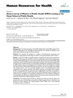
of methylation for each of the 3 analyzed CpG sites
(CpG1, CpG2, CpG3) was expressed as the percentage of
methylated cytosines over the sum of methylated and
unmethylated cytosines (Figure 1). Within- and between-
run variations for the determination of LINE-1 methyla-
tion through the pyrosequencing assay utilized have been
previously described [14].
Quantitative RT-PCR analysis of LINE-1 mRNA expression
Real-time quantitative RT-PCR analyses were performed
essentially as described [15]. Briefly, total RNA was
digested with RNAse-free DNAse ( Roche Diagnostics,
Milan, Italy) to remove contaminating g enomic DNA.
Synthesis of cDNA was performed on 1 μgtotalRNA
using MMLV reverse transcriptase (Invitrogen, Milan,
RFS
CpG1
CpG2
CpG3
3’5’
X58075
A
B
CpG1 CpG2 CpG3
Figure 1 LINE-1 bisulfite pyrosequencing assay. A. The region of the LINE-1 sequence [GenBank:X58075] utilized for the design of the assay is
reported. Vertical bars indicate individual CpG sites. Horizontal lines indicate forward (F), reverse (R) and sequencing (S) primers. Vertical arrows
indicate the CpG sites (CpG1, CpG2, CpG3) analyzed by pyrosequencing (adapted from [14]). B. Representative pyrograms for the methylation of
LINE-1 repetitive sequences. Yellow shadowing highlights the 3 target regions (CpG1, 2, 3) in the pyrograms. T and C peaks indicate
unmethylated and methylated cytosines, respectively. Accordingly, % of LINE-1 methylation at each site is defined by the % of the C base. Upper
and lower panels are representative of short-term cultures of CM cells with low and high LINE-1 methylation, respectively.
Sigalotti et al. Journal of Translational Medicine 2011, 9:78
/>Page 3 of 10
Italy) and random hexamer primers (Promega, Milan,
Italy), following manufacturers’ instructions. Real-time
quantitative RT-PCR reactions were conducted on the
ABI prism 7000 Sequence Detection System (Applied Bio-
systems, Milan, Italy), utilizing 20 ng retrotranscribed total
RNA in a final volume of 25 μl 1 X SYBR Green Master
Mix (Applied Biosystems). Relative quantification of
LINE-1 mRNA was performed with the aid of the DataAs-
sist v2.0 software (Applied Biosystems), using the b-acti n
house-keeping gene as endogenous control and normal
human PBMC as calibrator. The primers utilized for mea-
surement of LINE-1 (forward, GGCCAGTGTGTG
TGCGCACCG; reverse, CCAGGTGTGGGATATAGTCT
CGTGG) and of b-actin (forward, CGAGCGCGGCTA-
CAGCTT; reverse, CCTTAATGTCACGCACGATT)
mRNA expression were described previously [15,16].
Statistical analysis
The primary objective was to determine differences in
survival among various LINE-1 DNA methylation level
groups. In order to increase statistical power, sample
has been divided in two groups of the same size using
median as threshold: CpG1 (<25.68, ≥ 25.68), CpG2
(<27.26, ≥27.26), and CpG3 (<40.46, ≥40.46). For simpli-
city groups have been defined as LINE-1 hypomethy-
lated (patients with a LINE-1 methyl ation <median) and
hyper-methylated (patients with a LINE-1 methylatio n
≥median). The characteristics including age, gender, pri-
mary tumor localization, Breslow thickness, Clark level,
and ulceration of the primary tumor, number of lymph
nodes involved, and pre-operative serum LDH values
were examined. Survival time was calculated in months
from the date of stage IIIC diagnosis until the date of
death. According with the specific goals of the analysis,
we did not classify the deaths considering their cause.
Patien ts were censored at the last follow-up date or the
last date the patient was last known to be alive. Median
survival duration was determined by the Kaplan-Meier
method [17]. Cumulative survival by DNA meth ylation
level was evaluated using the log-rank test. P values
were two sided and values <0.05 were considered to be
statistically significant . Cox proportional hazard method
[18] was used to examine the effect of DNA methylation
level on survival and results were presented as Hazard
Ratios (HR) with corresponding 95% Confidence Inter-
vals (CI). LINE-1 methylation was also entered in the
mod el as a continuous variable with the unit set at 10%
of methylation. A stepwise regression (forward selection)
was conducted to select variables to add in our models.
Correlation between LINE-1 methylation and mRNA
expression was evaluated by Spearman’ s rank correla-
tion. The statistical analyses were carried out using the
SAS Software version 9.13 (SAS Institute Inc., Cary,
North Carolina, USA).
Results
Patients
The study was conducted on CM patients who under-
went radical lymph node dissection for stage III disease
at the Centro di Riferimento Oncologico National Can-
cer Institute between 1991 and 2007. Patients diagnosed
with a stage IIIC disease, and for whom a short-term
cell culture had been successfully generated from the
surgically removed autologous neoplastic tissue, were
included in the study. Table 1 summarizes the 42
patients under study and their clinico-pathologic charac-
teristics at presentation.
Extent of LINE-1 methylation in CM patients
To define if CM undergoes changes in the overall con-
tent of 5-methylcytosine, bisulfite pyrosequencing ana-
lyses (Figure 1) were utilized to measure the extent of
Table 1 Characteristics of the 42 AJCC stage IIIC
melanoma patients
Variable n. patients %
Age, years
Median 54
Range 29-83
Gender
Male 27 64
Female 15 36
Localization of primary tumor
extremities 14 33
trunk 23 55
head & neck 3 7
NA* 2 5
Breslow thickness of primary tumor
≤2.0 mm 13 31
>2.0 mm 22 52
NA 7 17
Clark level of primary tumor
1- 3 12 29
4-5 24 57
NA 6 14
Ulceration of primary tumor
No 10 24
Yes 30 71
NA 2 5
N. lymph nodes involved
1921
>1 33 79
LDH
Low
†
28 67
High 11 26
NA 3 7
*NA, not available.
†
low LDH is established as LDH values ≤ 0.8 times the upper limit of normal;
high LDH is defined as LDH values > 0.8 times the upper limit of normal.
Sigalotti et al. Journal of Translational Medicine 2011, 9:78
/>Page 4 of 10
methylation of LINE-1 repetitive elements in the 42
shor t-term CM cell cultur es under study. Data obtained
identified largely heterogeneous levels of methylation of
the LINE-1 elements in CM cells from stage IIIC
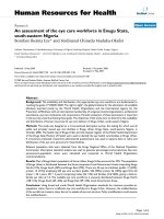
patients (CpG1: median 25.68%, range 12.45%-54.05%;
CpG2: median 27.26%, range 16.50%-49.43%; CpG3:
median 40.46%, range 28.10%-64.15%; Figure 2), demon-
strating that highly variable alterations in the overall
level of genomi c DNA occur in CM. In contrast, homo-
geneous and high levels of methylation at each of the
investigated CpG sites were measured in normal human
melanocytes (CpG1: median 62.82%, range 60.43%-
67.53%; CpG2: median 52.57%, range 51.37%-52.87%;
CpG3: median 65.77%, range 62.40%-67.33%) and in
PBMC isolated from healthy donors (CpG1: median
78.0%, range 67.8%-84 .2%; CpG2: median 54.7%, range
51.4%-56.8%; CpG3: median 67.9%, range 66.2%-73.3%).
Prognostic value of LINE-1 methylation in CM patients
The highly heterogeneous levels of LINE-1 methylation
observed in CM cells from stage IIIC patients led us to
investigate whether they correlated with a different clini-
cal outcome of patients under study.
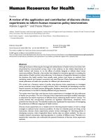
Kaplan-Meier analysis indicated that median OS for
stage IIIC CM patients under analysis was 15 .3 months
(95% CI, 11.0-31.5; Figure 3). To evaluate the assoc iation
between LINE-1 methylation status and OS, patients were
divided according to the median value of methylation of
each analyzed CpG site (CpG1 = 25.68%; CpG2 = 27.26%;
CpG3 = 40.46%). Patients were defined as having hypo-
methylated or hypermethylated LINE-1 sequences,
depending on the methylation level being below or above
the median value for each group, respectively. Kaplan-
Meier analysis showed a trend toward an increased OS
rate for patients with hypomethylated CpG1, however, the
difference did not reach statistical signific ance (P = 0.22,
log-rank = 1.51; Figure 3). On the other hand, a significant
survival advantage was observed in patients with CpG2 <
27.26% as compared to patients with CpG2≥27.26% (P =
0.04, log-rank = 4.14) (Figure 3). Similarly, the survival
rate of patients with CpG3 < 40.46% was significantly
higher than that of patients with CpG3≥40.46% (P = 0.01,
log-rank = 6.39) (Figure 3). In line with these data, median
OS of patients with hypomethylated CpG1, CpG2 and
CpG3 sites was 24.3, 31.5, and 31.9 months, respectively,
as compared to 15.3, 11.5, and 11.5 months of patients
with hypermethylated LINE-1 CpGs (Figure 3, Table 2).
Accordingly, the 5 year OS was 39%, 43%, and 48% for
patients with hypomethylated CpG1, CpG2, and CpG3
sites, respectively, as compared to 16%, 13%, and 7% of
patients with hypermethylated LINE-1 CpGs (Table 2).
Cox univariate analysis was carried out to identify
patient ch aracteristics and clinico-pathologic factors that
predicted survival. Among all factors examined, including
age, gender, localization of primary tumor, Breslow thick-
ness, Clark level and ulceration of primary tumor, number
of lymph nodes involved, and level of pre-operative LDH,
only CpG2 methylation (HR = 2.12 for CpG2≥27.26% vs.
CpG2 < 27.26; 95% CI: 1.01-4.4 4; P = 0.04) and CpG3
methylation (HR = 2.63 for CpG2≥40.46% vs. CpG2 <
40.46; 95% CI: 1.21-5.69; P = 0.01) were associated with
statistically significant differences in OS (Table 3). A step-
wise regression (forward selection) did not point t o any
independent varia ble to add in our models, thus, only
unadjusted HRs are reported in tables. When LINE-1
methylation was analyzed as a continuous variable, a sta-
tistically significant inverse association emerged between
OS and an increase of 10% of methylation of CpG1 (HR =
1.51; 95%CI:1.06-2.16; P = 0. 02), CpG2 (HR = 1.60; 95%
CI:1.02-2.52; P = 0.04) and CpG3 (HR = 1.49; 95%CI:1.06-
2.09; P = 0.02) (Table 3). The above reported statisti cally
significant increased risk of death associated with LINE-1
hyp ermethylation suggests a potential robust association
between methylation at CpG2 and CpG3 and OS, even if
the power of our analyses is below 25%.
Expression of LINE-1 mRNA in CM patients
To provide an initial evaluation of whether the products
encoded by the LINE-1 re petitive elements might have a
direct role in determining the different survival of CM
CpG1 CpG2 CpG3
20 30 40 50 60
% methylation
Figure 2 LINE-1 methylation in stage IIIC CM patients. LINE-1
methylation at 3 CpG sites (CpG1, CpG2, CpG3) was evaluated by
bisulfite pyrosequencing analysis in short-term cultures of CM cells
generated from neoplastic lesions of 42 stage IIIC melanoma
patients. All cells were analyzed at 6
th
in vitro passage. Separate box
plots have been generated for each of the CpG sites under analysis.
Black horizontal bars represent the median values of methylation for
each group.
Sigalotti et al. Journal of Translational Medicine 2011, 9:78
/>Page 5 of 10
patients with neoplastic cells having different LINE-1
methylation statuses, quantitative RT-PCR analyses were
utilized to measure the level of LINE-1 mRNA in the 42
shor t-term CM cell cultur es under study. Data obtained
revealed heterogeneous levelsofLINE-1mRNAinthe
CM cell cultures from stage IIIC CM patients (median
0.65, range 0.12-1.97); h owever, no significant correla-
tion was observed between levels of expression of LINE-
1 transcripts and methylation at either CpG1, CpG2 or
CpG3 sites (Figure 4).
Table 2 OS of stage IIIC CM patients according to LINE-1 methylation
LINE1 CpG site # events/# patients* Extent methylation
†
Median OS (95%CI)
‡
5 year OS (%)
CpG1 13/21 <25.68 24.3 (11.1-inf) 39
17/21 ≥25.68 15.3 (6.8-26.9) 16
CpG2 12/21 <27.26 31.5 (12.5-inf) 43
18/21 ≥27.26 11.5 (6.8-20.9) 13
CpG3 11/21 <40.46 31.9 (13.1-inf) 48
19/21 ≥40.46 11.5 (9.2-20.6) 7
* number of patients who died (# events) and total number of patients in the group (# patients) are reported;
† Patients were divided according to the % of methylation of the specified CpG site being < or ≥ the median % methylation measured in the examined patients’
population;
‡
Survival functions were calculated by the Kaplan-Meier method. Data are reported as Median OS in months, together with the corresponding 95% Confidence
Intervals (CI).
Percent Survival
time (months)
M edian= 15.3 months
A B
Percent Survival
time (months)
M edian= 24.3 months
M edian= 15.3 months
Log-Rank=1.51; p=0.22
C
Percent Survival
time (months)
M edian= 31.5 months
M edian= 11.5 months
Log-Rank=4.14; p=0.04
D
Percent Survival
time (months)
M edian= 31.9 months
M edian= 11.5 months
Log-Rank=6.39; p=0.01
Figure 3 Kaplan-Meier analysis of CM patients survival according to LINE-1 methylation. LINE-1 methylation at 3 CpG sites (CpG1, CpG2,
CpG3) was evaluated by bisulfite pyrosequencing analysis in short-term cultures of CM cells generated from neoplastic lesions of 42 stage IIIC
melanoma patients. Cells were analyzed at 6
th
in vitro passage. Kaplan- Meyer function for OS was calculated for CM patients either unstratified
(A) or stratified according to median methylation of CpG1 (B), CpG2 (C) or CpG3 (D) site of LINE-1 elements. Dashed and solid lines refer to
patients with LINE-1 methylation below or above the median, respectively. Vertical bars represent censored patients. Cumulative survival by LINE-
1 methylation level was evaluated using the Log-Rank test, reported P values were two sided.
Sigalotti et al. Journal of Translational Medicine 2011, 9:78
/>Page 6 of 10
Discussion
In this study we demonstra te that the global level of
LINE-1 methylation o f short-term tumor cell cultures
grown from patients with nodal disease is a significant
predictor of OS in stage IIIC CM patients. This finding
is of remarkable clinical relevance, since, to the best of
our knowledge, it provides the first evidence of a mole-
cular marker capable of differentiating the prognosis of
CM patients in this high-risk substage. These results are
of particular emphasis given the conduct of this study in
subjects within a single clinically well-defined clinico-
pathological staging sub-group, which has become the
focus of several ongoing clinical trials in the US and
Europe (i.e., ECOG intergroup trial E4697, EORTC trial
18071, GSK trial 111482 “DERMA”).
Genomic DNA hypomethylation has been proposed to
have an important impact on tumor biology through the
generation of chromosomal instability, reactivation of
transposable elements, and loss of imprinting [5]. Thus, a
negative correlation between genomic hypomethylation
and survival of CM patients could have been expected.
Instead, we found that hypomethylation of LINE-1 ele-
ments at CpG2 or CpG3 sites was associated with a signif-
icantly better OS, as demonstrated by Kaplan-Meier
analysis and log-rank test. The positive prognostic value of
LINE-1 hypomethylation we have identified in CM is in
sharp contrast with data most recently obtained in colon
and ovarian cancer patients, in which LINE-1 hypomethy-
lation in neoplastic tissues was associated with a poorer
prognosis [10,11]. This discrepancy, however, is not com-
pletely surprising. Indeed, data generated on hematologic
malignancies showed that LINE-1 hypomethylation can be
either a poor or a good prognostic factor, depending on
the patient being affected by chronic myeloid leukemia or
acute lymphoblastic leukemia, respectively [19,20]. Thus,
the different behavior of CM, with respect to the other
solid tumors so far investigated, might further suggest that
the underlying biological effect(s) of LINE-1
hypomethylation on patients’ outcome could depend on
the tumor histotype. Nevertheless, it should be empha-
sized that our findings are generated from patients in the
same clinico- pathological stage of disease, while the stu-
dies on ovari an and colon cancer were conducted on the
heterogeneous patients population as a whole, and did not
investigate the prognostic potential of LINE-1 methylation
in specific clinically defined stages of disease. Thus, it
remains to be demonstrated whether this different study
approach might contribute to the observed discrepancy.
Furthermore, it cannot be ruled out that in the different
sources of neoplastic material analyzed, the presence of
varying proportions of contaminating normal cells in neo-
plastic tissues, as well as the different methodological
approaches employed might contribute to conclusions
that may differ from those we have reached in these stu-
dies. In this context, our use of short-term CM cultures
has the advantage of eliminating contaminating normal
cells, yet representing the methylation status of neoplastic
cells of the fresh autologous lesion. In fact, similar levels of
LINE-1 methylation were observed between short-term
cultures and autologous uncultured CM cells that were
purified by anti-HMW-MAA immunomagnetic beads
from tumor cell suspensions that were avail able from 10
patients (data not shown).
The mechanism(s) through which LINE-1 hypomethy-
lation affects survival of CM patients remains to be fully
expl ored; however, some speculat ion can be made, based
on recent data in the literature. Tellez et al [21] have
demonstrated that higher levels of LINE-1 methylation
correlate with an increased number of aberrantly hyper-
methylated tumor suppressor genes (TSG) in cultured
melanoma cel l lines. This notion has gained further sup-
port from our most recent observation showing a direct
correlation between higher LINE-1 methylatio n and
increased genome-wi de gene methylation, measured
through CpG island microarrays (Sigalotti and Maio,
manuscript in preparation) . Thus, epigenetic inactivation
Table 3 Cox analysis of the influence of LINE-1 methylation on OS of stage IIIC CM patients
LINE1 CpG site # events/# patients* Extent methylation
†
HR
‡
95% CI; P value HR
cont.
§
95% CI; P value
CpG1 13/21 <25.68 1** 1.51 1.06-2.16; 0.02
17/21 ≥25.68 1.57 0.76-3.24; 0.22
CpG2 12/21 <27.26 1 1.60 1.02-2.52; 0.04
18/21 ≥27.26 2.12 1.01-4.44; 0.04
CpG3 11/21 <40.46 1 1.49 1.06-2.09; 0.02
19/21 ≥40.46 2.63 1.21-5.69;. 0.01
* number of patients who died (# events) and total number of patients in the group (# patients) are reported;
† Patients were divided according to the % of methylation of the specified CpG site being <or ≥ the median % methylation measured in the examined patients’
population;
‡
Cox proportional hazard method was used to examine the effect of LINE-1 methylation on OS. Results were presented as Hazard Ratios (HR) with corresponding
95% Confidence Intervals (CI);
§
LINE-1 methylation was also evaluated as continuous variable. The HR value is that of the LINE-1 methylation relative to an increase of 10%;
** set as reference.
Sigalotti et al. Journal of Translational Medicine 2011, 9:78
/>Page 7 of 10
of TSG might account for more aggressive disease we
have observed in patien ts with elevated LINE-1 methyla-
tion in their neoplastic cells. This hypothesis is in accor-
dance with initial studies reporting a negative association
between survival and the presence of hypermethylated
ER-a, RASSF1A, RAR-b2, or MINT31 DNA in neopla stic
tissues or sera of stage III/IV CM patient s [22-24]. On
the other hand, hypomethylation, and consequent tran-
scriptional activation, of LINE-1 elemen ts might per se
reduce the tumorigenic potential of neoplastic cells by
triggering apoptosis and a senescence-like state through
the activity of the second open reading frame of LINE-1
[25]. In our findings, this seems not to be the case, since
the lack of correlation between methylation and mRNA
expression of LINE-1 elements, suggests that LINE-1 pro-
ducts may not be the driving force for the observed
increased OS of LINE-1 hypomethylated patients. Geno-
mic DNA hypomethylation has also been associat ed with
the de novo expression of tumor associated antigens
belonging to the Cancer Testis Antigen (CTA) class by
neoplastic cells of different histotype, including mela-
noma stem cells [26-29], and we have recently identified
a significant correlation between a hypomethylated status
of LINE-1 elements and increased levels and total num-
ber of CTA concomitantly expressed in short-term cul-
tures of CM cells (Sigalotti and Maio, unpublished).
Besides, pharmacologic DNA hypomethylation has been
consistently demonstrated to increase immunogenicity
and immune recognition of cancer cells through the up-
regulation of different molecules involved in antigen pro-
cessing and present ation, including HLA class I antigens
and co-stimulatory molecules [30,31]. Thus, it is intri-
guing to speculate that a better immune r ecognition of
LINE-1 hypomethylated CM cells might contribute to the
improved survival of these patients. This hypothesis may
find indirect support from most recent gene expression
profiling studies that identified the expression of
“ immune-related” genes in the tumor as a marker of
good prognosis in stage III-IV CM [32-34].
Conclusion
Irrespective of the underlying biological mechanism(s) trig-
gered by LINE-1 hypomethylation, the prognostic value of
LINE-1 methylation here identified for stage IIIC CM
patients bears several important practical clinical implica-
tions. Among these, the goal to provide CM patients with
improved clinico-pathological sub-stage and/or follow-up-
procedures would be enhanced using LINE-1 methylation
status, and these findings might be used to select and/or
stratify patients for adjuvanttreatmentbasedonthemethy-
lation level of LINE-1 in their tumors. In addition, the sig-
nificant positive p rognosis of LINE-1 hypomethylated
patients should prompt the incorporation of this in new
studies aimed at understanding whether pharmacologic
DNA hypomethylation [35] could be regarded as a feasible
chemoprevention approach in the initial phases of disease
and/or in patients at high-risk of disease recurrence.
Our present findings will be further investigated in
prospective multicenter studies in which the prognostic
significance and the predictive value for diff erent treat-
ments of CM will be validated. Providing further sup-
port to our initial data will finally allow to establish the
appropriateness of adding the evaluation of LINE-1
methylation into the routine clinico-pathological
0
10
20
30
40
50
60
70
0.00 0.50 1.00 1.50 2.00 2.50
0
10
20
30
40
50
60
70
0.00 0.50 1.00 1.50 2.00 2.50
0
10
20
30
40
50
60
70
0.00 0.50 1.00 1.50 2.00 2.50
LINE-1 mRNA
% LINE-1 methylation % LINE-1 methylation % LINE-1 methylation
CpG1
CpG2
CpG3
Rho=-0.10
P=0.52
Rho=-0.05
P=0.75
Rho=-0.13
P=0.40
Figure 4 Association between methylation and mRNA
expression of LINE-1 elements in stage IIIC CM patients. Short-
term cultures of CM cells generated from neoplastic lesions of 42
stage IIIC melanoma patients were evaluated for LINE-1 methylation
at 3 CpG sites (CpG1, CpG2, CpG3) and for LINE-1 mRNA expression
by bisulfite pyrosequencing and quantitative RT-PCR analyses,
respectively. All cells were analyzed at 6
th
in vitro passage.
Methylation at each investigated LINE-1 CpG site is reported as %,
level of LINE-1 mRNA expression is reported relative to the value
measured in PBMC obtained from healthy donors, used as
reference. Correlation between LINE-1 methylation and mRNA
expression was evaluated by Spearman’s rank correlation test,
reported P values were two sided.
Sigalotti et al. Journal of Translational Medicine 2011, 9:78
/>Page 8 of 10
ascertainment of CM patients, in order to help persona-
lizing their comprehensive clinical management.
List of Abbreviations Used
CI: Confidence Intervals; CM: cutaneous melanoma; CTA: Cancer Testis
Antigen; ER-α: Estrogen Receptor-α; HLA: Human Leukocyte Antigen; HMW-
MAA: High Molecular Weight-Melanoma Associated Antigen; HR: Hazard
Ratio; LINE-1: Long Interspersed Nucleotide Element-1; MINT31: Methylated
IN Tumors locus 31; OS: overall survival; RASSF1A: Ras Association (RalGDS/
AF-6) domain Family member 1A; RAR-β2: Retinoic Acid Receptor-β2; TSG:
tumor suppressor genes.
Acknowledgements and Funding
This work was supported in part by grants from the Associazione Italiana per
la Ricerca sul Cancro (IG 6038 to MM and MFAG 9195 to LS), Fondazione
Monte dei Paschi di Siena, the Harry J. Lloyd Charitable Trust, the Istituto
Superiore di Sanità, and SPORE P50CA121973.
Author details
1
Cancer Bioimmunotherapy Unit, Centro di Riferimento Oncologico, Istituto
di Ricovero e Cura a Carattere Scientifico, Aviano, Italy.
2
Biostatistics and
Epidemiology Unit, Centro di Riferimento Oncologico, Istituto di Ricovero e
Cura a Carattere Scientifico, Aviano, Italy.
3
Breast Surgery Unit, Centro di
Riferimento Oncologico, Istituto di Ricovero e Cura a Carattere Scientifico,
Aviano, Italy.
4
University of Pittsburgh School of Medicine, Pittsburgh,
Pennsylvania, USA.
5
Division of Medical Oncology and Immunotherapy,
Department of Oncology, University Hospital of Siena, Istituto Toscano
Tumori, Siena, Italy.
Authors’ contributions
LS participated in acquiring laboratory data, data analysis and interpretation,
study coordination, and drafted the manuscript. EF performed the
pyrosequencing analyses, and contributed in data acquisition and analysis.
EB performed the statistical analyses. AC, GP, FC contributed in cellular
biology procedures, molecular assays and data acquisition. SC, contributed in
data interpretation. SM participated in acquisition of clinical data and data
interpretation. JMK participated in data interpretation and manuscript
drafting. MM conceived of the study, participated in its design and
coordination, and contributed in producing the final draft of the manuscript.
All authors read and approved the final manuscript.
Competing interests
LS and MM have applied for a patent based on the findings reported in this
manuscript. All other authors declare no competing interests.
Received: 16 March 2011 Accepted: 26 May 2011
Published: 26 May 2011
References
1. MacKie RM, Hauschild A, Eggermont AMM: Epidemiology of invasive
cutaneous melanoma. Ann Oncol 2009, 20:vi1-vi7.
2. Bhatia S, Tykodi SS, Thompson JA: Treatment of metastatic melanoma: an
overview. Oncology (WillistonPark) 2009, 23:488-496.
3. Balch CM, Buzaid AC, Soong SJ, Atkins MB, Cascinelli N, Coit DG,
Fleming ID, Gershenwald JE, Houghton A Jr, Kirkwood JM, et al: Final
version of the American Joint Committee on Cancer staging system for
cutaneous melanoma. JClin Oncol 2001, 19:3635-3648.
4. Jennings L, Murphy GM: Predicting outcome in melanoma: where are we
now? BrJ Dermatol 2009.
5. Esteller M: Epigenetics in cancer. NEnglJ Med 2008, 358:1148-1159.
6. Gama-Sosa MA, Slagel VA, Trewyn RW, Oxenhandler R, Kuo KC, Gehrke CW,
Ehrlich M: The 5-methylcytosine content of DNA from human tumors.
NuclAcids Res 1983, 11:6883-6894.
7. Yang AS, Estecio MR, Doshi K, Kondo Y, Tajara EH, Issa JP: A simple method
for estimating global DNA methylation using bisulfite PCR of repetitive
DNA elements. Nucleic Acids Res 2004, 32:e38.
8. Cho NY, Kim JH, Moon KC, Kang GH: Genomic hypomethylation and CpG
island hypermethylation in prostatic intraepithelial neoplasm. Virchows
Arch 2009, 454:17-23.
9. Park SY, Yoo EJ, Cho NY, Kim N, Kang GH: Comparison of CpG island
hypermethylation and repetitive DNA hypomethylation in premalignant
stages of gastric cancer, stratified for Helicobacter pylori infection.
JPathol 2009, 219:410-416.
10. Pattamadilok J, Huapai N, Rattanatanyong P, Vasurattana A, Triratanachat S,
Tresukosol D, Mutirangura A: LINE-1 hypomethylation level as a potential
prognostic factor for epithelial ovarian cancer. IntJ GynecolCancer 2008,
18:711-717.
11. Ogino S, Nosho K, Kirkner GJ, Kawasaki T, Chan AT, Schernhammer ES,
Giovannucci EL, Fuchs CS: A cohort study of tumoral LINE-1
hypomethylation and prognosis in colon cancer. JNatlCancer Inst 2008,
100:1734-1738.
12. Altomonte M, Gloghini A, Bertola G, Gasparollo A, Carbone A, Ferrone S,
Maio M: Differential expression of cell adhesion molecules CD54/CD11a
and CD58/CD2 by human melanoma cells and functional role in their
interaction with cytotoxic cells. Cancer Res 1993, 53:3343-3348.
13. Ausubel FM, Brent R, Kingston RE, et al: Current Protocols in Molecular
Biology New York: John Wiley & Sons; 1998.
14. Aparicio A, North B, Barske L, Wang X, Bollati V, Weisenberger D, Yoo C,
Tannir N, Horne E, Groshen S, et al: LINE-1 methylation in plasma DNA as
a biomarker of activity of DNA methylation inhibitors in patients with
solid tumors. Epigenetics
2009, 4:176-184.
15.
Fratta E, Sigalotti L, Colizzi F, Covre A, Nicolay HJ, Danielli R, Fonsatti E,
Altomonte M, Calabro L, Coral S, Maio M: Epigenetically regulated clonal
heritability of CTA expression profiles in human melanoma. J Cell Physiol
2010, 223:352-358.
16. Aporntewan C, Phokaew C, Piriyapongsa J, Ngamphiw C, Ittiwut C,
Tongsima S, Mutirangura A: Hypomethylation of intragenic LINE-1 represses
transcription in cancer cells through AGO2. PLoS One 2011, 6:e17934.
17. Kaplan EL, Meier P: Nonparametric Estimation from Incomplete
Observations. Journal of the American Statistical Association 1958,
53:457-481.
18. Cox DR: Regression models and life-tables (with discusssion). J Roy Statist
Soc B 1972, 34:187-220.
19. Roman-Gomez J, Jimenez-Velasco A, Agirre X, Castillejo JA, Navarro G,
Garate L, Jose-Eneriz ES, Cordeu L, Barrios M, Prosper F, et al: Promoter
hypermethylation and global hypomethylation are independent
epigenetic events in lymphoid leukemogenesis with opposing effects on
clinical outcome. Leukemia 2006, 20:1445-1448.
20. Roman-Gomez J, Jimenez-Velasco A, Agirre X, Cervantes F, Sanchez J,
Garate L, Barrios M, Castillejo JA, Navarro G, Colomer D, et al: Promoter
hypomethylation of the LINE-1 retrotransposable elements activates
sense/antisense transcription and marks the progression of chronic
myeloid leukemia. Oncogene 2005, 24:7213-7223.
21. Tellez CS, Shen L, Estecio MR, Jelinek J, Gershenwald JE, Issa JP: CpG island
methylation profiling in human melanoma cell lines. Melanoma Res 2009,
19:146-155.
22. Mori T, O’Day SJ, Umetani N, Martinez SR, Kitago M, Koyanagi K, Kuo C,
Takeshima TL, Milford R, Wang HJ, et al: Predictive utility of circulating
methylated DNA in serum of melanoma patients receiving
biochemotherapy. J Clin Oncol 2005, 23:9351-9358.
23. Mori T, Martinez SR, O’Day SJ, Morton DL, Umetani N, Kitago M,
Tanemura A, Nguyen SL, Tran AN, Wang HJ, Hoon DS: Estrogen receptor-
alpha methylation predicts melanoma progression. Cancer Res 2006,
66:6692-6698.
24. Tanemura A, Terando AM, Sim MS, van Hoesel AQ, de Maat MF, Morton DL,
Hoon DS: CpG island methylator phenotype predicts progression of
malignant melanoma. Clin Cancer Res 2009, 15:1801-1807.
25. Wallace NA, Belancio VP, Deininger PL: L1 mobile element expression
causes multiple types of toxicity. Gene 2008, 419:75-81.
26. De Smet C, De Backer O, Faraoni I, Lurquin C, Brasseur F, Boon T: The
activation
of human gene MAGE-1 in tumor cells is correlated with
genome-wide demethylation. Proc NatlAcadSciUSA 1996, 93:7149-7153.
27. Sigalotti L, Coral S, Nardi G, Spessotto A, Cortini E, Cattarossi I, Colizzi F,
Altomonte M, Maio M: Promoter methylation controls the expression of
MAGE2, 3 and 4 genes in human cutaneous melanoma. JImmunother
(1997) 2002, 25:16-26.
28. Woloszynska-Read A, Mhawech-Fauceglia P, Yu J, Odunsi K, Karpf AR:
Intertumor and intratumor NY-ESO-1 expression heterogeneity is
associated with promoter-specific and global DNA methylation status in
ovarian cancer. ClinCancer Res 2008, 14:3283-3290.
Sigalotti et al. Journal of Translational Medicine 2011, 9:78
/>Page 9 of 10
29. Sigalotti L, Covre A, Zabierowski S, Himes B, Colizzi F, Natali PG, Herlyn M,
Maio M: Cancer testis antigens in human melanoma stem cells:
expression, distribution, and methylation status. J Cell Physiol 2008,
215:287-291.
30. Sigalotti L, Covre A, Fratta E, Parisi G, Colizzi F, Rizzo A, Danielli R,
Nicolay HJ, Coral S, Maio M: Epigenetics of human cutaneous melanoma:
setting the stage for new therapeutic strategies. J Transl Med 2010, 8:56.
31. Fonsatti E, Nicolay HJ, Sigalotti L, Calabro L, Pezzani L, Colizzi F,
Altomonte M, Guidoboni M, Marincola FM, Maio M: Functional up-
regulation of human leukocyte antigen class I antigens expression by 5-
aza-2’-deoxycytidine in cutaneous melanoma: immunotherapeutic
implications. Clin Cancer Res 2007, 13:3333-3338.
32. John T, Black MA, Toro TT, Leader D, Gedye CA, Davis ID, Guilford PJ,
Cebon JS: Predicting clinical outcome through molecular profiling in
stage III melanoma. Clinical cancer research: an official journal of the
American Association for Cancer Research 2008, 14:5173-5180.
33. Jonsson G, Busch C, Knappskog S, Geisler J, Miletic H, Ringner M,
Lillehaug JR, Borg A, Lonning PE: Gene expression profiling-based
identification of molecular subtypes in stage IV melanomas with
different clinical outcome. Clinical cancer research: an official journal of the
American Association for Cancer Research 2010, 16:3356-3367.
34. Mandruzzato S, Callegaro A, Turcatel G, Francescato S, Montesco MC,
Chiarion-Sileni V, Mocellin S, Rossi CR, Bicciato S, Wang E, et al: A gene
expression signature associated with survival in metastatic melanoma.
Journal of translational medicine 2006, 4:50.
35. Sigalotti L, Fratta E, Coral S, Cortini E, Covre A, Nicolay HJ, Anzalone L,
Pezzani L, Di Giacomo AM, Fonsatti E, et al: Epigenetic drugs as
pleiotropic agents in cancer treatment: biomolecular aspects and clinical
applications. J Cell Physiol 2007, 212:330-344.
doi:10.1186/1479-5876-9-78
Cite this article as: Sigalotti et al.: Methylation levels of the “long
interspersed nucleotide element-1” repetitive sequences predict survival
of melanoma patients. Journal of Translational Medicine 2011 9:78.
Submit your next manuscript to BioMed Central
and take full advantage of:
• Convenient online submission
• Thorough peer review
• No space constraints or color figure charges
• Immediate publication on acceptance
• Inclusion in PubMed, CAS, Scopus and Google Scholar
• Research which is freely available for redistribution
Submit your manuscript at
www.biomedcentral.com/submit
Sigalotti et al. Journal of Translational Medicine 2011, 9:78
/>Page 10 of 10