Báo cáo sinh học: " Effects of transplantation with bone marrowderived mesenchymal stem cells modified by Survivin on experimental stroke in rats" potx
Bạn đang xem bản rút gọn của tài liệu. Xem và tải ngay bản đầy đủ của tài liệu tại đây (4.06 MB, 10 trang )
RESEARC H Open Access
Effects of transplantation with bone marrow-
derived mesenchymal stem cells modified by
Survivin on experimental stroke in rats
Nan Liu
1*
, Yixian Zhang
1,2
, Lin Fan
3
, Mingzhou Yuan
4
, Houwei Du
1
, Ronghua Cheng
1
, Deshan Liu
1
and Feifei Lin
1
Abstract
Background: This study was performed to determine whether injury induced by cerebral ischemia could be
further improved by transplantation with bone marrow-derived mesenchymal stem cells (MSCs) modified by
Survivin (SVV).
Methods: MSCs derived from bone marrow of male Sprague-Dawley rats were infected by the self-inactive
lentiviral vector GCFU carrying green fluorescent protein (GFP) gene and SVV recombinant vector (GCFU-SVV). In
vitro, vascular endothelial growth factor (VEGF) and basic fibrobl ast growth factor (bFGF) were detected in infected
MSCs supernatants under hypoxic conditions by ELSIA. In vivo, experiments consisted of three groups, one
receiving intravenous injection of 500 μl of phosphate-buffered saline (PBS) without cells (control group) and two
groups administered the same volume solution with either three million GFP-MSCs (group GFP) or SVV/GFP-MSCs
(group SVV). All animals were submitted to 2-hour middle cerebral artery occlusion (MCAO) and then reperfusion.
Differentiation and survival of the transplanted MSCs were determined by confocal microscope. Western blot was
used to detect the expression of VEGF and bFGF in ischemic tissue. A 2,3,5-triphenyltetrazolium chloride (TTC)
staining was used to assess the infarct volume. Evaluation of neurological function was performed using a
modified Neurological Severity Score (mNSS).
Results: In vitro, modification with SVV further increased secretion of VEGF and bFGF under hypoxic condition. In
vivo, only very few transplantated cells co-expressed GFP and NeuN. The survival transplanted cells in the group
SVV was 1.3-fold at 4 days after transplantation and 3.4-fold higher at 14 days after trans plantation, respectively,
when compared with group GFP. Expression of VEGF and bFGF in the ischemic tissue were further up-regulated by
modification with SVV. Moreover, modification with SVV further reduced the cerebral infarct volume by 5.2% at 4
days after stroke and improved post-stroke neurolog ical function at 14 days after transplantation.
Conclusion: Modification with SVV could further enhance the therapeutic effects of MSCs possibly through
improving the MSCs survival capacity and up-regulating the expression of protective cytokines in the ischemic
tissue.
Background
Despite the advances in medical, thrombolytic and sur-
gical treatment, the treatment of cerebral infarction still
lacks an ideal method. Previous studies have shown that
MSCs could differentiate into potential neuron-like cells
bothinvivoandinvitro[1,2],suggestingthatMSCs
transplantation could improve neurological function
after cerebral ischemia, and the efficacy is closely related
to the nu mber of MSCs grafted [3]. However, the survi-
val rate of simple transplantation of MSCs in ischemic
tissue is very low [4]. Recent research has demonstrated
that the combining of apoptosi s inhibitors with MSC s
or anti-apoptosis gene-modified MSCs for transplanta-
tion promoted better recovery of neurological function
after cerebral ischemia [5-7], which suggests that anti-
apoptosis strategies for the MSCs transplantation might
break through the limitation of current MSCs strategies
for the treatment of cerebral infarction. Survivin (SVV)
* Correspondence:
1
Department of Neurology, Union Hospital, Fujian Medical University, Fuzhou
350001, P.R. China
Full list of author information is available at the end of the article
Liu et al. Journal of Translational Medicine 2011, 9:105
/>© 2011 Liu et al; licensee BioMed Central Ltd. This is an Open Access article distributed under the terms of the Creative Commons
Attribution Lice nse (http://c reativecommons.org/licenses/by/2.0), which permits unrestricted use, distribution, and reproduction in
any medium, provided the original work is properly cited.
is a special new member of the inhibitor of apoptosis
protein family (IAP). A study by Fan et al. has demon-
strated that transplantation with survivin-engineered
MSCs can further improve t he cardiac performance of
rats after myocardial infarction by en hancing survival of
the transplanted cells [8]. However, it is unclear whether
such MSCs could result in better therapeutic effects for
strokeinrats.Inthispaper,wetrytoinvestigatethe
effects of transplantation with MSCs modified by SVV
on an experimental stroke model performed in rats.
Methods
Animal ethics
The investigation conformed to the Principles of
Laboratory Animal Care formulated by the National
Society for Medical Research and the Guide for the
Care and Use of Laboratory Animals published by the
U.S. National Institutes of Hea lth (NIH Publication, No.
86-23, revised 1985). The investigators responsible for
molecular, histological andfunctionalstudieswere
blinded to the treatment groups.
Preparation and characterization of MSCs
MSCs were prepared from rat bone marrow as
described by Friedenstein et al [9]. In brief, we eutha-
nized Sprague Dawley (SD) rats weighted 80-100 g and
harvested bone marrow. Bone marrow cells were intro-
duced into 100-mm dishes and cultured in complete
medium, consisting of Dulbecco’s Modified Eagle’s Med-
ium (DMEM; Sigma) containing 10% fetal bovine serum
and antibiotics: 100 U/ml penicillin G, 100 mg/mg
streptomycin, and 0.25 mg amphotericin B. Culture
medium was replaced every three days and floating cells
were discarded. Following two passes, the attached cells
were divided into three ne w flasks and c ultured until
the cell density of the colonies grew to approximately
90% confluence. These cells were analyzed by fluores-
cence-activated cell sorting (FACS) as described pre-
viou sly [10]. A fter blocking for nonspecific binding with
buffer containing 1% bovine serum albumin, the cells
were incubated for 20 minutes at 4°C with the following
antibodies: anti-CD29, Phycoerythrin (PE), anti-CD106,
PE, (Biolegend). anti-CD44, luorescein isothiocyanate
(FITC), anti-CD14, FITC and a nti-CD45, FITC (AbD
Serotec). The matched isotype contro ls were purchase d
from AbD Serotec or Biolegend. At least 1 × 10
4
cells
per sample were acquired and analyzed.
MSCs differentiation assay
The differentiation of MSCs in vitro towards the adipo-
genic and the osteogeni c lineage as previously describe d
[11,12]. Briefly, for adipocyte differentiation, MSCs was
cultured 3 weeks with adipogenic medium, containing
10
-6
M dexamethasone, 10 μg/ml insulin and 100 μg/ml
3-isobutyl-1-methylxantine (Sigma). For Osteoblast dif-
ferentiation, MSCs was cultured 3 weeks with osteo-
genic medium, containing 10
-7
M dexamethasone, 50 μg/
ml ascorbic acid and 10 mM b-glycero phosphate
(Sigma). Oil-red-O and von kossa dyes were employed
to identify adipocytes, osteoblasts respectively.
SVV recombinant lentiviral vector construction
Human SVV recombi nant lentiviral vector was con-
structed using previous method [8]. Briefly, the full-
length human SVV cDNA without termination codon
was amplified by polymerase chain reaction (PCR) from
pUC18-SVV and inserted into the Age I site of the
GCFU plasmid to form a GFP/SVV fusion gene. The
identity of SVV cDNA obtained in this manner was con-
firmed by sequencing and comparing it with the Gene
Bank sequence NM_001168.2. The primer sequence was
forward, 5’ -GATGATGACGACAAACCGGTCATG
GGTGCCCCGACGTTG-3’ and reverse, 5’ -TCAC-
CATGGTGGCGACCGGTTTATCCATGGCAGCCA
GCTG-3’. The SVV recombinant lentiviral vector was
prepared using Lipofectmaine 2000 transfection
technology.
MSCs gene modification
For passage 1 MSCs were infected by lentivirus with a
multiplicity of infection (MOI) of 8 [8]. The MSC s
infected with SVV recombinant lentivirus were defined
as SVV/GFP-MSCs and the MSCs infected with mock
lentivirus were defined as GFP-MSCs. To achieve the
optimal gene transfer, polybrene (a final concentration
of 8 μg/ml) was used. All MSCs were expanded to 3
passes, and then used for transplantation. The efficiency
of gene transduction was assessed with FACS.
SVV expression in modified MSCs
The survivin e xpression was detected by immunofluor-
escence staining. In brief, the 3
rd
passage transfected
MSCs were plated onto fibronectin-coated chamber
slides, fixed with 4% paraformaldeh yde (Sigma) for 10
minu tes at roo m temperature, and washed twice in 0.01
M phosphate-buffered saline (PBS, GIBCO). Slides were
blockedwithgoatserumfor20minutesandincubated
overnight with mouse anti-human Survivin antibody
(AbCam) at 4°C. A fter that, the slides were incubated
with Texas-Red fluorescent anti-mouse secondary anti-
body (Santa Cruz) for 30 minutes at 4°C. Between steps
the slides were washed with PBS. A 1:500 dilution of
primary antibod y against human SVV and a 1:500 dilu-
tion of secondary antibody were used, respectively. Cells
were examined by fluorescencemicroscopy (Leica Co,
Germany).
Liu et al. Journal of Translational Medicine 2011, 9:105
/>Page 2 of 10
Vascular endothelial growth factor (VEGF) and basic
fibroblast growth factor (bFGF) secretion in MSCs under
hypoxic conditions
After the 3
rd
passage infected MSCs completed adher-
ence, they were inc ubated for 24 hours at 37°C in a
humidified modular hypoxia chamber (Billups Rothen-
berg) containing 95% nitrogen and 5% carbon dioxide (n
= 4 in each group). Subsequently, the supernatants were
collected for analysis. Commercial VEGF or bFGF
ELISA (enzyme-linked immunoso rbent assay) kits (R&D
Systems Inc. Minneapolis, USA) was used to quantify
the concentration of VEGF and bFGF in each of the
samples. The supernatant from MSCs cultured in nor-
mal condition was used for control. Any experiment
was repeated for three times.
Animal Model
Adult male Sprague-Dawley rats weighing 220-250 g
were used in this study. A middle cerebral artery occlu-
sion (MCAO) was established with the modified Longa
method [13]. Rats were initially anesthetized with 10%
chloral hydrate. Rectal temperature was controlled at
37°C with a feedback-regulated water heating system.
The right common carotid artery, external carotid artery
(ECA), and internal carotid artery were exposed. A 3.0
monofilament nylon suture (18.5 mm, determined by
animal weight), with its tip rounded by heating near a
flame, was advanced from the ECA into the lumen of
the internal carotid artery until it blocked the origin of
the middle cerebral artery (MCA). 2 hours after MCAO ,
animals were reanesthetized with halothane, and reper-
fusion was performed by withdrawal the suture until th e
tip cleared the lumen of the ECA.
Transplantation
MSCs transplantation was performed as a method
reported in prev ious study [7]. Briefly, after 2-hour mid-
dle cerebral artery occlusion (MCAO) and 24-hour
reperfusion, Rats were grouped into three groups which
received a 500 μl injection of e ither phosphate-buffered
saline (PBS) without cells (group control, n = 18) or
containing three million GFP-MSCs (group GFP , n =
30)orSVV/GFP-MSCs(groupSVV,n=30)viatail
vein.
Double Immunofluorescence Staining
In order to identify survival and differentiate of the
transplanted MSCs, a method of double immunofluores-
cent staining was used. Rats in the GFP and SVV groups
were euthanized with 10% chloral hydrate at 4 days (n =
6 i n each group) or 14 days (n = 6 in each group) after
transplantation. For pre paration of frozen sections, rats
were perfused transcardially with normal saline and the
brain samples were removed immediately. Blocks
corresponding to coronal coordinates form bregma -1 to
1 mm were obtained and frozen rapidly in liquid nitro-
gen. A series of 6-um-thick sections was obtained.
Thereafter, the frozen s ections were rewarmed at room
temperature for 45 minutes to 1 hour, and were concu-
bated overnight at a dilution of 1:200 with FITC labeled
goat anti-GFP (AbCam) and rabbit anti-rats Neuronal
nuclei (NeuN, which is a marker of neuron.) (DA KO),
and then incubated for 45 minutes using a secondary
antibody of goat anti-rabbi t/mouse IgG c onjug ated with
TAXES (S anta Cruz) for detecting NeuN at 37°C.
Between steps the slides were washed with 0.01M PBS.
Finally, the sections were used to detect the survival and
differentiation into neuron-like cells of the transplanted
MSCs by a laser scanning confocal microscope (Zeiss
Co., LSM510).
Western Blot for VEGF and bFGF in Injuried Cerebral
Tissues
Rats were euthanized with 10% chloral hydrate at 4 days
(n = 6 in ea ch group) or 14 days (n = 6 in each group)
after transplantation. The protein concentration from
injured cerebral tissues was determined using the
bicinchoninic acid (BCA) protein assay kits (Beyotime
Biotechnology, P.R. China). Thirty micrograms protein
were loaded on 10% acrylamide gel for electrophoresis
and were electroblotted onto a polyvinylide ne difluoride
membrane (PVDF, Invitrogen). The membranes were
then probed with mouse anti-VEGF (1:500) and anti-
bFGF (1:500), respectively, followed by incubation with
horseradish-peroxidase-conjugated sheep-anti-mouse
IgG (Bio-Rad Laboratories). Protein expression was
detected with an enhanced chemiluminescence detection
system (Amersham Pharmacia Biotech Inc) and b-actin
was used as a loading control. All bands from western
blot were analyzed using Image J software (version 1.6
NIH) to verify the relative level of VEGF and bFGF
defined as the optical density ration of VEGF or bFGF
over b-actin.
Measurement of Cerebral Infarction Volume
At 14 days after MSCs transplantation, rats in each
groups (n = 6) were used for evaluate cerebral infarction
volume. The brain samples were removed carefully and
dissected into five equally spaced coronal blocks using a
vibratome. The fresh brain slices were immersed in a
2% solution of 2, 3, 5-triphenyltetrazolium chloride
(TTC) (Sigma) in PBS (GIBCO) at 37°C for 30 minutes.
The cross-sectional area of infarction and non infarction
in each brain slice was measured using Image J analysis
software (version 1.6 NIH). The infarct volume was
indirectly determined by subtracting the volume of
intact tissue in the ipsilateral hemisphere from that in
the contralateral hemisphere.
Liu et al. Journal of Translational Medicine 2011, 9:105
/>Page 3 of 10
Evaluation of neurological function
Evaluation of neurological function was performed 1 day
and 14 days after transplantation in each groups (n = 6)
using a modified Neurological Severit y Score (mNSS)
[3]. The mNSS is a composite of the mot or (muscle sta-
tus and abnormal movement), sensory (visual, tactile,
and proprioceptive), and reflex tests. The neurological
function was graded on a scale of 0-18 (normal score 0,
maximal deficit score 18)
Statistical analysis
Data were presented as mean values and standard devia-
tion. A method of ANOVA (analysis of variance) with
Scheffe’s post hoc test was used to i dentify differences
among all groups. A P value of less than 0.05 was con-
sidered as statistical significance.
Result
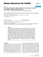
Phenotypic characterization and differentiation capacity
of cells
Cells were scattered in a number of colony distributions
3 days after planted. At day 8 ~ 9, the bottle was cov-
ered with long-spindle cells. Passaged cells (mostly spin-
dle cells) were uniformly distributed, and covered the
bottomevery4~5days.The3
rd
Passage MSCs highly
expressed the surface marker molecules CD29 (97.7%),
CD90 (100%) and CD106 (100%), and lowly expressed
the blood cell surface molecules CD14 (2.2%) and CD45
(2.6%) (Figure 1).
Cells were different iated in vitro using adipogenic and
oesteogenic induction media. Following 3 weeks of adi-
pogenic induction, the cells stained Oil red ‘O’ positive
showing lipid laden adipocyte phenotype. Similarly,
when induced with oesteogenic induction medium for 3
weeks, these cells showed oesteogensis upon staining
with von kossa for calcium deposits (Figure 1C, D).
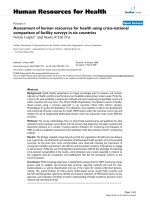
Efficiency of gene transduction and SVV expression
After infection with SVV recombinant len tivirus and
mock lentivirus, MSCs were over expressed GFP (Figure
2A, B), and the efficiency of gene transduction was simi-
lar to that of mock lentivirus (97.2% vs. 92.9%) (Figure
2F, G). The 3
rd
passage transfected MSCs were planted
on fibrone ctin-coated chamber slides for immunofluor-
escence microscopy. Expression of the SVV gene was
evident in SVV/GFP-MSCs (Figure 2D), but not in GFP-
MSCs (Figure 2C).
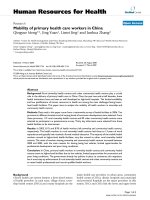
SVV enhanced the survival of Transplanted MSCs
The transplanted MSCs via tail vein were identified by
GFP. In the group SVV and the g roup GFP, the trans-
planted MSCs were distributed throughout the damaged
tissues, with the majority located close to the injured tis-
sue. Quantitative analysis sho wed that number of the
GFP-positive MSCs in the group SVV increased by
about 1.3-fold (101.8 ± 10.3 per high -power magnifica-
tion field [HPF] vs.76.8 ± 7.9 per HPF, P < 0.05) at 4
days after transplantation, and by 3.4-fold (61.3 ± 8.2
Figure 1 Phenotypic characterization and differentiation of cells:(A) The initial passage MSCs grew as a morphologically homogeneous
population of fibroblast-like cells, (B) The Passage 3 MSCs grew as whorls of densely packed spindle-shaped (scale bar = 200 um in A and B). (C)
Adipocyte differentiation of MSCs: Upon induction with adipocyte induction media cells showed adipocyte globules on oil red ‘O’ staining. (D)
Osteogenic differentiation of MSCs: Upon induction with osteogenic induction media cells showed calcium deposits on von kossa staining. (scale
bar = 100 um in C and D) (E-I): Flow cytometry analysis: MSCs expressed the markers molecules CD29, CD106, CD90 and negative for the blood
cell surface molecules CD45, CD14. The percentage of positivity was mentioned in the brackets.
Liu et al. Journal of Translational Medicine 2011, 9:105
/>Page 4 of 10
per HPF vs.17.8 ± 4.8 per HPF, P < 0.01) at 14 days
after transplantation when compared with in the group
GFP. There were very few GFP-positive cells coexpres-
sion NeuN in the cell transplantation groups (Figure 3).
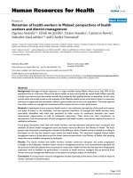
VEGF and bFGF expression in vitro and in vivo
In vitro, there was no differe nce in VEGF and bFGF con-
centration between GFP-M SCs and uninfected MSCs
(VEGF concentration: 760.7 ± 94.7 vs. 696.6 ± 79.1 P >
0.05, bFGF concentration: 678.6 ± 83.9 vs.607.9 ± 69.3 P
> 0.05). However, MSCs over expression of SVV
incr eased the secretion of VEGF (1093. 9 ± 93.3 P < 0.01)
and bFGF (868.9 ± 84.6 P < 0.01) when compared with
GFP-MSCs under hypoxic conditions (Figure 4D, E). In
vivo, The levels of VEGF and bFGF in the group GFP sig-
nificantly increased at 4 da ys (t he ratio of optical density
of VEGF over b-actin: 0.66 ± 0.12 vs. 0.42 ± 0.09, P <
0.05, the ratio of optical density of bFGF over b-a ctin:
0.41 ± 0.09 vs. 0.35 ± 0.07, P < 0.05) but no obvious
differences at 14 days (0.45 ± 0.15 vs.0.35 ± 0.07, P >
0.05; 0.32 ± 0.08 vs.0.27 ± 0.05, P > 0.05), w hen com-
pared with the group control. However, modification
with SVV further upregulated expression of VEGF and
bFGF. The levels of VEGF (0.91 ± 0.18 at 4 days after
transplantation, 0.83 ± 0.21 at 14 days after transplanta-
tion) and bFGF (0.82 ± 0.12 at 4 days after transplanta-
tion, 0.48 ± 0.10 at 14 days aft er transplantation) were
significantly higher t han those of in the group control
and the group GFP (p < 0.05 or p < 0.01) (Figure 4A-C).
Administration of SVV-MSCs decreases Infarct Volume
The pale stained area was determined to the infarct area
(Figure 5A). The infarct volume in the group control
(28.7% ± 3.8%) was significantly larger than that in the
group GFP (24.5% ± 2.3%, P < 0.05) and in the group SVV
(19.3% ± 2.8%, P < 0.01). When compared with the group
GFP, transplantation with SVV/GFP-MSCs further
reduced the infarct volume by 5.2% (P < 0.05) (Figure 5B).
Figure 2 Efficiency of ge ne transduc tion and SVV expression:(A): Expression of green fluorescent protein in GFP-MSCs. (B): Expression of
green fluorescent protein in SVV/GFP-MSCs. (scale bar = 100 um). (E-G): The efficiency of gene transduction was analyzed by FACS: (E) Control
MSCs, (F) GFP-MSCs, (G) SVV/GFP-MSCs. (C-D): SVV expression in gene modified MSCs, (C): no SVV expression in GFP-MSCs, (D): stronger SVV
expression in SVV/GFP-MSCs (scale bar = 50 um in A, B, C and D).
Liu et al. Journal of Translational Medicine 2011, 9:105
/>Page 5 of 10
Administration of SVV-MSCs improved neurological
function
TherewerenodifferenceinmNSSamongthegroup
SVV,groupGFPandgroupcontrolat1dayafterthe
transplantation (P = 0.77). Neurological deficits
improved in all groups at 14 days after transplantation.
Scores in grou p SVV (5.3 ± 0.81, P < 0.01) and group
GFP (6.8 ± 0.98, P < 0.01) were lower than those in the
control group (8.5 ± 0.83). When compared with the
group GFP, transplantation with SVV/GFP-MSCs
further reduced the scores (P < 0.01) (Figure 6).
Discussion
Our study showed that modification with SVV enhanced
survival of the transplanted MSCs, further upregulated
expression of VEGF and bFGF in the cerebral ischemic
Figure 3 Confocal images of brain sections from rats after MSCs transplantation.: ( A)4 days in group SVV, (B)4 days in group GFP, (C)14
days in group SVV, (D)14 days in group GFP, (Column1) GFP-positive cells (write arrows), (Column2) neuronal marker NeuN-positive cells(green
arrows). (Column3) GFP-positive MSCs (yellow arrows) expressed neuronal marker NeuN. (E) Quantitative analysis of the number of survival MSCs
at 4 and 14 days after transplantation. Data are mean ± S.D. (n = 6), Scale bar = 100 um. *P < 0.05,
#
P < 0.01.
Liu et al. Journal of Translational Medicine 2011, 9:105
/>Page 6 of 10
Figure 4 VEGF and bFGF expression in vitro and in vivo:(A) Western blot analysis was performed for VEGF and bFGF expression in injured
cerebral tissues at 4 days and 14 days after MSCs transplantation in group control, group GFP and group SVV, b-actin served as a loading
control. Quantitative analysis shows that the ratio of optical density for VEGF (B) or bFGF (C) in group SVV was significantly higher than those in
the group control and the group GFP. (D-E) ELSIA analysis for VEGF (D) and bFGF (E) in MSCs supernatants under hypoxic conditions, the lever
of VEGF and bFGF in MSCs modificated with SVV were higher than those in MSCs modificated with GFP and Control MSCs. *P < 0.05,
#
P < 0.01.
Liu et al. Journal of Translational Medicine 2011, 9:105
/>Page 7 of 10
tissues, reduced the infarct volume and finally further
improved the neurological functional recovery in a rat
model of stroke.
Previous studies have demonstrated that MSCs can
improve the neurological function after stroke by pro-
moting the nerve regeneration [14]. Very few trans-
planted MSCs co-expression GFP and NeuN were found
in our observati on. This is consistent with the results of
a study by Chen et al [15]. Although so few cells with
the neurons specific surface marker are detected, there
is no electrophysiology or other evidences which can
prove that these cells have the functions of the nerve
cells. Furthermore, their morphous was not similar as
the new neuron-like cells but as that before transplanta-
tion. Thus, we cannot provide a supportive evidence of
differentiation of the transplanted MSCs into n ew neu-
ron-like cells. On the other hand, we found that the
amount of the survival MSCs in the group GFP was
very few. Several factors may be involved in so low
capacity of survival of the transplanted MSCs, such as
the stro ng inflammatory and oxidative stress reaction, a
large a mount of pro-apoptosis factors and chemokines,
and the lethal effect on t he transplanted cells cause d by
ischemia-reperfusion injury for example. Inversely, the
amount of s urvival MSCs in the group SVV was signifi-
cantly more than that of the group GFP at 4 days and
else 14 days after transplantation. It indicated that the
SVV can improve the MSCs po st-transplantation survi-
val rate, which may be explained by powerful anti-apop-
tosis effect of SVV [16]. As reported in previous studies,
the high death rate of the transplanted MSCs in the
ischemic tissue limited the therapeutic effects [4,17]. In
our study, we also found that transplantation with GFP-
MSCs only imp roved neurological function marginally
when compared with group control. However, the score
of mNSS in the group SVV was signif icantly lower than
that of group GFP. It indicated that MSCs modified
with SVV can further improve the neurological function
after MACO. However, considering the results of confo-
cal observation, it is difficult to ascribe the improvement
of neurological function to differentiation.
Thus, we further investigated the effect of modifica-
tion for MSCs with SVV o n neuroprotective factors
such as VEGF and bFGF, which can promote vascular
regeneration and anti-apoptosis after cerebral ischemia
[15,18,19]. In vitro or in vivo , our results showed that
MSCs modified by SVV could enhance secretion of
VEGF and bFGF, uniformly. Previous studies have a lso
demonstrated that treatment of stro ke with MSCs
enhancing VEGF [19] and bFGF [ 15] expression. So, the
paracrine effect may be a major factor for the nerve
repair in the cere bral ischemic rats. Moreover, in group
SVV or group GFP, there was a similar trend b etween
up-regulation of these neurotrophic factors and the
transplanted MSCs survi val in the cerebral ischemic tis-
sue. This indic ated that enhancement of paracrine effect
Figure 5 Administration of SVV-MSCs decreases Infarct Volume:(A) Brain sections stained with TTC to visualize the ischemic lesions 14 days
after MSCs transplantation in group Control, group GFP and group SVV. (B) Quantitative analysis of the Infarct Volume. Data are expressed as the
mean ± SD (n = 6). Scale bar = 10 mm.
Figure 6 Transplantation with SVV-MSCs improved
neurological function: The score of mNSS on 1 and 14 days after
MSCs transplantation in group Control, group GFP and group SVV.
Data are expressed as the mean ± SD (n = 6). *P < 0.01.
Liu et al. Journal of Translational Medicine 2011, 9:105
/>Page 8 of 10
of MSCs for these neuroprotective factors may be indir-
ectly resulted from improvement of the transplanted
MSCs survival due to modification with SVV.
Finally, we found that, although modification with
SVV further reduced the infarct volume after MACO
when compared with transplantation with GFP-MSCs,
the extent of reduction was still relatively small, which
only led to reduction of 5.2% in average. This may be
explained by a method of transplantation via tail vein in
our study. Notwithstanding, there are several potential
mechanisms how MSC get through the blood brain bar-
rier (BBB) after stroke. At first, one of potential
mechanisms is passive translocation of MSCs to the
brain p arenchyma through a disrupted BBB after stoke.
The second, active transendothelial migration of MSCs,
similar as t he recruitment of leukocytes and monocytes
from the bloodstream to an inflammation site, is
expected to be involved in the engraftment of MSCs
transplanted via intravenous injection. After stroke,
many inflammation cytokines and chemokines were
released into peripheral blood including vascular cell
adhesion molecule 1, p-selectin, CXCR4 and SDF-1,
which promote the adhesion of MSCs to the endothe-
lium or induce the migration of MSCs to the ischemic
tissue in the brain [20-22]. However, in previous studies,
it has been demonstrated that the transplanted cells
may be detained by lung, spleen, sinus hepaticus, or
other organs so that only parts of them could reach the
damaged region to exert an action of reparation for
ischemic cerebral tissue [3,23]. Thus, further study aim-
ing at an optimal method of transplantation should be
required. Meanwhile, the re were several limitations in
our study: (1) whether SVV change property of stem
cells which differentiate into neuronal lineage cells is
still not determined; (2) how SVV up-regulates expres-
sion of VEGF and bFGF, and how these cytokines
improve the neurological function were not investigated;
(3) how other organs detain the transplanted MSCs was
not determined. Even so, our study may be helpful to
extend our understanding for transplantation with
MSCs in stroke.
Conclusions
Modified with S VV could further enhance the therapeu-
tic effects of MSCs possibly through improving the
MSCs survival capacity and up-regulating the expression
of protective cytokines in the ischemic tissue.
Acknowledgements
We thank Dr Shuangmu Zhuo and Professor Jianxin Chen, Key Laboratory of
Optoelectronic Science and Technology for Medicine, Ministry of Education,
Fujian Normal University, for their technical assistance. This work was
supported in part by the Natural Science Foundation of Fujian Province of
China (2008J0282) and by the professorial academic Foundation of Fujian
Medical University (JS06077).
Author details
1
Department of Neurology, Union Hospital, Fujian Medical University, Fuzhou
350001, P.R. China.
2
Department of Rehabilitation, Union Hospital, Fujian
Medical University, Fuzhou 350001, P.R. China.
3
Department of Cardiology,
Union Hospital, Fujian Medical University, Fuzhou 350001, P.R. China.
4
Department of Rheumatology, The First Affiliated Hospital, Fujian Medical
University, Fuzhou 350001, P.R. China.
Authors’ contributions
All authors have read and approved the final manuscript. NL conceived the
study and participated in its design, YZ and MZ participated in the design of
the study, performed the immunohistochemistry, animal experiment,
statistical analysis, and drafted the manuscript. LF carried out lentiviral vector
construction, DL carried out the Western blot analysis, HD, RC, and FL
participated in refinement of experiment protocol and coordination and
helped in drafting the manuscript.
Competing interests
The authors declare that they have no competing interests.
Received: 9 January 2011 Accepted: 6 July 2011 Published: 6 July 2011
References
1. Woodbury D, Schwarz EJ, Prockop DJ, Black IB: Adult bone marrow
stromal cells differentiate into neurons. J Neurosci Res 2000, 61:364-370.
2. Chopp M, Li Y: Treatment of neural injury with marrow stromal cells.
Lancet Neurol 2002, 1:92-100.
3. Chen J, Li Y, Wang L, Zhang Z, Lu D, Lu M, Chopp M: Therapeutic benefit
of intravenous administration of bone marrow stromal cells after
cerebral ischemia in rats. Stroke 2001, 32:1005-1011.
4. Mangi AA, Noiseux N, Kong D, He H, Rezvani M, Ingwall JS, Dzau VJ:
Mesenchymal stem cells modified with Akt prevent remodeling and
restore performance of infarcted hearts. Nat Med 2003, 9:1195-1201.
5. Chen J, Li Y, Wang L, Lu M, Chopp M: Caspase inhibition by Z-VAD
increase the survival of grafted bone marrow cells and improve s
functional outcome after MCAO rats. JNeurolSci2002,
199:2417-2434.
6. Wei L, Cui L, Snider BJ, Rivkin M, Yu SS, Lee CS, Adams LD, Gottlieb DI,
Johnson EM Jr, Yu SP, Choi DW: Transplantation of embryonic stem cells
overexpressing Bcl-2 promotes function recovery after cerebral ischemia.
Neurobiol Dis 2005, 19:183-193.
7. Hanabusa K, Nagaya N, Iwase T, Itoh T, Murakami S, Shimizu Y, Taki W,
Miyatake K, Kangawa K: Adrenomedullin Enhances Therapeutic Potency
of Mesenchymal Stem Cells After Experimental Stroke in Rats. Stroke
2005, 36:853-858.
8. Fan L, Lin C, Zhuo S, Chen L, Liu N, Luo Y, Fang J, Huang Z, Lin Y, Chen J:
Transplantation with survivin-engineered mesenchymal stem cells
results in better prognosis in a rat model of myocardial infarction. Eur J
Heart Fail 2009, 11:1023-1030.
9. Friedenstein AJ, Petrakova KV, Kurolesova , Frolova GP: Heterotopic of bone
marrow analysis of precursor cells for osteogenic and hematopoietic
tissues. Transplantation 1968, 6:230-247.
10. Nagaya N, Fujii T, Iwase T, Ohgushi H, Itoh T, Uematsu M, Yamagishi M,
Mori H, Kangawa K, Kitamura S: Intravenous administration of
mesenchymal stem cells improves cardiac function in rats with acute
myocardial infarction through angiogenesis and myogenesis. Am J
Physiol Heart Circ Physiol 2004, 287:H2670-H2676.
11. Pittenger MF, Mackay AM, Beck SC, Jaiswal RK, Douglas R, Mosca JD,
Moorman MA, Simonetti DW, Craig S, Marshak DR: Multilineage potential
of adult human mesenchymal stem cells. Science 1999, 284:143-147.
12. Krampera M, Pasini A, Rigo A, Scupoli MT, Tecchio C, Malpeli G, Scarpa A,
Dazzi F, Pizzolo G, Vinante F: HB-EGF/HER-1 signalling in bone marrow
mesenchymal stem cells: inducing cell expansion and reversibly
preventing multi-lineage differentiation. Blood 2005, 106:59-66.
13. Longa EZ, Weinstein PR, Carlson S, Cummins R: Reversible middle cerebral
artery occlusion without craniectomy in rats. Stroke 1989, 20:84-91.
14. Tohill M, Mantovani C, Wiberg M, Terenghi G: Rat bone marrow
mesenchymal stem cells express glial markers and stimulate nerve
regeneration.
Neurosci Lett 2004, 362:200-203.
15.
Chen J, Li Y, Katakowski M, Chen X, Wang L, Lu D, Lu M, Gautam SC,
Chopp M: Intravenous Bone Marrow Stromal Cell Therapy Reduces
Liu et al. Journal of Translational Medicine 2011, 9:105
/>Page 9 of 10
Apoptosis and Promotes Endogenous Cell Proliferation After Stroke in
Female Rat. J Neuro Res 2003, 73:778-786.
16. Shin S, Sung BJ, Cho YS, Kim HJ, Ha NC, Hwang JI, Chung CW, Jung YK,
Oh BH: An anti-apoptoric protein human surviving is a direct inhibitor of
caspase-3 and 7. Biochemistry 2001, 40:1117-1123.
17. Zhu W, Chen J, Cong X, Hu S, Chen X: Hypoxia and serum deprivation-
induced apoptosis in mesenchymal stem cells. Stem Cells 2006,
24:416-425.
18. Sun Y, Jin K, Xie L, Childs J, Mao XO, Logvinova A, Greenberg DA: VEGF-
induced neuroprotection, neurogenesis, and angiogenesis after focal
cerebral ischemia. J Clin Invest 2003, 1843-1851.
19. Chen J, Zhang ZG, Li Y, Wang L, Xu YX, Gautam SC, Lu M, Zhu Z, Chopp M:
Intravenous administration of human bone marrow stromal cells
induces angiogenesis in the ischemic boundary zone after stroke in rats.
Circ Res 2003, 92:692-699.
20. Rüster B, Göttig S, Ludwig RJ, Bistrian R, Müller S, Seifried E, Gille J,
Henschler R: Mesenchymal stem cells display coordinated rolling and
adhesion behavior on endothelial cells. Blood 2006, 108:3938-3944.
21. Segers VF, Van Riet I, Andries LJ, Lemmens K, Demolder MJ, De Becker AJ,
Kockx MM, De Keulenaer GW: Mesenchymal stem cell adhesion to cardiac
microvascular endothelium: activators and mechanisms. Am J Physiol
Heart Circ Physiol 2006, 290:H1370-H1377.
22. Leu S, Lin YC, Yuen CM, Yen CH, Kao YH, Sun CK, Yip HK: Adipose-derived
mesenchymal stem cells markedly attenuate brain infarct size and
improve neurological function in rats. J Transl Med 2010, 8:63.
23. Lee RH, Pulin AA, Seo MJ, Kota DJ, Ylostalo J, Larson BL, Semprun-Prieto L,
Delafontaine P, Prockop DJ: Intravenous hMSCs improve myocardial
infarction in mice because cells embolized in lung are activated to
secrete the anti-inflammatory protein TSG-6. stem cell 2009, 5:54-63.
doi:10.1186/1479-5876-9-105
Cite this article as: Liu et al.: Effects of transplantation with bone
marrow-derived mesenchymal stem cells modified by Survivin on
experimental stroke in rats. Journal of Translational Medicine 2011 9:105.
Submit your next manuscript to BioMed Central
and take full advantage of:
• Convenient online submission
• Thorough peer review
• No space constraints or color figure charges
• Immediate publication on acceptance
• Inclusion in PubMed, CAS, Scopus and Google Scholar
• Research which is freely available for redistribution
Submit your manuscript at
www.biomedcentral.com/submit
Liu et al. Journal of Translational Medicine 2011, 9:105
/>Page 10 of 10