Báo cáo sinh học: " High Chromosome Number in hematological cancer cell lines is a Negative Predictor of Response to the inhibition of Aurora B and C by GSK1070916" potx
Bạn đang xem bản rút gọn của tài liệu. Xem và tải ngay bản đầy đủ của tài liệu tại đây (926.97 KB, 10 trang )
RESEARC H Open Access
High Chromosome Number in hematological
cancer cell lines is a Negative Predictor of
Response to the inhibition of Aurora B and C by
GSK1070916
Christopher Moy
*
, Catherine A Oleykowski, Ramona Plant, Joel Greshock, Junping Jing, Kurtis Bachman,
Mary Ann Hardwicke, Richard Wooster and Yan Degenhardt
Abstract
Background: Aurora kinases play critical roles in mitosis and are being evaluated as therapeutic targets in cancer.
GSK1070916 is a potent, selective, ATP competitive inhibitor of Aurora kinase B and C. Translation of predictive
biomarkers to the clinic can benefit patients by identifying the tumors that are more likely to respond to therapies,
especially novel inhibitors such as GSK1070916.
Methods: 59 Hematological cancer-derived cell lines were used as models for response where in vitro sensitivity to
GSK1070916 was based on both time and degree of cell death. The response data was analyzed along with
karyotype, transcriptomics and somatic mutation profiles to determine predictors of response.
Results: 20 cell lines were sensitive and 39 were resistant to treatme nt with GSK1070916. High chromosome
number was more prevalent in resistant cell lines (p-value = 0.0098, Fisher Exact Test). Greater resistance was also
found in cell lines harboring polyploid subpopulations (p-value = 0.00014, Unpaired t-test). A review of NOTCH1
mutations in T-ALL cell lines showed an association between NOTCH1 mutation status and chromosome number
(p-value = 0.0066, Fisher Exact Test).
Conclusions: High chromosome number associated with resistance to the inhibition of Aurora B and C suggests
cells with a mechanism to bypass the high ploidy checkpoint are resistant to GSK1070916. High chromosome
number, a hallmark trait of many late stage hematological malignancies, varies in prevalence among hematological
malignancy subtypes. The high frequency and relative ease of measurement make high chromosome number a
viable negative predictive marker for GSK1070916.
Background
Aurora kinases are an evolutionarily conserved protein
family required for a variety of mitotic functions including
chromosomal segregation, cell division events , and cyto-
kinesis. Aurora Kinase B (AURKB) is a serine/threonine
kinase and a component of the chromosome passenger
complex (CPC) responsible for regulation of cytokinesis
during mitosis. Aurora B localizes to the centromeres dur-
ing prometaphase and to the spindle midphase region dur-
ing anaphase onset to form a complex with survivin and
the inner centromere protein (INCENP) for regulation
and activation [1]. Aurora C is closely related to Aurora B
with overlapping functions and similar localization
patterns [2].
Aurora kinases are over expressed in both solid and
hematological malignancies [3-8] and Aurora A
(AURKA) has been reported amplified in numerous
malignancies [9-11]. Since Aurora kinases are exclusively
expressed in proliferating cells, Aurora B inhibitors are
anticipated to have reduced side effects such as neuro-
toxicity commonly associated with chemotherapies
affecting tubulin in non-dividing cells (e.g. taxanes, vinca
alkaloids). These features make Aurora kinases attractive
* Correspondence:
GlaxoSmithKline Oncology Research, Cancer Metabolism, 1250 Collegeville
Road, Collegeville, PA 19426, USA
Moy et al. Journal of Translational Medicine 2011, 9:110
/>© 2011 Moy et al; licensee BioMed Central Ltd. This is an Open Access article distributed under the terms of the Creati ve Commons
Attribution License (http://c reativecommons.org/licenses/by/2.0), which permits unrestricted use, distribution, and reproduction in
any medium, provided the original work is properly cited.
cancer targets for therapeutics and multiple Aurora
kinase inhibitors are currently being studied in early
phase I and II trials [12].
GSK1070916 is a selective inhibitor of AURKB/C and
has demonstrated anti-proliferative characteristics in
vitro and in vivo for both solid tumors as well as hema-
tological malignancies [13-15]. For many hematological
malignancies , few treatment al ternatives have been
developed in recent years, and for many tumor subtypes
such as Acute Myeloid Leukemia (AML) and Non-
Hodgkin’ s Lymphoma (NHL), significant challenges
remain. As with solid tumors, identification of predictive
biomarkers can accelerate the clinical development of
therapies for hematological m alignancies through the
identification of the tumors most likely to respond. One
successful story of predictive biomarkers for hematologi-
cal malignancies is Imatinib (Gleevec) and the BCR-ABL
translocation commonly found in Chronic Mylogenous
Leukemia (CML).
Here, we report t he evaluation of 67 hematological
tumor cell lines to identify predictive biomarkers for
GSK1070916. The cell line response data was compared
to the mutation patterns in the cell lines, gene expres-
sion patterns and t he karyotypes of the cell lines. High
chromosome number in the cell lines was associated
with resistance to GSK1070916. Furthermore, treatment
with GSK1070916 generally elicited a polyploidy pheno-
type in the hematological cell lines; as has been seen
with Aurora B inhibit ors. Conveniently, it is standard
clinical practice to perform karyotyping on hematologi-
cal cancer cells an d chromosome number can serve as a
resistance marker for patient response to GSK1070916.
Methods
Cell Line Panel
Cell lines were purchased from the American Type Cul-
ture Collection [ATCC] and the German Resource Cen-
tre for Biological Material [DSMZ] and grown to
standard culture media recommended by the vendor.
The majority of the cell lines were used within 6 months
of acquisition and no re-authentication was performed.
For the DSMZ cell bank STR DNA typing is performed
for authentication and numerous authentication tests are
performedattheATCCcellbank(STR,Sequencing,
SNP fingerprinting). Four cell lines in the panel (PLB-
985, SKO-007, J.RT3-T3.5, CEM/C1) were excluded from
analyses (data still provided in Additional File 1) since
they are subcl ones derived from parental cell lines
already on the panel (HL-60, U266, Jurkat, CCRF-CEM).
There are also four cell lines (GA10, 1A2, TO 175.t,
HUNS1) that are commercially available but not been
published as new cell lines so their characterization may
be incomplete. Cell cycle rates (doubling times) are also
provided for each cell line [Additional File 1, Table S1].
Cellular Proliferation Assays
Cells were seeded in 96-well white flat bottom plates
(NUNC #136102) in the recommended growth media
and incubated at 37°C in 5% CO2 overnight. The follow-
ing day, 2-fold serial dilutions of GSK1070916 starting at
10 or 20 μM for a 20 point titration curve were added to
the cell plates. The final DMSO concentration in all wells
was 0.2%. At the time of compound addition, one set of
cell plates was treated with CellTiter-Glo (Promega
#G7 573) to determine the number of cells present at the
start of the treatment (T = 0). Following 6-7 day incuba-
tion with GSK1070916, CellTiter-Glo reagent was added
using a volume equivalent to the cell culture volume in
the wells. Plates were shaken and incubated at room tem-
perature for approximately 30 minutes and the chemilu-
minescent signal determined using the Envison 2100
(Perkin Elmer). For analysis of cell growth inhibi tion, the
datawasplottedasthepercentoftheDMSO-treated
control samples and the data was fit using the IDBS
XLfit4 software for data analysis. Values from wells with
no cells wer e subtracted from all samples for background
correction.
Cell Cycle Analysis
Cells were seeded in 96-well plates in the recommended
growth media and incubated at 37°C in 5% CO2 over-
night. The following day, three fold serial dilutions from
556 nM to 7 nM of GSK1070916 were added and the
plates incubated for 24, 48 and 72 h ours. After com-
pound treatment, the cells were processed for cell cycle
analysis using the detergent-trypsin Vindelov method
(Vindeløv, 1983). Briefly, the treated cells were washed
with PBS and suspended in 25 μl of citrate buffer
(40 mM Trisodium Citrate, 250 mM Sucrose, 5% DMSO,
pH 7.6) for 2 minutes. Next 100 μlofSolutionA
(0.03 mg/ml Trypsin, 3.4 mM Trisodium Citrate, 0.5 mM
Tris Base, 0.1% NP40, 0.522 mg/ml spermine) was added
followed by the addition of 100 ul of solution B (0.5 mg/
ml Trypsin inhibitor (Sigma T-9003), 0.1 mg/ml of Rnase
A, 3.4 mM Trisodium Citrate, 0.5 mM Tris Base, 0.1%
NP40, 0.522 mg/ml spermine) for 10 minutes. The sam-
ples were then stain ed with the addition of 100 μlof
Solution C (0.208 mg/ml propidium iodide, 1.682 mg/ml
spermine, 3.4 mM Trisodium Citrate, 0.5 mM Tris Base,
0.1% NP40) for 10 minutes in the dark. These steps were
all performed at room temperature while slowly shaking.
The stained samples were analyzed for their DNA con-
tent using a BD Biosciences FACScan Cytometer. For
each sample 3000 events were acquired on the BD
Bioscience FAScan flow cytometer and no gating was
applie d. The instrument settings were applied so that the
2N-DNA peak on FL2-area histogram for each DMSO
treated cell line was aligned at 200 fluorescent units.
FL2-Area histo grams were used to determine DNA
Moy et al. Journal of Translational Medicine 2011, 9:110
/>Page 2 of 10
content and analyzed using FlowJo software (Tree Star)
which incorporates the Watson pragmatic algorithm.
Histograms were plotted as number of cellular events
versus FL-2-Area. DNA content was divided into 5
regions, sub-2N DNA, 2N DNA, 2N to 4N DNA, 4N
DNA and >4N DNA and the percentage of cellula r
events in each of the five regions quantified.
Defining Cell Sensitivity
An analysis of cell line sensitivity to GSK1070916 was per-
formed with the data generated from screening cell lines
in cellular proliferation assays and from cell cycle analyses.
Cell lines were classified into one of three categories based
on the time when the majority of cells contained sub-2N
DNA (cell death) as determined by cell cycle analysis.
“Early” responders were defined as cell lines in which the
majority of cells contained sub-2N DNA within 48 hours
after compound treatment, “intermediate” required a 72
hour exposure, and “ late” responders re quired greater
than or equal to a 96 hour exposure with GSK1070916 for
the majority of cells to contain sub2N DNA. Furthermore,
the Ymin (minimum response of the dose response curve)
and the T = 0 values (the number of cells at Time zero)
were determined from the cellular proliferation assays
with GSK1070916. Ymin values represent the bottom of
theresponsecurveanddefinethelargesteffectofthe
compound. These Ymin values are evaluated relative to
the number of cells at time zero using a Ymin/T0 ratio.
Response curves with values significantly below 1.0 are
considered cytotoxic while those above 1.0 are considered
cytostatic. Using the cell cycle response data and the
Ymin/T0 ratios, “Sensitive” cell lines were defined as cell
lines which were classified as an “early” or “moderate”
responders to GSK1070916 treatment by cell cycle analysis
(FACs) with a Ymin/T0 ratio of ≤ 0.5. Cell lines were clas-
sified as “Resistant ” if they were “late” responders as
definedbythecellcycleanalysisandhadYmin/T=0
ratios of > 0.5. Cell lines that were discordant between the
two measures were considered ambiguous and excluded
from the analysis. EC50 values greater than 500 were con-
sidered “resistant” regardless of cell cycle or Ymin values.
[Additional File 1, Table S1]
Karyotype and Mutation Data
Karyotype data included both G-banding and Spectral
Karytoyping (SKY) was collected from a variety of public
sources including the DSMZ [16], ATCC [17], and the
NCBI Sky col lection [18]. These data contain important
karyotype information such chromosomal rearrange-
men ts, chromo somal additions and deletions, tra nsloca-
tions, modality (chromosome number) and ot her
notable structural c hanges in the genome. Karyotypes
were compiled with response profiles from GSK1070916
and reviewed for potential biomarker candidates.
[Additional File 1, Table S2]. Somatic mutation profiles
for genes implicated in tumorigene sis were collected
from the Catalogue of Somatic Mutations in Cancer
(COSMIC)[19] and are presented in Additional File 1,
Table S4.
Estimates of Patient Prevalence
To estimate the expected frequency of high chromo-
some number in t he patient population, we reviewed
the Mitelman Database of Chromosome Aberrations in
Cancer [20].
Transcriptomics
mRNA transcript expressi on was quantified by using the
Affymetrix U133 Plus2 GeneChips in triplicate. First, cell
lines were plated in triplicate and lysed in TRIzol. Lysates
were captured with chloroform and purified using QIA-
GEN RNeasy Mini Kit (QIAgen, Inc., Valencia, CA).
cDNA was prepared from 5 μgtotalRNAusingtheInvi-
trogen SuperScript Double-Stranded cDNA Synthesis Kit
(Invitrogen, Inc, Carlsbad, CA) and amplified using the
ENZO BioArray High-Yield RNA Transcript Labeling Kit
(Enzo Biochem, Inc. New York, NY). Finally, the samples
were fragmented and hybridized to the HG-U133Plus2
GeneChips, stained and scann ed accord ing to the manu-
facturer’s protocols. Transcript abundance was estimated
by normalizing all probe si gnal intensities were normal-
ized to a value of 150 using the mas5 algorithm in the
Affymetrix Microarray Analysis Suite 5.0. For subsequent
analysis, the average probe intensity was used for tripli-
cates. Values of mRNA abundance for Aurora A, B and C
are presented in Additional File 1, Table S4.
Kinase Screening
Enzymatic kinase screening assays for GSK7160916 were
performed by the Upstate Group tate.
com using the KinaseProfiler to determine activity
across a range of kinases including the ABL kinase
oncogene.
Results
In Vitro Response Data
Based on proliferation, most of the hematological cell
lines were responsive to GSK1070916 with a media n
EC50 of 7 nM. Since cancer cell death is a more desired
phenotype, the in vitro response of 91 hematological cell
lines were defined based on both time of response and
degree of cell death. 20/91 (22%) cell lines were desig-
nated sensitive and 39/91 (43%) cell lines were desig-
nated resistant (where sensitive a nd resistant is defined
in the Methods). Discordant values between prolifera-
tion and cell death were identified for 32 cell lines and
subsequently excluded, leaving 59 cell lines in the panel
for further analysis. The response of CML (4/6, 67%),
Moy et al. Journal of Translational Medicine 2011, 9:110
/>Page 3 of 10
Large B-Cell lymphomas (4/6, 67%) and B-Cell Acute
lymphocytic leukemia (4/6, 67%) subtypes were among
the more sensitive subtypes. Conversely, T-cell Acute
lymphoblastic leukemia (1/6, 17%) B-cell lymphomas
(1/8, 13%) and Myelom as (0/3, 0%) were more resistant
among the different subtypes. (Figure 1; Additional
File 1, Table S1).
Modal Chromosome Number
In the analysis of the impact of chromosome number on
response, we found that most cell lines that were
approximately triploid or greater in chromosome number
(3n, > 6 9) were less sensitive to GSK1070916 (Figure 2).
This relationship with high chromosome number and
resistant phenotype was apparent in most hematological
subtypes, with exception of two cell lines, an AML line
(HL-60) and a CML line (EM-2). Notably, three CML
lines with hyperdiploidy (>2n) and hypertriploidy (>3n)
still showed sensitive response (HL-60, EM-2, KU-812).
In addition to inhibiting Aurora B and C, GSK1070916
also has activity for ABL (Additional File 1, Table S6)
which potentially contributes to the sensitivity observed
in these cell lines.
Comparison of the two response phenotypes for
modal chromosome number, using a chromosome
count of (3n) as the cutoff, showed a difference in the
response between the two cell line populations (p-value
= 0.0098, two-tailed Fisher Exact Test; Table 1). Using
the in-vitro data as a model for evaluating diploid chro-
mosome number as potential marker for patient selec-
tion provided reasonably high sensitivity in predicting
response rates (16/18 = 89%) but a lower specificity in
predicting those patients that would not respond to
treatment (13/27 = 48%). Not surprisingly, the negative
predictive value for low chromosome number was
higher (NPV = 14/16 = 88%) compared to the positive
predictive value (PPV = 16/33 = 49%).
Polyploidy in Tumor Subpopulations
In addition to the data for the primary chromosome
number, as used in Figure 2 , karyotype data can be
reviewed for percentage of polyploidy in cell su bpopula-
tions. For instance , the karyo type data for the TANOUE
cell line has a chromosome modal number of 48 for the
primary population of cells, but also 12% of the cell
population was polyploid (See Additional File 1, Table
S2 for karyotype data). To evaluate the effect these sub-
populations may have on response, we reviewed the
ploidy of cell subpopulations for cell lines with low/
diploid chromosome number (<50) in the primary popu-
lation (Figure 3). Interestingly, with the limited subset of
karyotyp e data available, we found that the average per-
centage of polyploid subpopulations was substantially
higher for the resistant cell lines compared to sensitiv e
cell lines in the panel. (7.9% vs. 1.2%, n = 28, p-value =
0.00014, Unpaired t-test, 95%, CI 0.0284- 0.1044) (Addi-
tional File 1, Table S3).
GSK1070916 Treatment Generates Polyploid Phenotype
Treatment of cancer cells with GSK1070916 yielded
phenotypes with polyploid DNA content resulting from
chromosome replication without nuclear or cell division.
A sensitive and diploid T-ALL cell line MOLT16, and a
polyploid and resistant T-ALL cell line CTV-1 were
treated with increasing concentrations of GSK1070916
for different time periods, and a flow cytometry study
was performed. For the sensitive cell line MOLT16, a
population of pol yploid cells emerged within 24 hrs and
maintained their growth with increasing drug concentra-
tion. However, over longer period of drug treatment (48
hr and 72 hr), the percentage of polyploid cells were sig-
nificantly reduced, and there was a simultaneous
increase of sub-G1 population representing dead cells,
suggesting that the polyploid cells developed earlier
were not being tolerated and subsequently died. This is
in contrast to CTV-1, which exhibited much higher
levels of polyploidy cells and low cell death throughout
the study. (Figure 4)
Genetics Analysis
The background genetics of the hematological cell line
panel was reviewed in relation to Aurora inhibition by
GSK1070916. Expression profiles of Aurora A, B, and C
were evaluated in terms of res ponse to Aurora inhibi-
tion and no association was observed (p-value = 0.79
and 0.96 respectively, unpaired t-test, Additional File 1,
Table S4).
In our response dataset, we observed 6 of the 7 T-
ALL cell lines with high chromosome number also had
Figure 1 Response profile of G SK1070916 for he matological
cell lines using cell cycle analysis and cell death measures to
determine sensitivity and resistance. Cell lines that are early and
moderate responders by cell cycle analysis with a Ymin/T0 ratio ≤
0.5 were considered sensitive (see METHODS).
Moy et al. Journal of Translational Medicine 2011, 9:110
/>Page 4 of 10
mutations in NOTCH1. To investigate this further, we
collected additional mutation data from public databases
for T-ALL cell lines (Additional File 1, Table S4). For
this dataset, a notable association with NOTCH1 and
high modal chromosome number was identified (Table
2, n = 23, p-value 0.0066, two-tailed Fisher Exact Test).
Prevalence of High Chromosome Modality in Patient
Population
To estimate the expected frequency of high chromo-
some modality in a prospective patient population, we
reviewed the Mi telman Database of Chromosome Aber-
rations in Cancer (see METHODS). The most prevalent
cases of hig h chromoso me modality were found in
Hodgkin’s Lymphoma, Myeloma, and B-cell Acute Lym-
phocytic Leukemia. Conversely, AML and T-cell Acute
Lymphoblastic Leukemia subtypes had a lower preva-
lence of high chromosome modality (Table 3a).
For the GSK1070916 inhibitor, one prospective target
patient p opulation is Non-Hodgkin’s B-cell L ymphoma.
To ascertain the relative frequency of high chromosome
modality in this patient population, frequency data for
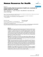
Figure 2 Response vs. Chromosome Number. Response profile of GSK1070916 for various hematological cell line tumor types (n = 45). Those
cell lines that were responsive to treatment are on the left and those that were resistant are on the right. Higher chromosome numbers is more
prevalent for the less sensitive phenotypes.
Table 1 Response to GSK107916 among populations of
cells with high and low modal chromosome number in a
2 × 2 contingency table
Sensitive Resistant Total
Diploid (~2n) 16 13 33
High Modality (>3n) 21412
Total 18 27 45
Moy et al. Journal of Translational Medicine 2011, 9:110
/>Page 5 of 10
each subtype of B-cell lymphoma was collected and
reviewed. The distribution of high chromosome mod al-
ity was varied with Diffuse Large B-Cell, Follicular, and
Mantle lymphoma subtypes having higher frequencies
compared to Burkitt and MALT NHL subtypes
(Table 3b).
Discussion
Karyotyping is a standard clinical practice for hematolo-
gical malignancies, and the cytogenetics of the disease
not only helps with diagnosis, but often provides prog-
nostic values [21-23 ]. With karyoty pe data from these
cell lines, we discovered that high chromosome number
in cell lines were associated with resistance to
GSK1070916. As with other Aurora B inhibitors, treat-
ment with GSK1070916 generally e licited a polyploidy
phenotype in cell lines. Thi s suggests cancer cells with a
polyploid phenotype might have developed mechanisms
to bypass checkpoints for polyploidy and thus are resis-
tan t to Aurora inhibition. Our comprehensi ve review of
publicly available karyotype data revealed subtypes of
hematological malignancies with high frequencies of
polyploidy. Conveniently, it is standard clinical practice
to perform karyotyping on hematological cancer cells
and chromosome number can serve as an attractive
resistance marker for patient response enrichment for
GSK1070916 in malignancies such as NHL.
AnumberofAurorakinaseinhibitorsarealreadyin
clinical or preclinical development including GSK1070916,
VX-680, AZD1152, PHA-739358, AT9283 and CYC116
[24-28]. Aurora kinase Inhibitors have shown potential
efficacy for a variety of hematological tumor subtypes
including AML, ALL and CML [29-33]. As with other tar-
geted therapies, predictive biomarkers for GSK1070916
that could stratify patient populations can accelerate clini-
cal development and cell line models have proven to be
Figure 3 The response profile of GSK1070916 for cell lines with a primary diploid chromosome number (<50).Thepercentageof
polyploidy within subpopulations of these cells is provided on the y axis. Resistant cell lines appeared to have elevated polyploidy among cell
subpopulations.
Moy et al. Journal of Translational Medicine 2011, 9:110
/>Page 6 of 10
useful system for this purpose [34]. However, most of the
hematological cell lines in our panel exhibited high sensi-
tivity using proliferation as a measure of response. This
sensitive response profile is likely due to the continuous
proliferating nature of the established cell lines in tissue
culture. Since cancer cell death is a more desired response
in clinic, measures of cell death were used as the criteri a
to categorize response to GSK107016.
Using these criteria, our cell line panel exhibited sensi-
tivity with GSK1070916 in a broad range of leukemias
(AML, B-ALL, and CML) and two subtypes of NHL
(Burkitt’s, Large B-Cell Lymphoma). These findings are
generally consistent with response profiles observed
with other Aurora inhibitors [29,31,33] and suggests
these disease subtypes can serve as important predictors
of response.
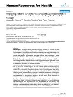
Figure 4 Cell cycle distribution from fluorescent-activated cell sorting ( FACs) analysis of T-ALL cell lines after treatment with
GSK1070916 at 24, 46, and 72 hours. (a) MOLT-16 was sensitive to GSK1070916 and showed increasing amounts of sub-2N DNA (blue)
indicating cell death.(b) In contrast, CTV-1 had higher amounts of 4N DNA or greater (light blue, green) which increased with prolonged
exposure to GSK1070916, generating a large multinucleated resistant phenotype.
Table 2 Association of NOTCH1 mutation status to high
modal chromosome number in T-ALL cell lines
WT Mutant NOTCH1 Total
Diploid (~2n) 72 9
High Modality (>3n) 21214
Total 10 14 23
Moy et al. Journal of Translational Medicine 2011, 9:110
/>Page 7 of 10
Genetic and cytogenetic information for the cell lines
were used to discover genetic markers with predictive
value. Cell lines with the polyploid phenotype were asso-
ciated with resistance to GSK1070916. This observation
was particularly striking in the response profile for T-
ALL cells in which a majority of cells (5/6) had both high
chromosome number and resistance to GSK1070916
with the sensitive cell line (MOLT-16) also having the
low chromosome phenotype. Not surprisingly, three
CML lines with hyperdiploidy (>2n) and hypertriploidy
(>3n) still maintained a sensitive response profile. The
sensitivity observed in CML cell lines, even with the poly-
ploid phenotype, was not unexpected since GSK1070916
inhibits ABL, and aurora kinase inhibitors that also inhi-
bit ABL can be considered a potential therapeutic alter-
native for patients resistant to Imatinib [35].
Cell lines and tumors can often exhibit heterogeneous
genetic backgrounds from diverse subpopulations. Upon
examination of the cell lines with low primary chromo-
some number, we found a higher proportion of polyploidy
among cell subpopulations in the resistant group. For
instance, in our panel of B-cell lymphoma cell lines, 6 of
the 7 cell lines were resistant to GSK1070916 and con-
tained low chromosome number in the primary popula-
tion of cells. However, when in reviewing the ploidy
content in the cell subpopulations in this tumor type, we
observe high ploidy content in numerous B-cell lymphoma
lines (e.g. REC-1, 25% polyploidy). This further under-
scores the significance of the general observation between
polyploidy and resistance. For these data, we hypothesize
there is a selective growth advantage for the subpopulation
of cells with the polyploid phenotype during Aurora inhi-
bition. This may represent a resistance mechanism that
potentially can develop upon prolonged drug treatment
with Aurora inhibitors. These findings warrant further
investigation about the relationship of chromosome num-
ber in primary and secondary populations of the tumor
during and after treatment to monitor potential evolving
resistance.
Inhibition of Aurora B does not inhibit cell cycle pro-
gression but rather enters and exits mitosis with normal
kinetics, with cells re-replicating their genome [36].
Treatment of cancer cells with GSK1070916 typically
yields a polyploid phenotype resulting from chromosome
replication without nuclear or cell division. Our FACS
analysis of GSK1070916 treatment shows that for sensi-
tive cells, p olyploid cell populations would develop dur-
ing earlier time points and would be killed upon longer
drug incubation. For resistant cell lines, however, poly-
ploid cell populations were tolerated over time and sig-
nificantly less cell death was observed. To maintain
genome integrity, cells generally have developed mechan-
isms/checkpoints to prevent polyploidy [37]. It can be
hypothesized that for cells that are primarily polyploid,
they have developed mechanisms to bypass these check-
points to tolerate polyploidy and therefore can evade cell
death by AURKB/C inhibition. One of these mechanisms
could be p53 dependent tetraploidy checkpoint [38-40].
Interestingly, excl uding cell lines with high chromosome
content (chromosome number >50 or polyploidy in >5%
of cell population), 4/5 sensitive lines were reported wild-
type for p53 while 3/4 resistant lines were p53 mutant
(Additional File 1, Table S5). These data further suggests
that inactivation of polyploidy checkpoints might contri-
bute to resistance during AURKB inhibition.
The expression profile for Aurora B and C in our panel
did not show any relationship with response to
GSK1070916 (Additional File 1, Table S4). However, since
the expression data in our panel does not reflect the rela-
tive expression of the Aurora genes at the time of mitosis,
the relationship of Aurora expression and response to
GSK1070916 is still unclear. In a s ubse quent analysis of
the background genetics, we found NOTCH1 mutation
status to be associated with high chromosome number in
T-ALL cells. In concordance with these findings, 3 of 4
resistant T-ALL cell lines with polyploidy also had muta-
tions in NOTCH1. While there was one AML cell line
(ML- 2) with a NOTCH1 mutation which appeared to be
tetraploidy and was resistant to GSK1070916, a majority
of cell lines that were not T-ALL cell lines were wild-type
for NOTCH1. Since the association of NOTCH1 mutation
status with response to GSK1070916 was beyond the
scope of this study, no further data was collec ted to fully
confirm this relationship. While NOTCH activation has
been reported to be associated with tetraploidy and chro-
mosomal instability in meningiomas [41], the specific
mechanism by which these mutations may play in the for-
mation of the observed polyploid phenotype in T-ALL
cells has yet to be determined. Interestingly, NOTCH sig-
naling has also been considered to play a role in cancer
stem cell regulation [42] but it is unclear what role the
polyploid phenotype may play for these cell types.
Estimates of patient prevalence for a biomarker are cri-
tical for determ ining the appropriate patient selection
strategy. These estimates of prevalence can provide gui-
dance on the number of patients needed to screen for the
Table 3 Estimated frequency of high modality in major
hematological patient populations
Tumor Type >2n >3n Total Cases
AML 4.6% 1.5% 14,611
B-ALL 25.0% 2.0% 3,769
NHL - B-Cell 14.8% 8.2% 3,542
NHL - T-Cell 7.2% 5.1% 1,497
Hodgkins 48.8% 30.3% 244
T-ALL 5.9% 3.5% 1,130
Myeloma 39.8% 8.3% 1,561
Moy et al. Journal of Translational Medicine 2011, 9:110
/>Page 8 of 10
marker and the subtypes of the disease that are most
likely to provide a positive or negative response. The pre-
valence of the high modal chromosome number in
patients can be estimated using cytogenetic data publicly
available from the Mitelman database. We found the fre-
quency of high chromosome number is generally higher
among lymphoma compared to leukemia malignancies.
While the Hodgkin’s lymphoma subtype has an elevated
frequency of high chromosome modality in its patient
population, the NHL subtypes represent a population of
patients with a significant unmet medical need. Further
review of NHL subtypes showed that Follicular and Dif-
fuse Large B-Cell are the most promising as candidate
NHL subtypes for using high chromosome number as a
marker of negative response to Aurora inhibition. A
review of NOTCH mutations in the COSMIC data-
base [19] for T-ALL tumors show a mutation frequency
of 40% suggesting that T-ALL may also be a potentially
attractive subtype for patient stratification.
Conclusions
Identification of cytogenetic abnormalities u sing karyo-
typing for prognosis and treatment of hematological
malignancies has been a standard diagnostic tool for
many years [43-46]. Detection of polyploidy in cells,
with its ease of measurement, low costs, and b iological
relevance as a negative predictor of response to Aurora
inhibition, can be a powerful tool to enrich patients that
can potentially respond to GSK1070916.
Additional material
Additional file 1: Additional Table S1. Response Data for treatment of
cells with GSK1070916. Response is designated through evaluation of
Cell Cycle Analysis (FACs), Ymin/T0 and EC50 values (See METHODS).
Additional Table S2. Available Karyotype data for Cell lines treated with
GSK1070916. Additional Table S3. Among cell lines with low native
modal chromosome number (< 50), the estimated polyploidy in the
subpopulation of cells are reviewed in terms of response to Aurora
inhibition by GSK1070916. Additional Table S4. Background Genetics
data for Cell lines treated with GSK1070916. Additional Table S5.
Review of Cell lines in panel with low native chromosome number (<
50) and low polyploid in subpopulations (< = 5%). Additional Table S6.
Percent inhibition from Kinase screen of GSK1070916 for human and
mouse ABL oncogene at 0.3 uM and 10 uM
Acknowledgements
No special acknowl edgements.
Authors’ contributions
CAO, RP carried out the cell cycle and response studies. CM participated in
the design of the study and performed the statistical analysis. YD, MAH, CM
conceived of the study, and participated in its design and coordination and
helped to draft the manuscript. All authors read and approved the final
manuscript.
Competing interests
The authors declare that they have no competing interests.
Received: 6 April 2011 Accepted: 15 July 2011 Published: 15 July 2011
References
1. Wheatley SP, Carvalho A, Vagnarelli P, Earnshaw WC: INCENP is required
for proper targeting of Survivin to the centromeres and the anaphase
spindle during mitosis. Curr Biol 2001, 11:886-890.
2. Tang CJ, Lin CY, Tang TK: Dynamic localization and functional
implications of Aurora-C kinase during male mouse meiosis. Dev Biol
2006, 290:398-410.
3. Bischoff JR, Anderson L, Zhu Y, Mossie K, Ng L, Souza B, Schryver B,
Flanagan P, Clairvoyant F, Ginther C, et al: A homologue of Drosophila
aurora kinase is oncogenic and amplified in human colorectal cancers.
EMBO J 1998, 17:3052-3065.
4. Ikezoe T, Takeuchi T, Yang J, Adachi Y, Nishioka C, Furihata M, Koeffler HP,
Yokoyama A: Analysis of Aurora B kinase in non-Hodgkin lymphoma. Lab
Invest 2009, 89:1364-1373.
5. Ikezoe T, Yang J, Nishioka C, Tasaka T, Taniguchi A, Kuwayama Y,
Komatsu N, Bandobashi K, Togitani K, Koeffler HP, Taguchi H: A novel
treatment strategy targeting Aurora kinases in acute myelogenous
leukemia. Mol Cancer Ther 2007, 6:1851-1857.
6. Lee EC, Frolov A, Li R, Ayala G, Greenberg NM: Targeting Aurora kinases
for the treatment of prostate cancer. Cancer Res 2006, 66:4996-5002.
7. Li D, Zhu J, Firozi PF, Abbruzzese JL, Evans DB, Cleary K, Friess H, Sen S:
Overexpression of oncogenic STK15/BTAK/Aurora A kinase in human
pancreatic cancer. Clin Cancer Res 2003, 9:991-997.
8. Smith SL, Bowers NL, Betticher DC, Gautschi O, Ratschiller D, Hoban PR,
Booton R, Santibanez-Koref MF, Heighway J: Overexpression of aurora B
kinase (AURKB) in primary non-small cell lung carcinoma is frequent,
generally driven from one allele, and correlates with the level of genetic
instability. Br J Cancer 2005, 93:719-729.
9. Meza-Zepeda LA, Kresse SH, Barragan-Polania AH, Bjerkehagen B,
Ohnstad HO, Namlos HM, Wang J, Kristiansen BE, Myklebost O: Array
comparative genomic hybridization reveals distinct DNA copy number
differences between gastrointestinal stromal tumors and
leiomyosarcomas. Cancer Res 2006, 66:8984-8993.
10. Park HS, Park WS, Bondaruk J, Tanaka N, Katayama H, Lee S, Spiess PE,
Steinberg JR, Wang Z, Katz RL, et al: Quantitation of Aurora kinase A gene
copy number in urine sediments and bladder cancer detection. J Natl
Cancer Inst 2008, 100:1401-1411.
11. Staff S, Isola J, Jumppanen M, Tanner M: Aurora-A gene is frequently
amplified in basal-like breast cancer. Oncol Rep 2010, 23:307-312.
12. Gautschi O, Heighway J, Mack PC, Purnell PR, Lara PN, Gandara DR: Aurora
kinases as anticancer drug targets. Clin Cancer Res 2008, 14:1639-1648.
13. Hardwicke MA, Oleykowski CA, Plant R, Wang J, Liao Q, Moss K,
Newlander K, Adams JL, Dhanak D, Yang J, et al: GSK1070916, a potent
Aurora B/C kinase inhibitor with broad antitumor activity in tissue
culture cells and human tumor xenograft models. Mol Cancer Ther 2009,
8:1808-1817.
14. Adams ND, Adams JL, Burgess JL, Chaudhari AM, Copeland RA,
Donatelli CA, Drewry DH, Fisher KE, Hamajima T, Hardwicke MA, et al:
Discovery of GSK1070916, a potent and selective inhibitor of Aurora B/C
kinase. J Med Chem 2010, 53:3973-4001.
15. Anderson K, Lai Z, McDonald OB, Stuart JD, Nartey EN, Hardwicke MA,
Newlander K, Dhanak D, Adams J, Patrick D, et al: Biochemical
characterization of GSK1070916, a potent and selective inhibitor of
Aurora B and Aurora C kinases with an extremely long residence time1.
Biochem J 2009, 420:259-265.
Table 4 Prevalence of high modality in NHL B-Cell
Lymphoma subtypes
NHL Subtype >2n >3n Total Cases
Diffuse Large 27.5% 13.7% 1225
Follicular 18.3% 8.0% 1330
Mantle 9.7% 7.7% 402
Burkitt 6.4% 2.0% 659
MALT 5.9% 3.5% 340
Moy et al. Journal of Translational Medicine 2011, 9:110
/>Page 9 of 10
16. Drexler H: Guide to Leukemia-Lymphoma Cell Lines. 2005.
17. American Type Culture Collection. [].
18. Knutsen T, Gobu V, Knaus R, Padilla-Nash H, Augustus M, Strausberg RL,
Kirsch IR, Sirotkin K, Ried T: The interactive online SKY/M-FISH & CGH
database and the Entrez cancer chromosomes search database: linkage
of chromosomal aberrations with the genome sequence. Genes
Chromosomes Cancer 2005, 44:52-64.
19. Bamford S, Dawson E, Forbes S, Clements J, Pettett R, Dogan A, Flanagan A,
Teague J, Futreal PA, Stratton MR, Wooster R: The COSMIC (Catalogue of
Somatic Mutations in Cancer) database and website. Br J Cancer 2004,
91:355-358.
20. Mitelman Database of Chromosome Aberrations and Gene Fusions in
Cancer. [ />21. Bacher U, Schnittger S, Haferlach C, Haferlach T: Molecular diagnostics in
acute leukemias. Clin Chem Lab Med 2009, 47:1333-1341.
22. Ferrara F, Palmieri S, Leoni F: Clinically useful prognostic factors in acute
myeloid leukemia. Crit Rev Oncol Hematol 2008, 66:181-193.
23. Zenz T, Mertens D, Dohner H, Stilgenbauer S: Molecular diagnostics in
chronic lymphocytic leukemia - pathogenetic and clinical implications.
Leuk Lymphoma 2008, 49:864-873.
24. Carpinelli P, Ceruti R, Giorgini ML, Cappella P, Gianellini L, Croci V,
Degrassi A, Texido G, Rocchetti M, Vianello P, et al: PHA-739358, a potent
inhibitor of Aurora kinases with a selective target inhibition profile
relevant to cancer. Mol Cancer Ther 2007, 6:3158-3168.
25. Dawson MA, Curry JE, Barber K, Beer PA, Graham B, Lyons JF, Richardson CJ,
Scott MA, Smyth T, Squires MS, et al: AT9283, a potent inhibitor of the
Aurora kinases and Jak2, has therapeutic potential in myeloproliferative
disorders. Br J Haematol 2010, 150:46-57.
26. Harrington EA, Bebbington D, Moore J, Rasmussen RK, Ajose-Adeogun AO,
Nakayama T, Graham JA, Demur C, Hercend T, Diu-Hercend A, et al: VX-
680, a potent and selective small-molecule inhibitor of the Aurora
kinases, suppresses tumor growth in vivo. Nat Med 2004, 10:262-267.
27. Wang S, Midgley CA, Scaerou F, Grabarek JB, Griffiths G, Jackson W,
Kontopidis G, McClue SJ, McInnes C, Meades C, et al: Discovery of N-
phenyl-4-(thiazol-5-yl)pyrimidin-2-amine aurora kinase inhibitors. J Med
Chem 2010, 53:4367-4378.
28. Yang J, Ikezoe T, Nishioka C, Tasaka T, Taniguchi A, Kuwayama Y,
Komatsu N, Bandobashi K, Togitani K, Koeffler HP, et al: AZD1152, a novel
and selective aurora B kinase inhibitor, induces growth arrest, apoptosis,
and sensitization for tubulin depolymerizing agent or topoisomerase II
inhibitor in human acute leukemia cells in vitro and in vivo. Blood 2007,
110:2034-2040.
29. Fei F, Stoddart S, Groffen J, Heisterkamp N:
Activity of the Aurora kinase
inhibitor VX-680 against Bcr/Abl-positive acute lymphoblastic leukemias.
Mol Cancer Ther 2010, 9:1318-1327.
30. Noronha G, Cao J, Chow CP, Dneprovskaia E, Fine RM, Hood J, Kang X,
Klebansky B, Lohse D, Mak CC, et al: Inhibitors of ABL and the ABL-T315I
mutation. Curr Top Med Chem 2008, 8:905-921.
31. Gontarewicz A, Balabanov S, Keller G, Panse J, Schafhausen P, Bokemeyer C,
Fiedler W, Moll J, Brummendorf TH: PHA-680626 exhibits anti-proliferative
and pro-apoptotic activity on Imatinib-resistant chronic myeloid
leukemia cell lines and primary CD34+ cells by inhibition of both Bcr-
Abl tyrosine kinase and Aurora kinases. Leuk Res 2008, 32:1857-1865.
32. Gontarewicz A, Balabanov S, Keller G, Colombo R, Graziano A, Pesenti E,
Benten D, Bokemeyer C, Fiedler W, Moll J, Brummendorf TH: Simultaneous
targeting of Aurora kinases and Bcr-Abl kinase by the small molecule
inhibitor PHA-739358 is effective against imatinib-resistant BCR-ABL
mutations including T315I. Blood 2008, 111:4355-4364.
33. Walsby E, Walsh V, Pepper C, Burnett A, Mills K: Effects of the aurora
kinase inhibitors AZD1152-HQPA and ZM447439 on growth arrest and
polyploidy in acute myeloid leukemia cell lines and primary blasts.
Haematologica 2008, 93:662-669.
34. Sharma SV, Haber DA, Settleman J: Cell line-based platforms to evaluate
the therapeutic efficacy of candidate anticancer agents. Nat Rev Cancer
2010, 10:241-253.
35. Gontarewicz A, Brummendorf TH: Danusertib (formerly PHA-739358)–a
novel combined pan-Aurora kinases and third generation Bcr-Abl
tyrosine kinase inhibitor. Recent Results Cancer Res 2010, 184:199-214.
36. Gautschi O, Mack PC, Davies AM, Lara PN, Gandara DR: Aurora kinase
inhibitors: a new class of targeted drugs in cancer. Clin Lung Cancer 2006,
8:93-98.
37. Mazzino A, Muratore-Ginanneschi P, Musacchio S: Scaling properties of the
two-dimensional randomly stirred Navier-Stokes equation. Phys Rev Lett
2007, 99:144502.
38. Margolis RL: Tetraploidy and tumor development. Cancer Cell 2005,
8:353-354.
39. Andreassen PR, Lohez OD, Margolis RL: G2 and spindle assembly
checkpoint adaptation, and tetraploidy arrest: implications for intrinsic
and chemically induced genomic instability. Mutat Res 2003, 532:245-253.
40. Margolis RL, Lohez OD, Andreassen PR: G1 tetraploidy checkpoint and the
suppression of tumorigenesis. J Cell Biochem 2003, 88:673-683.
41. Baia GS, Stifani S, Kimura ET, McDermott MW, Pieper RO, Lal A: Notch
activation is associated with tetraploidy and enhanced chromosomal
instability in meningiomas. Neoplasia 2008, 10:604-612.
42. Androutsellis-Theotokis A, Leker RR, Soldner F, Hoeppner DJ, Ravin R,
Poser SW, Rueger MA, Bae SK, Kittappa R, McKay RD:
Notch signalling
regulates stem cell numbers in vitro and in vivo. Nature 2006,
442:823-826.
43. Aldoss IT, Weisenburger DD, Fu K, Chan WC, Vose JM, Bierman PJ,
Bociek RG, Armitage JO: Adult Burkitt lymphoma: advances in diagnosis
and treatment. Oncology (Williston Park) 2008, 22:1508-1517.
44. Johnson NA, Savage KJ, Ludkovski O, Ben-Neriah S, Woods R, Steidl C,
Dyer MJ, Siebert R, Kuruvilla J, Klasa R, et al: Lymphomas with concurrent
BCL2 and MYC translocations: the critical factors associated with
survival. Blood 2009, 114:2273-2279.
45. Jares P, Colomer D, Campo E: Genetic and molecular pathogenesis of
mantle cell lymphoma: perspectives for new targeted therapeutics. Nat
Rev Cancer 2007, 7:750-762.
46. Fleishman EV, Sokova OI, Popa AV, Shneider MM, Kirichenko OP,
Konstantinova LN, Metel’kova NF: [Chromosomal translocation t(8,21) in
acute myeloid leukemia of children: prognostic value of additional
karyotype abnormalities]. Vestn Ross Akad Med Nauk 2009, 9-16.
doi:10.1186/1479-5876-9-110
Cite this article as: Moy et al.: High Chromosome Number in
hematological cancer cell lines is a Negative Predictor of Response to
the inhibition of Aurora B and C by GSK1070916. Journal of Translational
Medicine 2011 9:110.
Submit your next manuscript to BioMed Central
and take full advantage of:
• Convenient online submission
• Thorough peer review
• No space constraints or color figure charges
• Immediate publication on acceptance
• Inclusion in PubMed, CAS, Scopus and Google Scholar
• Research which is freely available for redistribution
Submit your manuscript at
www.biomedcentral.com/submit
Moy et al. Journal of Translational Medicine 2011, 9:110
/>Page 10 of 10