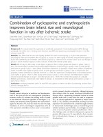
Báo cáo sinh học: "Combination of cyclosporine and erythropoietin improves brain infarct size and neurological function in rats after ischemic stroke" potx
Bạn đang xem bản rút gọn của tài liệu. Xem và tải ngay bản đầy đủ của tài liệu tại đây (8.99 MB, 14 trang )
RESEARC H Open Access
Combination of cyclosporine and erythropoietin
improves brain infarct size and neurological
function in rats after ischemic stroke
Chun-Man Yuen
1
, Cheuk-Kwan Sun
2
, Yu-Chun Lin
3
, Li-Teh Chang
4
, Ying-Hsien Kao
3
, Chia-Hung Yen
5
,
Yung-Lung Chen
6
, Tzu-Hsien Tsai
6
, Sarah Chua
6
, Pei-Lin Shao
7
, Steve Leu
8†
and Hon-Kan Yip
6,8*†
Abstract
Background: This study tested the superiority of combined cyclosporine A (CsA)-erythropoietin (EPO) therapy
compared with either one in limiting brain infarction area (BIA) and preserving neurological function in rat after
ischemic stroke (IS).
Methods: Fifty adult-male SD rats were equally divided into sham control (group 1), IS plus intra-peritoneal
physiological saline (at 0.5/24/48 h after IS) (group 2), IS plus CsA (20.0 mg/kg at 0.5/24h, intra-peritoneal) (group 3),
IS plus EPO (5,000IU/kg at 0.5/24/48h, subcutaneous) (group 4), combined CsA and EPO (same route and dosage as
groups 3 and 4) treatment (group 5) after occlusion of distal left internal caroti d artery.
Results: BIA on day 21 after acute IS was higher in group 2 than in other groups and lowest in group 5 (all p <
0.01). The sensorimotor functional test showed higher frequency of left turning in group 2 than in other groups
and lowest in group 5 (all p < 0.05). mRNA and protein expressions of apoptotic markers and number of apoptotic
nuclei on TUNEL were higher in group 2 than in other groups and lowest in group 1 and 5, whereas the anti-
apoptotic markers exhibited an opposite trend (all p < 0.05). The expressions of inflammatory and oxidized protein
were higher in group 2 than in other groups and lowest in group 1 and 5, whereas anti-inflammatory markers
showed reversed changes in group 1 and other groups (all p < 0.05). The number of aquaporin-4+ and glial
fibrillary acid protein+ stained cells were higher in group 2 as compared to other groups and lowest in groups 1
and 5 (all p < 0.01).
Conclusion: combined treatment with CsA and EPO was superior to either one alone in protecting rat brain from
ischemic damage after IS.
Background
Despite current advances in medicine and implementa-
tion of the state-of-the-art management guidelines,
ischemic stroke (IS) remains the leading cause of death
in the industrial countries regardless of etiologies [1-4].
Indeed, this unsavory clinical problem has vexed neurol-
ogists for decades. Not only the death but also the high
incidence of severe neurological impairment after IS
with permanent disability [5] that cause a tremendous
social economic burden worldwide. Although growing
data indicate that the newly developed thrombolytic
therapy offers a promising treatment option for some
patients with acute IS early after the onset of symptoms
[6,7], its clinical application is impeded by major limita-
tions [7-10]. Besides, thrombolytic therapy has been
reported to be associated with a relatively high incidence
of intracranial hemorrhage [10,11] contributing to its
notable mortality and morbidity. Accordingly, the treat-
ment of acute IS patients remains problematic. There-
fore, finding a safe and effective therapeutic regimen for
patients following acute IS, especially for those unsuita-
ble for thrombolytic therapy, is of utmost importance
for physicians.
* Correspondence:
† Contributed equally
6
Division of cardiology, Department of Internal Medicine, Kaohsiung Chang
Gung Memorial Hospital and Chang Gung University College of Medicine,
Kaohsiung, Taiwan
Full list of author information is available at the end of the article
Yuen et al. Journal of Translational Medicine 2011, 9:141
/>© 2011 Yuen et al; licensee BioMed Central Ltd. This is an Open Access article distributed under the terms of the Creative Common s
Attribution License (http:// creativecommons.o rg/licenses/by/2.0), which permits u nrestricted use, distribution, and reproduction in
any medium, provided the original work is properl y cited.
Not only has erythropoietin (EPO) therapy been
reported to enhance erythropoiesis in the treatment of
anemia, but it has also been shown to alleviate ische-
mia-related organ dysfunction through anti-ischemic
and cellular protective effects [12-15]. Our recent stu-
dies [16,17] have further shown that E PO therapy
remarkably improves neurological impairment in rat IS
model and c linical outcome in patients after acute IS.
Additionally, accumulating evidence from animal models
indicates that not only does cyclosporine A (CsA) pos-
sess immunosuppressive properties, but it is also a
potent inhibitor of mitochondrial permeability transition
pore (mPTP) that plays an important role in attenuating
ischemia-reperfusion injury [18-20]. Recently, a clinical
observational study [21] and an experimental investiga-
tion using a mini-pig animal model [22] d emonstrated
that administration of CsA after acute ST-segment ele-
vation myocardial infarction (STEMI) effectively limited
left ventricular infarct size. However, whether combined
therapy with CsA and EPO will maximize the ant i-
ischemic effect and further improve outcome after acute
IS remains uncertain. In view of the fact that there is no
effective therapy for the majority of patients with acute
ISandthatbothEPOandCsAhavebeenshownto
offer therapeutic benefit to this patient population, this
study investigated whether combined therapy with these
two drugs was superior to either one alone in reducing
brain infarction and impro ving neurological function in
a rat acute IS model.
Methods
Ethics
Allanimalexperimentalprocedures were approved by
the Institute of Animal Care and Use Committee at our
institute and performed in acc ordance with the Guide
for the Care and Use of Laboratory Animals (NIH publi-
cation No. 85-23, National Academy Press, Washington,
DC, USA, revised 1996).
Animal Model of Acute Ischemic Stoke and Corner Test
The protocol and procedure of using a rodent model of
acute IS has been described in details in our recent
report [23]. Adult male Sprague-Dawley rats, weighing
300-325 g (Charles River Technology, BioLASCO Tai-
wan Co., Ltd., Taiwan) were utilized in the current
study. All animals were an esthetized by chloral hydrate
(35 mg/kg i.p.) and placed in a supine position on a
warming pad at 37°C. After exposure of the left com-
mon carotid artery (LCCA) through a transverse neck
incision, a small incision was made on the LCCA
through which a nylon filament (0.28 mm in diameter)
was carefully advanced into the distal left internal caro-
tid artery for occlusion of left middle cerebral artery
(LMCA) to induce brain infarction of its blood-
supplying area. The nylon filament was removed three
hour s after occlusion, followed by closure of the muscle
and skin in layers. The rats were then placed in a por ta-
ble animal intensive care unit (ThermoCare
®
)for24
hours. The sensorimotor functional test (Corner test)
was done for each rat at baseline and on day 1 (24 h
afte r procedure), 3, 7, 14, and 21 after acute IS induc tion
as we recently described [16,23]. Briefly, the rat was
allowed to walk through a tunnel and then into a corner,
the angle of which was 60 degrees. To exit the corner,
the rat could turn either to left or right. The results were
recorded by a technician who was blind to the study
design. This test was repeated 10 to 15 times with at least
30 seconds between each trial. We recorded the number
of right and left turns from 10 successful trials for each
animal and used the results for statistical analysis.
Treatment Protocol
Ten sham-operated healthy rats served as normal con-
trols (group 1). The other 40 rats with acute IS were
equally divided into IS plus intra-peritoneal 1.0 mL phy-
siological saline (at 0.5, 24 and 48 hour after IS) (group
2, n = 10), IS plus CsA (20.0 mg/kg at 0.5 and 24 hour,
intra-peritoneal) (group 3, n = 10), IS plus EPO (5,000
IU/kg at 0.5, 24, and 48 hour, subcutaneous) (group 4, n
= 10), and combined CsA (20.0 mg/kg at 0.5 and 24
hour, intra-peritoneal) and EPO (5,000 IU/kg at 0.5, 24
and 48 hour, subcutaneous) treatment (group 5, n = 10).
Two rats died in group 2 and one rat died in each
other group (i.e. groups 3 to 5) during the procedure.
For the purpose of this study, additional rats were
added so that 10 animals in each group went through
the whole study.
The dosage of EPO and the timing of treatment were
based on previous literature and our recent report
[16,24], whereas the dosage of cyclosporine and the treat-
ment protocol were according to a previous report [25].
Specimen Collection and Preparation for Individual Study
Rats in all groups were euthanized on day 21 afte r IS
induction, and the brain of each rat was promptly
removed and immersed in cold saline. For immunohis-
tofluorescent (IHF) study, the brain tissue was rinsed
with PBS, embedded in OCT compound (Tissue-Tek,
Sakura, Netherlands) and snap-frozen in liquid nitrogen
before being stored at -80°C. Fo r immunohistochemical
(IHC) staining, the brain tissue was fixed in 4% formal-
dehyde and embedded in paraffin. Additionally, the
brain tissue of infarct area was collected for Western
blot, real-time PCR, and oxidative stress analyses.
Measurement of Brain Infarct Area
To evaluate the impact of CsA, EPO, and combined
EPO and CsA treatment on brain infarction, coronal
Yuen et al. Journal of Translational Medicine 2011, 9:141
/>Page 2 of 14
sections of the brain were obtained from six extra ani-
mals in groups 2 to 5 (n = 6 for each group) as 2 mm
slices. Each cross section of brain tissue was then
stained with 2% 3,5-Triphenyl-2H-Tetraz olium Chloride
(TTC) (Alfa Aesar) for BIA analysis. The methodology
has been described in details in our recent studies
[16,23]. Briefly, all brain sections were placed on a tray
with a scaled vertical bar to which a digital camera was
attached. The sections were photogr aphed from directly
above at a fixed height. The images obtained were then
analyzed using Image Tool 3 (IT3) image analysis soft-
ware (University of Texas, Health Science Center, San
Ant onio, UTHSCSA; Image Tool for Windows, Version
3.0, USA). BIA was identified as either whitish or pale
yellowish regions. Infarct region was further confirmed
by microscopic examination. The percentages of infarct
area were then calculated by dividing the area with total
cross-sectional area of the brain.
All measurements (i.e. Corner test and assessment of
BIA) were performed by a skillfu l senior technician
blinded to the treatment and non-treatment groups.
TUNEL Assay for Apoptotic Nuclei
For each rat, six sections of BIA were analyzed by an in
situ Cell Death Detection Kit, AP (Roche) according to
the manufacturer’s guidelines. Three randomly chosen
high-power fields (HPFs) (×400) were observed for
term inal deoxynu cleotidyl transferase-mediated 2’-deox-
yuridine 5’-triphosphate nick-end labeling (TUNEL)-
positive cells for each section. The m ean number of
apoptotic nuclei p er HPF for each animal was obtained
by dividing the total number of cells with 18.
Immunofluorescent Staining
Frozen sections (4 μm thick) were obtained from BIA of
each animal. The sections were fi xed with 4 % parafor-
maldehyde and permeated with 0.5% Triton X-100 and
then incubated with antibodies against NeuN (1:1000,
Millipore), GFAP (1:500, DAKO), PGC-1a (1:100, Santa
cruz), and AQP4 (1:200, abcam) at 4°C overnight. Alexa
Fluor488, Alexa Fluor568, or Alexa Fluor594-conjugated
goat anti-mouse or rabbi t IgG were used to localize sig-
nals. Sections were then counterstained with DAPI and
observed with a fluorescent microscope equipped with
epifluorescence (Olympus IX-40).
Western Blot Analysis for Bax, Cytochrome C, Caspase 3,
NADPH oxidase 1 (NOX-1), NOX-2, Inducible Nitric Oxide
Synthase (iNOS), and Endothelial (e)NOS
Equal amounts (50 mg) of protein extracts were loaded
and separated by SDS- PAGE using 12% acrylamide gra-
dients. After electrophoresis, the separated proteins
were transferred electrophoretically to a p olyvinylidene
difluoride (PVDF) membrane (Amersham Biosciences).
Nonspecific sites we re blocked by incubation of the
membrane in blocking buffer [5% nonfat dry milk in T-
TBS (TBS containing 0.05% Tween 20)] for overnight.
The membranes were incubated with the indicated pri-
mary antibodies (Bax, 1:1000, abcam; Cytochrome C,
1:2000, BD; Caspase, 1:3000, abcam; NOX-1, 1:1500,
Sigma; NOX-2, 1:500, Sigma; iNOS, 1:200, abcam;
eNOS, 1:1000, 1:500, abcam; Actin, 1:10000, Chemico n)
for 1 hr at room temperature. Horseradish peroxidase
-conjugated anti-rabbit or anti-mouse immunoglobulin
IgG (1:2000, Cell Signaling) was used as a second
antibody for 1 hr at room temperature. The washing
procedure was repeated eight times within 1h, and
immunoreactive bands were visualized by enhanced che-
miluminescence (ECL; Amersham B iosciences) and
exposure to Biomax L film (Kodak). For purposes o f
quantitation, ECL signals were digitized using Labwork
software (UVP).
Protocol for RNA Extraction
Lysis/binding buffer (High Pure RNA Tissue Kit, Roche,
Germany) 400 μL and an appropriate amount of frozen
brain tissues were added to a nuclease-free 1.5 mL
microcentrifug e tube, followed by disru ption and homo-
genization of the tissue by using a rotor-stator homoge-
nizer (Roche).
For each isolation, 90 mL DNase incubation buffer
was pipetted into a sterile 1.5 mL reaction tube, 10 mL
DNase I working solution was then added, mixed and
incubated for 15 min at 25°C. Wash buffer I 500 mL
was then added to the upper reservoir of the filter tube,
which was then centrifuged for 15 seconds at 8,000g.
Wash buffer II 300 mL was added to the upper reservoir
of the filter tube, which was centrifuged for 2 min full-
speedatapproximately13,000g. Elution Buffer 100 mL
was then added to the upper reservoir of the filter tube.
Finally, the tube assembly was centrifuged for 1 min at
8,000g, resulting in eluted RNA in the microcentrifuge
tube.
Real-Time Quantitative PCR Analysis
Real-time polymerase chain re action was conducted
using LighCycler TaqMan Master (Roche, Germany) in
a single capillary tube according to the manufacturer’s
guidelines for individual component concentrations.
Forward and reverse primers were each designed based
on individual exons of the target gene sequence to avoid
amplifying genomic DNA.
During PCR, the probe was hybridized to its comple-
mentary single-strand DNA sequence within the PCR
target. As amplification occurred, the probe was
degraded due to the exonuclease activity of Taq DNA
polymerase, thereby separating the quencher from
reporter dye during extension. During the entire
Yuen et al. Journal of Translational Medicine 2011, 9:141
/>Page 3 of 14
amplification cycle, light emission increased exponen-
tially. A positive result was determined by i dentifying
the threshold cycle value at which reporter dye emission
appeared above background. For normalization, the
housekeeping gene Pe ptidyl-prolyl cis-trans isomerasa
(Ppia, Cyclophilin A) was used as the reference gene.
Oxidative Stress Reaction of BIA
The Oxyblot Oxidized Protein Detection Kit was pur-
chased from Chemicon (S7150). The oxyblot procedure
was performed according to the previous study [26].
The 2,4-dinitrophenylhydrazine (DNPH) derivatization
was carried out on 6 μg of protein for 15 min according
to manufacturer’s instructions. One-dimensional electro-
phoresis was carried out on 12% SDS/polyacrylamide gel
after DNPH derivatization. Proteins were transferred to
nitrocellulose membranes which were then incubated in
the primary antibody solution (anti-DNP 1:150) for 2 h,
followed by incubation with second antibody solution
(1:300) for 1 h at room temperature. The washing
procedure was repeated eight times within 40 min.
Immunoreactive bands were visualized by enhanced che-
miluminescence (ECL; Amersham Biosciences) which
was then exposed to Biomax L film (Kodak). For quanti-
ficati on, ECL signals were digitized using Labwork soft-
ware (UVP). On each gel, a standard control sample was
loaded.
Statistical Analysis
Data were expressed as mean values (mean ± SD). Sta-
tis tical analysis was adequately performed by analysis of
variance, followed by Scheff e multiple-comparison post
hoc test. SAS statistical software for Windows version
8.2 was utilized. (SAS institute, Cary, NC). A probabil ity
value < 0.05 was considered statistically significant.
Results
Effect of Combined CsA and EPO on Infarction Area and
Neurological Function after Acute IS
The mortality rate [2 in group 2, 1 in each other group
(i.e. groups 3 to 5)] did not statistically differ among
groups 2 to 5 (p = 0.413). TTC staining of brain tissues
on day 21 after acute IS showed notably reduced BIA in
IS animals treated with CsA (group 3) and EPO (group
4) than in IS animals without treatment (group 2), and
further reduced after combined therapy with CsA and
EPO (group 5) than in group 3 and group 4 (Figure 1).
Corner test showed that, as compared with group 2, a
transient improvement in neurological function was
noted in groups 3 to 5 o n day 3 after acute IS (Figure
2A). However, corner test showed the attainment of a
steady state of neurological functional impairment on
day 7 and day 14 following acute IS in groups 2, 3 and
5 but an improvement in neurological function was
noted in group 3 as compared to groups 2, 4 and 5. Sig-
nificant improvement in neurological function became
apparent in groups 3 and 4 as compared with group 2,
and further improvement was noted in group 5 than in
group 2 on day 21 after acute IS (Figure 2B).
Attenuation of Inflammatory Response through
Combined Therapy with CsA and EPO
On day 21 following acute IS induction, the mRNA
expressions of tumor necrosis factor (TNF)-a and
matrix metalloproteinase (MMP)-9, two indicators of
inflammation, were notably higher in group 2 as com-
pared to other groups (Figure 3, A and 3B). In addition,
these two biomarkers were significantly higher in groups
3and4thaningroups1and5.Furthermore,TNF-a
expression was significantly higher in group 5 as com-
pared with group 1. However, the MMP-9 expression
showed no difference between groups 1 and 5. Addition-
ally, the protein expression of inducible nitric oxide
synthase (iNOS), an index of inflammation, was remark-
ably higher in group 2 than in other groups, notably
higher in groups 3 and 4 than in groups 1 and 5 , and
significantlyhigheringroup5thaningroup1(Figure
4A). Furthermore, the protein expression of NADPH
oxidase 1 (NOX-1), an index of reactive oxygen species
(ROS) formation, w as significantly higher in group 2
compare d to that in other groups and notably higher in
groups 3 and 4 than in groups 1 and 5, but it was simi-
lar between group 1 and group 5 (Figure 4B). On the
other hand, the protein expression of NOX-2 did not
differ among the 5 groups (Figure 4C). In contrast, the
protein expression of endothelial NOS (eNOS), in index
of anti-inflammation, was remarkably lower in group 2
than in other groups, notably lower in groups 3 and 4
than in groups 1 and 5, but no significant difference was
noted between group 1 and group 5 (Figure 4D).
Enhanced Reduction of Apoptosis and Oxidative Stress by
Combined Treatment with CsA and EPO
On day 21, the mRNA (Figure 3C) and protein expres-
sions (Figu re 5A) of caspase 3, one pro-apoptotic index,
weresubstantiallyhigheringroup2thaninother
groups. They were also markedly higher in groups 3 and
4 than in groups 1 and 5, but they did not show signifi-
cant difference b etween groups 1 and 5. Additionally,
the mRNA (Figure 3D) and mitochondrial protein
expressions (Figure 5B) of Bax, another pro-apoptotic
index, were substantially higher in group 2 than in other
groups, notably higher in groups 3 and 4 than in groups
1 and 5, and the mitochondrial protein expression sig-
nificantly higher in g roup 5 than in group 1. However,
the Bax mRNA ex pression only had a statistical trend of
notably higher in group 5 than in group 1. On the other
hand, the cytosolic protein expression of Bax (Figure
Yuen et al. Journal of Translational Medicine 2011, 9:141
/>Page 4 of 14
Figure 1 Ratios of infarct area to total coronal sectional area using TTC staining. (A to E) Identification of gross infarct area (green circles)
in animals with B) ischemic stroke (IS) (group 2), C) IS + cyclosporine (CsA) (group 3), D) IS + erythropoietin (EPO) (group 4) and E) IS +
combined CsA & EPO (group 5), respectively. (F) Significantly lower ratio of infarct area to total coronal sectional area in group 5 than in group
2, 3, and 4, and notably lower in group 3 and 4 than in group 2 (n = 6 for each group). * vs. other groups, p < 0.0001 (using ANOVA). Symbols
(*, †, ‡) indicate significance (at 0.05 level) (by Scheffe multiple-comparison post hoc test).
Yuen et al. Journal of Translational Medicine 2011, 9:141
/>Page 5 of 14
Figure 2 Assessment of neurological function with Corner test. A) TheresultsofCornertest(n=10)onday0,1,3,7,14,and21after
acute IS, showing a steady state of neurological functional impairment on day 3 to 14 following acute IS in group 2, 3, 4, and 5. B) Significant
improvement in neurological function noted in group 3, 4, and 5 compared with group 2 on day 21 after acute IS, and further improvement
observed in group 5 compared with group 3 and 4. * vs. other groups, p < 0.001 (at day 21). Symbols (*, †, ‡) indicate significance (at 0.05 level)
(by Scheffe multiple-comparison post hoc test).
Figure 3 Profiles of mRNA expression in infarct area. A) Tumor necrosis factor (TNF)-a mRNA expression was remarkably higher in group 2
than in other groups, notably higher in group 3 and 4 than in group 1 and 5, and significantly higher in group 5 than in group 1. † vs. other
groups, p < 0.0001 (ANOVA test). B) Matrix metalloproteinase (MMP)-9 mRNA expression markedly increased in group 2 than in other groups,
notably increased in group 3 and 4 than in group 1 and 5, but no remarkable difference between group 3 and 4 or between group 1 and 5. †
vs. other groups, p < 0.0001 (ANOVA test). C) & D) Substantially higher mRNA expressions of caspase 3 (C) and Bax (D) in group 2 than in other
groups, and significantly higher in group 3 and 4 than in group 1 and 5, but no notable difference between group 3 and 4 or between group 1
and 5. † vs. other groups, p < 0.0001 (ANOVA test). E) & F) Significantly lower mRNA expressions of Bcl-2 and PGC-1a in group 2 than in other
groups and markedly lower in group 3 and 4 than in group 1 and 5, but no difference between group 3 and 4 and between group 1 and 5. †
vs. other groups, p < 0.0001 (ANOVA test). G) Substantially higher mRNA expression of aquaporin-4 (AQP-4) in group 2 than in other groups and
remarkably higher in group 3 and 4 than in group 1 and 5, but no significant difference between group 3 and 4 or between group 1 and 5. †
vs. other groups, p < 0.001 (ANOVA test). Symbols (*, †, ‡, §) from A) to G) indicate significance (at 0.05 level) (by Scheffe multiple-comparison
post hoc test) (n = 6 for each group).
Yuen et al. Journal of Translational Medicine 2011, 9:141
/>Page 6 of 14
5C) was significantly lower in group 2 than in other
groups, notably lower in groups 3 and 4 than in group
1,butitshowednodifferencebetweengroups1and5
or among groups 3, 4 and 5.
The mRNA (Figure 3E) and protein expressions (Fig-
ure 5D) of Bcl-2, an indicator of anti-apoptosis, were
notably lower in group 2 than in other groups. The
expressions were also significantly lower in groups 3
and 4 than in groups 1 and 5 but without notable differ-
ence between groups 1 and 5. Furthermore, TUNEL
assay (Figure 6) showed that the number of apoptotic
nuclei was substantially increased in group 2 than in
other groups, remarkably higher in g roups 3 and 4 than
in groups 1 and 5, and significantly higher in group 5
than in group 1.
On day 21 following acute IS induction, Western blot-
ting (Figure 7, A and 7B) demonstrated a significantly
higher oxidative index in mitochondria in group 2 than
in other g roups. The oxidative index was also signifi-
cantly higher in groups 3 and 4 t han in groups 1 and 5,
and notably higher in group 5 compared with that in
group 1.
Better Preservation of Mitochondrial Cytochrome C after
Combined Therapy with CS and EPO against Acute IS
The protein expression of cytochrome C in mitochon-
dria (Figure 7C) was significantly reduced in group 2
compared to that in other groups, significantly lower in
groups 3 and 4 than in group 1, but it did not differ
among groups 3 to 5, or between groups 1 and 5. In
contrast , its cytosolic expression (Figure 7D) was signifi-
cantly enhanced in group 2 compared with other
groups, significantly elevated in groups 3 and 4 than in
groups 1 and 5, but it did not differ between group 1
group 5. These findings indicate that the expression of
Figure 4 Protein expression levels of inflammation and
oxidative stress-related in infarct area. A) and B) Remarkably
elevated protein expressions of inducible nitric oxide synthase
(iNOS) (A) and NADPH oxidase 1 (NOX-1) (B) in group 2 than in
other groups, notably higher in group 3 and 4 than in group 1 and
5, significantly increased in group 5 than in group 1, but no
difference between group 3 and 4. † vs. other groups, p < 0.001
(ANOVA test). C) No significant difference in NOX-2 protein
expression among all groups. D) Remarkably lower protein
expressions of endothelial (e)NOS in group 2 than in other groups,
notably lower in group 3 and 4 than in group 1 and 5, but no
difference between group 3 and 4. Similar eNOS protein expression
noted between group 1 and group 5. † vs. other groups, p < 0.001
(ANOVA test). Symbols (*, †, ‡, §) from A) to D) indicate significance
(at 0.05 level) (by Scheffe multiple-comparison post hoc test) (n = 6
for each group).
Figure 5 Protein expression levels of apoptosis-related in
infarct area. A) Caspase 3 protein expression was notably higher in
group 2 than in other groups, notably higher in group 3 and 4, but
no significant difference between group 3 and 4 and between
group 1 and 5. † vs. other groups, p < 0.0001 (ANOVA test). B)
Significantly higher mitochondrial protein expression of Bax in
group 2 than in other groups. Significant elevation also noted in
group 3 and 4 compared with that in group 1 and 5, and notably
higher in group 5 than in group 1, but no remarkable difference
between group 3 and 4. † vs. other groups, p < 0.001 (ANOVA test).
C) Cytosolic protein expression of Bax substantially lower in group 2
than in other groups, but no difference between group 1 and 5 or
among group 3, 4, and 5. † vs. other groups, p < 0.001 (ANOVA
test). D) Bcl-2 protein expression notably lower in group 2 than in
other groups, significantly lower in group 3 and 4 than in group 1
and 5, but no significant difference between group 1 and 5 or
between group 3 and 4. † vs. other groups, p < 0.001 (ANOVA test).
Symbols (*, †, ‡,§)inA) to D) indicate significance (at 0.05 level)
(by Scheffe multiple-comparison post hoc test).
Yuen et al. Journal of Translational Medicine 2011, 9:141
/>Page 7 of 14
Figure 6 TUNEL assay for indentifying apoptotic nuclei in brain infarct area. The number o f apoptotic nuclei (yellow arrows) notably
higher in group 2 (B) than in group 1 (A), group 3 (C), group 4 (D) and group 5 (E), significantly higher in group 3 and 4 than in group 1 and
5, and significantly higher in group 5 than in group 1, but no significant difference between group 3 and 4. Scale bars in right lower corner
represent 20 μm (400x). † vs. other groups, p < 0.001 (ANOVA test). Symbols (*, †, ‡, §) indicate significance (at 0.05 level) (by Scheffe multiple-
comparison post hoc test).
Yuen et al. Journal of Translational Medicine 2011, 9:141
/>Page 8 of 14
cytochrome C, an index of energy supply and storage in
mitochondria, was relatively well-preserved in groups 3
to 5 as compared with that in group 2, and was more
preservedingroup5ascomparedtogroups3and4.
Additionally, the increase in cytosolic cytochrome C
content also suggests significant mitochondrial damage
with cytochrome C release into the cytosol in the brain
of group 2 animals.
Further Reduction in Expressions of Glial Fibrillary Acid
Protein (GFAP) and Aquaporin-4 (AQP-4) and Preservation
of Neural PGC-1a in Infarct Brain after Combined Therapy
with CsA and EPO
The mRNA expression of peroxisome proliferator-acti-
vated receptor-g coactivator-1a (PGC-1a)(Figure3F),
which is a transcriptional coactivator for regulating lipid
catabolism, oxidative metabolism, mitochondrial meta-
bolism and biogenesis, was notably lower in group 2
than in other groups and significantly lower in groups 3
and4thaningroups1and5,butitdidnotdiffer
between groups 3 and 4 or between groups 1 and 5.
Conversely, AQP-4 mRNA expression (Figure 3G), an
indicator of brain edema, was substantially increased in
group 2 compared to that in othe r groups and notably
increased in groups 3 and 4 than in groups 1 and 5, but
it was similar between groups 3 and 4 or between
groups 1 and 5.
Immunofluorescent staining showed that the expres-
sion of GFAP (Figure 8, A-E, white arrows), the princi-
pal intermediate filament of ma ture astrocytes, was
remarkably higher (Figure 8G) in group 2 compared to
that in other groups, significantly higher in groups 3
and 4 than in groups 1 and 5, and notably higher in
group 5 compared to that in group 1. In addition, AQP-
4 (Figure 8, A-E, yellow arrows) was substantially
increased(Figure8F)ingroup2thaninothergroups,
notably increased in groups 3 and 4 than in groups 1
and 5, but no significant difference was noted between
groups 1 and 5. Conversely, neuronal e xpression of
PGC-1a, an index of mitochondrial integrity (Figure 9,
A-E, doubly labeled by yellow and white arrows), was
remarkably lower (Figure 9G) in groups 2 than in other
groups, notably lower in group 3 and 4 than in groups 1
and 5, and significantly lower in group 5 as compared
with that in group 1.
Discussion
Combined Therapy with Cyclosporine and EPO Provided
Additional Benefits of Limiting Brain Infarct Size and
Improving Recovery of Neurological Function
The most important finding in the current study was
that TTC staining of the brain tissue on day 21 after
acute IS showed that the BIA was remarkably reduced
in IS animals treated with either CsA (group 3) or EPO
(group 4) than in IS animals without treatment (group
2). These findings imply that CsA or EPO therapy sig-
nificantly reduce BIA after IS. Moreove r, corner test
showed a significant improvement in neurological func-
tioningroups3and4thaningroup2onday21after
acute IS. Interestingly, previous studies [12-15] have
demonstrated that EPO therapy significantly reversed
ischemia-related left ventricular dysfunction. In concert
with this finding, previous investigations by ot her
authors and our recent studies [16,24] have also shown
that EPO therapy markedly attenuated BIA and
improved neurological function in rat after acute IS.
Furthermore, our recent clinical trial [17] has shown
that EPO therapy substantially improved 90-day major
adverse neurological event. Our findings, therefore, are
consistent with those of previous studies [12-17].
Figure 7 Oxidative index and protein expression levels of
cytochrome (Cyt) C in brain infarct area. A) Oxidative index
determination by Western blotting of brain infarct area (BIA) (n = 6),
showing notably increased oxidative index, protein carbonyls, in BIA
of group 2 compared with other groups, notably higher in group 3
and 4 than in group 1 and 5, and significantly higher in group 5
than in group 1 on day 21 following acute IS. B) † vs. other groups,
p < 0.0001 (ANOVA test). C) Protein expression of mitochondrial
cytochrome C in brain infarct area (n = 6) markedly lower in group
2 than in other groups, notably lower in group 3 and 4 than in
group 1, but no notable difference among group 3,4, and 5, or
between group 1 and 5. † vs. other groups, p < 0.01 (ANOVA test).
D) Protein expression of cytosolic cytochrome C in BIA (n = 6)
markedly higher in group 2 than in other groups, notably higher in
group 3 and 4 than in group 1 and 5, but no significant difference
between group 3 and 4, or between group 1 and 5. † vs. other
groups, p < 0.01 (ANOVA test). Symbols (*, †, ‡, §) from B) to D)
indicate significance (at 0.05 level) (by Scheffe multiple-comparison
post hoc test).
Yuen et al. Journal of Translational Medicine 2011, 9:141
/>Page 9 of 14
Figure 8 Distribution of glial fibrillary acid protein (GFAP) and aquaporin- 4 (AQP-4) in brain infarct area. A) t o E) Immunofluorescent
staining (400 x) of glial fibrillary acid protein (GFAP) (white arrows) and aquaporin-4 (AQP-4) (yellow arrows) in brain infarct area (n = 6). Both
numbers of GFAG and AQP-4 remarkably higher in group 2 than in other groups, notably higher in group 3 and 4 than in group 1 and 5, and
significantly higher in group 5 than in group 1. F) and G) † vs. other groups, p < 0.0001 (ANOVA test). Symbols (*, †, ‡,§)in(F) and (G) indicate
significance (at 0.05 level) (by Scheffe multiple-comparison post hoc test). Scale bars in right lower corner represent 20 μm.
Yuen et al. Journal of Translational Medicine 2011, 9:141
/>Page 10 of 14
Figure 9 Distribution of Neural peroxisome proliferator-a ctivated receptor-g coactivator-1a (PGC-1a) i n brain infarct area. A) to E)
Immunofluorescent staining (400 x) of PGC-1a (yellow arrows) and neuron (white arrows) in brain infarct area (n = 6). Both numbers of PGC-1a
+ cells and neurons remarkably lower in group 2 than in other groups, significantly lower in group 3 and 4 than in group 1 and 5, and
significantly reduced in group 5 compared with group 1. F) and G) † vs. other groups, p < 0.0001 (ANOVA test). Symbols (*, †, ‡,§)in(F) and
(G) indicate significance (at 0.05 level) (by Scheffe multiple-comparison post hoc test). Scale bars in right lower corner represent 20 μm.
Yuen et al. Journal of Translational Medicine 2011, 9:141
/>Page 11 of 14
Interestingly, as compared with EPO, CsA therapy
(group 4) offered similar protection of the brain against
infarction/ischemia in the current study. Recent studies
[21,22,27,28] have shown that CsA therapy notably
reduced infarction size and improved ischemia-related
organ function in b oth animal experiments and clinical
observational studies. Thus, our findings strengthen
those of the studies [21,22,27,28]. Of import ance is that,
as compared with those group 3 and group 4, combined
therapy with CsA and EPO (group 5) further attenuated
BIA. These findings may exp lain the enhanced improve-
ment in neurological function in group 5 animals as
compared with those in group 3 and group 4. In this
way, the results of the present investigation extend the
findings of previous studies [12-17,21,22,27,28].
Combined therapy with EPO and tissue plasminogen
activator (tPA) for patients after acute IS has been
recently reported by Ehrenreich et al. [ 29]. Failure in
demonstrating additio nal benefits of combining EPO
with tPA i n improving clinical outcome of patients with
acute IS as compared with placebo-controls in that clini-
cal trial [29] may be due to tPA-associated bleeding
complication that outweighe d the benefit of EPO treat-
ment [17].
Combined Therapy of CsA and EPO Further Limited
Inflammatory Reaction, Generation of Reactive Oxygen
Species, and Oxidative Stress after Acute IS
Abundant studies have shown that innate immune
mechanisms are rapidly activated following acute arterial
obstructive syndrome (i.e. tissue injury and necrosis)
which, in turn, initiat e the complement cascade, inflam-
matory reaction, and ROS generation [16,23,30,31]. Addi-
tionally, inflammatory components of the ischemic
cascades further perpetuate cellular apoptosis and necro-
sis in ischemic region [15,16,22,23,30-33]. One important
finding of the present study is that the inflammatory
responses were markedly increased in group 2 animals
tha n in those in groups 3 to 5 on day 21 after acute IS.
Moreover , both ROS generation (NOX-1) and oxidative
stress were remarkably enhanced in group 2 animals than
in other groups on day 21 after acute IS. Another intri-
guing finding of the current study is that the expressions
of anti-inflammatory protein, eNOS, was substantially
reduced in group 2 than in other groups. Additional
important finding also includes the remarkably increased
number of GFAP-positive cells, an indicator of inflamma-
tory cells in ischemic brain, in group 2 and notable
reduction in groups 3 to 5 after treatment. Therefore,
our findings, in addition to corroborating those of pre-
vious reports [15,16,22,23,30-33], could at least partially
explain the poorer prognostic outcome in g roup 2 ani-
mals compared wit h those in groups 3 to 5. Besides, the
results of our study may support the proposal that both
CsA and EPO therapy are equally effective in protecting
the brain against ischemic damage after acute IS through
suppressing inflammation, generation of ROS, and oxida-
tive stress. Of importance is that combined therapy with
CsA and EPO was found to be superior to either one
alone in inhibiting the pro duction of inflammatory bio-
markers, ROS, and oxidative stress.
Possible Mechanisms of CsA and EPO Underlying
Improved Outcome after Acute Ischemic Stroke
The key role of EPO therapy in improving outcome after
acute IS has been mainly attributed to attenuation of
inflammation, oxidative stress, cellular apoptosis,
and enhancement of angiogenesis and neurogenesis
[16,17,24,34]. On the other hand, inhibition of inflamma-
tion, oxidative stress, cellular apoptosis, and mPTP open-
ing have been proposed to be the underlying mechanisms
involved in CsA-mediated protection against i schemia-
reperfusi on organ dysfunction [18-20,27,28]. In the cur-
rent study, not only were the inflammatory and oxidative
cascades found to be substantially diminished, but the
apoptotic markers were also substantially attenuated after
CsA and EPO therapy. Accordingly, the anti-apoptotic
index (Bcl-2) was notably enhanced following combined
therapy. In addition, reduction in the number of AQP4+
cells and preservation of the number of PGC-1a+neu-
rons in BIA were observed after CsA and EPO treatment.
Moreover, mitochondrial cytochrome C was better pre-
served in treated than in untreated animals after acute IS.
Therefore, our findings not only extend those of previous
studies [16-20,24,27,28,34], but they also provide novel
information on the superiority of combined therapy with
CsA and EPO compared with either agent alone in the
treatment of acute IS in an experimental setting. In con-
sideration of the f act that both EPO and CsA are fre-
quently and separately used in our daily clinical practice
for variety of disease entities, this pre-clinical information
may warrant the need for a prospective clinical trial in
evaluating the benefit of combined therapy with CsA and
EPO which have been widely used in dif ferent clinical
settings after acute IS.
Study Limitation
This study has limitations. First, since the current study
period was only 21 days, the long-term effect of com-
bined therapy with CsA and EPO on sens orimotor func-
tion in this experimental setting is unknown. Second,
this study did not investigate the safety of CsA dosage
so that the side-effects of CsA therapy remain unclear.
A balance between the benefits and risks of CsA use,
therefore, is still a major concern regarding the clinical
use of CsA in the setting of acute IS.
Yuen et al. Journal of Translational Medicine 2011, 9:141
/>Page 12 of 14
Conclusion
The results of the present study suggest that combined
therapy with CsA and EPO is superior to either agent
alone in reducing BIA and improving neurological func-
tion after acute IS. The proposed mechanisms underly-
ing the potential impacts of combined CsA and EPO in
rats after IS have been summarized in Figure 10.
Acknowledgements
This study was supported by a program grant from Chang Gung Memorial
Hospital, Chang Gung University (grant no. CMRPG 880431).
Author details
1
Division of Trauma, Department of Surgery, Kaohsiung Chang Gung
Memorial Hospital and Chang Gung University College of Medicine,
Kaohsiung, Taiwan.
2
Department of Emergency Medicine, E-Da Hospital, I-
Shou University, Kaohsiung, Taiwan.
3
Department of Medical Research, E-Da
Hospital, I-Shou University, Kaohsiung, Taiwan.
4
Basic Science, Nursing
Department, Meiho University, Pingtung, Taiwan.
5
Department of Life
Science, National Pingtung University of Science and Technology, Pingtung,
Taiwan.
6
Division of cardiology, Department of Internal Medicine, Kaohsiung
Chang Gung Memorial Hospital and Chang Gung University College of
Medicine, Kaohsiung, Taiwan.
7
Graduate Institute of Medicine, College of
Medicine, Kaohsiung Medical University, Kaohsiung, Taiwan.
8
Center for
Translational Research in Biomedical Sciences, Kaohsiung Chang Gung
Memorial Hospital and Chang Gung University College of Medicine,
Kaohsiung, Taiwan.
Authors’ contributions
All authors have read and approved the final manuscript.
CMY, CKS, YCL, SL, and HKY designed the experiment, performed animal
experiments, and drafted the manuscript. LTC, YHK, CHY, YLC, THT and PLS
were responsible for the laboratory assay and troubleshooting. SC, CKS, SL,
and HKY participated in refinement of experiment protocol and coordination
and helped in drafting the manuscript.
Competing interests
The authors declare that they have no competing interests.
Received: 15 June 2011 Accepted: 24 August 2011
Published: 24 August 2011
References
1. Hankey GJ: Stroke: how large a public health problem, and how can the
neurologist help? Arch Neurol 1999, 56:748-754.
2. Organization WH: World Health Report 1999-Making a Difference Geneva,
Switzerland: World Health Organization; 1999.
3. Association AH: Stroke Statistics Dallas, Dallas, TX: American Heart
Association; 2002.
4. Bacigaluppi M, Pluchino S, Martino G, Kilic E, Hermann DM: Neural stem/
precursor cells for the treatment of ischemic stroke. J Neurol Sci 2008,
265:73-77.
5. Andres RH, Choi R, Steinberg GK, Guzman R: Potential of adult neural
stem cells in stroke therapy. Regen Med 2008, 3:893-905.
6. Wardlaw JM, Murray V, Berge E, Del Zoppo GJ: Thrombolysis for acute
ischaemic stroke. Cochrane Database Syst Rev 2009, CD000213.
7. Wahlgren N, Ahmed N, Davalos A, Ford GA, Grond M, Hacke W,
Hennerici MG, Kaste M, Kuelkens S, Larrue V, et al: Thrombolysis with
alteplase for acute ischaemic stroke in the Safe Implementation of
Thrombolysis in Stroke-Monitoring Study (SITS-MOST): an observational
study. Lancet 2007, 369:275-282.
8. Adams HP Jr, del Zoppo G, Alberts MJ, Bhatt DL, Brass L, Furlan A,
Grubb RL, Higashida RT, Jauch EC, Kidwell C, et al: Guidelines for the early
management of adults with ischemic stroke: a guideline from the
American Heart Association/American Stroke Association Stroke Council,
Clinical Cardiology Council, Cardiovascular Radiology and Intervention
Council, and the Atherosclerotic Peripheral Vascular Disease and Quality
of Care Outcomes in Research Interdisciplinary Working Groups: the
American Academy of Neurology affirms the value of this guideline as
an educational tool for neurologists. Stroke 2007, 38:1655-1711.
9. Bravata DM: Intravenous thrombolysis in acute ischaemic stroke:
optimising its use in routine clinical practice. CNS Drugs 2005, 19:295-302.
10. Sandercock P, Berge E, Dennis M, Forbes J, Hand P, Kwan J, Lewis S,
Lindley R, Neilson A, Wardlaw J: Cost-effectiveness of thrombolysis with
recombinant tissue plasminogen activator for acute ischemic stroke
assessed by a model based on UK NHS costs. Stroke 2004, 35:1490-1497.
11. Thomalla G, Sobesky J, Kohrmann M, Fiebach JB, Fiehler J, Zaro Weber O,
Kruetzelmann A, Kucinski T, Rosenkranz M, Rother J, Schellinger PD: Two
tales: hemorrhagic transformation but not parenchymal hemorrhage
after thrombolysis is related to severity and duration of ischemia: MRI
Figure 10 The proposed mechanisms underlying the protective actions of cyclosporine and erythropoietin in rats after ischemic
stroke.
Yuen et al. Journal of Translational Medicine 2011, 9:141
/>Page 13 of 14
study of acute stroke patients treated with intravenous tissue
plasminogen activator within 6 hours. Stroke 2007, 38:313-318.
12. Calvillo L, Latini R, Kajstura J, Leri A, Anversa P, Ghezzi P, Salio M, Cerami A,
Brines M: Recombinant human erythropoietin protects the myocardium
from ischemia-reperfusion injury and promotes beneficial remodeling.
Proc Natl Acad Sci USA 2003, 100:4802-4806.
13. Moon C, Krawczyk M, Ahn D, Ahmet I, Paik D, Lakatta EG, Talan MI:
Erythropoietin reduces myocardial infarction and left ventricular
functional decline after coronary artery ligation in rats. Proc Natl Acad Sci
USA 2003, 100:11612-11617.
14. Hirata A, Minamino T, Asanuma H, Fujita M, Wakeno M, Myoishi M,
Tsukamoto O, Okada K, Koyama H, Komamura K, et al: Erythropoietin
enhances neovascularization of ischemic myocardium and improves left
ventricular dysfunction after myocardial infarction in dogs. J Am Coll
Cardiol 2006, 48:176-184.
15. Chua S, Leu S, Lin YC, Sheu JJ, Sun CK, Chung SY, Chai HT, Lee FY, Kao YH,
Wu CJ, et al: Early Erythropoietin Therapy Attenuates Remodeling and
Preserves Function of Left Ventricle in Porcine Myocardial Infarction. J
Investig Med 2011, 59:574-586.
16. Yuen CM, Leu S, Lee FY, Yen CH, Lin YC, Chua S, Chung SY, Chai HT,
Sheu JJ, Ko SF, et al: Erythropoietin markedly attenuates brain infarct size
and improves neurological function in the rat. J Investig Med 2010,
58:893-904.
17. Yip HK, Tsai TH, Lin HS, Chen SF, Sun CK, Leu S, Yuen CM, Tan TY, Lan MY,
Liou CW, et al: Effect of erythropoietin on level of circulating endothelial
progenitor cells and outcome in patients after acute ischemic stroke. Crit
Care 2011, 15:R40.
18. Argaud L, Gateau-Roesch O, Muntean D, Chalabreysse L, Loufouat J,
Robert D, Ovize M: Specific inhibition of the mitochondrial permeability
transition prevents lethal reperfusion injury. J Mol Cell Cardiol 2005,
38:367-374.
19. Kim JS, Jin Y, Lemasters JJ: Reactive oxygen species, but not Ca2+
overloading, trigger pH- and mitochondrial permeability transition-
dependent death of adult rat myocytes after ischemia-reperfusion. Am J
Physiol Heart Circ Physiol 2006, 290:H2024-2034.
20. Argaud L, Gateau-Roesch O, Chalabreysse L, Gomez L, Loufouat J, Thivolet-
Bejui F, Robert D, Ovize M: Preconditioning delays Ca2+-induced
mitochondrial permeability transition. Cardiovasc Res 2004, 61:115-122.
21. Piot C, Croisille P, Staat P, Thibault H, Rioufol G, Mewton N, Elbelghiti R,
Cung TT, Bonnefoy E, Angoulvant D, et al: Effect of cyclosporine on
reperfusion injury in acute myocardial infarction. N Engl J Med 2008,
359:473-481.
22. Sheu JJ, Chua S, Sun CK, Chang LT, Yen CH, Wu CJ, Fu M, Yip HK: Intra-
coronary administration of cyclosporine limits infarct size, attenuates
remodeling and preserves left ventricular function in porcine acute
anterior infarction. Int J Cardiol 2011, 147:79-87.
23. Leu S, Lin YC, Yuen CM, Yen CH, Kao YH, Sun CK, Yip HK: Adipose-derived
mesenchymal stem cells markedly attenuate brain infarct size and
improve neurological function in rats. J Transl Med 2010, 8:63.
24. Wang L, Zhang Z, Wang Y, Zhang R, Chopp M: Treatment of stroke with
erythropoietin enhances neurogenesis and angiogenesis and improves
neurological function in rats. Stroke 2004, 35:1732-1737.
25. Matsumoto S, Isshiki A, Watanabe Y, Wieloch T: Restricted clinical efficacy
of cyclosporin A on rat transient middle cerebral artery occlusion. Life Sci
2002, 72:591-600.
26. Leu S, Kao YH, Sun CK, Lin YC, Tsai TH, Chang LT, Chua S, Yeh KH, Wu CJ,
Fu M, Yip HK: Myocardium-derived conditioned medium improves left
ventricular function in rodent acute myocardial infarction. J Transl Med
2011, 9:11.
27. Wu L, Shen F, Lin L, Zhang X, Bruce IC, Xia Q: The neuroprotection
conferred by activating the mitochondrial ATP-sensitive K+ channel is
mediated by inhibiting the mitochondrial permeability transition pore.
Neurosci Lett 2006, 402:184-189.
28. Leger PL, De Paulis D, Branco S, Bonnin P, Couture-Lepetit E, Baud O,
Renolleau S, Ovize M, Gharib A, Charriaut-Marlangue C: Evaluation of
cyclosporine A in a stroke model in the immature rat brain. Exp Neurol
2010.
29. Ehrenreich H, Weissenborn K, Prange H, Schneider D, Weimar C,
Wartenberg K, Schellinger PD, Bohn M, Becker H, Wegrzyn M, et al:
Recombinant human erythropoietin in the treatment of acute ischemic
stroke. Stroke 2009, 40:e647-656.
30. Frangogiannis NG, Smith CW, Entman ML: The inflammatory response in
myocardial infarction. Cardiovasc Res 2002, 53:31-47.
31. Lambert JM, Lopez EF, Lindsey ML: Macrophage roles following
myocardial infarction. Int J Cardiol 2008, 130:147-158.
32. Kerschensteiner M, Meinl E, Hohlfeld R: Neuro-immune crosstalk in CNS
diseases. Neuroscience 2009, 158:1122-1132.
33. McColl BW, Allan SM, Rothwell NJ: Systemic infection, inflammation and
acute ischemic stroke. Neuroscience 2009, 158:1049-1061.
34. Jerndal M, Forsberg K, Sena ES, Macleod MR, O’Collins VE, Linden T,
Nilsson M, Howells DW: A systematic review and meta-analysis of
erythropoietin in experimental stroke. J Cereb Blood Flow Metab 2010,
30:961-968.
doi:10.1186/1479-5876-9-141
Cite this article as: Yuen et al.: Combination of cyclosporine and
erythropoietin improves brain infarct size and neurological function in
rats after ischemic stroke. Journal of Translational Medicine 2011 9:141.
Submit your next manuscript to BioMed Central
and take full advantage of:
• Convenient online submission
• Thorough peer review
• No space constraints or color figure charges
• Immediate publication on acceptance
• Inclusion in PubMed, CAS, Scopus and Google Scholar
• Research which is freely available for redistribution
Submit your manuscript at
www.biomedcentral.com/submit
Yuen et al. Journal of Translational Medicine 2011, 9:141
/>Page 14 of 14