Báo cáo sinh học: " EBV latent membrane protein 1 abundance correlates with patient age but not with metastatic behavior in north African nasopharyngeal carcinomas" pptx
Bạn đang xem bản rút gọn của tài liệu. Xem và tải ngay bản đầy đủ của tài liệu tại đây (717.08 KB, 7 trang )
BioMed Central
Page 1 of 7
(page number not for citation purposes)
Virology Journal
Open Access
Research
EBV latent membrane protein 1 abundance correlates with patient
age but not with metastatic behavior in north African
nasopharyngeal carcinomas
Abdelmajid Khabir
1
, Hela Karray
2
, Sandrine Rodriguez
3
, Mathieu Rosé
4
,
Jamel Daoud
5
, Mounir Frikha
6
, Tahia Boudawara
1
, Jaap Middeldorp
7
,
Rachid Jlidi
8
and Pierre Busson*
3
Address:
1
Laboratoire d'Anatomie et de Cytologie Pathologiques, Hôpital Universitaire Habib Bourguiba, 3029 Sfax, Tunisia,
2
Laboratoire de
Bactériologie-Virologie, Hôpital Universitaire Habib Bourguiba, 3029 Sfax, Tunisia,
3
UMR 8126 CNRS/IGR, Institut Gustave Roussy, 94805
Villejuif Cedex, France,
4
Département de Santé Publique, Institut Gustave Roussy, 94805 Villejuif Cedex, France,
5
Service de Radiothérapie,
Hôpital Universitaire Habib Bourguiba, 3029 Sfax, Tunisia,
6
Service de Chimiothérapie, Hôpital Universitaire Habib Bourguiba, 3029 Sfax,
Tunisia,
7
Dept of Pathology, Free University Hospital, De Boelelaan 1117, 1081 HV Amsterdam, The Netherlands and
8
Laboratoire Privé de
Pathologie, Cité-Jardin, 3029 Sfax, Tunisia
Email: Abdelmajid Khabir - ; Hela Karray - ; Sandrine Rodriguez - ;
Mathieu Rosé - ; Jamel Daoud - ; Mounir Frikha - ;
Tahia Boudawara - ; Jaap Middeldorp - ; Rachid Jlidi - ;
Pierre Busson* -
* Corresponding author
Abstract
Background: Undifferentiated nasopharyngeal carcinomas are rare in a majority of countries but they
occur at a high incidence in South China and to a lesser extent in North Africa. They are constantly
associated with the Epstein-Barr virus (EBV) regardless of patient geographic origin. In North Africa, the
distribution of NPC cases according to patient age is bi-modal with a large group of patients being around
50 years old (80%) and a smaller group below 25 years old. We and others have previously shown that
the juvenile form of NPC has distinct biological characteristics including a low amount of p53 and Bcl2 in
the tumor tissue and a low level of anti-EBV IgG and IgA in the peripheral blood.
Results: To get more insight on potential oncogenic mechanisms specific of these two forms, LMP1
abundance was assessed in 82 NPC patients of both groups, using immuno-histochemistry and semi-
quantitative evaluation of tissue staining. Serum levels of anti-EBV antibodies were simultaneously assessed.
For LMP1 staining, we used the S12 antibody which has proven to be more sensitive than the common
anti-LMP1 CS1-4 for analysis of tissue sections. In all NPC biopsies, at least a small fraction of cells was
positively stained by S12. LMP1 abundance was strongly correlated to patient age, with higher amounts of
the viral protein detected in specimens of the juvenile form. In contrast, LMP1 abundance was not
correlated to the presence of lymph node or visceral metastases, nor to the risk of metastatic recurrence.
It was also independent of the level of circulating anti-EBV antibodies.
Conclusion: The high amount of LMP1 recorded in tumors from young patients confirms that the juvenile
form of NPC has specific features regarding not only cellular but also viral gene expression.
Published: 20 April 2005
Virology Journal 2005, 2:39 doi:10.1186/1743-422X-2-39
Received: 04 April 2005
Accepted: 20 April 2005
This article is available from: />© 2005 Khabir et al; licensee BioMed Central Ltd.
This is an Open Access article distributed under the terms of the Creative Commons Attribution License ( />),
which permits unrestricted use, distribution, and reproduction in any medium, provided the original work is properly cited.
Virology Journal 2005, 2:39 />Page 2 of 7
(page number not for citation purposes)
Background
Nasopharyngeal carcinoma has a highly variable inci-
dence depending on the geographic area [1]. It is rare in
most countries including Europe and North America [1].
Very high incidence foci are located in South China (as
much as 25 per 100,000-year). In addition, there are large
areas of intermediate incidence including several coun-
tries of North Africa (Tunisia, Algeria and Morocco) and
South-East Asia (Vietnam, Indonesia)(between 3 and 8
per 100,000-year). The vast majority of NPCs are undiffer-
entiated (WHO type II and III). They are constantly asso-
ciated with EBV except for a few cases of differentiated
forms (WHO I) occuring in non-endemic areas, often
related to tobacco and alcohol consumption [2].
EBV-infection of epithelial cells often results in the pro-
duction of EBV particles; virus-cell interactions are pecu-
liar in NPC cells where EBV-infection is mainly latent [3].
The full length viral genome is contained in the nuclei of
all malignant cells which generally contain several copies
of EBV DNA in the form of circular extra-chromosomal
elements or episomes. Most viral genes – especially genes
involved in the productive viral cycle – are silent, in a very
large majority of tumor cells. Only a few viral genes com-
patible with EBV latency are consistently transcribed in
NPC. These genes encode small untranslated RNAs called
EBER 1 and 2 (Epstein-Barr encoded RNA) and a nuclear
protein called EBNA1 (Epstein-Barr nuclear antigen 1)
detected in all NPC biopsies and visualized in the major-
ity of malignant cells. Another EBV protein called LMP1
(Latent membrane protein 1) is frequently detected in
NPC biopsies but with wide variations between individ-
ual tumors. According to numerous reports from various
parts of the world, there are about 50 to 60 % NPC biop-
sies where LMP1 can be visualized in a majority of malig-
nant cells using conventional immuno-histo-chemistry
[4-7]. Recent reports have shown that other EBV proteins
– LMP2 and the BARF1 protein – are often expressed in
NPC biopsies, probably also with wide quantitative varia-
tions but this remains to be substantiated [8,9]. All these
viral products EBERs, EBNA1, LMP1, LMP2 and BARF1
(BamH1 A open Reading Frame 1) have oncogenic activ-
ity in experimental systems and are suspected to contrib-
ute to the malignant phenotype of NPC cells [3,9].
Another aspect of EBV association with NPC is the pres-
ence of aberrant levels of circulating antibodies directed
against viral proteins, in particular against EBNA1 and
lytic cycle antigens, such as EA (early antigen) and VCA
(Viral Capsid Antigen) but with low antibody levels
against LMP1 [10-13]. Although viral lytic cycle proteins
are usually not detected in malignant cells there is a rela-
tionship between the tumor mass and the concentration
of anti-VCA and EA in the blood. A likely explanation of
this paradox could be that a very small fraction of malig-
nant cells entering the lytic productive cycle is sufficient to
trigger and sustain antibody response although these cells
are not easily detected on tissue sections [14].
While in South China, most NPC patients are between 40
and 60 years old, in North Africa, the distribution of NPC
according to age is bi-modal. Beside the main peak of inci-
dence around 50 (80% cases), there is a secondary peak
between the age of 10 and 25 (20% cases). Previous
reports have shown that the juvenile forms of NPC have
some specific clinical features, sometimes reminiscent of
malignant lymphomas [15,16]. For example, young NPC
patients have a higher rate of lymph node metastases than
adult patients and they are subjected to earlier recur-
rences. On the other hand, there is a good presumption
that young NPC patients are cured when the complete
remission last more than one year [15]. We and others
have previously reported that the juvenile and adult forms
of NPC have distinct biological characteristics. P53 and
Bcl2 are more abundantly expressed in the adult forms
whereas c-kit is more frequently detected in the juvenile
form [16-18]. There are also reports showing that anti-
VCA and EA antibodies are less abundant in the juvenile
form suggesting a lower rate of escape from viral latency
in tumors from youg patients [13,19]. LMP1 whose
expression is highly variable in NPC specimens is sus-
pected to play a role not only in oncogenesis but also in
the maintenance of latency [20]. Therefore the aim of this
study was to combine investigations on LMP1 expression
with assessment of anti-VCA and EA antibodies in the two
age groups of North African NPCs. We have found that
LMP1 is expressed at a higher level in the juvenile form of
NPC. However there is no direct relationship between
LMP1 abundance and a low level of circulating anti-VCA
and EA antibodies.
Results
Patients and tumor specimens
Primary NPC biopsy samples were collected with
informed consent from 82 patients, prior to any treat-
ment, in the Sfax University Hospital, between January
1993 and December 1999. The ages ranged from 10 to 77
years (mean age: 43 years). Twenty two (27%) patients
were less than thirty years old. The clinical stage of the dis-
ease was determined according to the TNM classification
of the AJCC/UICC (1997). Five (6%) patients were at
stage II, twenty (25%) patients were at stage III and fifty
seven (69%) were at stage IV. NPC histological type was
determined on tissue sections according to the World
Health Organisation (WHO) classification, resulting in
the following distribution : 1/82 keratinising squamous
cell carcinoma (SCC, WHO type 1, 1.2%), 52/82 non-
keratinizing carcinoma (NKC, WHO type 2, 63%) and 29/
82 undifferentiated carcinomas (UC, WHO type 3, 35%).
All patients were treated by irradiation of the nasopharynx
Virology Journal 2005, 2:39 />Page 3 of 7
(page number not for citation purposes)
and/or cervical lymph nodes. Fifty one (62%) were first
treated by induction chemotherapy. The follow-up period
which was the time between the last day of radiation ther-
apy and either the day of death or the date of the last
examination varied from 1 to 116 months.
LMP1 expression in tumor cells and correlations with
clinical data
Immunohistochemistry using the anti-LMP1 antibody
S12 resulted in highly heterogenous staining between
tumors from different patients. It was assessed using a
scoring system based on the percentage of positive cells
and the intensity of staining. Scores of LMP1 varied from
2 to 12 with a mean of 7.6 (+/- 2.6 SD)(Fig. 1 and Table
1). LMP1 staining was also highly heterogeneous within
the tumor tissue for each single patient. Both types of het-
erogeneity did not simply result from the presence of the
EBV-negative infiltrating lymphocytes. There were true
variations in the amount of LMP1 staining visible in
malignant cells, from one patient to another and within a
given tumor. We found no NPC specimens with complete
absence of S12 staining. Even when staining was minimal,
a fraction of cells were nevertheless LMP1-positive with
moderate intensity, thus resulting in a score of 2. In con-
trast, we found a complete absence of staining on sections
of lung or laryngeal carcinomas used as negative controls,
resulting in a minimal score of 0 (Fig. 1 and data not
shown). In the NPC sections with minimal LMP1 stain-
ing, we found no specific features of the rare LMP1-posi-
tive malignant cells, in terms of cell morphology or
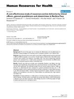
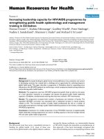
LMP1 immunostaining on tissue sections of NPC samplesFigure 1
LMP1 immunostaining on tissue sections of NPC samples. A. Intense and diffuse LMP1 expression in an NPC biopsy
from a 47 year old patient (score 12, 400X) B. Intense LMP1 expression in a limited area in an NPC biopsy from a 17 year old
patient (score 7, 600X) C. Moderate and diffuse LMP1 expression in an NPC biopsy from a 44 year patient (score 8, 400X) D.
Absence of LMP1 expression in a lung carcinoma biopsy (score 0, 600 X)
A
B
C
D
Virology Journal 2005, 2:39 />Page 4 of 7
(page number not for citation purposes)
relationship with tumor vessels, lymphoid infiltrate or
foci of necrosis.
We attempted to find relationships of the LMP1 score
with various clinical parameters. We found a highly signif-
icant influence of patient age on LMP1 score (p =
0.004)(Table 1). In contrast, we found no relationships
with lymph node or extra-nodal metastases at initial
examination neither with the occurrence of a metastatic
relapse. There was also no relationship with the WHO his-
tological type (Table 1).
Lack of correlations between LMP1 expression and levels
of serum anti-EBV antibodies
As previously reported in other studies, the serum profile
of anti-EBV antibodies was not identical in the two age
groups of NPC patients [13,21]. Serum levels of anti-VCA
and EA IgG were significantly lower in the juvenile form
whereas the anti-EA and VCA IgA were undetectable (<10)
in majority of young patients (Table 2). Because LMP1 is
known to antagonize entry in the lytic cycle in some
experimental models we hypothesized that LMP1 might
block production of EA and VCA in NPC cells and there-
fore prevent an increase of circulating antibodies directed
to these viral proteins [20]. With this in mind we
attempted to find an inverse relationship between the lev-
els of anti-VCA and -EA IgG and IgA on one hand and the
level of LMP1 expression in the tumor tissue on the other
hand. Using univariate analysis, a significant inverse rela-
tionship was found only between the level of LMP1
expression and the level of serum anti-EA IgA (Table 3; p
= 0.012). However, this result was not confirmed by mul-
tivariate analysis including patient age and title of anti-EA
IgA as co-variables. In other words, both LMP1 amounts
in the tumor tissue and titles of serum anti-EA IgA are
strongly influenced by patient age but there is no direct
link between these 2 parameters.
Discussion
Heterogeneity in LMP1 expression in NPC biopsies has
been noticed since early studies based on Western blot-
ting. LMP1 amounts can vary from traces only detectable
after long exposure of the immunoblots to high levels
comparable to those found in EBV-transformed B-lym-
phocytes [22,23]. For this reason, the rate of NPC speci-
mens recorded as LMP1-postive is highly dependent on
the sensitivity of the method used for its detection. For
example when using RT-PCR with one round of PCR
amplification, LMP1 products are detected in only a frac-
tion of NPC biopsies; in contrast, the percentage of posi-
Table 1: Variations of the LMP1 score according to clinical and histo-pathological data
Number of Specimens Mean Score (SD
a
)p
b
Sex
Male 57 7.5 (2.6) p = 0.47
Female 25 7.9 (2.8)
Age
< 30 22 9.0 (2.4) p = 0.004
≥ 30 60 7.1 (2.6)
Histological type
c
SCC 1 7.1 (3.1) p = 0.42
NKC 52 8.0 (2.3)
UC 29 7.6 (2.7)
TNM
d
T2 + T3 40 7.2 (2.6) p = 0.18
T4 42 8.0 (2.7)
N0 21 7.4 (2.9) p = 0.67
N+ 61 7.7 (2.6)
M0 73 7.8 (2.5) p = 0.83
M+ 9 7.6 (2.7)
Metastatic relapse
+ 24 7.5 (2.8) p = 0.94
- 47 7.6 (2.6)
NA
e
11
a
SD: standard deviation
b
Based on the Student t-test
c
Histological type : SCC : squamous cell carcinoma, NKC : non-keratinizing carcinoma,
UC : undifferentiated carcinoma.
d
Clinical staging: primary tumor extension classified T2, T3 or T4 according to AJCC/UICC (1997);
regional lymph node extension classified N0 in the absence of clinical or radiological evidence of lymph node invasion at the initial workup, N+
in the other cases; metastatic status defined as M0 in the absence of clinical or radiological evidence of distant metastasis at the initial workup,
M+ in the other cases (synchronous metastases).
e
NA : not applicable.
Virology Journal 2005, 2:39 />Page 5 of 7
(page number not for citation purposes)
tive samples is often close to 100% when making a second
round of PCR using nested primers [24,25]. The same
applies to investigations by immuno-histo-chemistry
(IHC). According to a recent report by Dietz et al., the per-
centage of LMP1-postive NPCs markedly increases when
using a tyramid-enhancement process instead of conven-
tional tissue staining [26].
In contrast to our study, all previous articles reporting
LMP1 detection in NPCs by conventional IHC have
recorded a fraction of about 40% specimens as LMP1-neg-
ative tumors [4-7]. In most cases, these groups of LMP1-
negative tumors were in fact made of 2 categories : speci-
mens with complete absence of LMP1-positive cells and
specimens with a percentage of stained cells below an
arbitrary threshold of 5 or 10%. In our study, we have
found no biopsy completely devoid of LMP1-positive
cells. This is probably due to the fact that we have used the
S12 antibody which is more sensitive in staining of tissue
sections than the CS1-4 antibody from Dako [27]. Hence,
to our knowledge, CS1-4 was used in all previous investi-
gations of LMP1 expression in NPC biopsies [4-7]. In
addition, we have chosen not to consider any threshold of
minimal LMP1 expression; LMP1 staining has been
scored even when the protein was visible in a very small
fraction of malignant NPC cells.
A large series of studies performed in vitro have produced
an impressive amount of data suggesting that LMP1 can
induce various phenotypic changes consistent with a met-
astatic behavior. For example in transfected cells, LMP1
can induce the production of the c-Met receptor and of the
metallo-protease MMP9 as well as the down-regulation of
the E-cadherin [5,28,29]. In this context, it is surprising to
find no relationship between LMP1 score and the pres-
ence of lymph node or visceral metastases at initial exam-
ination or the risk of metastatic recurrence. In this regard,
our data are in contrast with two previous reports showing
a relationship between LMP1 expression and the fre-
quency of metastases [5,30]. However more recently, Jeon
et al. have found a relationship between LMP1 expression
and MMP9 expression but not between LMP1 and the
presence of metastases [6]. Investigations of LMP1
expression on novel prospective series of NPC patients
using the S12 monoclonal antibody might be useful to
solve these discrepancies.
Table 2: Variations of anti-VCA and -EA Ig titles according to patient ages
Age category (patient number)
EBV-antibody titles < 30 years (n = 21) ≥ 30 years (n = 47) p
a
IgG VCA < 320 7 (33,3%) 4 (8,5%) 0,03
IgG EA < 40 12 (57,1%) 6 (12,8%) 2.7 × 10
-4
IgA VCA < 10 15 (75%) 7 (14,9%) < 10
-5
IgA EA < 10 16 (76,2%) 12 (25,5%) <10
-3
a
Based on the Fisher exact test
Table 3: Variations of the LMP1 score according to serum levels of anti-EBV antibodies
Number of Specimens Mean Score (SD
a
)p
b
IgA VCA title
< 10 22 8.4 (2.9) p = 0.10
≥ 10 45 7.2 (2.7)
ND
c
15
IgA EA title
< 10 28 8.6 (2.8) p = 0.012
≥ 10 40 6.9 (2.5)
ND
c
14
a
SD: standard deviation
b
Based on the Student t-test
c
ND : not determined
Virology Journal 2005, 2:39 />Page 6 of 7
(page number not for citation purposes)
Conclusion
The most striking finding of this study is the observation
of a higher level of LMP1 expression in the juvenile form
of NPC. It provides clear evidence that this clinical form
has specific biological features not only in terms of cellu-
lar gene expression but also in terms of latent viral gene
expression. From previous studies it was known that anti-
VCA and EA IgG and IgA were at a low level in the juvenile
form by contrast with the adult form of NPC [13,19]. This
observation was confirmed by our own data. However, we
found no direct relationship between LMP1 expression
and a low level of anti-VCA and EA IgG and IgA. In futures
studies, it will be important to investigate in both age-
groups of NPCs the status of other EBV-proteins which are
suspected to be expressed in this malignancy with a rather
heterogenous pattern, for example LMP2A, LMP2B and
the BARF1 protein [8,9]. Another issue will be to investi-
gate the anti-LMP1 immune response in the juvenile form
of NPCs for example the status of circulating anti-LMP1
antibodies [11].
Methods
Pathological diagnosis and immunohistochemical staining
of LMP1
All tumor specimens were fixed in Bouin's fixative (75 %
saturated picric acid, 25 % formalin, 5% glacial acetic
acid) and paraffin-embedded for ligth microscopy and
immunohistochemistry. The diagnosis was based on mor-
phological examination after Hematoxylin and Eosin
staining. It was further assisted by immuno-staining of
Leucocyte Common Antigen and cytokeratin in 29 cases,
in order to facilitate the differential diagnosis with a
malignant lymphoma or a sarcoma. Tumor sections from
all 82 NPC patients were stained with the anti-LMP1 S12
monoclonal antibody. In addition, two squamous carci-
nomas of the larynx and one squamous lung carcinoma
were also stained with S12 and used as negative controls.
Five µm sections attached on silanized slides were de-
waxed in xylene, rehydrated in graded ethanol, covered
with 10 mM citrate buffer (pH 6) and heated in a micro-
wave oven for two consecutive 10 minute periods, at 500
W. They were then incubated for 15 to 30 minutes with
the purified primary antibody S12 (0.5 to 1 µg/
ml)[27,31]. Primary antibody binding was visualized
with biotin-labelled secondary antibodies and a streptavi-
din-peroxidase complexe using di-aminobenzidine as a
chromogenic substrate (LSAB system, Dako).
Scoring method
Immuno-staining was scored on the basis of the approxi-
mate percentage of positive tumor cells and the relative
immunostaining intensity. Sections from each biopsies
were read and scored independently by two pathologists
(AK and RJ) who were blinded to the patient clinical data.
Five consecutive microscope fields were analyzed. The dif-
ferences in scores between the two observers were
resolved at a conference microscopy (AK, RJ and TB). The
following grading system was adopted to score the
number of positive tumor cells: 0, none seen in the sec-
tion; 1, presence of positive cells even rare but not exceed-
ing 25%; 2, 26 to 50% positive cells; 3, 51 to 75%; and 4,
76 to 100%. Immuno-staining intensity was rated as fol-
lows: 0, none; 1, weak; 2, moderate; and 3, intense. When
the staining intensity was heterogeneous, each compo-
nent of the tumor were scored independently and the
results were summed. For example, when a specimen con-
tained 50% of the tumor cells with moderate intensity (2
× 2 = 4), 25% of tumor cells with intense immunostaining
(1 × 3 = 3), and 25% of cells with weak intensity (1 × 1 =
1), the score was 4 +3 +1 = 8. The maximal possible score
was twelve.
Serological analysis
Serum samples were collected from 68 out of the 82
patients at initial diagnosis. IgG and IgA antibodies to EBV
EA and VCA were titrated by indirect immunofluores-
cence on Raji and P3HR1 cells, respectively [13,32].
Statistical analysis
LMP1 immunostaining scoring results were expressed as
means (standard deviation, SD) and compared using the
Student t-test. Variations of anti-EBV antibody titles
according to patient age were assessed using the Fisher
exact test. To assess relationships between LMP1 score, age
and anti-EBV antibody titles, multivariate analysis was
carried out using a linear multiple regression (Sas soft-
ware, version 8, SAS Institute Inc, Cary, NC, USA). All tests
were bilateral with a 5% level.
Competing interests
The author(s) declare that they have no competing
interests.
Authors' contributions
AK, RJ and TB made pathological diagnosis, immunohis-
tochemistry and scoring of immunostaining, HK carried
out assessment of serum EBV-antibodies, PB and SR par-
ticipated in the design and coordination of the study and
helped to draft the manuscript, MR performed the statisti-
cal analysis, JD and MF gathered clinical data, JM purified
the S12 antibody and set up conditions for its use in
immunohistochemistry. All authors read and approved
the final manuscript.
Acknowledgements
This study was supported by a cooperative grant from the French CNRS
and Tunisian DGRST (n° 17963) and by a grant from the "Comité du Cher"
of the French "Ligue Nationale contre le Cancer".
Virology Journal 2005, 2:39 />Page 7 of 7
(page number not for citation purposes)
References
1. Busson P, Keryer C, Ooka T, Corbex M: EBV-associated nasopha-
ryngeal carcinomas: from epidemiology to virus-targeting
strategies. Trends Microbiol 2004, 12:356-360.
2. Nicholls JM, Agathanggelou A, Fung K, Zeng X, Niedobitek G: The
association of squamous cell carcinomas of the nasopharynx
with Epstein-Barr virus shows geographical variation remi-
niscent of Burkitt's lymphoma. J Pathol 1997, 183:164-168.
3. Raab-Traub N: Epstein-Barr virus in the pathogenesis of NPC.
Semin Cancer Biol 2002, 12:431-441.
4. Niedobitek G, Fahraeus R, Herbst H, Latza U, Ferszt A, Klein G, Stein
H: The Epstein-Barr virus encoded membrane protein
(LMP) induces phenotypic changes in epithelial cells. Virchows
Arch B Cell Pathol Incl Mol Pathol 1992, 62:55-59.
5. Horikawa T, Sheen TS, Takeshita H, Sato H, Furukawa M, Yoshizaki
T: Induction of c-Met proto-oncogene by Epstein-Barr virus
latent membrane protein-1 and the correlation with cervical
lymph node metastasis of nasopharyngeal carcinoma. Am J
Pathol 2001, 159:27-33.
6. Jeon YK, Lee BY, Kim JE, Lee SS, Kim CW: Molecular characteri-
zation of Epstein-Barr virus and oncoprotein expression in
nasopharyngeal carcinoma in Korea. Head Neck 2004,
26:573-583.
7. Shao JY, Ernberg I, Biberfeld P, Heiden T, Zeng YX, Hu LF: Epstein-
Barr virus LMP1 status in relation to apoptosis, p53 expres-
sion and leucocyte infiltration in nasopharyngeal carcinoma.
Anticancer Res 2004, 24:2309-2318.
8. Heussinger N, Buttner M, Ott G, Brachtel E, Pilch BZ, Kremmer E,
Niedobitek G: Expression of the Epstein-Barr virus (EBV)-
encoded latent membrane protein 2A (LMP2A) in EBV-
associated nasopharyngeal carcinoma. J Pathol 2004,
203:696-699.
9. Seto E, Yang L, Middeldorp J, Sheen TS, Chen JY, Fukayama M, Eizuru
Y, Ooka T, Takada K: Epstein-Barr virus (EBV)-encoded
BARF1 gene is expressed in nasopharyngeal carcinoma and
EBV-associated gastric carcinoma tissues in the absence of
lytic gene expression. J Med Virol 2005, 76:82-88.
10. Meij P, Vervoort MB, Aarbiou J, van Dissel P, Brink A, Bloemena E,
Meijer CJ, Middeldorp JM: Restricted low-level human antibody
responses against Epstein-Barr virus (EBV)-encoded latent
membrane protein 1 in a subgroup of patients with EBV-
associated diseases. J Infect Dis 1999, 179:1108-1115.
11. Xu J, Ahmad A, D'Addario M, Knafo L, Jones JF, Prasad U, Dolcetti R,
Vaccher E, Menezes J: Analysis and significance of anti-latent
membrane protein-1 antibodies in the sera of patients with
EBV-associated diseases. J Immunol 2000, 164:2815-2822.
12. Fachiroh J, Schouten T, Hariwiyanto B, Paramita DK, Harijadi A, Har-
yana SM, Ng MH, Middeldorp JM: Molecular diversity of Epstein-
Barr virus IgG and IgA antibody responses in nasopharyngeal
carcinoma: a comparison of Indonesian, Chinese, and Euro-
pean subjects. J Infect Dis 2004, 190:53-62.
13. Karray H, Ayadi W, Fki L, Hammami A, Daoud J, Drira MM, Frikha M,
Jlidi R, Middeldorp JM: Comparison of three different serologi-
cal techniques for primary diagnosis and monitoring of
nasopharyngeal carcinoma in two age groups from Tunisia. J
Med Virol 2005, 75:593-602.
14. Zhang JX, Chen HL, Zong YS, Chan KH, Nicholls J, Middeldorp JM,
Sham JS, Griffin BE, Ng MH: Epstein-Barr virus expression within
keratinizing nasopharyngeal carcinoma. J Med Virol 1998,
55:227-233.
15. Daoud J, Toumi N, Bouaziz M, Ghorbel A, Jlidi R, Drira MM, Frikha
M: Nasopharyngeal carcinoma in childhood and adolescence:
analysis of a series of 32 patients treated with combined
chemotherapy and radiotherapy. Eur J Cancer 2003,
39:2349-2354.
16. Khabir A, Ghorbel A, Daoud J, Frikha M, Drira MM, Laplanche A, Bus-
son P, Jlidi R: Similar BCL-X but different BCL-2 levels in the
two age groups of north African nasopharyngeal carcinomas.
Cancer Detect Prev 2003, 27:250-255.
17. Khabir A, Sellami A, Sakka M, Ghorbel AM, Daoud J, Frikha M, Drira
MM, Busson P, Jlidi R: Contrasted frequencies of p53 accumula-
tion in the two age groups of North African nasopharyngeal
carcinomas. Clin Cancer Res 2000, 6:3932-3936.
18. Bar-Sela G, Kuten A, Ben-Eliezer S, Gov-Ari E, Ben-Izhak O: Expres-
sion of HER2 and C-KIT in nasopharyngeal carcinoma: impli-
cations for a new therapeutic approach. Mod Pathol 2003,
16:1035-1040.
19. Sbih-Lammali F, Clausse B, Ardila-Osorio H, Guerry R, Talbot M,
Havouis S, Ferradini L, Bosq J, Tursz T, Busson P: Control of apop-
tosis in Epstein Barr virus-positive nasopharyngeal carci-
noma cells: opposite effects of CD95 and CD40 stimulation.
Cancer Res 1999, 59:924-930.
20. Prince S, Keating S, Fielding C, Brennan P, Floettmann E, Rowe M:
Latent membrane protein 1 inhibits Epstein-Barr virus lytic
cycle induction and progress via different mechanisms. J Virol
2003, 77:5000-5007.
21. Sbih-Lammali F, Djennaoui D, Belaoui H, Bouguermouh A, Decaussin
G, Ooka T: Transcriptional expression of Epstein-Barr virus
genes and proto-oncogenes in north African nasopharyngeal
carcinoma. J Med Virol 1996, 49:7-14.
22. Young LS, Dawson CW, Clark D, Rupani H, Busson P, Tursz T, John-
son A, Rickinson AB: Epstein-Barr virus gene expression in
nasopharyngeal carcinoma. J Gen Virol 1988, 69 ( Pt
5):1051-1065.
23. Fahraeus R, Fu HL, Ernberg I, Finke J, Rowe M, Klein G, Falk K, Nils-
son E, Yadav M, Busson P, et al.: Expression of Epstein-Barr virus-
encoded proteins in nasopharyngeal carcinoma. Int J Cancer
1988, 42:329-338.
24. Chen F, Hu LF, Ernberg I, Klein G, Winberg G: Coupled transcrip-
tion of Epstein-Barr virus latent membrane protein (LMP)-1
and LMP-2B genes in nasopharyngeal carcinomas. J Gen Virol
1995, 76 ( Pt 1):131-138.
25. Brooks L, Yao QY, Rickinson AB, Young LS: Epstein-Barr virus
latent gene transcription in nasopharyngeal carcinoma cells:
coexpression of EBNA1, LMP1, and LMP2 transcripts. J Virol
1992, 66:2689-2697.
26. Dietz A, Logothetis CA, Helbig M, Flechtenmacher C, Rudat V, Doll-
ner R, Wallner F, Bosch FX: Prognostic Impact of EBV-Related
LMP-1, Histologic Type, and Environmental Factors in
Nasopharyngeal Carcinoma in a German Population. Onkolo-
gie 2004, 27:345-350.
27. Jiwa NM, Oudejans JJ, Dukers DF, Vos W, Horstman A, van der Valk
P, Middledorp JM, Walboomers JM, Meijer CJ: Immunohistochem-
ical demonstration of different latent membrane protein-1
epitopes of Epstein-Barr virus in lymphoproliferative
diseases. J Clin Pathol 1995, 48:438-442.
28. Tsai CN, Tsai CL, Tse KP, Chang HY, Chang YS: The Epstein-Barr
virus oncogene product, latent membrane protein 1, induces
the downregulation of E-cadherin gene expression via activa-
tion of DNA methyltransferases. Proc Natl Acad Sci U S A 2002,
99:10084-10089.
29. Takeshita H, Yoshizaki T, Miller WE, Sato H, Furukawa M, Pagano JS,
Raab-Traub N: Matrix metalloproteinase 9 expression is
induced by Epstein-Barr virus latent membrane protein 1 C-
terminal activation regions 1 and 2. J Virol 1999, 73:5548-5555.
30. Hu LF, Chen F, Zhen QF, Zhang YW, Luo Y, Zheng X, Winberg G,
Ernberg I, Klein G: Differences in the growth pattern and clini-
cal course of EBV-LMP1 expressing and non-expressing
nasopharyngeal carcinomas. Eur J Cancer 1995, 31A:658-660.
31. Mann KP, Staunton D, Thorley-Lawson DA: Epstein-Barr virus-
encoded protein found in plasma membranes of trans-
formed cells. J Virol 1985, 55:710-720.
32. Henle W, Henle G, Zajac BA, Pearson G, Waubke R, Scriba M: Dif-
ferential reactivity of human serums with early antigens
induced by Epstein-Barr virus. Science 1970, 169:188-190.