Báo cáo sinh học: " Actively replicating West Nile virus is resistant to cytoplasmic delivery of siRNA" ppt
Bạn đang xem bản rút gọn của tài liệu. Xem và tải ngay bản đầy đủ của tài liệu tại đây (1.1 MB, 13 trang )
BioMed Central
Page 1 of 13
(page number not for citation purposes)
Virology Journal
Open Access
Research
Actively replicating West Nile virus is resistant to cytoplasmic
delivery of siRNA
Brian J Geiss
1
, Theodore C Pierson
4
and Michael S Diamond*
1,2,3
Address:
1
Departments of Medicine, Washington University School of Medicine, 660 South Euclid Avenue, Box 8051, St. Louis, MO 63110, USA,
2
Molecular Microbiology, Washington University School of Medicine, 660 South Euclid Avenue, Box 8051, St. Louis, MO 63110, USA,
3
Pathology
& Immunology, Washington University School of Medicine, 660 South Euclid Avenue, Box 8051, St. Louis, MO 63110, USA and
4
Department of
Microbiology, University of Pennsylvania, Philadelphia, PA, 19104, USA
Email: Brian J Geiss - ; Theodore C Pierson - ;
Michael S Diamond* -
* Corresponding author
Abstract
Background: West Nile virus is an emerging human pathogen for which specific antiviral therapy
has not been developed. Recent studies have suggested that RNA interference (RNAi) has
therapeutic potential as a sequence specific inhibitor of viral infection. Here, we examine the ability
of exogenous small interfering RNAs (siRNAs) to block the replication of West Nile virus in human
cells.
Results: WNV replication and infection was greatly reduced when siRNA were introduced by
cytoplasmic-targeted transfection prior to but not after the establishment of viral replication. WNV
appeared to evade rather than actively block the RNAi machinery, as sequence-specific reduction
in protein expression of a heterologous transgene was still observed in WNV-infected cells.
However, sequence-specific decreases in WNV RNA were observed in cells undergoing active viral
replication when siRNA was transfected by an alternate method, electroporation.
Conclusion: Our results suggest that actively replicating WNV RNA may not be exposed to the
cytoplasmic RNAi machinery. Thus, conventional lipid-based siRNA delivery systems may not be
adequate for therapy against enveloped RNA viruses that replicate in specialized membrane
compartments.
Background
West Nile virus (WNV) is a significant human and veteri-
nary mosquito-borne pathogen that has rapidly spread
across North America. Humans develop a febrile illness
and a small subset progress to meningitis or encephalitis
syndromes [1]. Currently, no specific therapy or vaccine
has been approved for treatment or prophylaxis of WNV
infection in humans.
WNV is an enveloped virus with an 11-kilobase positive
strand RNA genome. It is translated directly from the
genomic RNA as a single polyprotein and cleaved by cel-
lular and viral proteases into ten mature proteins, three
structural (C, M, and E) and seven non-structural (NS1,
NS2A, NS2B, NS3, NS4A, NS4B, and NS5) proteins [2,3].
Virus entry occurs by endocytosis after the E protein inter-
acts with cellular receptor(s). Genomic viral RNA traffics
to the endoplasmic reticulum (ER), where WNV protein
Published: 28 June 2005
Virology Journal 2005, 2:53 doi:10.1186/1743-422X-2-53
Received: 28 May 2005
Accepted: 28 June 2005
This article is available from: />© 2005 Geiss et al; licensee BioMed Central Ltd.
This is an Open Access article distributed under the terms of the Creative Commons Attribution License ( />),
which permits unrestricted use, distribution, and reproduction in any medium, provided the original work is properly cited.
Virology Journal 2005, 2:53 />Page 2 of 13
(page number not for citation purposes)
translation and RNA replication occur [4]. The positive
strand genomic WNV RNA that associates with the ER is
competent for translation and transcription of negative
strand RNA. WNV and related flaviviruses induce ER
membrane proliferation and reorganization, and replicat-
ing viral RNA has been observed at these membranous
structures [5-7]. Disruption of WNV protein translation
and/or RNA replication blocks the viral lifecycle and
aborts infection.
RNA interference (RNAi) is a cellular process that specifi-
cally degrades RNA within the cytoplasm of cells in a
sequence-specific manner [8]. RNAi occurs in plants [9],
nematodes [10], parasites [11,12], insects [13], and mam-
malian cells [14,15] and is believed to function as a regu-
lator of cellular gene expression and possibly as an innate
defense against RNA viruses [16]. RNAi uses double
stranded RNA (dsRNA) to target and degrade sequence-
specific single-stranded RNA. The cytoplasmic ribonucle-
ase DICER recognizes and cleaves long dsRNA molecules
into 21 to 30 base pair small interfering RNA (siRNA)
molecules; these associate with the RNA Induced Silenc-
ing Complex (RISC) to target and degrade complementary
single-stranded RNA molecules [8].
RNAi has been used as a method to transiently disrupt var-
ious gene products to study their function [14,15,17-20].
Many mammalian viruses appear susceptible to treatment
with exogenous siRNA. Cells that express virus-specific
siRNA are resistant to infection by WNV [21], poliovirus
[22,23], influenza A [21,24], HIV [25] and hepatitis C
[26,27]in vitro. Administration of siRNAs in vivo has mod-
estly reduced hepatitis B antigen production [28,29] and
influenza A virus infection [30,31]. The sequence specific
activity of siRNA against viruses has led to great interest in
its potential as a new class of antiviral therapy. Nonethe-
less, there may be limitations with this approach as in vivo
RNAi has not been demonstrated as effective as post-expo-
sure therapy.
Previously, we demonstrated that transgenic expression of
a sequence-specific siRNA prior to infection could effi-
ciently inhibit WNV replication [21]. However, for a
WNV-specific siRNA to be effective as a post-exposure
therapeutic, it would need to inhibit infection in cells that
are actively replicating WNV RNA. In this study, we evalu-
ated the efficacy of siRNA against WNV that has already
initiated active replication. Although cytoplasm-directed
transfection of cells with siRNA prior to infection effi-
ciently blocked WNV infection, administration after infec-
tion had little efficacy. Unlike plant viruses that encode
active suppressors of RNA interference [32-34], WNV did
not appear to actively inhibit the RNAi response, but
rather avoided degradation by replicating in a manner
that was inaccessible to the RNAi machinery.
Results
In vitro generated siRNA inhibits WNV infection in cells
We have previously demonstrated that plasmid expressed
hairpin siRNA efficiently inhibited infection of WNV in
mouse and human cell lines [21]. Because a therapeutic
application of exogenously delivered rather than plasmid-
expressed siRNA may be more clinically relevant, we
assessed the inhibitory activity of in vitro transcribed hair-
pin siRNA against WNV infection. A 21-nucleotide region
of the WNV capsid gene (nucleotides 312–332; Cap) was
initially targeted, as this region is conserved among all
WNV strains and lacks homology to known cellular genes.
To demonstrate the specificity of Cap siRNA, a hairpin
siRNA that targets the Influenza A M2 gene (nucleotides
18–38, M2) [21] and a mutated version of Cap siRNA
(Cap Mut) that had 4 changes were also designed (Table
1). Our in vitro transcription strategy employed partially
duplexed oligonucleotides containing a double stranded
T7 promoter sequence (Fig 1A).
Human Huh7.5 cells were used because they were effi-
ciently transfected with siRNA and infected with WNV.
Huh-7.5 were transfected with Cap or Cap-Mut siRNA,
infected with a New York strain of WNV at 18 hours after
transfection, and analyzed 48 hours post-infection for lev-
els of viral RNA by RT-PCR (Fig 1B). Pretreatment of
Huh7.5 cells with Cap siRNA resulted in approximately 1
log reduction of WNV RNA, whereas pretreatment of
Huh7.5 cells with Cap-Mut siRNA showed no significant
reduction of WNV RNA. To confirm that RNAi also
decreased WNV antigen production, siRNA-transfected
Huh7.5 cells were examined for WNV envelope protein
expression at 48 hours post infection. Approximately 70%
of mock or TKO treated Huh-7.5 cells were positive for
WNV antigen, levels comparable to that observed in cells
transfected with either M2 (57% positive) or Cap-Mut
(59% positive) siRNAs (Fig 1C). In contrast, less than 3%
of Cap siRNA transfected cells stained positive for WNV E
antigen. Thus, in vitro generated sequence-specific hairpin
siRNA efficiently and specifically blocked WNV RNA and
antigen production in mammalian cells.
To demonstrate that siRNAs targeting different regions of
the WNV genome could inhibit infection, multiple siR-
NAs were designed spanning the nonstructural genes of
WNV (Fig 1D). Two siRNAs (5497 and 6349) targeted to
the nonstructural proteins reduced WNV envelope expres-
sion by at least 4-fold. Despite using an siRNA prediction
algorithm, many of the siRNAs demonstrated little ability
to inhibit envelope protein expression, possibly due to
secondary structure in the WNV genomic RNA. Interest-
ingly, treatment with combinations of siRNA did not
show appreciably greater inhibition than treatment with
either siRNA alone (data not shown).
Virology Journal 2005, 2:53 />Page 3 of 13
(page number not for citation purposes)
Timing of siRNA treatment affects effectiveness against
WNV
siRNA therapy in a clinical setting likely would require
treatment after WNV infection has occurred. Because of
this, we assessed the ability of siRNA to block WNV RNA
before and after infection (Fig 2A). Huh7.5 cells were
transfected with siRNAs 18 hours before infection or at 10
hours after infection. Cells were not transfected at very
early times after infection because the TKO transfection
reagent interfered with the ability of WNV virus to infect
cells in a time-dependent manner (B. Geiss and M. Dia-
mond, unpublished observation). By ~10 hours post-
infection virus had entered cells and were no longer
affected by the TKO reagent. Total RNA was harvested 48
hours post-infection and analyzed for genomic WNV RNA
content. As expected, pretreatment of cells with Cap
siRNA, but not Cap Mut siRNA, resulted in a 10-fold
reduction in WNV RNA levels. Strikingly, addition of Cap
siRNA 10 hours after infection resulted in no reduction of
WNV RNA. The lack of inhibitory effect of RNAi at late
times after infection was not due to the emergence of
resistant mutants: sequence analysis of multiple viral iso-
lates at 48 hours post-infection from cells that had been
transfected with Cap or 6349 siRNA demonstrated no
mutations in the targeted viral sequences (data not
shown). Thus, WNV, in contrast to poliovirus [35], did
not appear to mutate to evade siRNA-triggered
degradation.
The rate of viral replication does not affect RNAi
resistance
The establishment of siRNA resistance could in part, be
due to the ability of a rapidly replicating WNV to saturate
the RNAi degradation machinery. To test if the replication
rate affected siRNA resistance, we used an attenuated line-
age II WNV that contains a GFP marker gene inserted into
the 3' UTR and replicates more slowly than wild-type lin-
eage I or II WNV [36]. Because the nucleotide sequence of
the lineage II WNV was different than the lineage I WNV,
a new sequence-specific siRNA was designed (6337) that
targeted the analogous region on NS3 as siRNA 6349.
Huh7.5 cells were transfected with Cap-Mut, 6349, or
6337 at 18 hours prior to or 10 hours after infection with
the attenuated lineage II WNV, and viral RNA content was
determined at 48 hours post-infection (Fig 2B). As
expected, pretreatment with either Cap Mut or the lineage
I-specific 6349 siRNA did not inhibit replication, whereas
pretreatment with 6337 siRNA strongly blocked replica-
tion (~ 60-fold). In contrast, treatment with any of the
three siRNAs 10 hours after infection demonstrated no
inhibitory effect. Thus, a WNV strain with a lower replica-
tion rate similarly resisted the inhibitory effects of RNAi
soon after replication was established.
The mode of introduction of siRNA affects the ability to
establish RNAi
Based on the timing experiments, the establishment of
resistance to RNAi correlated with the onset of WNV RNA
Table 1: Small interfering RNA
Name Virus Start Nucleotide Target Sequence
Cap WNV Lineage I 312 gaacaaacaaacagcgatgaa
Cap-Mut WNV Lineage I 312 gaagaaagaaagaccgatgaa
M2 Influenza A M2 18 ggtcgaaacgcctatcagaaa
3110 WNV Lineage I 3110 gggcagttctgggtgaagt
3317 WNV Lineage I 3317 ctacggtcaccctgagtga
4119 WNV Lineage I 4119 gaggagcaagtctgctatgc
4823 WNV Lineage I 4823 gtgtcaaggaggatcgact
5039 WNV Lineage I 5039 gacggtgatgtgattgggct
5497 WNV Lineage I 5497 gcagcaagaggttacattt
6337 WNV Lineage II 6337 gttgaagtcatcacgaagt
6349 WNV Lineage I 6349 gtggaagtcatcacgaagc
6915 WNV Lineage I 6915 caacgagatgggttggcta
7353 WNV Lineage I 7353 gaagaacgctgtagtggat
7693 WNV Lineage I 7693 ggacgcaccttgggagaggt
8892 WNV Lineage I 8892 ggtcaacagcaatgcagct
8898 WNV Lineage I 8898 cagcaatgcagctttgggt
9095 WNV Lineage I 9095 gaagcagagccatttggtt
9607 WNV Lineage I 9607 gggaaaggacccaaagtca
10355 WNV Lineage I 10355 gagagatatgaagacacaac
siRNA were generated against 19–21 nucleotide sequences corresponding to the target region of different parts of the New York 1999 WNV
genome. Sequences were chosen using the SciTools RNAi design program and compared against the GenBank database to exclude sequences that
may affect cellular genes.
Virology Journal 2005, 2:53 />Page 4 of 13
(page number not for citation purposes)
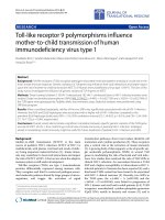
WNV is susceptible to siRNA pretreatmentFigure 1
WNV is susceptible to siRNA pretreatment. A. Scheme for generation of small hairpin siRNAs. B. Cap siRNA specifi-
cally inhibits WNV RNA accumulation. Huh7.5 cells were mock transfected or transfected with Cap siRNA or Cap Mut
siRNA. Eighteen hours later cells were infected with WNV at MOI 0.1. Forty-eight hours later cells were collected and total
RNA was recovered. WNV RNA was measured by quantitative real time RT-PCR. The results are an average of three inde-
pendent experiments and the error bars indicate standard error of the mean. C. Capsid siRNA specifically inhibits WNV E
protein expression. Huh7.5 cells were mock transfected or transfected with M2 siRNA, Cap siRNA, or Cap Mut siRNA as
described above. Forty-eight hours after infection, cells were collected and processed for flow cytometry using anti-WNV
envelope protein antibody E1. The results are one representative example of three independent experiments. D. Inhibitory
activity of different WNV-specific siRNA. Huh7.5 cells transfected with the indicated siRNA, infected with WNV, and then ana-
lyzed for viral antigen as described in Materials and Methods. The fold inhibition was calculated after dividing the percentage of
antigen positive cells from mock-transfected cells by the percentage of antigen positive cells from siRNA transfected cells. The
results are one representative example of two independent experiments.
Virology Journal 2005, 2:53 />Page 5 of 13
(page number not for citation purposes)
replication [37]. Shortly after infection, flaviviruses
induce vesicular membrane proliferation that becomes
the site of viral RNA replication [6,7,38]. Because the
lipid-based transfection reagent targets nucleic acids to the
cytoplasm (Mirus Corp, personal communication), the
presence of additional membranes between the viral RNA
and the cytoplasm could prevent the siRNA from reaching
the actively replicating WNV RNA complex. Electropora-
tion, in contrast, transiently opens pores in cellular mem-
branes [39] allowing nucleic acids and cytoplasmic
components to cross membranous structures such as the
nucleus, endoplasmic reticulum, and potentially the
membranous vesicles induced by WNV.
To determine if the mode of delivery of siRNA affected
RNAi resistance, we tested whether siRNA could inhibit
replication of a persistently replicating subgenomic
lineage I WNV replicon; this cell line (Huh7.5-Rep)
expresses the non-structural but lacks the structural pro-
teins of WNV (Fig 3A). Huh7.5-Rep cells were transfected
with 6337 and 6349 siRNAs using a lipid-based reagent or
by electroporation, cultured, and assayed for reduction of
NS3 antigen expression 72 hours later (Fig 3B). Replicon-
expressing cells that were transfected with siRNA using the
lipid-based reagent showed no significant reduction in
viral protein or RNA. In contrast, using electroporation,
NS3 protein levels were reduced by approximately 10-fold
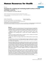
WNV becomes resistant to RNAi after infectionFigure 2
WNV becomes resistant to RNAi after infection. A. Huh7.5 cells were mock transfected or transfected with Cap or
Cap Mut siRNA at the indicated times before or after WNV infection. Forty-eight hours after infection cells were harvested
and WNV RNA levels were determined by quantitative real-time RT-PCR. The results are an average of three independent
experiments and error bars indicate standard error of the mean. B. Induction of RNAi resistance by an attenuated lineage II
WNV. Inset. Genomic structure of the attenuated lineage II WNV, which includes an IRES-controlled GFP insertion in the 3'
UTR. 6337 denotes the target region of the lineage II specific siRNA. Attenuated WNV becomes resistant to siRNA after
infection is established. Huh7.5 cells were transfected with Cap Mut, 6349, or 6337 siRNA at the indicated times prior to or
after infection. Forty-eight hours after infection total RNA was collected and viral RNA was assessed as in Fig 1.
Virology Journal 2005, 2:53 />Page 6 of 13
(page number not for citation purposes)
by 6349 siRNA, but not by the lineage II-specific 6337
siRNA. The reduction of NS3 antigen levels correlated
with ~7.5-fold decreases in replicon RNA levels in the
presence of 6349 (Fig 3C).
Cellular localization of siRNA
The preceding data suggested that lipid-mediated transfec-
tion delivered siRNA into the cytoplasm, whereas electro-
poration at least transiently exposed replicating viral RNA
to the RNAi response. However, it remained unclear
whether the amount of siRNA delivered or the localiza-
tion determined its ability to inhibit WNV RNA. To assess
the relative amount and distribution of siRNA after trans-
fection or electroporation, Huh7.5 cells were transfected
or electroporated with Cy5-labeled Cap siRNA and
analyzed 18 hours later for Cy5 expression by flow
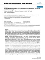
Mode of siRNA introduction influences WNV RNAi susceptibilityFigure 3
Mode of siRNA introduction influences WNV RNAi susceptibility. A. Huh7.5-Rep cells. (Top) Diagram of the genetic
structure of the pWN5'Pur replicon. (Bottom) Flow cytometric analysis of Huh7.5 cells that express the pWN5'Pur replicon.
Only non-structural proteins (e.g., NS1 and NS3 but not E) are expressed. B. siRNA treatment of Huh7.5-Rep cells. Huh7.5-
Rep cells were mock-transfected, transfected with TKO reagent complexed with 6337 or 6349 siRNAs, or electroporated
with 6337 or 6349 siRNAs. Three days later, cells were processed for viral NS3 protein expression by flow cytometry using
anti-NS3 antibody (right). Fold inhibition of NS3 antigen production was determined using the formula (% NS3 positive mock
electroporated / % NS3 positive siRNA electroporated). C. RNA analysis of Huh7.5-Rep cells electroporated with siRNA.
Huh7.5-Rep cells were electroporated with 6349 or 7353 siRNA as described in Materials and Methods. Three days later, total
cellular RNA was collected and viral RNA was assessed. Fold inhibition was determined by dividing the amount of viral RNA in
mock electroporated samples to the amount of viral RNA in siRNA electroporated samples. The results are an average of
three independent experiments and error bars indicate standard error of the mean.
Virology Journal 2005, 2:53 />Page 7 of 13
(page number not for citation purposes)
cytometry and localization by fluorescence microscopy
(Fig 4). Although all transfected cells were positive for Cy5
fluorescence, the signal was significantly higher in lipid-
transfected cells than in electroporated cells (Fig 4A, geo-
metric mean fluorescence intensity 4370 compared to
766). Microscopic analysis of lipid-transfected cells
showed Cy5 signal primarily in the cytoplasm, with exclu-
sion of Cy5 from the nucleus (Fig 4B, middle panels). In
contrast, Cy5 signal was observed diffusely throughout
the cell after electroporation (Fig 4B, right panels), sug-
gesting that electroporation effectively delivered siRNA
across intracellular membranes. Taken together, our data
suggests that the cellular localization of siRNA appears
more important than the absolute amount of siRNA deliv-
ered into the cell in determining its effectiveness against
actively replicating WNV RNA.
Localization of siRNAFigure 4
Localization of siRNA. Huh7.5 cells were transfected with Cy5 labeled Cap siRNA using the lipid TKO reagent (middle pan-
els) or by electroporation (right panels). Eighteen hours later, cells were collected and analyzed for Cy5 fluorescence by (A)
flow cytometry or (B) fluorescence microscopy as described in Materials and Methods. Arrows denote the position of the
nucleus. The gain in the Cy5 micrograph of electroporated cells was increased to compensate for lower levels of intracellular
siRNA as compared to lipid-transfected samples.
Virology Journal 2005, 2:53 />Page 8 of 13
(page number not for citation purposes)
RNAi against other mRNAs is intact in WNV-infected cells
Although the mode and location of siRNA introduction
could affect the sensitivity of actively replicating WNV to
RNAi, we speculated that WNV could additionally evade
RNAi by directly inhibiting one or more steps of the RNAi
pathway. Targeted inhibition of the RNAi pathway has
been observed in plant viruses, and has been recently
reported with several mammalian viruses including
LaCrosse virus, adenovirus, and influenza A virus [32-
34,40-43]. To determine if WNV replication directly atten-
uated the RNAi response, we tested the efficiency of
siRNA-mediated inhibition of Influenza A virus M2 pro-
tein expression in cells that actively replicated WNV
genomes (Fig 5). Mock-infected Huh7.5 cells, WNV-
infected Huh7.5 cells (10 hours post-infection), and
Huh7.5-Rep cells were co-transfected with an M2 expres-
sion plasmid and either Cap or M2 siRNA. Twenty-four
hours later, cells were analyzed for M2 expression by flow
cytometry. As expected, transfection of Cap siRNA did not
significantly affect the expression of M2 in Huh7.5,
Huh7.5-Rep, or WNV infected Huh7.5 cells. However,
transfection of M2 siRNA effectively reduced the expres-
sion of the M2 protein in all cell types, including those
that actively replicated WNV RNA. Thus, WNV replication
per se did not affect the establishment of RNAi of a heter-
ologous gene.
Viral translation is necessary for WNV RNAi resistance
The experiments above suggest that resistance to RNAi by
WNV occurs in the setting of ongoing viral replication.
During the de novo infection of a cell, translation of the
input positive viral RNA strand is required before replica-
tion occurs [2]. To more finely dissect the kinetics of RNAi
resistance with respect to the initiation of RNA replica-
tion, we added puromycin, a reversible inhibitor of pro-
tein chain elongation. Because puromycin inhibits
cellular and viral protein translation, we first assessed how
it independently affected the establishment of RNAi. Cap
or Cap Mut siRNA were transfected into cells in the pres-
ence of puromycin. Four hours later, cells were infected
with WNV for an additional four hours, and then free
virus and puromycin were removed by serial washing of
infected monolayers. Forty-eight hours after initial infec-
tion, cells were analyzed for WNV envelope protein
expression (Fig 6A). When translation of full-length viral
protein was reduced by puromycin, Cap siRNA more
effectively inhibited WNV antigen production (90-fold
versus 10-fold reduction), suggesting that cellular transla-
tion was not necessary for priming the RNAi response and
that delay of onset of WNV translation enhanced the effi-
ciency of siRNA-mediated inhibition. Importantly, the
inhibition was sequence-specific as no significant
decrease in viral antigen expression was observed with the
Cap Mut siRNA.
To define the kinetics of RNAi resistance around the time
of initial viral replication, a puromycin time course was
performed (Fig 6B). Cells were infected with WNV at MOI
0.01 and puromycin was added to Huh7.5 cells at differ-
ent times (-1, 0, 1, 2, 3, 4, 5, 6, 7, or 8 hours) before or
after infection. At 9 hours after WNV infection, all cells
were transfected with Cap or Cap Mut siRNA and puromy-
cin was removed from the medium. Cells treated with
puromycin from -1 to 1 hours post-infection were greatly
protected against WNV infection. However, significant
attenuation of RNAi emerged when puromycin was added
just four hours after infection. These results suggest that
the induction of RNAi resistance by WNV depends on
translation of viral polyprotein and occurs as early as 4
hours after infection.
Discussion
In this paper we examine the ability of exogenous siRNA
to inhibit the replication of WNV. Developing strategies
for specific inhibition of WNV is an important goal as no
current therapy exists for infected individuals. siRNA has
been proposed as a potential therapy against several
RNAi is active in WNV infected cellsFigure 5
RNAi is active in WNV infected cells. RNAi of influenza
M2 gene in cells that replicate WNV RNA. Huh7.5, Huh7.5-
Rep, and WNV infected Huh7.5 cells (8 hours post infection)
were transfected with pCM2 and Cap or M2 siRNA as
described in Materials and Methods. 24 hours later cells were
processed by flow cytometry for M2 expression using anti-
body 14C2. The percentage of M2 inhibition was calculated
according to the following formula: (1 – (% M2 expression of
siRNA-transfected cells / % M2 expression in cells trans-
fected with transfection vehicle only) × 100). The results are
an average of three independent experiments and error bars
indicate standard error of the mean.
Virology Journal 2005, 2:53 />Page 9 of 13
(page number not for citation purposes)
viruses, and we have previously demonstrated that plas-
mid based RNAi is effective against WNV in vitro [21].
Here, we tested the ability of exogenously generated
siRNA to inhibit WNV infection, as this reagent may be
more practical for clinical use because there is little possi-
bility of adverse integration into a patient's genome.
Using a conventional lipid-based delivery system that tar-
gets siRNA to the cytoplasm, we confirmed that pretreat-
ment of cells prevented infection. However, resistance to
RNAi was observed when siRNA was delivered after viral
translation and replication had commenced. In contrast,
when siRNA was delivered by electroporation, a
technique that allows macromolecules to pass across
intracellular membranes, it reduced viral replication in a
sequence-specific manner even if active replication was
already underway. The data in this manuscript provide a
first description of flavivirus resistance to RNAi during
infection, and suggests a possible mechanism: WNV
resists exogenously-introduced siRNA by replicating in a
compartment that is sequestered behind cellular
membranes.
Poliovirus is a positive strand RNA virus that replicates its
genome in the cytoplasm of infected cells [44] and
although susceptible to siRNA treatment, may relieve the
selective pressure from siRNA by accumulating mutations
in the targeted region [22,23,35]. In contrast, despite
sequencing multiple independent isolates, we were una-
ble to identify any mutations in siRNA-targeted regions in
WNV-infected or replicon-expressing cells that were
exposed to inhibitory siRNA. Also with poliovirus, some
of the RNAi resistance could be overcome by administra-
tion of multiple inhibitory siRNA to disparate regions of
the genome [35]. However, this was not observed with
WNV, as simultaneous delivery of multiple inhibitory
siRNA did not affect the resistance to RNAi in WNV-
WNV RNAi resistance is dependent of viral translation early in infectionFigure 6
WNV RNAi resistance is dependent of viral translation early in infection. A. Puromycin does not interfere with
RNAi. Huh7.5 cells were mock-treated or treated with 6 µg/ml puromycin and transfected with Cap or Cap Mut siRNA for 4
hours. Cells were washed twice, the puromycin replaced, and cells were infected with WNV at MOI 0.1. Four hours later cells
were washed twice and replaced with medium that lacked puromycin. Forty-eight hours after infection, cells were collected
and analyzed for WNV envelope protein expression by flow cytometry. Fold inhibition was calculated as described above. The
results are an average of three independent experiments and error bars indicate standard error of the mean. B. Puromycin
time course. Huh7.5 cells were infected with WNV (MOI = 0.01), and puromycin was added at the indicated times before or
after infection. At 9 hours post-infection cells were washed and transfected with Cap or Cap Mut siRNA. Forty-eight hours
later, WNV envelope protein expression was assessed by flow cytometry. Fold inhibition was calculated as described in Fig 3B.
The results are an average of three independent experiments and error bars indicate standard error of the mean.
Virology Journal 2005, 2:53 />Page 10 of 13
(page number not for citation purposes)
infected or replicon expressing cells. Thus, unlike poliovi-
rus, WNV does not appear evade RNAi by mutating its tar-
get sequences.
WNV polyprotein translation and RNA replication within
hours of infection [45]. Treatment of cells with the protein
chain elongation inhibitor puromycin confirmed that
establishment of RNAi resistance depended on translation
of the infectious viral RNA, and that this occurred within
the first four hours of infection. Electroporation of siRNA
into cells expressing actively replicating WNV replicons
aborted replication, suggesting both a mechanism and a
means to overcome WNV-induced RNAi resistance. None-
theless, it is possible that the method of delivery inde-
pendently affects the ability of the siRNA to prime the
RNAi response. The route of delivery differs between TKO
transfection and electroporation (endosome versus direct
transfer across membranes), and a proportion of TKO
transfected siRNA may remain in endosomes for extended
periods of time after transfection. However, even though
five-fold more siRNA was detected in cells transfected by
the TKO method, no inhibition was observed in cells that
had ongoing replication of WNV RNA. We favor an alter-
native explanation in which WNV replication complexes
are physically sequestered in a de novo generated special-
ized membranous compartment that is inaccessible to the
cytoplasmic RNAi machinery. However, if siRNA gains
access to these compartments (e.g., by electroporation)
the RNAi machinery can be primed for sequence-specific
degradation of viral RNA. Consistent with this, several
studies have indicated that the reorganization and prolif-
eration of endoplasmic reticulum membranes induced by
flaviviruses is essential for efficient replication [6,7,38].
Uchil and Satchidanandam [46] proposed a model of
flavivirus RNA replication in which viral dsRNA is
enclosed within a double membrane structure; such a
model could explain our findings. When siRNA is
introduced by transfection prior to WNV infection, the
cytoplasmic RNAi machinery becomes primed, and effi-
ciently degrades infectious viral RNA after nucleocapsid
penetration but before translation. In contrast, when
siRNA is introduced by lipid-based transfection several
hours after infection, replicating viral RNA is sequestered
from the cytoplasm where the RNAi response is primed,
allowing near-normal levels of replication to occur. Dur-
ing electroporation, however, siRNAs may be delivered
across membranes and into the lumen of the viral replica-
tion compartment. How the Dicer and RISC components
gain access into the replication compartment remains
unknown. Although some cytoplasmic proteins may
translocate across membranes during electroporation, the
large size of the RNAi machinery may limit transport
across membrane structures. We speculate that a small
amount of Dicer and RISC gains access to the lumen of the
replication compartment during its formation, and
become activated when siRNA are delivered via electropo-
ration. Clearly, additional experiments are necessary to
confirm the precise mechanism.
Because treatment with siRNA in vivo would occur after an
infection has been established, post-infection administra-
tion of siRNA in cell culture may reasonably predict the
therapeutic utility of siRNA against individual viruses.
Although many recent reports, including our own [21-
27], have documented that pretreatment of cells with
siRNA effectively aborts infection, few studies have exam-
ined the effects of siRNA treatment on established virus
infection in vitro or in vivo. For example, siRNA adminis-
tration into mice 5 hours after Influenza A infection only
modestly reduced viral titers [31]. Several groups have
recently demonstrated that electroporation of hepatitis C
(HCV)-specific siRNA reduced HCV RNA replication in
cells expressing subgenomic replicon [26,27,47-50],
results that are consistent with ours. In contrast, one study
reported that siRNA transfection with oligofectamine, a
lipid-based reagent, modestly reduced HCV protein
expression and RNA replication in HCV-replicon express-
ing cells [50]. The disparity among results with lipid-
based transfection systems may be reagent-based, as oli-
gofectamine is reported to deliver a fraction of the siRNA
across membranes (Invitrogen, personal communication)
and thus, may transport small amounts of siRNA into the
HCV replication compartment.
Conclusion
The data presented here suggests that actively replicating
WNV avoids the RNAi response by replicating in a manner
that is inaccessible to cytoplasm-targeted delivery of
siRNA. Consistent with this, we observed little therapeutic
effect of siRNA against WNV in vivo in mice (B. Geiss, M.
Diamond, unpublished observation). No protection
against WNV was observed when mice were treated with
siRNA 24 hours after infection [51]. This lack of siRNA-
mediated therapeutic effect in vivo correlates with the
induction of siRNA resistance that we observe in vitro.
Future studies will address the role of flavivirus-induced
membrane reorganization in RNAi resistance, and deter-
mine whether this mechanism is a common feature of
other positive strand enveloped RNA viruses. Such infor-
mation may inform the development of alternate delivery
systems that allow siRNA to efficiently cross intracellular
membranes and inhibit actively replicating enveloped
viruses.
Materials and methods
Cells, viruses, and plasmids
Baby hamster kidney cells (BHK21-15 [52]) and human
Huh-7.5 hepatoma cells (gift from C. Rice, New York, NY
[53]) were cultured in Dulbecco's Modified Eagle Medium
with 10% fetal bovine serum as previously described [52].
Virology Journal 2005, 2:53 />Page 11 of 13
(page number not for citation purposes)
The lineage I (3000.0259, New York 2000) and the line-
age II WNV strains have been described previously [54-
56].
The lineage I WNV subgenomic replicon plasmid
pWN5'Pur was generated from a genomic clone of the
New York 1999 strain (plasmids pWN-AB1 and pWN-
CG) provided by R. Kinney (Centers for Disease Control,
Fort Collins, CO). pWN5'Pur was generated by deleting
WNV nucleotides 181–2379 and fusing the first 31 amino
acids of the capsid protein followed by the FMDV 2A
autocleavage peptide [57] and the puromycin N-acetyl
transferase (PAC) gene [58]. The EMCV IRES [53] was
placed downstream of the PAC stop codon, so that trans-
lation of the WNV structural proteins begins at nucleotide
2380 (Methionine 794). The lineage II WNV genomic
clone containing a Not I restriction site or an IRES-driven
GFP have been described [36,56].
DNA template for replicon RNA transcription was pre-
pared by linearization of pWN5'Pur with Xba I restriction
endonuclease followed by phenol:chloroform extraction
and ethanol precipitation. Replicon RNA was generated
using the Amplicap T7 High Yield Message Maker kit (Epi-
center Technologies, Madison WI). T7 RNA transcripts
were electroporated into Huh7.5 cells as described below
to generate Huh7.5-Rep cells. Huh7.5-Rep cells were sta-
bly selected with 5 µg/ml puromycin (Sigma-Aldrich, St.
Louis MO). Reverse transcriptase PCR was performed as
previously described [59,60] using primers 3026F
(5'TGACTCGAAGATCATTGGAA) and 4496R (5'ATCCAT-
ATCTTCCAAGGTGC). Plasmid pCAGGS M2 was
described previously [21].
siRNA production, RNA and DNA transfection
siRNA were generated in vitro by run-off transcription
from a partially double-stranded oligonucleotide
template. Oligonucleotides that contained the T7 RNA
polymerase promoter (5'AAATTTAATACGACTCACTATA)
were annealed to a 75-mer oligonucleotide, which con-
tained an antisense T7 RNA polymerase promoter
sequence, 19–21 nucleotides corresponding to the target
sequence (Table 1), a 10 nucleotide loop region, 19–21
nucleotides complementary to the target sequence, and
two adenine residues (5'AA (sense 21) AACCAGAAGA
(antisense 21) TATAGTGAGTCGTATTAAATTT). Targeted
sequences were chosen using the SciTools RNAi design
program (Integrated DNA Technology, Coralville, IA) and
compared against the GenBank database to exclude
sequences that may affect cellular genes. Polyacrylamide
gel electrophoresis (PAGE)-purified oligonucleotides
were purchased from Integrated DNA Technologies (Cor-
alville, IA).
siRNA were transcribed using the MegaShortScript T7
Transcription Kit (Ambion, Austin, TX) according to the
manufacturers recommendations with the exception that
200 additional units of T7 RNA Polymerase (Ambion)
were included in each 20 µl reaction. RNA transcription
reactions were carried out at 37°C for 1.5 hours, treated
with DNAse I for 15 min, extracted with phenol and chlo-
roform, desalted over ChromaSpin TE-10 columns (BD
Biosciences, Palo Alto, CA), and stored at -80°C. siRNA
were quantified by PAGE gel electrophoresis and UV spec-
troscopy. Cy5 labeled siRNA was generated by adding 1
mM Cy5-UTP (Amersham Biosciences, Piscataway, NJ) to
transcription reactions. Huh7.5 cells were transfected at
various times before or after infection with 1 µg siRNA
using 5 µl Trans-IT TKO reagent (Mirus Corp., Madison,
WI) according to the manufacturer's instructions. Electro-
porations were performed using a BTX ElectroSquarePora-
tor as described [53]. In all electroporation experiments, 5
× 10
6
cells were electroporated in the presence of 50 µg
siRNA. TKO transfected cells were washed twice with fresh
DMEM media before infection with WNV. Viral antigen
expression in WNV-infected cells was analyzed by flow
cytometry (FACSCalibur, Becton-Dickinson) using the
anti-WNV envelope E1 monoclonal antibody [61]. Mon-
oclonal antibodies against NS1, (9NS1; K. Chung and M.
Diamond, unpublished data) and NS3, (clone E1E6; R.
Beatty and E. Harris, unpublished data) were used to
detect viral antigen in cells that expressed WNV replicons.
Goat-anti-mouse IgG -FITC (Sigma-Aldrich, St. Louis MO)
was used to detect primary antibodies. Plasmid DNA
transfections were performed using Trans-IT LT1 reagent
(Mirus Corp., Madison WI) at a ratio of 8 µl transfection
reagent / 1 µg plasmid DNA according to the manufactur-
ers recommendation. For experiments involving co-trans-
fection of plasmid and siRNA, plasmid DNA and siRNA
were separately complexed with the appropriate transfec-
tion reagent, mixed together and incubated at room tem-
perature for 15 minutes, and added to cells.
Quantitative real-time reverse transcriptase PCR
WNV infected samples were collected at 48 hours post-
infection, and total RNA was isolated using RNEasy RNA
extraction columns (Qiagen, Valencia, CA) according to
the manufacturer's instructions. Real-time reverse
transcriptase PCR and quantitation of WNV transcripts
was performed as previously described [62]. Quantitation
of Lineage I replicon RNA was performed using a primer-
probe set directed towards the 3' UTR WNV genome (for-
ward primer 5'AGAGTGCAGTCTGCGATAGTGC; probe 5'
Fam ACAAAGGCAAACCAACGCCCCA TAMRA; reverse
primer 5'CCTTTCGCCCTGGTTAACA). Quantitation of
Lineage II genome was performed using a primer set
directed towards the 3' UTR of the Lineage II WNV
genome (forward primer
5'AGAGTGCAGTCTGCGATAGTGC; probe 5' FAM
Virology Journal 2005, 2:53 />Page 12 of 13
(page number not for citation purposes)
ACAAAGGCAAAACATCGCCCCA TAMRA; reverse primer
5'CCCTTCTCCCTGGTTAACA).
Fluorescence microscopy
Cy5-Cap transfected or electroporated Huh7.5 cells were
plated onto Lab-Tek glass slides (Nalge Nunc, Naperville
IL) and incubated for 18 hours at 37°C. Cells were fixed
in cold 4% paraformaldehyde, washed extensively, per-
meabilized with 0.5% Triton X-100, and mounted in Pro-
long Gold Plus DAPI mounting reagent (Molecular
Probes, Eugene OR). Slides were visualized and digitally
captured using a Zeiss Axioskop microscope (Zeiss Micro-
imaging, Thornwood, NY).
Competing interests
The author(s) declare that they have no competing
interests.
Authors' contributions
BJG designed and constructed the subgenomic WNV rep-
licons, designed and performed all experiments, and
helped draft the manuscript. TCP designed, constructed,
and tested the WNV-GFP clone and critically reviewed the
manuscript. MSD and BJG designed the study, and MSD
helped draft and critically review the manuscript.
Acknowledgements
We thank Richard Kinney for the WNV plasmids pWN-AB1 and pWN-
CG, Andy Pekosz and Matt McCown for the plasmid pCAGGS M2 and for
monoclonal antibody 14C2, and Robert Beatty and Eva Harris for mono-
clonal antibody E1E6. We would also like to thank Elizabeth Moulton for
helping to set up the siRNA production system, Keril Blight for use of the
electroporation apparatus, Andy Pekosz and Robert Doms for critical
review of this manuscript and helpful discussions, and the Blight, Olivo,
Leib, Pekosz, Diamond, Klein, and Morrison laboratories for helpful discus-
sions. This work was supported by a grant from the NIH (1 U01 AI53870).
BG was supported by a Ruth L. Kirschstein National Research Service
Award, 5 T32 AI07172-25,
References
1. Petersen LR, Marfin AA: West Nile virus: a primer for the
clinician. Ann Intern Med 2002, 137:173-179.
2. Lindenbach: Flaviviridae: The Viruses and Their Replication.
In Fields Virology Volume 1. 4th edition. Philadelphia, Lippincott, Wil-
liams, and Wilkins; 2001:991-1041.
3. Chambers TJ, McCourt DW, Rice CM: Production of yellow fever
virus proteins in infected cells: identification of discrete poly-
protein species and analysis of cleavage kinetics using region-
specific polyclonal antisera. Virology 1990, 177:159-174.
4. Mackenzie JM, Jones MK, Young PR: Immunolocalization of the
dengue virus nonstructural glycoprotein NS1 suggests a role
in viral RNA replication. Virology 1996, 220:232-240.
5. Mackenzie JM, Westaway EG: Assembly and maturation of the
flavivirus Kunjin virus appear to occur in the rough endoplas-
mic reticulum and along the secretory pathway,
respectively. J Virol 2001, 75:10787-10799.
6. Westaway EG, Khromykh AA, Mackenzie JM: Nascent flavivirus
RNA colocalized in situ with double-stranded RNA in stable
replication complexes. Virology 1999, 258:108-117.
7. Westaway EG, Mackenzie JM, Kenney MT, Jones MK, Khromykh AA:
Ultrastructure of Kunjin virus-infected cells: colocalization
of NS1 and NS3 with double-stranded RNA, and of NS2B
with NS3, in virus-induced membrane structures. J Virol 1997,
71:6650-6661.
8. Meister G, Landthaler M, Dorsett Y, Tuschl T: Sequence-specific
inhibition of microRNA- and siRNA-induced RNA silencing.
Rna 2004, 10:544-550.
9. Que Q, Jorgensen RA: Homology-based control of gene expres-
sion patterns in transgenic petunia flowers. Dev Genet 1998,
22:100-109.
10. Fire A, Xu S, Montgomery MK, Kostas SA, Driver SE, Mello CC:
Potent and specific genetic interference by double-stranded
RNA in Caenorhabditis elegans. Nature 1998, 391:806-811.
11. Ngo H, Tschudi C, Gull K, Ullu E: Double-stranded RNA induces
mRNA degradation in Trypanosoma brucei. Proc Natl Acad Sci
U S A 1998, 95:14687-14692.
12. Ullu E, Tschudi C, Chakraborty T: RNA interference in proto-
zoan parasites. Cell Microbiol 2004, 6:509-519.
13. Hoa NT, Keene KM, Olson KE, Zheng L: Characterization of
RNA interference in an Anopheles gambiae cell line. Insect
Biochem Mol Biol 2003, 33:949-957.
14. Paddison PJ, Caudy AA, Bernstein E, Hannon GJ, Conklin DS: Short
hairpin RNAs (shRNAs) induce sequence-specific silencing in
mammalian cells. Genes Dev 2002, 16:948-958.
15. Paddison PJ, Caudy AA, Hannon GJ: Stable suppression of gene
expression by RNAi in mammalian cells. Proc Natl Acad Sci U S
A 2002, 99:1443-1448.
16. Waterhouse PM, Wang MB, Lough T: Gene silencing as an adap-
tive defence against viruses. Nature 2001, 411:834-842.
17. Elbashir SM, Lendeckel W, Tuschl T: RNA interference is medi-
ated by 21- and 22-nucleotide RNAs. Genes Dev 2001,
15:188-200.
18. Elbashir SM, Harborth J, Lendeckel W, Yalcin A, Weber K, Tuschl T:
Duplexes of 21-nucleotide RNAs mediate RNA interference
in cultured mammalian cells. Nature 2001, 411:494-498.
19. Garrus JE, von Schwedler UK, Pornillos OW, Morham SG, Zavitz KH,
Wang HE, Wettstein DA, Stray KM, Cote M, Rich RL, Myszka DG,
Sundquist WI: Tsg101 and the vacuolar protein sorting path-
way are essential for HIV-1 budding. Cell 2001, 107:55-65.
20. Sui G, Soohoo C, Affar el B, Gay F, Shi Y, Forrester WC: A DNA
vector-based RNAi technology to suppress gene expression
in mammalian cells. Proc Natl Acad Sci U S A 2002, 99:5515-5520.
21. McCown M, Diamond MS, Pekosz A: The utility of siRNA tran-
scripts produced by RNA polymerase i in down regulating
viral gene expression and replication of negative- and posi-
tive-strand RNA viruses. Virology 2003, 313:514-524.
22. Gitlin L, Karelsky S, Andino R: Short interfering RNA confers
intracellular antiviral immunity in human cells. Nature 2002,
418:430-434.
23. Saleh MC, Van Rij RP, Andino R: RNA silencing in viral infections:
insights from poliovirus. Virus Res 2004, 102:11-17.
24. Ge Q, McManus MT, Nguyen T, Shen CH, Sharp PA, Eisen HN, Chen
J: RNA interference of influenza virus production by directly
targeting mRNA for degradation and indirectly inhibiting all
viral RNA transcription. Proc Natl Acad Sci U S A 2003,
100:2718-2723.
25. Jacque JM, Triques K, Stevenson M: Modulation of HIV-1 replica-
tion by RNA interference. Nature 2002, 418:435-438.
26. Wilson JA, Jayasena S, Khvorova A, Sabatinos S, Rodrigue-Gervais IG,
Arya S, Sarangi F, Harris-Brandts M, Beaulieu S, Richardson CD: RNA
interference blocks gene expression and RNA synthesis from
hepatitis C replicons propagated in human liver cells. Proc
Natl Acad Sci U S A 2003, 100:2783-2788.
27. Randall G, Grakoui A, Rice CM: Clearance of replicating hepati-
tis C virus replicon RNAs in cell culture by small interfering
RNAs. Proc Natl Acad Sci U S A 2003, 100:235-240.
28. McCaffrey AP, Nakai H, Pandey K, Huang Z, Salazar FH, Xu H, Wie-
land SF, Marion PL, Kay MA: Inhibition of hepatitis B virus in
mice by RNA interference. Nat Biotechnol 2003, 21:639-644.
29. Giladi H, Ketzinel-Gilad M, Rivkin L, Felig Y, Nussbaum O, Galun E:
Small interfering RNA inhibits hepatitis B virus replication in
mice. Mol Ther 2003, 8:769-776.
30. Tompkins SM, Lo CY, Tumpey TM, Epstein SL: Protection against
lethal influenza virus challenge by RNA interference in vivo.
Proc Natl Acad Sci U S A 2004, 101:8682-8686.
31. Ge Q, Filip L, Bai A, Nguyen T, Eisen HN, Chen J: Inhibition of influ-
enza virus production in virus-infected mice by RNA
interference. Proc Natl Acad Sci U S A 2004, 101:8676-8681.
Publish with BioMed Central and every
scientist can read your work free of charge
"BioMed Central will be the most significant development for
disseminating the results of biomedical research in our lifetime."
Sir Paul Nurse, Cancer Research UK
Your research papers will be:
available free of charge to the entire biomedical community
peer reviewed and published immediately upon acceptance
cited in PubMed and archived on PubMed Central
yours — you keep the copyright
Submit your manuscript here:
/>BioMedcentral
Virology Journal 2005, 2:53 />Page 13 of 13
(page number not for citation purposes)
32. Chen J, Li WX, Xie D, Peng JR, Ding SW: Viral virulence protein
suppresses RNA silencing-mediated defense but upregulates
the role of microrna in host gene expression. Plant Cell 2004,
16:1302-1313.
33. Kasschau KD, Xie Z, Allen E, Llave C, Chapman EJ, Krizan KA, Car-
rington JC: P1/HC-Pro, a viral suppressor of RNA silencing,
interferes with Arabidopsis development and miRNA
unction. Dev Cell 2003, 4:205-217.
34. Qu F, Ren T, Morris TJ: The coat protein of turnip crinkle virus
suppresses posttranscriptional gene silencing at an early ini-
tiation step. J Virol 2003, 77:511-522.
35. Gitlin L, Stone JK, Andino R: Poliovirus Escape from RNA Inter-
ference: Short Interfering RNA-Target Recognition and
Implications for Therapeutic Approaches. J Virol 2005,
79:1027-1035.
36. Pierson TC, Diamond MS, Ahmed AA, Valentine LE, Davis CW, Sam-
uel MA, Hanna SL, Puffer BA, Doms RW: An infectious West Nile
virus that expresses a GFP reporter gene. Virology 2005,
334:28-40.
37. Takegami T, Hotta S: Synthesis and localization of Japanese
encephalitis virus RNAs in the infected cells. Microbiol Immunol
1990, 34:849-857.
38. Mackenzie JM, Jones MK, Westaway EG: Markers for trans-Golgi
membranes and the intermediate compartment localize to
induced membranes with distinct replication functions in fla-
vivirus-infected cells. J Virol 1999, 73:9555-9567.
39. Weaver JC: Electroporation: a general phenomenon for
manipulating cells and tissues. J Cell Biochem 1993, 51:426-435.
40. Joost Haasnoot PC, Cupac D, Berkhout B: Inhibition of virus rep-
lication by RNA interference. J Biomed Sci 2003, 10:607-616.
41. Li WX, Li H, Lu R, Li F, Dus M, Atkinson P, Brydon EW, Johnson KL,
Garcia-Sastre A, Ball LA, Palese P, Ding SW: Interferon antagonist
proteins of influenza and vaccinia viruses are suppressors of
RNA silencing. Proc Natl Acad Sci U S A 2004, 101:1350-1355.
42. Lu S, Cullen BR: Adenovirus VA1 noncoding RNA can inhibit
small interfering RNA and MicroRNA biogenesis. J Virol 2004,
78:12868-12876.
43. Soldan SS, Plassmeyer ML, Matukonis MK, Gonzalez-Scarano F: La
Crosse Virus Nonstructural Protein NSs Counteracts the
Effects of Short Interfering RNA. J Virol 2005, 79:234-244.
44. Baltimore D, Eggers HJ, Franklin RM, Tamm I: Poliovirus-induced
RNA polymerase and the effects of virus-specific inhibitors
on its production. Proc Natl Acad Sci U S A 1963, 49:843-849.
45. Lindenbach BD, Rice CM: trans-Complementation of yellow
fever virus NS1 reveals a role in early RNA replication. J Virol
1997, 71:9608-9617.
46. Uchil PD, Satchidanandam V: Architecture of the flaviviral repli-
cation complex. Protease, nuclease, and detergents reveal
encasement within double-layered membrane
compartments. J Biol Chem 2003, 278:24388-24398.
47. Sen A, Steele R, Ghosh AK, Basu A, Ray R, Ray RB: Inhibition of
hepatitis C virus protein expression by RNA interference.
Virus Res 2003, 96:27-35.
48. Zhang J, Yamada O, Sakamoto T, Yoshida H, Iwai T, Matsushita Y, Shi-
mamura H, Araki H, Shimotohno K: Down-regulation of viral rep-
lication by adenoviral-mediated expression of siRNA against
cellular cofactors for hepatitis C virus. Virology 2004,
320:135-143.
49. Kronke J, Kittler R, Buchholz F, Windisch MP, Pietschmann T, Barten-
schlager R, Frese M: Alternative approaches for efficient inhibi-
tion of hepatitis C virus RNA replication by small interfering
RNAs. J Virol 2004, 78:3436-3446.
50. Takigawa Y, Nagano-Fujii M, Deng L, Hidajat R, Tanaka M, Mizuta H,
Hotta H: Suppression of hepatitis C virus replicon by RNA
interference directed against the NS3 and NS5B regions of
the viral genome. Microbiol Immunol 2004, 48:591-598.
51. Bai F, Wang T, Pal U, Bao F, Gould LH, Fikrig E: Use of RNA inter-
ference to prevent lethal murine west nile virus infection. J
Infect Dis 2005, 191:1148-1154.
52. Diamond MS, Roberts TG, Edgil D, Lu B, Ernst J, Harris E: Modula-
tion of Dengue virus infection in human cells by alpha, beta,
and gamma interferons. J Virol 2000, 74:4957-4966.
53. Blight KJ, Kolykhalov AA, Rice CM: Efficient initiation of HCV
RNA replication in cell culture. Science 2000, 290:1972-1974.
54. Ebel GD, Dupuis AP, Ngo K, Nicholas D, Kauffman E, Jones SA, Young
D, Maffei J, Shi PY, Bernard K, Kramer LD: Partial genetic charac-
terization of West Nile virus strains, New York State, 2000.
Emerg Infect Dis 2001, 7:650-653.
55. Wengler G, Gross HJ: Studies on virus-specific nucleic acids
synthesized in vertebrate and mosquito cells infected with
flaviviruses. Virology 1978, 89:423-437.
56. Yamshchikov V, Mishin V, Cominelli F: A new strategy in design of
+RNA virus infectious clones enabling their stable propaga-
tion in E. coli. Virology 2001, 281:272-280.
57. de Felipe P: Skipping the co-expression problem: the new 2A
"CHYSEL" technology. Genet Vaccines Ther 2004, 2:13.
58. Vara J, Malpartida F, Hopwood DA, Jimenez A: Cloning and expres-
sion of a puromycin N-acetyl transferase gene from Strepto-
myces alboniger in Streptomyces lividans and Escherichia
coli. Gene 1985, 33:197-206.
59. Diamond MS, Shrestha B, Marri A, Mahan D, Engle M: B cells and
antibody play critical roles in the immediate defense of dis-
seminated infection by West Nile encephalitis virus. J Virol
2003, 77:2578-2586.
60. Lanciotti RS, Kerst AJ, Nasci RS, Godsey MS, Mitchell CJ, Savage HM,
Komar N, Panella NA, Allen BC, Volpe KE, Davis BS, Roehrig JT:
Rapid detection of west nile virus from human clinical speci-
mens, field-collected mosquitoes, and avian samples by a
TaqMan reverse transcriptase-PCR assay. J Clin Microbiol 2000,
38:4066-4071.
61. Oliphant T, Engle M, Nybakken GE, Doane C, Johnson S, Huang L,
Gorlatov S, Mehlhop E, Marri A, Chung KM, Ebel GD, Kramer LD,
Fremont DH, Diamond MS: Development of a humanized mon-
oclonal antibody with therapeutic potential against West
Nile virus. Nat Med 2005, 11:522-530.
62. Diamond MS, Shrestha B, Mehlhop E, Sitati E, Engle M: Innate and
adaptive immune responses determine protection against
disseminated infection by West Nile encephalitis virus. Viral
Immunol 2003, 16:259-278.