Báo cáo sinh học: " HBx M130K and V131I (T-A) mutations in HBV genotype F during a follow-up study in chronic carriers" potx
Bạn đang xem bản rút gọn của tài liệu. Xem và tải ngay bản đầy đủ của tài liệu tại đây (637.58 KB, 10 trang )
BioMed Central
Page 1 of 10
(page number not for citation purposes)
Virology Journal
Open Access
Research
HBx M130K and V131I (T-A) mutations in HBV genotype F during
a follow-up study in chronic carriers
Bernal León*
1
, Lizeth Taylor
1
, Minor Vargas
2
, Ronald B Luftig
5
,
Federico Albertazzi
3
, Libia Herrero
4
and Kirsten Visona
1
Address:
1
International Center for Medical Research and Training, Louisiana State University ICMRT-LSU, San José, Costa Rica,
2
Pathology
Department, San Juan de Dios Hospital, CCSS, Costa Rica,
3
Molecular Biology Center, Universidad of Costa Rica,
4
Virology Department,
Microbiology School, Universidad of Costa Rica and
5
Microbiology, Immunology & Parasitology Department, School of Medicine, Louisiana State
University, USA
Email: Bernal León* - ; Lizeth Taylor - ; Minor Vargas - ;
Ronald B Luftig - ; Federico Albertazzi - ; Libia Herrero - ;
Kirsten Visona -
* Corresponding author
Abstract
Background: Around 400 million people worldwide are chronically infected with Hepatitis B virus (HBV). An
estimated 10% of these chronic patients develop progressive liver damage including cirrhosis and Hepatocellular
Carcinoma (HCC). The HBx gene encodes a protein of 154 amino acids which is a transactivator and has been
associated with HBV pathogenesis. A change in the amino acid sequences at positions 130 and 131 in the HBV-X
protein (M130K and V131I) produced by T-A point mutations at the nucleic acids level has been associated with
severe liver damage and HCC in patients from China and Africa. Further, such changes have been proposed as a
prognostic marker for progressive liver damage and HCC. The purpose of this study was to determine if T-A
mutations are present in HBV chronic carriers with genotype F (the major genotype in Costa Rica) and further,
if these mutations are associated with HBV disease progression in Costa Rica HBV patients from 1972 to 1985.
Results: Serum samples from 50 HBV positive individuals were amplified and directly sequenced, 48 belonged to
genotype F, 1 from genotype D and another was classified as D or E.
T-;A mutations were absent in 17 acute patients who recovered, but was present in 12 of 29 chronic carrier
samples (42.8%), in one sample the T-A mutations were detected as early as 29 days after clinical onset of disease.
In 17 carriers with available liver biopsies, T-;A mutations were found in 8 sera of 13 (61.5%) classified as
moderate or severe, and none in 4 biopsies with mild liver damage. However, it was not possible to demonstrate
a statistical association between the presence of T-A mutations and moderate/severe liver damage, using a Fischer
exact test, 1 tail, p = 0.05.
In 4 patients HCC was diagnosed, and 2 of them presented the T-A mutations in their sera.
Conclusion: T-A mutations were found in HBV genotype F in chronic carriers but not in patients who recovered
from acute infection. These mutations could be developing early during infection although the possibility of
infection with the mutant virus could not be excluded.
More studies are necessary to establish if the T-A mutation can be used as a prognostic marker for severity of
liver disease in patients infected with HBV.
Published: 04 August 2005
Virology Journal 2005, 2:60 doi:10.1186/1743-422X-2-60
Received: 05 April 2005
Accepted: 04 August 2005
This article is available from: />© 2005 León et al; licensee BioMed Central Ltd.
This is an Open Access article distributed under the terms of the Creative Commons Attribution License ( />),
which permits unrestricted use, distribution, and reproduction in any medium, provided the original work is properly cited.
Virology Journal 2005, 2:60 />Page 2 of 10
(page number not for citation purposes)
Background
The hepatitis B virus (HBV) is a small double stranded
DNA virus that produces a chronic infection in 2–10% of
adults and in approximately 90% of infected infants.
Approximately 10% of these chronic patients develop
progressive liver damage including cirrhosis and Hepato-
cellular Carcinoma (HCC)[1]. The mechanism by which
HBV progression to liver cirrhosis and/or HCC occurs is
not clear, however many studies suggest that the X protein
(HBx) is related to this process. HBx has been associated
with a variety of biological functions. As a transcriptional
transactivator, it can regulate transcription of a wide diver-
sity of viral and cellular promoters [2,3]. HBx overlaps
with regions of crucial importance for viral replication
such as: the direct repeat sequences DR1 and DR2, the
preC/C gene promoter and the enhancer II region. There
are controversial results about the consequence of muta-
tions in this region and its relationship with pathogenesis.
A study carried out in Korea determined that mutations in
the core promoter have little effect on viral load and the
HBeAg status [4]. In contrast, another study points out
that changes in HBx especially in the core promoter region
may alter HBV gene expression [5]. Among other altera-
tions observed in the HBx gene are deletions and one of
the most common is the 8 bp deletion between nucle-
otides 1763–1770 [6], which has been described to
decrease the virus replication [7,8]. These deletions in
HBx as in other HBV genes have also been related to devel-
opment of cirrhosis in long term renal transplant patients
[9].
Natural mutations in the HBx gene have been related to
progression to chronic disease as a consequence of the
rescission of anti proliferative and apoptotic effects, which
might produce uncontrolled growth and contribute to
multistep hepatocarcinogenesis [10].
A double point mutation with a transversion nucleotide
from adenine to thymine at nucleotide 1762, K130M with
a transition from adenine to guanine at position 1764
V131I (T-A mutations), has been found more frequently
in patients with hepatic tumors than in asymptomatic
chronic patients from China [11,12] and Africa [13]. In
East Asia where genotype C is the most common geno-
type, it has been reported that the T-A mutation occurs
more frequently in relation to this genotype [14]. HBV is
classified worldwide into eight genotypes designated A to
H, with a specific geographical distribution [15-17].
Genotype F has been described as the HBV genotype of
the Amerindians. In Central America a study determined
79% of samples belong to genotype F [18] and in Costa
Rica genotype F is the most common, while the overall
prevalence of HBV is considered low (0.5 – 1%).
From 1972 to 1985 a study on the natural history of HBV
was done in San Ramón and Palmares, two adjacent Costa
Rican counties [19]. In this study 488 cases of HBV were
diagnosed, 80% with an age range between 5 and 40
years. In the group ≤ 5 years old 33% became chronic car-
riers and in the group > 5 years only 4.7% did. The 77.7%
cases were primary HBV infections and the rest were due
to household contacts. The purpose of this study was to
analyze the presence of T-A mutations in the HBx gene for
this population; the time which at they occur and if they
are related to hepatic injure. Furthermore, the presence of
other mutations in this gene were also observed
Results
PCR detection rate
Of the 77 selected samples, 18 were from group A, 14
from group B and 45 from group C; overall, 50 samples
(64.9%) could be amplified and sequenced. Of these fifty,
17 (94.4%) were from group A (recovered patients), 12
(85.7%) from group B (paired samples – known onset),
and 21(46.6%) from group C (chronic patient with
unknown onset). The sensitivity of the nested PCR was
8000 copies/ml.
T-A mutations were present in chronic HBV carriers but
not in acute recovered patients
Table 1 shows the mutation rate of T-A in HBx for M130K
and V131I amongst the three study groups. The T-A muta-
tions were not present in any of the 17 sequences from
Table 1: Distribution of the T-A mutations leading to (K130M and V131I) in the study groups.
GROUP MUTATIONS
T-A mutations V131I alone
#/n (%) #/n (%)
A 0/17 - -
B 5/8 (62.5) 2*/8
C** 7/20 (31.8) 3/20
* One of these two sample had V131I mutation in the first sample and TGA mutations later after a five year interval. One sample presented a
deletion in that position
** One sample presented a deletion in the T-A position
Virology Journal 2005, 2:60 />Page 3 of 10
(page number not for citation purposes)
group A, where the average days in which samples were
taken was 17 days ranging from 3–33 days. Of 8 chronic
patients in group B, the T-A mutations were identified in
5 (62.5%) of the sequenced samples and V131I alone was
detected in two. In one of the patients, T-A mutations were
detected at day 29 after clinical onset. Four patients were
not considered in the distribution of T-A mutations, since
the follow-up samples could not be amplified. From
group C the T-A mutations were detected in 7 of the 21
sequenced samples, and V131I alone in 3 samples.
Biopsy results and T-A mutations distribution
Of the 29 chronic carrier samples from groups B and C
sequenced during the chronic phase, 18 patients had a
liver biopsy characterized using the Knodell Index (KI).
Five (26%) patients had a KI ≤ 2 points, (mild liver lesions
with fatty deposits), 9 (47%) had KI between 3 and 4
points (moderate lesions) and 4 (21%) had a KI > 4
points (severe lesions). These are shown in fig. 1a, 1b and
1c respectively.
Of the 5 carriers with biopsy classified as KI ≤ 2, one sam-
ple had an 8 bp deletion that included the T-A mutations
site and another sample the V131I mutation alone. In the
group with KI > 2 points (moderate/severe) T-A mutations
were present in 8 (61.5%) of the sequenced samples
(Table 2).
Table 3 reveals HBV carrier biopsies with KI > 2, age of the
carrier at time of biopsy and sample collection, TSGO/
TSGP levels and HBeAg/anti-HBe status.
According with statistics of the Costa Rican National
Tumor Registry (NTR), four patients included in this study
died from HCC during the last 2 decades and two of these
had the presence of T-A mutations.
8 bp delections represent 8 % of the total samples
Four samples of the 50 samples (1 from group B and 3
from group C) presented 8 bp deletions at positions 389
to 397 nt of the HBx gene; the core promoter region, cor-
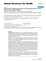
Figure 1
(A) – Persistent chronic hepatitis, Knodell index ≤ 2. Photomicrograph of liver showing chronic hepatitis with minimal
activity. Hepatocytes showing regenerative features are seen, with minimal inflammation and scattered ground- glass hepato-
cytes. Cobblestone arrangement (diffuse regeneration) with Hadziyannis cells and without necrosis or fibrosis. (H&E 250×).
(B) – Mild lobular chronic hepatitis, Knodell index 3–4. Photomicrograph of liver showing chronic hepatitis with mild
activity. Spotty hepatocyte necrosis is seen in a lobular pattern with focal lymphocytic infiltration. Lesions are characterized by
focal necrosis, conserved sinusoidal and trabecular patterns, lobular, portal, and focal lymphocytic infiltrated. (H&E 400×). (C)
– Moderate lobular chronic hepatitis, Knodell index > 4. Photomicrograph of liver showing chronic hepatitis with mod-
erate activity. There is portal chronic inflammation, focal interface hepatitis and periportal fibrous septa. Portal chronic swollen
periportal apoptosis, post-necrosis fibrous interportal bridges. Nodular regeneration (pre-cirrhosis). (H&E 250×)
Virology Journal 2005, 2:60 />Page 4 of 10
(page number not for citation purposes)
responding to 1763–1770 nt of the complete genome. Fig
2 shows the sequence and the band patterns of samples;
6541 (group C), 6290 (group C), 6516 (group C) and 467
(group B). To confirm that these deletions were not a PCR
artifact, the samples were further digested by SspI. Of the
four samples presenting the deletion only 2 were corrob-
orated by SspI, both samples (467 and 6516) were re-
amplified from the PCR1 product.
Mutations observed in HBV acute infected patients that
recovered versus chronic carriers
The percentage of the most frequent polymorphism
found in the study as well as the consensus sequences of
each of the population selected for statistical analysis are
shown in Table 4. Group A presented more amino acid or
nucleotide variability than the other groups, however, in
acute phase samples from group B, 50% of these had com-
mon mutations at position 12 (T12A).
Samples Genotype
Of the total 50 samples sequenced ; 48 belonged to geno-
type F, one sample belonged to genotype D subtype adw,
and the other to subtype ay, which was classified as geno-
type E by a web-based genotyping tool and as D by phyl-
ogenetic tree analysis (data not shown).
Discusion
T-A mutations were not found in any of 17 samples from
HBV patients who had recovered; a similar result had been
obtained in a study with self-limited acute hepatitis [20].
However, another study showed T-A mutations during the
acute phase in one out of 11 from genotype A, none of the
5 patients from genotype B and 4 out of 27 from genotype
C [21]. The significance of this finding needs to be further
studied.
T-A mutations were found in 12 (41.3%) of 29 samples
from chronic carriers. In one carrier the mutations were
detected 29 days after onset, with the probability that this
carrier could have been directly infected with HBV con-
taining the T-A mutations. In the 23 acute phase samples,
T-A mutations were not detected and therefore the possi-
bility to have an initial infection with T-A in other popu-
lations appears to be low. However, Kobayashi et al
, has
shown in their study a higher prevalence of the T-A muta-
tions in chronic patients during the acute phase than in
acute self limited HBV infection in patients infected with
genotypes C, A and B [21].
In chronic carriers, with a liver biopsy classified as moder-
ate or severe, T-A mutations were present in 61.5% (8/13)
and none in 4 biopsies classified as mild. However this
result was not statistically significant based on the Fisher
exact test, 1 tail, p = 0.05, probably due to the small sam-
ple size in the groups. Other studies have shown a better
correlation between the presence of T-A mutations and
patients with fulminant hepatitis, severe exacerbation
[20] or liver cirrhosis [22] especially with genotypes A or
C when compared with asymptomatic carriers [12-14]. In
agreement with the literature T-A mutations seem to
Sample deletions treated with Ssp I restriction enzymesFigure 2
Sample deletions treated with Ssp I restriction
enzymes. Recognition site of the enzyme SspI in the
sequences with 8 bp deletion (left). In the right, samples with
presumed deletions were run in a 3% agarose gel. Each pair
of lines have the same sample treated with and without the
Ssp I enzyme. An HIV sample having the AATATT site was
used as positive control in lanes 1 and 2, sample 1430 (616
bp) lanes 3 and 4 (negative control), 1000 bp ladder marker
lane 5, sample 6290 lanes 6 and 7, sample 467 lanes 8 and 9
sample 6516 lines 10 and 11, sample 6541 lanes 12 and 13.
The samples 467 and 6516 treated with SspI presented two
bands of 507 and 109 bp, lanes 8 and 10 (arrows) confirming
the deletion. Details in sequence are:
Position HBV 1758 1763 1768 1773 1778 1783 1788
nt | | | | | | |.
X
gene 385 395 405 415
2737M_5-09 CAGGTTAAAGGTCTTTGTATTAGGAGGCTGTAGGCA
6604m_969- CAGGTTAATGATCTTTGtatTAGGAGGctgTAGGCa
6516m_90-0 CAGGTTAAA TATTAGGAGGCTGTAGGCA
6541m_27-0 CAGGTtAAA TATTAGGAGGCTGTAGGCA
6290m_1232 CAGGTTAAA TATTAGGAGGCTGTAGGCA
467h_969-0 CAGGttAAA TATTAGGAGGCTGTAGGCA
Consensus CAGGTTAAATATTAGGAGGCTGTAGGCA
SspI recognition site stop codon
Virology Journal 2005, 2:60 />Page 5 of 10
(page number not for citation purposes)
appear more frequently in genotypes C [23,24] and A [13]
than D [25,26] or B [27].
In this study, V131I also occurred alone in 5 samples
(17%) of the 29 chronic patients; this event has been com-
monly reported by others [6,14,25,27,28]; nevertheless
M130K alone is very unusual. It has been described in 1 of
12 fulminant hepatitis patients [20] and in 1 genotype B
strain [27]. In one of the paired samples from this study
and in another from reference [6], the V131I mutation
appears in time before the methionine change at position
130.
In a Korean study T-A mutations were found in 32% (13/
41) of HBV carriers, and a triple mutation G1714A,
C1718T, A1721G was found in 27% (11/41) patients [4].
In our study wild type (wt) HBV strain nucleotide were
found in the 1714 and 1718 positions, but the mutation
A1721G was found in genotype F samples and not in two
samples with other genotypes. Again, T-A mutations are
common in all genotypes while other mutations seem to
be more related to specific genotypes.
No association could be established between the presence
of T-A mutations and HBeAg status (Table 3), similar to
other published data [4,24]. Of the four samples with the
8 bp deletion only (467 and 6516), two were re-amplified
from the PCR1 product and corroborated by enzyme
restriction digestion, which demonstrates that the dele-
tion was not a PCR artifact. This 8 bp deletion in the T-A
site has been reported previously [6,8,9,27,29] and it has
been associated with a low viral load [7,8,29]. Different
clones isolated from several patients showed a heteroge-
neous population of strains including T-A mutations, wt
strains as well as the 8 bp deletion. This could be a possi-
ble reason why we observed different results in amplified
samples of the initial PCR products with an 8 deletion
than in the reanalyzed two samples where the deletion
was not detected.
An interesting fact is that these deletions alter the X open
reading frame, changing K130N and introducing an iso-
leucine in the 131 site and a stop codon in the
position135.
The polymorphic differences observed between the
sequence of acute HBV recovered patients and chronic car-
riers are related to the genetic diversity of strain more than
the study group classification (A,B,C). All sequences iso-
lated in this study belong to genotype F with the exception
of 2. Using blast searches sequences from genotype F can
be divided in AY090455 – 1889 NIC sequences similar to
those which are related to South American sequences and
the AY090456- 1980HCR sequences similar to those
which are related to Central America sequences. The pol-
ymorphism observed in the nucleotides as well as the
amino acids in these groups may be due to a variability
present in the group related to the South American
sequences.
Many efforts have been made in order to clarify the role of
viral variants in the pathogenesis of HBV infection; and
still there is no final consensus. T-A mutations have been
proposed as possible prognostic markers for liver disease
progression [14] however, more studies are needed to elu-
cidate the role of the T-A mutations and its relation to
HBV diversity and disease outcome.
Conclusion
According to our results, T-A mutations were frequently
observed in HBV chronic carriers, but were not found in
acute recovered patients.
T-A mutations are frequent in all genotypes while other
mutations seem to be more related to specific genotypes.
T-A mutations may appear early during HBV infection
although the possibility of initial infection cannot be
excluded.
Methods
Study population
Samples were obtained from a study of HBV in San
Ramón and Palmares, Costa Rica areas outside of the cap-
ital city, San José, between 1972–1985 [19]. Based on
Table 2: Correlation between Knodell Index (KI) and HBx-T-A mutations.
MUTATIONS
T-A mutations V131I alone
Results KI #/n (%) #/n (%)
≤ 2 0/4 - 1/4 -
> 2 8/13 * (61.5) -
* One sample presented a deletion in the T-A position Fisher exact test, 1 tail, M130K p = 0.05, V131I p = 0.24
Virology Journal 2005, 2:60 />Page 6 of 10
(page number not for citation purposes)
serological markers and history of clinical onset, three
groups were established: Group A, included 18 samples
from acute cases who recovered from the infection; they
presented initially as HBsAg positive, anti-IgM HBc posi-
tive and had elevated ALT levels. A patient was catalogued
as a chronic carrier if HBsAg was present more than 6
months after the onset of disease. Group B, included 14
paired samples from chronic patients with known onset;
Table 3: Characterisation of samples with biopsies considered moderate and severe and patients who died from HCC.
Sex/Group Patient
ident
Sample Id/Time of
sample collection
after onset or
study initiation
Age at time of
sample
collection
Patient age at
time of biopsy
collection
Knodell Index K130M/V131I HBeAg/Anti-HBe TSGO/TSGP
M/B 950-08 865M/2 y 4 -/- +/- 50/32
6217M/12 y 14 15 2+1 = 3 +/+ -/-
M/B 671-10 445E/19 d 8 -/- +/- 74/83
5252M/9Y 17 18 2+2 = 4 +/+ -/+ 32/18
M/C 266-03 3751M/8 m 35 -/- -/+ 32/13
6400M/6 y 40 40 3+3 = 6 +/+ -/- 36/13
M/B 496-04 6461E/2 y 12 -/+ -/- 45/40
4904M/7 y 17 19 2+1 = 3 +/+ -/+ 28/25
M/B 969-04 467H/23 d 24 deletion -/- 500/550
6604M/7 y 31 32 HCC 5+2 = 7 +/+ -/- ND/18
M/C 1232-06 1481M/10 m 12 -/+ -/+ 36/21
6290M/9 y 22 23 2+1= 3 -/- -/+
M/C 65-35 6891M/11 y +/+ -/+ 55/50
20 20 3+0 = 3
M/C 158-01 6151M/11 y +/+ -/+ 36/16
33 35 1+2 = 3
M/C 921-07 6403M/10 y +/+ -/- 28/21
31 32 3+1 = 4
M/C 673-04 6572M/8 y -/- -/- ND/9
25 26 3+1 = 4
M/C 5-02 6067M/9 y 2+2 = 4 -/- -/+ 32/16
53 54
M/C 671-06 6593M/15 y -/- -/- ND/13
26 26 4+3 = 7
M/C 671-09 5251M/9y 2+4 = 6 -/- -/+ 28/18
16 19
M/B 1688-16 3254H/3 d 3 HCC -/- -/- 475/225
16 6653M/3 y 6 NB +/+ -/- ND/36
M/B 1400-01 575H/2 d 65 HCC -/- -/- 610/1200
5433M/4 Y 69 T -/- +/- 55/55
6825H/7 y 72 -/- +/- ND/55
M/C 1205-15 5399M/7 y 35 HCC -/- -/+ 32/28
ND = Not done, HCC = Hepatocellular carcinoma, T = tumor tissue only.
Virology Journal 2005, 2:60 />Page 7 of 10
(page number not for citation purposes)
with at least 3 years difference between the samples.
Group C included 45 chronic patients with unknown date
of onset. Twenty-nine patients had liver biopsy results, 4
from group B and 25 from group C.
The samples from all groups were negative by anti HAV
IgM or anti- HCV [31] and were kept frozen.
This project was approved by the Ethical Committee of
the Universidad of Costa Rica.
Table 4: Major sequence polymorphisms found in the groups studied.
Amino acid-Position-mutation Frequency (%) Consensus sequences
L5M 28 Group A: 17 recovered patients
Q8K 22
T12A 36
S29P 38
S31P 30
S33P 30
V37I 33
P40S 30 Group B: 7 acute-chronic patients
MAARLCCQLDP-RDVLCLRPVGAESRGRSLSGSLGAVPPPSPSAVPADDGSHLSLRGLPV
CSFSSAGPCALRFTSARRMETTVNAPRSLPTVLHKRTLGLSGRSMTWIEDYIKDCVFKDW
EELGEEIRLKVFVLGGCRHKLVCSPAPCNFFTSA*
D48N 25
R87W 30
R103W 33
T106P 25 Group B: 32 chronic patients
MAARLCCQLDPTRDVLCLRPVGAESRGRSLSGSLGAVPPPSPSAVPADDGSHLSLRGLPV
CSFSSAGPCALRFTSARRMETTVNAPRSLPTVLHKRTLGLSGRSMTWIEDYIKDCVFKDW
EELGEEIRL- - FVLGGCRHKLVCSPAPCNFFTSA*
D110E 33
K130M 24
V131I 27
Deleted nt 8 Consensus Deletion group 8 bp
MAARLCCQLDPTRDVLCLRPVGAESRGRSLSGSLGAVPPPSPSAVPADDGSHLSLRGLPV
CSFSSAGPCALRFTSARRMETTVNAPRSLPTVLHKRTLGLSGRSMTWIEDYIKDCVFKDW
EELGEEIRLNIRRL*
390-397
end codon
135 aa
Hyphens in the consensus sequence represent the amino acid polymorphism associated with the left column. The predicted consensus amino acid
sequence was obtained with Bioedit Software from the nucleotide sequence of the sample study.
| | | | | | | | | | | |
5 15 25 35 45 55
MAAR-CC-LDP-RDVLCLRPVGAESRGR-L-G-LGA-PP-SPSAVPA-DGSHLSLRGLPV
| | | | | | | | | | | |
65 75 85 95 105 115
CSFSSAGPCALRFTSARRMETTVNAP-SLPTVLHKRTLGLSG-SM-WIE-YIKDCVFKDW
| | | | | | |.
125 135 145 155
EELGEEIRLKVFVLGGCRHKLVCSPAPCNFFTSA*
Virology Journal 2005, 2:60 />Page 8 of 10
(page number not for citation purposes)
Biopsy classification Pathology
The inflammatory activity of Knodell in Chronic Persist-
ent Hepatitis (CPH) between 1 and 2 points, is repre-
sented by a uniform and diffuse cobblestone arrangement
of swollen hepatocytes, with compressed sinusoids; some
of which show Hadziyannis cells containing abundant
HBsAg.
Lobular Chronic Hepatitis (LCH) is between 2 and 6
points with an intact lobular architecture, perivenular cell
swelling, focal hepatocytolysis and a variable degree of
inflammatory activity [32]. Further, these lesions are char-
acterized by focal necrosis, abnormal hepatocytes and
scattered passive fibrous interportal bridges.
In this study the Knodell Index (KI) was used as follows:
≤ 2 points was considered mild liver lesion, 3 and 4 mod-
erate and > 4 as severe liver damage.
PCR Methods
Primers were chosen from conserved regions of the fol-
lowing HBV genotypes sequence obtained from GenBank.
Genotype A subtype adw2 (AF297625) and (AF373066),
genotype B (AF121243), genotype C subtype adr
(AB033550), subtype adw (AB033557), genotype D sub-
type ayw (AF280817), genotype E (AB032431), genotype
F (AB036919), genotype G (AB064310) and (AF160501).
Outer primers selected were: sense (1182–1200)
5'GTTTGCTGACGCAACCCCC3' and the antisense
5'CAATGTCCATGCCCCAAAGC3' (1891–1910). The
expected amplified product size was 728 bp. Inner prim-
ers: sense 5'GATCCATACTGCGGAACTCC3' (1263–
1282) and antisense 5'AGCTTGGAGGCTTGAACAGT3'
(1859–1878).
Genomic DNA was extracted from 200 µl of serum using
the QIAamp DNA mini Kits (Qiagen
®
U.S.A.) according to
manufacturer's instructions.
Nested PCR was performed using a thermocycler (Perkin-
Elmer).
For the first PCR, 10 µl of the extracted product were
added to a total of 50 µl of reaction volume containing 2.5
units of Taq (Promega
®
5 units/µl), 3.5 mM of MgCl
2
,
0.092 nmoles/µl of primers final concentration, 0.4
mmolar/µl of each dNTP. This amplification was per-
formed at 94°C for 3 min followed by 40 cycles at 94°C
for 1 min, 50°C for 1 min and 72°C for 1 min, with a final
extension of 4 min to 72°C.
For the nested PCR, 5 µl of product from the first PCR
were added to 50 µl of reaction, with a final concentration
of MgCl
2
, 2.5 mM and 0.080 nM of primers. Cycling
conditions for the second round were 94°C for 3 min, 40
cycles to 94°C for 0.40 min, 55°C for 0.40 min and 72°C
for 1.30 min. The final extension was 72°C for 4 min.
Nested products with a size of 616 bp were corroborated
by 2% agarose gel electrophoresis stained with ethidium
bromide.
Dilutions of 1:10 of a commercial CPG
®
DNA plasmid
with 10
5
copies/µl of the total HBV genome were prepared
and used as control as well as to determine the limit detec-
tion (sensitivity) of the PCR system.
Sequencing conditions
Nested PCR product (616 bp) was run on 1% agarose gels
and the expected band was cut and purified by a Qiagen
column system following manufacturer's instructions.
An Open Gene™ sequencer system (Visible Genetics) was
used. For sequencing the following primers were labeled
with cy 5.0 and cy 5.5 dyes: Sense 5' 5cy55
GTTTYGCTCGCAGCMGGTC3' y = c/t, m = c/a (1292–
1310) and antisense 5'-5cy5
CTTGAACGATRGGACATGAAC3' R = a/g (1848–1868).
Primers were diluted to a concentration of 3 pM in TE
buffer. All reagents were used according to manufacturer's
instructions. The first denaturation step was at 94°C for
2:30 min followed by 35 cycles of 0:30 min at 94°C, 0:30
min at 50°C, 1 min at 70°C and a final extension step at
72°C for 7 min. Finally, 1.5 µl of each sample was run in
a polyacrylamide gel at 1500 volts for 90 min.
A consensus sequence of the genotype F strain (NCBI
AB036919, AB036905, X75658) was used as our wild type
sequence.
Genotype sequencing
The HBx gene sequences were compared with homologue
sequences obtained from the GeneBank data base using
the BLAST program [33]. The genotype was determined
using a web- based genotyping tool for viral sequences
[34]. The subtype of some of the samples was determined
previously by specific antibodies available in our
laboratory.
Restriction Enzyme digestion
In order to corroborate an 8 bp deletion observed in some
sequences, a restriction enzyme SspI was used (New Eng-
land, BioLabs
INC,
). As a positive control a sample from
HIV having the same recognition site was used and a HBx
sample with the wild type sequence was employed as a
negative control. Ten µl of each purified product from the
nested PCR were dispensed into two different vials of 200
µl. In one vial 1 µl of SspI enzyme (5000 units/ml), 2 µl
Virology Journal 2005, 2:60 />Page 9 of 10
(page number not for citation purposes)
of enzyme buffer (New England, BioLabs
INC,
) and 7 µl of
water were added; while in the other vial the enzyme was
omitted. All samples were heated at 37°C for 90 minutes
and run in a 3% agarose gel. Results were visualized with
ethidium bromide.
Statistical analysis
The Fisher's exact test was used to evaluate the relation-
ship between two discrete and dichotomy variables. The t
test, for independent samples, was used to analyze contin-
uous variables when it was necessary. A new dichotomy
variable for hepatic damage was built into biopsy results
and using data from the Costa Rican National Tumor
Registry (NTR); by division into "mild damage" and
"moderate/severe damage". The relative risk (RR) was cal-
culated with a 95% confidence interval. All analyzes were
done with the JMP 4 software version 4.0.4 A BUSINESS
UNIT OF SAS Copyright
©
1989 – 2001 SAS Institute Inc.
(all rights reserved) and Epiinfo software CDC.
Competing interests
The author(s) declare that they have no competing
interests.
Authors' contributions
BL, FA, KV experimental design planning research
BL, MV laboratory: molecular and pathology work,
respectively
BL, FA statistical analysis
BL, KV editing
LH, LT, RBL contributed to manuscript content and edit-
ing of drafts
Acknowledgements
This research was supported by Ministerio de Ciencia y Tecnología (Minis-
try of Science and technology), Consejo Nacional para Investigaciones
Científicas y Tecnológicas (National Council for Science Research and
Technology) and Organización Panamericana de la Salud (Health Panamer-
ican Organization) grant.
The authors thank all the persons that kindly collaborated in the revision of
the manuscript, particularly to Dr. Joseph Schwarzman, Professor of
Pathology, Dartmouth-Hitchcock Medical Center, Lebanon, NH, USA for
his appropriate comments and to Ms. Virginia Larrad for editorial
assistance.
References
1. Beasley RP, Hwang LY, Lin CC, Chien CS: Hepatocelullar carci-
noma and hepatitis B virus: a prospective study of 22707
men in Taiwan. Lancet 1981, 2:1129-33.
2. Bergametti F, Sitterlin D, Transy C: Turnover of Hepatitis B Virus
X protein is regulated by a Damaged DNA-Binding
Complex. J Virol 2002, 76:6495-6501.
3. Lee H, Yun Y: HBx protein of Hepatitis B virus activates Jak1-
STAT signaling. J Biol Chem 1998, 273:25510-25515.
4. Chun YK, Kim JY, Woo HJ, Oh SM, Kang I, Ha J, Kim SS: No signif-
icant correlation exist between core promoter mutations,
viral replication and liver damage in chronic hepatitis B
infection. Hepatology 2000, 32:1154-1162.
5. Buckwold VE, Xu Z, Chen M, Yen TS, Ou JH: Effects of a naturally
occurring mutation in the hepatitis B virus basal core pro-
moter on precore gene expression and viral replication. J
Virol 1996, 70:5845-5851.
6. Horikita M, Itoh S, Yamamoto K, Shibayama T, Tsuda F, Okamoto H:
Differences in the entire nucleotide sequence between hep-
atitis B virus genomes from carriers positive for antibody to
hepatitis B e antigen with and without active disease. J Med
Virol 1994, 44:96-103.
7. Sallam TA, Tong CY: Two distinct types of hepatitis B virus
core promoter variants in Yemeni blood donors. J Med Virol
2002, 68:328-34.
8. Schlager F, Schaefer S, Metzler M, Gratzki N, Lampert F, Gerlich WH,
Repp R: Quantitative DNA fragment analysis for detecting
low amounts of hepatitis B virus deletion mutants in highly
viremic carriers. Hepatology 2000, 32:1096-105.
9. Preikschat P, Gunther S, Reinhold S, Will H, Budde K, Neumayer H,
Kruger DH, Meisel H: Complex HBV populations with muta-
tions in core promoter, C gene and Pre S region are associ-
ated with development of cirrhosis in long-term renal
transplant recipients. Hepatology 2002, 35:466-477.
10. Sirma H, Giannini C, Poussin K, Paterlini P, Kremsdorf D, Brechot C:
Hepatitis B Virus X mutants, present inhepatocellular carci-
noma tissue abrogate both the antiproliferative and transac-
tivation effects of HBx. Oncogene 1999, 26:4848-59.
11. Hsia CC, Yuwen H, Tabor E: Hot spot mutations in hepatitis B
virus X gene in hepatocelullar carcinoma. Lancet 1996,
348:625-626.
12. Fang ZL, Ling R, Wang SS, Nong J, Huang CS, Harrison TJ: HBV core
promoter mutations prevail in patients with hepatocellular
carcinoma in Guangxi, China. J Med Virol 1998, 56:18-24.
13. Batista M, Kramvis A, Kew M: High prevalence of 1672T 1764G
mutations in the basic core promoter of Hepatitis B virus
isolated from black Africans with hepatocellular carcinoma
compared with asymptomatic carriers. Hepatology 1999,
29:946-953.
14. Lindh M, Hannoun C, Dhillon AP, Norkrans G, Horal P: Core Pro-
moter Mutations and Genotypes in Relation to Viral Replica-
tion and Liver Damage in East Asian Hepatitis B Virus
Carriers. J Inf Dis 1999, 179:775-82.
15. Norder H, Hammas B, Lee SD, Courouce AM, Mushahwar IK, Magn-
ius L: Genetic relatedness of hepatitis B viral strains of
diverse geographical origin and natural variations in the pri-
mary structure of the surface antigen. J Gen Virol 1993,
74:1341-1348.
16. Norder H, Courouce AM, Magnius LO: Complete genomes, phy-
logenetic relatedness, and structural proteins of six strains of
the hepatitis B virus, four of which represent two new
genotypes. Virology 1994, 218:214-223.
17. Arauz-Ruiz P, Norder H, Robertson BH, Magnius L: Genotype H: a
new American genotype of hepatitis B virus revealed in Cen-
tral America. J Gen Virol 2002, 83:2059-2073.
18. Arauz-Ruiz P, Norder H, Visoná K, Magnius L: Genotype F prevails
in HBV infected patients of Hispanic origin in Central Amer-
ica and may carry the precore stop mutant. J Med Virol 1997,
51:305-312.
19. Visoná K, Eduarte C, Zamora E, Salazar L: Estudio epidemiológico
de las hepatitis virales en San Ramón y Palmares de 1972–
1985. Acta Médica Costarricense 1989, 33:69-77.
20. Honda A, Yokusaka O, Suzuki K, Saisho H: Detection of mutations
in hepatitis B virus enhancer 2/core promoter and x protein
regions in patients with fatal hepatitis B virus infection. J Med
Virol 2000, 62:167-176.
21. Kobayashi M, Arase Y, Ikeda K, Tsubota A, Suzuki Y, Saitoh S, Koba-
yashi M, Suzuki F, Akutas N, Hosaka T, Someya T, Matsuda M, Sato J,
Miyakawa Y, Kumada H: Wild type precore and core promoter
sequences in patients with acute self limited or chronic Hep-
atitis B. Scand Journal Gastroenterol 2004, 1:53-59.
22. Cho SW, Shin YJ, Hahm KB, Jin JH, Kim YS, Kim HJ: Analysis of the
precore and core promoter DNA Sequence in liver tissues
from patients with Hepatocellular carcinoma. J Korean Med
1999, 14:424-30.
Publish with BioMed Central and every
scientist can read your work free of charge
"BioMed Central will be the most significant development for
disseminating the results of biomedical research in our lifetime."
Sir Paul Nurse, Cancer Research UK
Your research papers will be:
available free of charge to the entire biomedical community
peer reviewed and published immediately upon acceptance
cited in PubMed and archived on PubMed Central
yours — you keep the copyright
Submit your manuscript here:
/>BioMedcentral
Virology Journal 2005, 2:60 />Page 10 of 10
(page number not for citation purposes)
23. Fang ZL, Yang J, Ge X, Zhuang H, Gong J, Li R, Ling R, Harrison TJ:
Core promoter mutations (A
1762
T and G
1764
A) and viral
genotype in chronic hepatitis B and hepatocellular carci-
noma in Guangxi, China. J Med Virol 2002, 68:33-40.
24. Ni YH, Chang MH, Hsu HY, Tsuei DJ: Longitudinal study on
mutation profilies of core promoter and precore regions of
the hepatitis B virus genome in children. Ped Res 2004,
56:396-399.
25. Vernard V, Corsaro D, Kajzer C, Bronowicki J, Faou A: Hepatitis B
virus X gene variability in French-born patients with chronic
hepatitis and hepatocellular carcinoma. J Med Virol 2000,
62:177-184.
26. Hannoun C, Horal P, Lindh M: Long -term mutation rates in the
hepatitis B virus genome. J Gen Virol 2000, 81:75-83.
27. Bläckberg J, Kidd-Ljunggren K: Mutations within the hepatitis B
virus genome among hepatitis B patients with Hepatocellu-
lar carcinoma. J Med Virol 2003, 71:18-23.
28. Gandhe SS, Chadha MS, Walimbe AW, Arankalle VA: Hepatitis B
virus: prevalence of precore/core promoter mutants in dif-
ferent clinical categories of indian patients. Viral Hepatitis 2003,
10:367-382.
29. Li KS, Yamashiro T, Sumie A, Terao H, Mifune K, Nishizono A: Hep-
atitis B virus harboring nucleotide deletions in the core pro-
moter region and genotype B correlate with low viral
replication activity in anti-HBe positive carriers. J Clin Virol
2001, 23:97-106.
30. Kuang SY, Jackson PE, Wang JB, Lu PX, Munoz A, Qian GS, Kensler
TW, Groopman JD: Specific mutations of hepatitis B virus in
plasma predict liver cancer development. Proc Natl Acad Sci U
S A 2004, 101:3575-80.
31. Palacios A, Taylor L, Haue L, Luftig RB, Visona KA: Development of
low cost peptide-based anti-Hepatitis C virus screening and
confirmatory assays: Comparison with commercially availa-
ble tests. J Med Virol 1999, 58:221-226.
32. Peters RL: Hepatocellular carcinoma Volume Chapter 8. Edited by:
Okuda K, Peters RL. John Wiley; 1976.
33. Altschul SF, Gish W, Miller W, Myers E, Lipman D: Basic local align-
ment search tool. J Mol Biol 1990, 215:403-410.
34. Rozanov M, Plikat U, Chappey C, Kochergin A, Tatusova TA: Web-
based genotyping resource for viral sequences. Nucleic Acids
Research 2004, 32(Web Server):w654-659.