Báo cáo sinh học: " Development of an in vitro cleavage assay system to examine vaccinia virus I7L cysteine proteinase activity" pptx
Bạn đang xem bản rút gọn của tài liệu. Xem và tải ngay bản đầy đủ của tài liệu tại đây (457.51 KB, 8 trang )
BioMed Central
Page 1 of 8
(page number not for citation purposes)
Virology Journal
Open Access
Research
Development of an in vitro cleavage assay system to examine
vaccinia virus I7L cysteine proteinase activity
Chelsea M Byrd
1
and Dennis E Hruby*
1,2
Address:
1
Molecular and Cellular Biology Program, Oregon State University, 220 Nash Hall, Corvallis, Oregon, 97331, USA and
2
Siga
Technologies, 4575 SW Research Way, Suite 230, Corvallis, Oregon, 97333, USA
Email: Chelsea M Byrd - ; Dennis E Hruby* -
* Corresponding author
Abstract
Through the use of transient expression assays and directed genetics, the vaccinia virus (VV) I7L
gene product has been implicated as the major maturational proteinase required for viral core
protein cleavage to occur during virion assembly. To confirm this hypothesis and to enable a
biochemical examination of the I7L cysteine proteinase, an in vitro cleavage assay was developed.
Using extracts of VV infected cells as the source of enzyme, reaction conditions were developed
which allowed accurate and efficient cleavage of exogenously added core protein precursors (P4a,
P4b and P25K). The cleavage reaction proceeded in a time-dependent manner and was optimal
when incubated at 25°C. I7L-mediated cleavage was not affected by selected inhibitors of
metalloproteinases, aspartic acid proteinases or serine proteinases (EDTA, pepstatin, and PMSF,
respectively), but was sensitive to several general cysteine proteinase inhibitors (E-64, EST,
Iodoacetic acid, and NEM) as well as the I7L active site inhibitor TTP-6171 [C. Byrd et al., J. Virol.
78:12147–12156 (2004)]. Finally, in antibody pull down experiments, it could be demonstrated that
monospecific αI7L serum depleted the enzyme activity whereas control sera including αG1L,
directed against the VV metalloproteinase, did not. Taken together, these data provide biochemical
evidence that I7L is a cysteine proteinase which is directly involved in VV core protein cleavage.
Furthermore, establishment of this I7L-mediated in vitro cleavage assay should enable future studies
into the enzymology and co-factor requirements of the proteolysis reaction, and facilitate antiviral
drug development against this essential target.
Background
The Orthopoxviridae include vaccinia virus, camelpox,
cowpox, ectromelia, monkeypox, raccoonpox, skunkpox,
taterapox, volepox, and variola. Viruses in this family are
the cause of numerous diseases including smallpox (vari-
ola), and recent human outbreaks of monkeypox.
Orthopoxviruses are large double-stranded DNA viruses
that are unique amongst DNA viruses in that they repli-
cate exclusively within the cytoplasm of infected cells.
Vaccinia virus (VV) is the most extensively studied virus in
this group and is the prototypic member. The genome of
VV is predicted to encode over 200 open reading frames.
VV expresses its genetic information in three stages, as
early, intermediate, and late genes. The early genes, which
account for approximately half of the genome and are
transcribed prior to DNA replication, encode many of the
proteins involved in viral DNA replication and intermedi-
ate gene expression. The intermediate genes, of which
only a handful have been identified, are expressed after
the onset of DNA replication, and encode proteins that
Published: 16 August 2005
Virology Journal 2005, 2:63 doi:10.1186/1743-422X-2-63
Received: 21 April 2005
Accepted: 16 August 2005
This article is available from: />© 2005 Byrd and Hruby; licensee BioMed Central Ltd.
This is an Open Access article distributed under the terms of the Creative Commons Attribution License ( />),
which permits unrestricted use, distribution, and reproduction in any medium, provided the original work is properly cited.
Virology Journal 2005, 2:63 />Page 2 of 8
(page number not for citation purposes)
are activators of late gene expression. The late genes
encode many proteins required for the transcription of
early genes, the viral structural proteins and the enzymes
necessary to process these proteins into their mature form.
Many viruses use proteolytic processing as a key step in
their developmental cycle. RNA viruses and retroviruses
commonly undergo formative proteolysis in which large
polyproteins are cleaved by viral encoded proteinases to
produce the structural and nonstructural proteins
required for morphogenesis. DNA viruses such as poxvi-
ruses and adenoviruses commonly use another type of
proteolysis, called morphogenic proteolysis where precur-
sor proteins are first synthesized and then cleaved by viral
proteinases to produce the mature form of the protein.
The mature protein then plays an essential role in virion
formation. During VV assembly, as the spherical imma-
ture virions (IVs) are maturing into the first infectious
form of vaccinia virus, intracellular mature virus (IMV), a
series of events takes place including proteolytic process-
ing of viral core proteins [1-4].
Our laboratory has worked to identify and characterize
the proteinases of VV in order to understand their regula-
tion, function, and biochemistry, with a long term goal of
developing inhibitors of these enzymes as antiviral drugs.
The gene product of the I7L open reading frame recently
has been suggested to be the core protein proteinase of VV
through the use of an in vivo trans processing assay [5,6].
I7L is an essential late gene, as shown through tempera-
ture sensitive mutant viruses [7,8] and conditional lethal
mutant viruses [9,10] where under non-permissive condi-
tions, viral morphogenesis is blocked prior to the forma-
tion of IMV. I7L is predicted to be a 47 kDa cysteine
proteinase that cleaves the major core protein precursors
P4a, P4b, and P25K, products of the A10L, A3L, and L4R
open reading frames respectively, at a novel Ala-Gly-Xaa
cleavage site with cleavage occurring after the glycine resi-
due [5,6]. I7L also is likely to be responsible for cleavage
of the A17 membrane protein, at an Ala-Gly-Ala site [9].
This consensus Ala-Gly-Xaa cleavage site of vaccinia is
similar to that used for both the adenovirus and African
swine fever virus proteinases which cleave after the second
glycine in a Gly-Gly-Xaa motif [11,12].
Comparative sequence analysis has suggested that the VV
I7L proteinase is related to the ASFV and adenovirus
cysteine proteinases and may form a new family of
SUMO-1 related enzymes [13,12]. The nucleophilic
cysteine is responsible for cleavage and is activated by the
imidazol group of the catalytic histidine residue. Substrate
specificity is determined by the substrate binding pocket
and is unique for each proteinase. Several critical residues
have been identified as being necessary for enzymatic
activity of I7L including the catalytic triad residues [6].
Based on the identification of the catalytic residues and
the predicted structure of the I7L proteinase, a new class
of small molecule inhibitors was developed that are capa-
ble of inhibiting the replication of VV, and were found to
specifically target I7L through the generation of drug
resistant mutant viruses with the mutations mapping to
I7L [14].
To date, direct studies on the enzymology of I7L-mediated
proteolysis have not been possible due to the absence of a
suitable biochemical assay. In the experiments reported
here, we describe the development of an in vitro I7L-medi-
ated cleavage assay. We have used this system to obtain
both biochemical and immunological data to prove that
I7L is directly involved in cleavage of the major VV core
protein precursors. Having this assay available will now
facilitate biochemistry of the I7L enzyme and identifica-
tion of all the required reaction components to be
undertaken.
Results
To date, all studies of VV I7L activity have been carried out
indirectly in transfected/infected tissue culture cells.
Although this approach has provided some important
insights into I7L biology, it is limited with respect to the
study of I7L enzymology and identification of all the cis
and trans factors required for substrate identification and
catalysis. In order to approach these questions, we have
sought to develop an in vitro cleavage assay for I7L. Thus
far, the obvious approaches of expressing and purifying
I7L from prokaryotic and eukaryotic expression vectors
and combining with peptides or proteins containing a
canonical A-G-X cleavage site have not been successful
(data not shown), perhaps due to either the lack of essen-
tial co-factors or inappropriate assay conditions. As an
alternative approach, we sought to develop a cleavage
assay using infected cell extracts as the source of I7L activ-
ity and labeled core protein precursors made in vitro as the
substrate. If successful, this system would provide the
starting point for a dissection of the essential reaction
components.
In vitro Processing of Core Protein Precursors
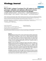
The three major core protein precursors P4a (A10L), P4b
(A3L), and P25K (L4R) which are known to be cleaved to
a mature form (Figure 1) were cloned into plasmid vectors
driven off of a T7 promoter to be used as a source of sub-
strate for the assay. To investigate the ability of I7L to
cleave the P4a, P4b, and P25K substrates in vitro, we have
used a system where the substrates are produced from an
in vitro transcription and translation assay using rabbit
reticulocyte lysates and then mixed with I7L expressed
from virus infected cells. BSC40 cells are infected with
ts16, a temperature sensitive mutant virus in which the
responsible mutation maps to I7L. The virus infected cells
Virology Journal 2005, 2:63 />Page 3 of 8
(page number not for citation purposes)
are incubated at the non-permissive temperature and
transfected with plasmids expressing either wild-type I7L
(pI7L) or I7L with the catalytic histidine residue mutated
to an alanine (pI7LH241A). The extracts are prepared as
described in the Materials and Methods. The extracts are
mixed and incubated with the substrates for 3 hrs and
then analyzed through SDS-PAGE and chemiluminescent
detection. As shown in Figure 2, a specific band corre-
sponding to unprocessed P4a (top panel), P4b (middle
panel), or P25K (bottom panel) is produced when the
substrate is run alone. When mixed with cellular extracts,
or extracts from cells infected with ts16 at the non-permis-
sive temperature and transfected with mutant I7L, no
cleavage products are observed. However, when mixed
with extracts from either cells infected with ts16 at the per-
missive temperature or cells infected with ts16 at the non-
permissive temperature transfected with wild-type I7L, the
cleaved products 4a, 4b, and 25K are observed. Substrates
with mutated A-G-X sites were not cleaved indicating that
cleavage was occurring at the correct sites (data not
shown). For the rest of the reported studies, P25K was
used as the source of substrate since it gave the best cleav-
age profile.
Processing Kinetics of Core Protein Precursors
To determine the optimal temperature and kinetics of
processing of the core protein precursors in the in vitro
cleavage assay, a time course of I7L-mediated processing
at various temperatures was performed. As shown in Fig-
ure 3A, at 0°C, no processing was observed during the 20
hr time period. At 25°C, a gradual increase in the amount
of P25K cleavage product was observed starting at 15 min
and increasing throughout the 20 hr incubation period
(Fig. 3B). Compared with the rate of cleavage at 25°C,
cleavage was slower at 30°C (Fig. 3C), starting around 30
min and increasing through the 20 hr period, but never to
the same level as at 25°C. Processing is greatly reduced at
37°C with only a faint processed band ever appearing (Fig
3D).
Influence of Thiol Reagents on the Protease Activity
Based on its sequence similarity to the adenovirus pro-
tease, the African swine fever virus protease, and an ubiq-
uitin-degrading enzyme in yeast, as well as the identity of
a catalytic triad composed of histidine, cysteine, and
aspartic acid, I7L has been classified as a cysteine protein-
ase. The thiol reagents dithiothreitol (DTT) and cysteine
have been shown to enhance the cleavage activity of the
adenovirus protease in an in vitro peptide cleavage assay
[15]. To determine whether these agents have a similar
effect on the activity of I7L, they were added to the in vitro
assay in a final concentration from 0–10 mM. However,
no increase in cleavage activity was observed with the
addition of either DTT or cysteine (data not shown). It is
possible that once purified recombinant enzyme is pro-
duced these thiol reagents may increase its activity.
Effect of Inhibitors on Protease Activity and
Characterization as a Cysteine Proteinase
The in vitro assay allowed us to test the effects of various
protease inhibitors, as well as specific small molecule
inhibitors on the activity of I7L. As shown in Figure 4 and
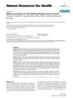
Schematic representation of the major core protein precursor cleavage productsFigure 1
Schematic representation of the major core protein precursor cleavage products. The vaccinia virus genome is
represented depicting three of the major core protein precursors, the gene products of the L4R, A10L, and A3L open reading
frames, P25K, P4a, and P4b respectively. The precursors are shown being cleaved into their mature form. Molecular mass is
indicated.
Virology Journal 2005, 2:63 />Page 4 of 8
(page number not for citation purposes)
Table 1, the metalloproteinase inhibitor ethylenediami-
netetraacetic acid (EDTA), the aspartic proteinase
inhibitor pepstatin, and the serine proteinase inhibitor
phenylmethanesulfonyl (PMSF) had no detectable effect
on cleavage activity. The cysteine proteinase inhibitors
iodoacetic acid (IA) and N-ethylmaleimide (NEM) effi-
ciently blocked I7L mediated proteolysis of P25K. The
cysteine proteinase inhibitors E-64 and EST were shown
to inhibit protease activity at a relatively high concentra-
tion, but not at the lower concentration tested. This is con-
sistent with what has been observed for both the
adenovirus protease [16], and the African swine fever
virus protease [17]. The failure of E-64 to inhibit protease
activity at the lower concentration tested, and the location
of the active site residues may suggest that each of these
enzymes are not conventional papain-like enzymes, but
may be a new family of cysteine proteinases. The cysteine
protease inhibitor leupeptin also failed to inhibit protease
activity, although this lack of inhibition was also observed
with the adenovirus proteinase [16].
Next we wanted to determine if the small molecule I7L
inhibitors previously developed as antiviral drug candi-
dates [14] could be shown to specifically inhibit the activ-
ity of I7L in the in vitro assay. The compound TTP-6171
has been shown to inhibit viral replication in tissue cul-
ture, with drug resistant virus mutations mapping to I7L
[14]. Here we see that this compound along with TTP-
1021, which was also found to inhibit I7L in tissue
culture, inhibits the processing of P25K in vitro. However
the compound TTP-0961, which was not found to gener-
ate resistant mutants in the I7L gene (data not shown),
does not inhibit cleavage. These results demonstrate that
this assay can be used for the screening of specific I7L
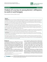
In vitro proteolytic processing of P4a, P4b, and P25KFigure 2
In vitro proteolytic processing of P4a, P4b, and P25K.
1 µl of TNT produced substrate either P4a (A), P4b (B), or
P25K (C) was mixed with 5 µl of Hepes buffer and 14 µl of
enzyme extracts, either from uninfected cells, or cells
infected with ts16 at the permissive or non-permissive tem-
perature. At the non-permissive temperature, plasmid borne
I7L, either wild-type (pI7L) or mutant I7L (pI7LH241A) was
transfected in as the source of enzyme. The reaction was
incubated at 29°C for 3 hrs before being stopped by the
addition of SDS sample buffer. Molecular weight is indicated
on the left and the core protein precursor and product on
the right. Lane 1 is substrate alone, lane 2 is substrate mixed
with cellular extracts and lanes 3–5 are substrate mixed with
the enzyme extract indicated.
P25K
25K
P4b
4b
30
66
97
66
P4a
4a
cell ts16-31° ts16-41° ts16-41 °
pI7LH241A pI7L
12345
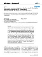
Processing kinetics of P25KFigure 3
Processing kinetics of P25K. Samples were incubated at
either 0°C (A), 25°C (B), 30°C (C), or 37°C (D) for up to 20
hrs, harvested at the indicated times and the reaction
stopped by the addition of SDS sample buffer. Incubation
temperature is indicated on the left and P25K precursor and
25K mature product are indicated on the right.
Virology Journal 2005, 2:63 />Page 5 of 8
(page number not for citation purposes)
inhibitors and confirms that this class of molecules targets
I7L.
Effects of I7L antibody competition on cleavage
To directly demonstrate that the cleavage observed in the
in vitro assay requires the presence of I7L, increasing con-
centrations of I7L specific antiserum were added to the
enzyme extracts overnight, and then the complex was pre-
cipitated with Protein A sepharose beads to deplete the
extract of I7L and any associated co-factors. As shown in
Figure 5, both of the I7L antisera tested inhibited cleavage
of P25K while an antiserum targeting a different VV gene
product, G1L, did not inhibit cleavage.
Discussion
In this report, a cell-free transcription and translation sys-
tem was used to develop an in vitro cleavage assay for the
VV cysteine proteinase I7L. Proteolytic activity was
obtained by co-expression of I7L in ts16 infected cells at
the non-permissive temperature. Each of the major core
protein precursors, P4a, P4b, and P25K, were shown to be
cleaved to their mature products by I7L using the in vitro
assay. Evidence that this cleavage is specific to I7L was
shown through the fact that expressing a mutant form of
I7L resulted in the inability to cleave the core protein pre-
cursors. Antibody pull down experiments with αI7L
supported the conclusion that I7L plays a direct role in the
proteolytic reaction.
A time course of processing at various temperatures indi-
cated that for this particular assay, the optimal tempera-
ture for the reaction to be carried out at is 25°C with
processing beginning as soon as 15 minutes after addition
of enzyme and increasing as time progresses. The cleavage
reaction was never driven to completion and this may be
due to a lack of replenishing co-factors or the enzyme may
have been used up in the reaction. It was surprising that
the optimal reaction temperature was 25°C instead of
37°C which is the optimal growth temperature for VV in
cell culture. One possible explanation is that I7L is present
at high concentrations in the extract and one can measure
marginal activity at low temperature, whereas at higher
temperatures other proteinases are activated which
degrade the I7L enzyme.
Known cysteine protease inhibitors such as E-64, iodoace-
tic acid, and NEM were shown to inhibit the in-vitro cleav-
age reaction while the metalloproteinase inhibitor EDTA,
the aspartic acid protease inhibitor pepstatin, and the
serine protease inhibitor PMSF all failed to inhibit the
cleavage reaction indicating that the enzyme responsible
for cleavage is a cysteine protease. Interestingly the
cysteine protease inhibitors leupeptin, and low concentra-
tions of E-64 did not inhibit the reaction. These cysteine
protease inhibitors were also not shown to be effective
against either the African Swine Fever Virus protease [17]
or the adenovirus protease [16], further providing support
for the theory that these enzymes may form a new family
of cysteine proteases that differ from papain-like cysteine
proteases.
Of particular interest, the small molecule inhibitors
designed to fit into the active site pocket of I7L and previ-
ously shown to inhibit viral replication [14], were found
to be active in inhibiting the in vitro cleavage reaction
described here. A related compound (TTP-0961) that was
not found to map to I7L was not able to abolish cleavage.
This indicated that this assay may be useful for high-
Effect of inhibitors on in vitro processingFigure 4
Effect of inhibitors on in vitro processing. Various con-
centrations of protease inhibitors were added to the in vitro
processing assay for 6 hr at 29°C. The first lane is P25K
expressed alone with no extract added. The second lane is
P25K mixed with cellular extracts and the third lane is P25K
mixed with I7L enzyme extracts. Each of the remaining lanes
has P25K mixed with I7L enzyme extracts plus indicated
inhibitor. Ethylenediaminetetraacetic acid (EDTA) was used
at 1 mM. Pepstatin A, Pep, was used at 10 µM. Phenlymeth-
anesulfonyl fluoride (PMSF) was used at 1 mM. N-(trans-
Epoxysuccinyl)-L-leucine 4-guanidinobutylamide trans-Epoxy-
succinyl-L-leucylamido(4-guanidino)butane (E-64) and a
related product EST, were both used at 10 µM and 100 µM
concentrations. Iodoacetic acid (IA) was used at 1 mM. Leu-
peptin (Leu) was used at 1 mM, and N-ethlymaleimide (NEM)
was used at 2.5 mM. The concentrations of TTP-6171, TTP-
1021, and TTP-0961 are indicated. The table indicates the
concentration of inhibitor used and whether cleavage activity
was observed.
Virology Journal 2005, 2:63 />Page 6 of 8
(page number not for citation purposes)
throughput screening of compounds to identify those that
have specific activity for I7L.
Conclusion
Until this point, all work demonstrating that I7L is the
core protein proteinase has been done through transient-
expression assays and the use of conditional lethal viruses
in tissue culture [9,5,6,10]. The data obtained has indi-
cated that I7L is essential for these processing activities, it
did not rule out the possibility that some other factor or
enzyme was also required for this activity to occur.
Through the use of an in vitro assay we have shown that
I7L is capable of cleaving the core protein precursors but
that an additional co-factor is required for this activity to
occur since expression of the enzyme through cell-free
translation produced inactive enzyme. The co-factor(s)
necessary for cleavage have yet to be determined. How-
ever, having the assay described in this report available
will now enable a reductive analysis to be conducted to
identify all the essential components of the reaction and
to study their individual biochemical characteristics.
Methods
Cells and Viruses
BSC
40
cells [18] were grown in Eagle's minimal essential
medium containing 5% fetal calf serum (FCS) (Sigma, St.
Louis, MO), 2 mM glutamine (Invitrogen, Carlsbad, CA),
and 15 µg/ml gentamicin sulfate (Invitrogen) in a 37°C
incubator with 5% CO
2
. Purified ts16 Vaccinia virus was
prepared as described [19]. Escherichia coli strains were
grown in Luria-Bertani broth or on Luria-Bertani medium
containing 1.5% agar and ampicillin at 50 µg/ml.
Plasmids
The A10L (P4a) gene was amplified by polymerase chain
reaction using oligonucleotides KH10 (5' CATGCCAT-
GGATGATGCCTATTAAGTCAATAGTTACT CTT-3') and
KH11 (5'-CCGCTCGAGTTATTCATCATCAAAAGAGACA-
GAGTC-3'), digested with NcoI and XhoI, and cloned into
the pTM1 vector, yielding pTM-P4a which utilizes a T7
promoter for expression. The A3L (P4b) gene was ampli-
fied using oligonucleotides KH08 (5'-CATGCCATGGAT-
GGAAGCCGTGGTCAATAG-3') and KH09 (5'-
Table 1: Effect of inhibitors on in vitro processing.
Inhibitor Name Concentration Inhibit Cleavage
Metalloproteinase EDTA 1 mM No
Aspartic acid proteinase Pepstatin 10 µMNo
Serine proteinase PMSF 1 mM No
Cysteine proteinase E-64 10 µMNo
E-64 100 µMYes
EST 10 µMNo
EST 100 µMYes
IA 1 mM Yes
Leupeptin 1 mM No
NEM 2.5 mM Yes
TTP inhibitors TTP-6171 50 µMYes
TTP-6171 20 µMYes
TTP-1021 50 µMYes
TTP-1021 20 µMYes
TTP-0961 50 µMNo
TTP-0961 20 µMNo
Effect of antibody competition on in vitro processingFigure 5
Effect of antibody competition on in vitro processing.
Lane 1 is P25K expressed alone. Lane 2 is P25K mixed with
I7L enzyme extracts. Lane 3 is P25K mixed with I7L extracts
that have been diluted with Hepes buffer and treated with
Sepharose beads. Lanes 4, 5, and 6 are P25K mixed with I7L
extracts that have been incubated overnight with different
I7L antiserum (indicated on each lane), treated with Sepha-
rose beads and the antibody complex removed by centrifuga-
tion. Lane 7 is P25K mixed with I7L extracts incubated with
G1L antiserum as above.
Virology Journal 2005, 2:63 />Page 7 of 8
(page number not for citation purposes)
TCCCCCGGGCTAAAAATAGTTCTGTAATAT-
GTCTAGCGCT-3'), digested with NcoI and SmaI, and
cloned into the pTM1 vector to yield pTM-P4b. The L4R
(P25K) gene was amplified using oligonucleotides DN51
(5'-CATGCCATG GATGAGTCTACTGCTAGAAAAC-3')
and KH07 (5'-CCGCTCGAGTCAATCCTTT GTCG-3'),
digested with NcoI and XhoI, and cloned into the pTM1
vector to yield pTM-P25K. The pI7L and pI7LH241A plas-
mids were described in Byrd et al., 2002 [5].
Preparation of polyprotein or proteinase-containing
extracts
Confluent monolayers of BSC
40
cells in 6-well plates were
infected with ts16 VV at a multiplicity of infection of 2
plaque-forming units per cell and transfected with 2 µg of
plasmid DNA (either pI7L, or pI7LH241A) using DMRIE-
C (Invitrogen) following the manufacturer's indications.
Infected cells were incubated either at the permissive tem-
perature of 31.5°C or the non-permissive temperature of
39°C. Cells were harvested at 24 h post-infection by
pipetting up and down to lift the cells from the surface.
The infected cells were centrifuged at 10,000 × g for 10
min, the supernatant was aspirated off, and the pellet was
resuspended in 500 µL homogenization buffer containing
20 mM HEPES (pH 7.4), 0.28 M sucrose, 2 mM EDTA.
This was passed through a 25-gauge syringe 15 times. The
homogenate was centrifuged at 700 × g for 5 min to sepa-
rate the nuclei and unbroken cells from the supernatant.
The supernatant was centrifuged at 100,000 × g for 30 min
at 4°C to separate the membrane/particulate material
from the supernatant. The supernatant was used as the
source of enzyme.
Coupled TNT reactions with T7 RNA polymerase were
performed according to the manufacturer's instructions
(Promega Corporation, Madison, Wisconsin) as a source
of substrate. Briefly, the TNT reactions were performed at
30°C in a final volume of 25 µL with 1 µg of plasmid
DNA, using the non-radioactive Transcend label (bioti-
nylated lysine residues are incorporated in the protein)
provided with the kit for detection of protein.
In vitro cleavage assay
Reactions were performed at the indicated temperature in
a final volume of 20 µL containing 1 µL of substrate, 13
µL of enzyme extract, and 6 µL of 20 mM HEPES (pH 7.4)
buffer, pH 7.4. After the indicated times, the reaction was
stopped by the addition of SDS sample buffer, and the
samples were subjected to SDS-polyacrylamide gel
electrophoresis. The results were analyzed by immunob-
lotting following the instructions provided by the TNT kit.
Inhibitor studies
For inhibitor studies, the reactions described above were
incubated for 6 hr in the presence or absence of the fol-
lowing protease inhibitors: 1 mM phenylmethanesulfonyl
fluoride (PMSF) (Sigma), 10 µM Pepstatin A (Sigma), 1
mM ethylenediaminetetraacetic acid (EDTA) (Sigma), 10
µM or 100 µM N-(trans-Epoxysuccinyl)-L-leucine 4-gua-
nidinobutylamide trans-Epoxysuccinyl-L-leucylamido(4-
guanidino)butane (E-64) (Sigma), 1 mM iodoacetic acid
(Sigma), 10 µM or 100 µM Leupeptin (Roche, Indianapo-
lis, IN), 2.5 mM N-ethylmaleimide (NEM) (Sigma). For
I7L specific inhibition studies, the reactions described
above were incubated for 6 hr in the presence or absence
of TTP-6171, TTP-1021, or TTP-0961 [14] at 5 µM or 20
µM final concentrations.
Antibody competition studies
For the antibody competition studies, 25 µl of I7L or G1L
specific antiserum was added to 25 µL of enzyme extract
on a rotating shaker overnight at 4°C. ProteinA: Sepha-
rose beads (Amersham Biosciences, Uppsla, Sweden)
were added for 3 hrs and the antibody complex was cen-
trifuged to pull down the I7L enzyme. The supernatant
was used as the source of extract in the in vitro assay
described above. As a control, enzyme extract was mixed
with buffer instead of antibody and treated with beads in
a similar manner.
Competing interests
The author(s) declare that they have no competing
interests.
Authors' contributions
CMB conceived the study, conducted all the experiments
and wrote the manuscript. DEH coordinated the research
efforts and edited the paper. Both authors read and
approved the final manuscript.
Acknowledgements
We would like to thank Kady Honeychurch for constructing pTM:L4R,
pTM:A3L, and pTM:A10L, Rich Condit for providing ts16, and TransTech
Pharma for supplying TTP-6171, TTP-1021, and TTP-0961. This work was
funded by NIH grant AI-060160.
References
1. Moss B, Rosenblum EN: Protein cleavage and poxvirus morpho-
genesis: tryptic peptide analysis of core precursors accumu-
lated by blocking assembly with rifampicin. J Mol Biol 1973,
81:267-269.
2. Silver M, Dales S: Biogenesis of vaccinia: interrelationship
between post-translational cleavage, virus assembly, and
maturation. Virology 1982, 117:341-356.
3. VanSlyke JK, Franke CA, Hruby DE: Proteolytic maturation of
vaccinia virus core proteins: identification of a conserved
motif at the N termini of the 4b and 25K virion proteins. J
Gen Virol 1991, 72:411-416.
4. VanSlyke JK, Whitehead SS, Wilson EM, Hruby DE: The multistep
proteolytic maturation pathway utilized by vaccinia virus
P4a protein: a degenerate conserved cleavage motif within
core proteins. Virology 1991, 183:467-478.
5. Byrd CM, Bolken TC, Hruby DE: The vaccinia virus I7L gene
product is the core protein proteinase. J Virol 2002, 76:8973-6.
Publish with BioMed Central and every
scientist can read your work free of charge
"BioMed Central will be the most significant development for
disseminating the results of biomedical research in our lifetime."
Sir Paul Nurse, Cancer Research UK
Your research papers will be:
available free of charge to the entire biomedical community
peer reviewed and published immediately upon acceptance
cited in PubMed and archived on PubMed Central
yours — you keep the copyright
Submit your manuscript here:
/>BioMedcentral
Virology Journal 2005, 2:63 />Page 8 of 8
(page number not for citation purposes)
6. Byrd CM, Bolken TC, Hruby DE: Molecular dissection of the vac-
cinia virus I7L core protein proteinase. J Virol 2003,
77:11279-11283.
7. Ericsson M, Cudmore S, Shuman S, Condit RC, Griffiths G, Locker JK:
Characterization of ts16, a temperature-sensitive mutant of
vaccinia virus. J Virol 1995, 69:7072-7086.
8. Kane EM, Shuman S: Vaccinia virus morphogenesis is blocked
by a temperature-sensitive mutation in the I7 gene that
encodes a virion component. J Virol 1993, 67:2689-2698.
9. Ansarah-Sobrinho C, Moss B: Role of the I7 protein in proteo-
lytic processing of vaccinia virus membrane and core
components. J Virol 2004, 78:6335-6343.
10. Byrd CM, Hruby DE: A conditional-lethal vaccinia virus mutant
demonstrates that the I7L gene product is required for vir-
ion morphogenesis. Virol J 2005, 2:4.
11. Webster A, Russell S, Talbot P, Russell WC, Kemp GD: Character-
ization of the adenovirus proteinase: substrate specificity. J
Gen Virol 1989, 70:3225-3234.
12. Andres G, Alejo A, Simon-Mateo C, Salas ML: African swine fever
virus protease, a new viral member of the SUMO-1-specific
protease family. J Biol Chem 2001, 276:780-787.
13. Li SJ, Hochstrasser M: A new protease required for cell-cycle
progression in yeast. Nature 1999, 398:246-51.
14. Byrd CM, Bolken TC, Mjalli AM, Arimilli MN, Andrews RC, Rothlein
R, Andrea T, Rao M, Owens KL, Hruby DE: New class of
orthopoxvirus antiviral drugs that block viral maturation. J
Virol 2004, 78:12147-12156.
15. Webster A, Hay RT, Kemp GD: The adenovirus protease is acti-
vated by a virus-coded disulphide-linked peptide. Cell 1993,
72:97-104.
16. Webster A, Russell WC, Kemp GD: Characterization of the ade-
novirus proteinase: development and use of a specific pep-
tide assay. J Gen Virol 1989, 70:3215-3223.
17. Rubio D, Alejo A, Rodriguez I, Salas ML: Polyprotein processing
protease of African swine fever virus: purification and bio-
chemical characterization. J Virol 2003, 77:4444-4448.
18. Raczynski P, Condit RC: Specific inhibition of vaccinia virus
growth by 2'-O-methyladenosine: isolation of a drug-resist-
ant virus mutant. Virology 1983, 128:458-62.
19. Hruby DE, Guarino LA, Kates JR: Vaccinia virus replication. I.
Requirement for the host-cell nucleus. J Virol 1979, 29:705-15.