Báo cáo sinh học: " Novel type I interferon IL-28A suppresses hepatitis C viral RNA replication" pptx
Bạn đang xem bản rút gọn của tài liệu. Xem và tải ngay bản đầy đủ của tài liệu tại đây (593.02 KB, 12 trang )
BioMed Central
Page 1 of 12
(page number not for citation purposes)
Virology Journal
Open Access
Research
Novel type I interferon IL-28A suppresses hepatitis C viral RNA
replication
Haizhen Zhu
1
, Mike Butera
1
, David R Nelson
2
and Chen Liu*
1
Address:
1
Department of Pathology, Immunology and Laboratory Medicine, University of Florida, P. O. Box 100275, Gainesville, Florida 32610,
USA and
2
Department of Medicine, University of Florida, P. O. Box 100275, Gainesville, Florida 32610, USA
Email: Haizhen Zhu - ; Mike Butera - ; David R Nelson - ;
Chen Liu* -
* Corresponding author
Abstract
Interferon alpha (IFN-α)-based therapy is the currently approved treatment for chronic hepatitis
C viral infection. The sustained antiviral response rate is approximately 50% for genotype-1
infection. The major challenge to the HCV community is to improve antiviral efficacy and to reduce
the side effects typically seen in IFNα-based therapy. One of the strategies is to identify new
interferons, which may have better efficacy and less undesirable side effects. In this report, we
examined the role of IL-28A (IFN λ2), a novel type I IFN, in suppression of human hepatitis C viral
RNA replication. We have cloned both the human genomic DNA and cDNA of IL-28A, and
evaluated their biological activity using HCV RNA replicon cell culture system. The results show
that IL-28A effectively inhibits HCV subgenomic RNA replication in a dose-dependent manner.
Treatment of human hepatoma cells with IL-28A activates the JAK-STAT signaling pathway and
induces the expression of some interferon-stimulated genes (ISGs), such as 6–16 and 1–8U. We
also demonstrate that IL-28A induces expression of HLA class I antigens in human hepatoma cells.
Moreover, IL-28A appears to specifically suppress HCV IRES-mediated translation. Although IL-
28A receptor shares one subunit with the IL-10 receptor, IL-10 treatment has no detectable effect
on IL-28A-induced antiviral activity. Interestingly, IL-28A can synergistically enhance IFNα antiviral
efficacy. Our results suggest that IL-28A antiviral activity is associated with the activation of the
JAK-STAT signaling pathway and expression of ISGs. The effectiveness of IL-28A antiviral activity
and its synergistic effect on IFN-α indicate that IL-28A may be potentially used to treat HCV
chronic infection.
Background
Interferon alpha (IFN-α), the prototype of type I inter-
feron, is widely used to treat human viral infections and
certain malignant tumors [1]. There are several subtypes
of type I interferons in humans, namely IFN-α, IFN-β,
IFN-ω, IFN-κ, IFN-tau, IFN-epsilon, IFN-zeta, and the
recently discovered IFN-λ [2,3]. At least 13 nonallelic IFN-
α genes, a single IFN-β gene, and a single IFN-ω gene were
identified on human chromosome 9 [4,5]. There are three
genes for IFNλ, named as IFN-λ1, IFN-λ2, and IFN-λ3
(also referred to as IL-29, IL-28A, and IL-28B, respec-
tively). Expression of these interferons is induced by viral
infection in the majority of nucleated cells. All the type I
interferons possess antiviral activity, but the antiviral effi-
cacy appears to vary significantly in subtypes [6,7]. They
play a critical role in the innate and adaptive immune
Published: 07 September 2005
Virology Journal 2005, 2:80 doi:10.1186/1743-422X-2-80
Received: 06 June 2005
Accepted: 07 September 2005
This article is available from: />© 2005 Zhu et al; licensee BioMed Central Ltd.
This is an Open Access article distributed under the terms of the Creative Commons Attribution License ( />),
which permits unrestricted use, distribution, and reproduction in any medium, provided the original work is properly cited.
Virology Journal 2005, 2:80 />Page 2 of 12
(page number not for citation purposes)
responses to viral infection [8]. Interferons exert their bio-
logical activities by binding to the heterodimeric receptor.
Current evidence suggests that all the type I interferons,
except for IFNλ, utilize the same cell membrane-bound
receptor, IFNAR, consisting of two subunits, IFNAR1 and
IFNAR2. The binding of the receptor by type I interferons
predominantly activates The JAK-STAT signaling pathway
[9], although other signaling pathways can also be acti-
vated in some types of cells [10,11]. Activation of the JAK-
STAT pathway leads to induction of the IFN-stimulated
gene factor 3 (ISGF), consisting of STAT1, STAT2, and
IFN-regulatory factor 9 (IRF-9), which serves as a tran-
scription complex to induce the expression of the down-
stream target genes, referred to as interferon-stimulated
genes (ISG) [12,13]. In either virus-infected or non-
infected cells, IFNs induce the transcription of more than
1000 genes [14,15], some of which have been shown to
possess direct antiviral properties [16-18]. Moreover,
recent studies suggest that type I interferons have an
impact on adaptive immunity by regulating MHC class I
antigen expression, stimulating dendritic cell maturation
[19], and increasing the function of the natural killer (NK)
cells [20].
The three members of novel IFNλ have several unique fea-
tures: 1. The sequence homology of IL-28 and other type I
interferons is only 15–19%; 2. These genes contain
introns; 3. They bind a specific heterodimeric receptor:
one subunit belonging to the class II receptor family and
the other subunit is identical to the IL-10 receptor subunit
2; 4. The receptor expression exhibits dramatic variations
in different tissues; and, 5. The genes are located on chro-
mosome 19 (q13.13). Despite these unique features of
IFN-λ, initial studies have demonstrated that these inter-
ferons can be activated by double-stranded RNA and viral
infection in cell cultures [2,3]. These interferons sup-
pressed the replication of vesicular stomatitis virus (VSV)
and encephalomyocarditis virus (ECMV) in human cell
lines, activated the JAK-STAT pathway, and induced
expression of some ISGs, which are similar to all the other
type I interferons. Thus, it is important to thoroughly
investigate these interferons, and to explore the possibility
of potential clinical application.
Hepatitis C viral (HCV) infection is a global health prob-
lem. It infects more than 170 million people worldwide
and 4 million people in the United States [21]. There is no
effective vaccine available [22], and the current treatment
is the combination therapy with interferon alpha (IFN)
and a nucleotide analog, Ribavirin. The best response rate
for genotype 1 infection, the predominant viral strain in
the United States, is about 50% [23-25]. Moreover, IFN
treatment carries significant side effects, partially due to
the broad range of IFN biological activities [26]. Unfortu-
nately, the mechanisms of interferon antiviral action, as
well as the mechanisms of viral interferon resistance, are
still poorly characterized. Thus, a major challenge to the
HCV community is to improve therapy for IFN nonre-
sponders, and to reduce its side effects. One of the strate-
gies is to identify new interferons or biological molecules,
which may have better efficacy and less undesirable side
effects.
In this report, we examined the role of IL-28A in suppres-
sion of human HCV RNA replication. We cloned both the
human genomic DNA and cDNA of IL-28A, and evaluated
their biological activities, cell signaling pathway, and gene
induction using HCV RNA replicon cell culture system.
We also examined the interactions of IL-28A, IFN, and IL-
10.
Results
Cloning of IL-28A genomic DNA and cDNA
To clone the cDNA of IL-28A, we designed primers
according to the available sequence information in Gen-
bank (NM_172138
). Total RNA was isolated from normal
human splenic tissue, followed by RT-PCR amplification.
An intense and specific DNA fragment of 1.6 kb in size
was identified, which was larger than the predicted 0.7 kb
for IL-28A cDNA. DNA sequence analysis confirmed that
this fragment represented the genomic DNA of IL-28A
gene (Fig. 1). This 1.6 kb PCR reaction product must be
derived from the residual DNA in the total RNA prepara-
tion. Consistent with this assumption, extensive DNase I
treatment of the RNA preparation eliminated the amplifi-
cation. Because we could not amplify the cDNA fragment
using this approach with several attempts, we, therefore,
decided to clone the cDNA fragment using the IL-28A
genomic clone. First, the IL-28A genomic DNA was cloned
into the expression vector pEF/V5-His-TOPO to construct
the plasmid, pTOPO-IL-28A. We then transfected
pTOPO-IL-28A into Huh-7 cells, followed by RNA isola-
tion and vigorous DNase I digestion. RT-PCR was per-
formed using the same pair of primers described above. A
0.7 kb cDNA fragment was readily obtained, which corre-
sponded to the predicted cDNA size of IL-28A. We
sequenced and analyzed both the genomic fragment and
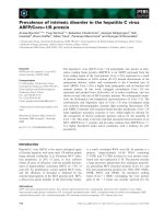
its cDNA. As shown in Fig. 1A, there are five introns and
six exons in the IL-28A gene. The first ATG starts from
nt.53 and ends at nt.655 (Fig 1B). The predicted amino
acid sequence was identical to that published by Sheppard
et al. [3].
IL-28A exhibits anti-HCV activity
Since IL-28A is a new member of type I interferon family
and IFNα is widely used to treat HCV infection, it is logical
to examine its anti-HCV activity. We first tested whether
the IL-28A DNA is functional in human hepatoma cells.
The eukaryotic expression vectors containing either the
genomic DNA or cDNA were transfected into a HCV
Virology Journal 2005, 2:80 />Page 3 of 12
(page number not for citation purposes)
subgenomic replicon cell line, GSB1. The control cells
were transfected by pTOPO vector without any insert
sequence. Forty-eight hours after transfection, total RNA
was isolated, followed by real-time RT-PCR analysis. As
shown in Fig. 2A, compared with the control plasmid-
transfected cells, the IL-28A transfected cells had signifi-
cantly lower viral RNA copy numbers. The viral suppres-
sion effect was also demonstrated by viral NS5A protein
expression, as determined by Western blot analysis (Fig.
2B). To further determine the effect of IL-28A secreted by
cells, we then tested the antiviral effect of the conditioned
medium. We subcloned IL-28A cDNA into pEF/V5-His-
TOPO vector and generated the plasmid, pTOPO-IL-
28A07. The plasmid pTOPO-IL-28A07 was transfected
into Huh-7 cells, and the supernatant was harvested after
72 hours of incubation. Varying amounts of the condi-
tioned-medium from Huh7 cells transfected with plasmid
pTOPO or pTOPO-IL-28A07 were added to GSB1 cells.
After 48 hours of incubation, total RNA was isolated from
the cells, followed by real-time RT-PCR analysis. As shown
in Fig 2C, the IL-28A-conditioned medium demonstrated
a significant inhibitory effect on HCV RNA replication in
a dose-dependent manner. Similar results were also
obtained using pTOPO-IL28A, the genomic expression
construct (data not shown). We then further examined the
effect of the recombinant IL-28A on HCV RNA replication
by incubating GSB1 cells with varying doses of rhIL-28A,
followed by total RNA extraction and real-time PCR anal-
ysis. As shown in Fig. 2D, the replication levels of HCV
RNA were significantly suppressed by rhIL-28A. Again, IL-
IL-28A gene structureFigure 1
IL-28A gene structure. A) Schematics of the exon-intron structure of the gene. The numbers indicate exon location. B)
Complete sequence of IL-28A genomic sequence (Accession number DQ126336). The bold nucleotides are the nucleotide
sequence of the exons (Accession number DQ126337).
Virology Journal 2005, 2:80 />Page 4 of 12
(page number not for citation purposes)
28A inhibits the viral RNA replication in a dose-depend-
ent manner, but the effective dose of rhIL28A is signifi-
cantly higher than recombinant IFN.
For simultaneous assessment of cap-dependent and HCV
IRES-dependent translation, Huh7 cells were transiently
transfected with a bicistronic reporter plasmid, pRL-HL,
encoding the Renilla and firefly luciferase cDNAs trans-
lated from the 5'cap and internally from the HCV IRES,
respectively, for 24 hours, followed by 24 hours of incu-
bation in medium alone or with medium containing
increasing amount of hrIL-28A. Cells were harvested, and
protein extracts were prepared, and a dual luciferase assay
using the luciferase assay system was performed. As
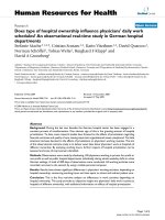
shown in Fig. 3, translation from the viral IRES elements
exhibited a dose-dependent suppression, while the cap-
dependent translation is not significantly affected by IL-
28A. These data suggest that IL-28A appears to specifically
inhibit HCV IRES-mediated translation without affecting
cap-mediated translation in the host cells.
IL-28A activates the JAK-STAT signaling pathway
It is known that type I interferons initiate cellular
responses at least partially through the JAK-STAT pathway.
All the human type I IFNs interact with the same receptor,
IFNAR [27]. When IFNs bind to specific cell surface recep-
tors on the host cells, the IFNAR receptor complex will
activate the JAK proteins, JAK1 and TYK2. The activated-
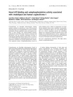
Effects of IL-28A on HCV RNA replication and protein expressionFigure 2
Effects of IL-28A on HCV RNA replication and protein expression. A) GSB cells were transfected by either control
plasmid (TOPO) or IL-28A genomic expression construct (TOPO-hIL28A). After 48 hours, total RNA was isolated, followed
by real-time PCR analysis with HCV-specific primers. The data represents the normalization with the internal control GADPH.
B) Western blot analysis of GSB1 cells transfected with the control plasmid (TOPO) or IL-28A expression construct (TOPO-
IL28A). The monoclonal antibody is specifically against HCV NS5A. The internal control is actin. C) Effect of IL-28A-condi-
tioned medium on HCV RNA replication in GSB cells. The conditioned medium was used to treat the cells for 48 hours, fol-
lowed by real-time RT-PCR analysis. D) Effect of recombinant IL-28A on HCV RNA replication in GSB cells. The relative HCV
RNA levels were normalized with the internal control GADPH. The error bars indicate the variations of three independent
assays.
Virology Journal 2005, 2:80 />Page 5 of 12
(page number not for citation purposes)
JAK proteins then phosphorylate STAT1, STAT2, and
STAT3. We hypothesized that the IL-28A-induced antiviral
effect in GSB1 cells would depend upon the activation of
the JAK-STAT signaling pathway. We, therefore, analyzed
the status of STAT1 and STAT3 in response to IL-28A stim-
ulation. Huh7 or GSB1 cells were treated with pTOPO-IL-
28A07 conditioned medium or rhIL-28A for 30 minutes,
followed by total protein extraction and Western blot
analysis using anti-p-STAT1, anti-p-STAT3, total STAT1
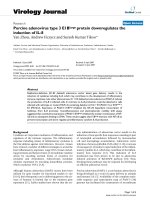
and STAT3 monoclonal antibodies. As shown in Fig. 4,
both phosphorylated STAT1 and STAT3 were detected in
cells treated with IL-28A. This result indicates that IL-28A
utilizes the similar JAK-STAT signaling pathway as the
IFN-α and IFN-β, despite receptor differences.
IL-28A induces interferon stimulated genes (ISGs)
expression
The transcription factor IFN-stimulated gene factor 3 com-
plex (ISGF3), consisting of phosphorylated STAT1, phos-
phorylated STAT2, and IRF-9/p48, translocates into the
nucleus and binds to IFN-stimulated response elements
(ISRE) within the promoters of ISGs [9]. Interferons exert
their biological function through induction of ISGs in the
cell. Therefore, it is possible that IL-28A provides antiviral
activity by induction of a subset of IFN-stimulated genes
(ISGs). To determine whether IL-28A can induce the ISGs,
total RNA was isolated from the cells treated by IL-28A-
conditioned medium from Huh7 cells transfected with
pTOPO-IL-28A, followed by semi-quantitative RT-PCR
analysis using gene specific primer sets for 6–16, 1–8U, 1–
8D, and IFIT1. As shown in Fig. 5, 6–16 and 1–8U were
significantly induced by IL-28A, while the gene IFIT1 was
not effectively induced. This observation suggests that IL-
28A is capable of inducing ISGs, but the gene profile may
not be identical to that of IFN.
IL-10 has no effect on the IL-28A-induced anti-HCV
activity
The IL-28A receptor complex consists of a ligand-binding
chain, IL-28R, and an accessory receptor chain, IL-10R2.
So it is logical to determine whether IL-10 interferes with
IL-28A in inhibiting HCV RNA replication. GSB1 cells
were treated with or without IL-28A (100 ng/mL and 300
ng/mL) in the presence or absence of 100 ng/mL IL-10.
After 72 hours of incubation, total RNA was isolated from
the cells, followed by real-time RT-PCR analysis. As shown
in Fig. 6, IL-10 did not have a significant effect on the IL-
28A-induced anti-HCV activity, while IL-28A can decrease
RNA replication,
IL-28A synergies with IFN-
α
in suppressing HCV RNA
replication
The above results indicated that IL-28A can signal through
the JAK-STAT pathway in a similar manner as to IFN-α. To
determine whether IL-28A enhances IFN-α-induced anti-
HCV RNA replication, we tested the effect of IL-28A on
Effects of IL-28A on CAP-dependent and HCV IRES-dependent translationFigure 3
Effects of IL-28A on CAP-dependent and HCV IRES-dependent translation. The GSB1 cells was transfected with
control plasmid or plasmid pRL-HL, which has different luciferases directed by either CAP- or HCV IRES. After 48 hours of
transfection, the cells were treated with varying doses of IL-28A. Cell extracts were made after 24 hours of incubation, fol-
lowed by luciferase determination. The data represents the average of three independent experiments. The open column indi-
cates CAP-mediated translation. The shadowed column indicates HCV IRES-mediated translation. The error bars indicate the
variations of three independent assays.
Virology Journal 2005, 2:80 />Page 6 of 12
(page number not for citation purposes)
IFN-α-induced anti-HCV RNA activity using GSB1 cell.
IFN-α was used at the dose of 50 U/mL, 100 U/mL with or
without 100 ng/mL IL-28A. After 24 hours of incubation,
cells were harvested and total RNA was isolated, followed
by real-time RT-PCR analysis using HCV-specific primers.
As shown in Fig. 7, the combination of IL-28A and IFN-α
reduced HCV viral RNA by 100-fold, while IFNα alone
reduced the virus by 15-fold and IL-28A alone by 6-fold.
Activation of STAT1 protein by phosphorylation is a criti-
cal step for IFN-α signaling pathway. In the next experi-
ment, we examined the effect of IL-28A on the IFN-α-
induced STAT1 phosphorylation by Western blotting. As
shown in Fig. 8, the levels of p-STAT1 were significantly
higher than those induced by IFN-α or IL-28A alone. In
addition, STAT1 remained phosphorylated for 8 h after
stimulation with IFN-α plus IL-28A, while STAT1 phos-
phorylation induced by IFN treatment alone decreased to
undetectable level (data not shown). The results indicate
that IL-28A can synergize with IFN-α in suppressing the
HCV RNA replication and inducing intracellular antiviral
signaling pathway.
IL-28A induces HLA class I antigen expression
Type I interferons are believed to play a role in immune
regulation. One of the mechanisms is through induction
of HLA class I antigen. To test whether IL-28A has such an
effect, we treated Huh7 cells with IL-28A-conditioned
medium from Huh7 cells transfected by plasmid pTOPO-
IL-28A, followed by flow cytometric analysis using anti-
HLA class I antigen. As shown in Fig. 9, treatment with IL-
28A induced HLA class I antigen production. The data
suggest that IL-28A has a similar capacity to induce class I
antigen production as other type I IFN.
The effect of IL-28A on JAK-STAT signaling pathwayFigure 4
The effect of IL-28A on JAK-STAT signaling pathway. GSB1 cells were treated with either IL-28A conditioned medium
(A) or recombinant rIL-28A (B) for 30 minutes, followed by protein extraction and Western blot analysis using antibodies as
indicated in the figure. Equal amounts (20 ug) of proteins were loaded in each lane and confirmed by detection of actin. STAT1
or STAT3 indicates total STAT protein. p-STAT1 or p-STAT3 indicates tyrosine phosphorylated form (activated STAT pro-
tein). The figures are representatives of at least four independent experiments.
Virology Journal 2005, 2:80 />Page 7 of 12
(page number not for citation purposes)
Discussion
Type I interferons play an essential role in innate immune
responses against viral infections. There are many sub-
types of type I interferons in humans, including the
recently identified IFN-λ, consisting of three members, λ1
(IL-29), λ2 (IL-28A), and λ3 (IL-28B). The most exten-
sively studied subtypes are IFN-α and IFN-β. There is
relatively little information available for IFN-λ. The major
difference between IFN-λ and the other type I IFNs is the
utilization of different receptors. Current type I interferon
therapy has significant side effects. Identification of novel
type I interferons with desirable clinical efficacy and less
side effects is needed. IFNλ s are potentially such
candidates.
In this report, we have cloned both the cDNA and the
gene of human IL-28A. Through a series experiments, we
have shown the biological effects of this protein on HCV
viral replication, its signaling events in human liver cells,
and its interaction with IFN-α and IL-10.
To clone this gene, we employed a RT-PCR approach
using total RNA extracted from spleen, liver, and periph-
eral blood mononuclear cells (PBMC). With extensive
Induction of interferon stimulated genes by IL-28A in GSB1 cellsFigure 5
Induction of interferon stimulated genes by IL-28A in GSB1 cells. GSB1 cells were treated by either control or 2-ml
IL-28A-conditioned medium for 12 hours. Total RNA was isolated, followed by RT-PCR analysis using a pair of gene-specific
primers and a pair of DADPH primers. The PCR amplification cycle is 25, which ensures PCR reaction in linear range. The PCR
products were analyzed in 1% agarose gel. M indicates the DNA molecular weight marker. The arrow indicates gene-specific
products. The bar indicates GADPH DNA fragment. The figure is a representative of two independent assays.
Effect of IL-10 on IL-28A-induced antiviral activityFigure 6
Effect of IL-10 on IL-28A-induced antiviral activity. The GSB1 cells were treated with varying doses of IL-10 or IL-28A
as indicated for 72 hours. Total RNA was isolated for real-time PCR analysis using HCV-specific primers. The data represents
the normalization with internal control GADPH. The error bars indicate the variations of three independent assays.
Virology Journal 2005, 2:80 />Page 8 of 12
(page number not for citation purposes)
effort, we could only obtain IL-28A genomic clones but
not cDNA, while we could readily amplify IFN-α and IFN-
β cDNA from the same RNA source. We confirmed that
the amplified genomic clones were derived from the resid-
ual DNA in the RNA preparation, since two rounds of
DNase I treatment eliminated the amplification. This
indicates that there is no detectable IL-28A expression in
these tissues at a normal physiological condition,
although it has been reported that IL-28A is expressed in
PBMCs from HCV-infected patients [28]. To obtain the
cDNA clone, we decided to clone the genomic DNA into
a expression vector, and then transfected it into Huh7
cells. Total RNA was extracted from the transfected cells
and RT-PCR was performed. The cDNA DNA fragment
was easily amplified using this approach. We noticed that
Kotenko et al. used a similar strategy to clone the first IL-
28A cDNA [2]. By comparing the cDNA and its gene, we
identified five introns and six extrons. So far, this is the
only type I interferon gene containing introns, while the
other type I IFNs encode within a single extron. The pres-
ence of multiple introns makes this gene more similar to
IL-10 gene family. Interestingly, the IL-28A receptor
shares one subunit with IL10 (IL-10Rβ). We know that IL-
10 and type I interferons play a different role during the
host immune responses to viral infections. The presence
of introns generally subjects the gene to an additional
gene expression control. According to Kotenco et al., the
IL-28A is predominantly expressed in the heart, liver and
spleen [2]. Whether the introns play any role in such rela-
tively tissue-restricted expression remains to be
investigated.
Effect of IL-28A on the antiviral activity of IFNαFigure 7
Effect of IL-28A on the antiviral activity of IFNα. Varying doses of IL-28A and IFNα2b, either alone or in combination,
were added to the GSB1 cells and incubated for 48 hours. Total RNA was isolated for real-time PCR analysis. The vertical axis
represents the fold of viral RNA reduction by IL-28A or IFN. The data represents the results of normalization with the internal
control GADPH.
Effect of IL-28A on IFNα-induced STAT1 activation in GSB1 cellsFigure 8
Effect of IL-28A on IFNα-induced STAT1 activation
in GSB1 cells. GSB1 cells were treated with IL-28A or/and
IFN as indicated. After 30 minutes of incubation, total protein
was extracted for Western blot analysis using antibodies
against total STAT1 (STAT1) or phosphorylation-specific
STAT1 (p-STAT1).
Virology Journal 2005, 2:80 />Page 9 of 12
(page number not for citation purposes)
After cloning this gene, we then showed that the gene
product, IL-28A, has similar biological properties as other
type I interferons. IL-28A resembles type I IFNs in its abil-
ity to induce anti-HCV activity through JAK-STAT signal-
ing pathway. As we have shown in Fig. 4, IL-28A activates
both STAT1 and STAT3. The IL-28A-mediated antiviral
activity is dose-dependent. Both the recombinant and the
gene product produced in liver cells are effective, though
the effective dose of the recombinant IL-28A is much
higher than the other type I interferons. Similar results
were recently reported by other laboratories [29,30].
We further analyzed the expression of ISGs using a RT-
PCR approach. Interestingly, at least one ISG cannot be
induced by IL-28A, while it can be readily induced by
IFNα. Moreover, by testing the effect of interferons on
cap-mediated translation and HCV IRES-mediated
translation, our preliminary data showed that IL-28A
appears to have a selective activity to inhibit HCV-IRES-
mediated translation, while it did not affect cap-mediated
translation. This observation is consistent with the fact:
even at higher dose (1000 ng/ml), IL-28A did not exhibit
antiproliferation activity in a human hepatoma cell line
(data not shown). These data suggest that IL-28A seems to
have at least some different biological activities as com-
pared with IFN-α. Whether these differences can be
employed to achieve therapeutic advantage remains to be
determined.
As we have mentioned above, the receptor for IL-28A
shares a common subunit with IL-10. Our previous study
showed that IL-10 did not have direct antiviral activity in
patients with chronic HCV infection [31]. We asked the
question whether the sharing of a receptor has any impact
on IL-28A activity. Our data suggests that IL-10 does not
have an antiviral effect in HCV replicon cells, nor does it
have any interference with IL-28A antiviral effect. Thus,
the significance of receptor sharing remains unknown.
Since IL-28A and other type I interferon use different
receptor for signaling transduction, we next examined the
combination effect of IL-28A and IFN-α. Interestingly,
combination of IL-28A and IFN-α exhibited synergistic
effect on JAK-STAT activation and anti-HCV activity. As
shown in Figure 7, combination of 50 U IFN-α and 100 ng
per milliliter IL-28A reduced HCV RNA by 40 folds, while
individual IFN-α and IL-28A reduced HCV RNA by 10-
fold and 6-fold, respectively. We do not know the precise
mechanism of this synergistic effect, though the STAT1
activation shows the similar synergistic effect (Fig. 8). It is
possible that the activation of one receptor may have
beneficial effect on the other receptor-mediated pathway.
It is also possible that the common downstream mole-
cules shared by both pathways can synergistically induced
by these two interferons. This synergistic effect has a
significant clinical implication. It is tempting to speculate
that combination of these two reagents may have
Effect of IL-28A on HLA class I antigen expression in Huh7 cellsFigure 9
Effect of IL-28A on HLA class I antigen expression in Huh7 cells. The Huh7 cells were treated with 2 ml IL-28A-con-
ditioned culture medium for 72 hours. The cells were then harvested and incubated with HLA class I antigen-specific antibody
labeled by FITC fluorescence, followed by flow cytometric analysis. The arrow-marked curve indicates control cells. The
arrowhead-marked curve indicates cells treated with IL-28A.
Virology Journal 2005, 2:80 />Page 10 of 12
(page number not for citation purposes)
therapeutic benefit for HCV therapy, particularly in the
setting of IFN resistance.
Type I interferons have an immunoregulatory function
[32,33]. One of the mechanisms is through induction of
HLA class I antigens [34]. We tested whether IL-28A has a
similar activity. Human hepatoma cells have relatively
lower HLA class I antigen expression comparing with nor-
mal hepatocytes [35]. Treatment of the hepatoma cells
increased class I antigen expression through flow cytomet-
ric study. Not only this shows that the IL-28A has immu-
noregulator effect, but the fact that IL-28A can induce HLA
class I antigen in tumor cells may implicate the role of IL-
28A in tumor immune therapy. It would be interesting to
see whether IL-28A is capable of promoting the host anti-
tumor immunity.
Conclusion
Our study shows the gene structure of IL-28A, its antiviral
effect on HCV, its signaling transduction pathway, and the
induction of ISGs. More importantly, we demonstrate the
synergistic effect of IL-28A and IFNα on anti-HCV activity,
which has a potential clinical application. IFN-α is cur-
rently used for the treatment for chronic HCV infection,
HBV infection, and many malignant tumors, including
hepatitis B, melanoma, hairy cell leukemia, and non-
Hodgkin's lymphoma. IL-28A is a potential therapeutic
agent to treat these clinical diseases.
Methods
Cell cultures, reagents and plasmids
The HCV subgenomic replicon cell line, GSB1, was a gift
from Dr. Christopher Seeger [36,37]. All cells were propa-
gated in DMEM supplemented with 10% FBS, 200 µM L-
glutamine, nonessential amino acids, penicillin and strep-
tomycin. Culture of the replicon cells has been previously
described [15]. The expression vector, pEF6/V5-His-
TOPO, was obtained from Invitrogen (Carlsbad, CA). The
HCV-NS5A-specific monoclonal antibody was generated
in the laboratory. Monoclonal antibodies against actin,
STAT1, STAT3 and phosphorylated STAT3 were obtained
from Santa Cruz Biotechnology (Santa Cruz, CA). The
antibodies against phosphorylated STAT1 were obtained
from Upstate (Charlottesville, VA). The secondary anti-
body goat anti-mouse or anti-rabbit IgG-HRP was from
Santa Cruz Biotechnology. Supersignal West Pico Chemi-
luminescent Substrate was purchased from Pierce Biotech-
nology, Inc. (Rockford, IL). Recombinant human IL-28A
(rhIL-28A) and hIL-10 were purchased from R&D Systems
(Mineanapolis, MN). The plasmid pRL-HL (a gift from Dr.
Lemon) is a bicistronic expression construct encoding
Renilla and firefly luciferase cDNAs translated from 5'cap
and internally from the HCV IRES (internal ribosome
entry site), respectively [38].
Amplification of human IL-28A DNA, cDNA, and plasmid
construction
RNA was isolated from human spleen. The human IL-28A
cDNA was amplified by RT-PCR from human spleen RNA
using two primers: 5'-GGGTGACAGCCTCAGAGTG-3', 5'-
ATAGCGACTGGGTGGCAATA-3'. Superscript One-Step
RT-PCR kit with platinum Taq according to the instruc-
tions (Invitrogen). The One-Step RT-PCR conditions were
as follows: 50°C, 30 min; 94°C, 4 min; followed with 40
cycles (95°C, 30 s; 55°C, 30 s; 72°C, 1 min;). The IL-28A
DNA was ligated into pEF6/V5-His-TOPO vector. The
expression vector pTOPO-IL-28A were transfected into
Huh7 cells using Lipofectin Reagent (Invitrogen) accord-
ing to the manufacturer's instruction. The total RNA was
purified from Huh7 cells transfected by pTOPO-IL-28A
for 48 hours and treated by DNase I. The human IL-28A
cDNA was generated by RT-PCR from the total RNA pre-
treated by DNase using the above primers. The reactions
were performed using 72°C, 7 mins. The expression vec-
tor pTOPO-hIL-28A 0.7 was constructed by inserting the
human IL-28A cDNA into pEF6/V5-His-TOPO.
Human IL-28A DNA Sequencing
The IL-28A DNA was amplified as described above. The
expression vector TOPO-hIL-28A was sequenced using
The BigDye Terminator V3.1 Kit from Applied Biosystems
(Foster City, CA). The reaction condition was: 96°C, 10 s;
50°C, 5 s; 60°C, 4 min, total 25 cycles. After that, 1/20
volume of 3 M sodium acetate (pH5.2) and 3 times
volume of ethanol were added, and incubated at -20°C
for 30 mins, followed by spinning down at 13000 g at
4°C for 30 mins. The DNA pellet was washed using 70%
ethanol and dried by vacuum. The sequence was detected
by ABI PRISM 377 DNA Sequencer (Applied Biosystems).
DNA transfection
The transfection protocol has been described previously
[39,40]. Briefly, GSB or Huh7 cells were transfected with
control plasmid pTOPO, pTOPO-IL-28A or pTOPO-IL-
28A07 plasmid using Lipofectin. In a 6-well tissue culture
plate, 1 × 10
5
GSB or Huh7 cells were seeded in 2 ml of
DMEM supplemented with serum and incubate at 37°C
in an incubator overnight. For each transfection, 2 µg of
DNA was used. The plasmid, pTOPO, pTOPO-IL-28A, or
pTOPO-IL-28A07 was transiently transfected into GSB or
Huh7 cells. The transfected cells were incubated for
another 48 hours before experiments.
Reverse Transcription and Polymerase Chain Reaction
(RT-PCR)
Total cellular RNA was purified from cells. After reverse
transcription, cDNA was used for PCR. The primers are for
6–16 (G1P3), forward 5'-AACCGTTTACTCGCTGCTGT-3,
reverse 5'-GCTGCTGGCTACTCCTCA-3'; for 1–8U, for-
ward 5'-CAAATGCCAGGAAAAGGAA-3', reverse 5'-ATA-
Virology Journal 2005, 2:80 />Page 11 of 12
(page number not for citation purposes)
CAGGTCATGGGCAGAGC; for 1–8D, forward 5'-
TGCCAGGAA GAGGAAACTGT-3', reverse 5'-CCTCAAT-
GATGCCTCCTGAT-3'; for IFIT1, forward 5'-TCTCAGAG-
GAGCCTGGCTAA-3', reverse 5'-AGTGGCTGATATCT
GGGTGC-3'; for GAPDH, forward 5-TCACCAGGGCT-
GCTTTTA-3', reverse 5'-TTCACACCCATGACGAACA-3'.
The PCR conditions were as follows: 94°C, 4 min; (95°C,
30 s; 55°C, 30 s; 72°C, 1 min;) × 40 cycles; 72°C, 7 mins.
The PCR product was detected on 2% agarose gel.
Quantitative Real-Time PCR
Total cellular RNA was isolated from cells as described
before. Real-time PCR was preformed as described previ-
ously [39]. Briefly, first-strand cDNAs were synthesized
from total cellular RNA by reverse transcription (20 µl of
reaction volume) using the Superscript II (50 U reverse
transcriptase per reaction) first-strand synthesis for RT-
PCR kit (Invitrogen) primed with oligo (dT)
12–18
(Invitro-
gen) according to the manufacturer's instructions. Fluoro-
phore-labeled LUX primers and their unlabeled
counterparts were obtained from Invitrogen. Reactions
were conducted in a 96-well spectrofluorometric thermal
cycler (ABI PRISM 7700 Sequence detector system,
Applied Biosystems). Fluorescence was monitored during
every PCR cycle at the annealing step. The primers for
HCV are: 5'-CGCTCAATGCCTGGAGATTTG-3', 5'-
GCACTCGCAAGCACCCTATC-3'; for GADPH: 5'-TGCT-
GGCGCTGAGTACGTC-3', 5'-GTGCAGGAGGCATT-
GCTGA-3'. PCR conditions were as follows: 50°C, 2 min;
95°C, 10 min; (95°C, 15 s; 60°C, 1 min) × 40 cycles.
Results were analyzed with SDS 2.0 software from Applied
Biosystems. Results for all experiments represent triplicate
determinations. Results are represented as means ± SD.
Western Blot Analysis
Equal numbers of cells were washed with PBS and lysed in
RIPA buffer as described previously [15]. Protein extrac-
tion from cells, electrophoresis and Western blot analysis
were described previously. Approximately 20 µg of pro-
tein were electrophoresed on a 8% SDS-polyacrylamide
gel and transferred to polyvinylidene difluoride mem-
brane (Bio-Rad). The membrane was incubated overnight
at 4°C in a block buffer (TBS containing 0.1% Tween 20
and 5% fat-free milk power). The blots were probed with
monoclonal antibodies specific for NS5A, STAT1, and
STAT3, p-STAT3, actin or polyclonal antibody specific for
p-STAT1 for 1 hour at room temperature. After being
washed 3 times for 30 min each with 0.1% Tween 20 in
TBS, the membrane was incubated with the secondary
antibody diluted in 5% fat-free milk in TBS containing
0.1% Tween 20 for 1 hour at room temperature and
washed 3 times as described above. Proteins were visual-
ized by using Supersignal West Pico Chemiluminescent
Substrate.
Flow cytometry
To detect the expression of MHC class I antigen, Huh7
cells were treated with IL-28A conditioned medium from
Huh7 cells transfected by plasmid pTOPO-IL-28A for 72
hours and their MHC class I expression was analyzed by
flow cytometry as previously described [41]. Cell surface
expression of the HLA class I antigens were detected using
class I antibody, followed by fluorescein isothiocyanate
(FITC)-conjugated goat anti-rabbit IgG. Ligand binding
was detected by flow cytometry.
Acknowledgements
We thank Drs. James Crawford, Jinxiong She, John Elyer, and Christopher
Seeger for the helpful discussion. The work was supported in part by the
Charles Trey MD Memorial liver scholar award from American Liver Foun-
dation and DK02958 from NIH to C.L.
References
1. Brassard DL, Grace MJ, Bordens RW: Interferon-alpha as an
immunotherapeutic protein. J Leukoc Biol 2002, 71:565-581.
2. Kotenko SV, Gallagher G, Baurin VV, Lewis-Antes A, Shen M, Shah
NK, Langer JA, Sheikh F, Dickensheets H, Donnelly RP: IFN-lamb-
das mediate antiviral protection through a distinct class II
cytokine receptor complex. Nat Immunol 2003, 4:69-77.
3. Sheppard P, Kindsvogel W, Xu W, Henderson K, Schlutsmeyer S,
Whitmore TE, Kuestner R, Garrigues U, Birks C, Roraback J,
Ostrander C, Dong D, Shin J, Presnell S, Fox B, Haldeman B, Cooper
E, Taft D, Gilbert T, Grant FJ, Tackett M, Krivan W, McKnight G,
Clegg C, Foster D, Klucher KM: IL-28, IL-29 and their class II
cytokine receptor IL-28R. Nat Immunol 2003, 4:63-68.
4. Chen J, Baig E, Fish EN: Diversity and relatedness among the
type I interferons. J Interferon Cytokine Res 2004, 24:687-698.
5. Gray PW, Goeddel DV: Structure of the human immune inter-
feron gene. Nature 1982, 298:859-863.
6. Stark GR, Kerr IM, Williams BR, Silverman RH, Schreiber RD: How
cells respond to interferons. Annu Rev Biochem 1998, 67:227-264.
7. Foster GR, Finter NB: Are all type I human interferons
equivalent? J Viral Hepat 1998, 5:143-152.
8. Samuel CE: Antiviral actions of interferons. Clin Microbiol Rev
2001, 14:778-809, table of contents.
9. Darnell JEJ, Kerr IM, Stark GR: Jak-STAT pathways and tran-
scriptional activation in response to IFNs and other extracel-
lular signaling proteins. Science 1994, 264:1415-1421.
10. Larner A, Reich NC: Interferon signal transduction. Biotherapy
1996, 8:175-181.
11. Parmar S, Platanias LC: Interferons: mechanisms of action and
clinical applications. Curr Opin Oncol 2003, 15:431-439.
12. Williams BR, Haque SJ: Interacting pathways of interferon
signaling. Semin Oncol 1997, 24:S9-70-S9-77.
13. Liu KD, Gaffen SL, Goldsmith MA: JAK/STAT signaling by
cytokine receptors. Curr Opin Immunol 1998, 10:271-278.
14. Der SD, Zhou A, Williams BR, Silverman RH: Identification of
genes differentially regulated by interferon alpha, beta, or
gamma using oligonucleotide arrays. Proc Natl Acad Sci U S A
1998, 95:15623-15628.
15. Zhu H, Zhao H, Collins CD, Eckenrode SE, Run Q, McIndoe RA,
Crawford JM, Nelson DR, She JX, Liu C: Gene expression associ-
ated with interferon alfa antiviral activity in an HCV replicon
cell line. Hepatology 2003, 37:1180-1188.
16. Kochs G, Haller O: Interferon-induced human MxA GTPase
blocks nuclear import of Thogoto virus nucleocapsids. Proc
Natl Acad Sci U S A 1999, 96:2082-2086.
17. Sen GC: Viruses and interferons. Annu Rev Microbiol 2001,
55:255-281.
18. Durbin RK, Mertz SE, Koromilas AE, Durbin JE: PKR protection
against intranasal vesicular stomatitis virus infection is
mouse strain dependent. Viral Immunol 2002, 15:41-51.
19. Buelens C, Bartholome EJ, Amraoui Z, Boutriaux M, Salmon I, Thiele-
mans K, Willems F, Goldman M: Interleukin-3 and interferon
beta cooperate to induce differentiation of monocytes into
Publish with BioMed Central and every
scientist can read your work free of charge
"BioMed Central will be the most significant development for
disseminating the results of biomedical research in our lifetime."
Sir Paul Nurse, Cancer Research UK
Your research papers will be:
available free of charge to the entire biomedical community
peer reviewed and published immediately upon acceptance
cited in PubMed and archived on PubMed Central
yours — you keep the copyright
Submit your manuscript here:
/>BioMedcentral
Virology Journal 2005, 2:80 />Page 12 of 12
(page number not for citation purposes)
dendritic cells with potent helper T-cell stimulatory
properties. Blood 2002, 99:993-998.
20. Marrack P, Kappler J, Mitchell T: Type I interferons keep acti-
vated T cells alive. J Exp Med 1999, 189:521-530.
21. Alter MJ: The epidemiology of acute and chronic hepatitis C.
Clin Liver Dis 1997, 1:559-68, vi-vii.
22. Lechmann M, Liang TJ: Vaccine development for hepatitis C.
Semin Liver Dis 2000, 20:211-226.
23. Fried MW, Shiffman ML, Reddy KR, Smith C, Marinos G, Goncales
FLJ, Haussinger D, Diago M, Carosi G, Dhumeaux D, Craxi A, Lin A,
Hoffman J, Yu J: Peginterferon alfa-2a plus ribavirin for chronic
hepatitis C virus infection. N Engl J Med 2002, 347:975-982.
24. Manns MP, McHutchison JG, Gordon SC, Rustgi VK, Shiffman M, Rein-
dollar R, Goodman ZD, Koury K, Ling M, Albrecht JK: Peginter-
feron alfa-2b plus ribavirin compared with interferon alfa-2b
plus ribavirin for initial treatment of chronic hepatitis C: a
randomised trial. Lancet 2001, 358:958-965.
25. Pawlotsky JM: The nature of interferon-alpha resistance in
hepatitis C virus infection. Curr Opin Infect Dis 2003, 16:587-592.
26. Matthews SJ, McCoy C: Peginterferon alfa-2a: a review of
approved and investigational uses. Clin Ther 2004, 26:991-1025.
27. Pestka S, Langer JA, Zoon KC, Samuel CE: Interferons and their
actions. Annu Rev Biochem 1987, 56:727-777.
28. Mihm S, Frese M, Meier V, Wietzke-Braun P, Scharf JG, Barten-
schlager R, Ramadori G: Interferon type I gene expression in
chronic hepatitis C. Lab Invest 2004, 84:1148-1159.
29. Robek MD, Boyd BS, Chisari FV: Lambda interferon inhibits hep-
atitis B and C virus replication. J Virol 2005, 79:3851-3854.
30. Brand S, Zitzmann K, Dambacher J, Beigel F, Olszak T, Vlotides G,
Eichhorst ST, Goke B, Diepolder H, Auernhammer CJ: SOCS-1
inhibits expression of the antiviral proteins 2',5'-OAS and
MxA induced by the novel interferon-lambdas IL-28A and IL-
29. Biochem Biophys Res Commun 2005, 331:543-548.
31. Nelson DR, Tu Z, Soldevila-Pico C, Abdelmalek M, Zhu H, Xu YL,
Cabrera R, Liu C, Davis GL: Long-term interleukin 10 therapy in
chronic hepatitis C patients has a proviral and anti-inflam-
matory effect. Hepatology 2003, 38:859-868.
32. Biron CA: Interferons alpha and beta as immune regulators
a new look. Immunity 2001, 14:661-664.
33. Tompkins WA: Immunomodulation and therapeutic effects of
the oral use of interferon-alpha: mechanism of action. J Inter-
feron Cytokine Res 1999, 19:817-828.
34. Girdlestone J: Regulation of HLA class I loci by interferons.
Immunobiology 1995, 193:229-237.
35. Kurokohchi K, Carrington M, Mann DL, Simonis TB, Alexander-Miller
MA, Feinstone SM, Akatsuka T, Berzofsky JA: Expression of HLA
class I molecules and the transporter associated with antigen
processing in hepatocellular carcinoma. Hepatology 1996,
23:1181-1188.
36. Guo JT, Bichko VV, Seeger C: Effect of alpha interferon on the
hepatitis C virus replicon. J Virol 2001, 75:8516-8523.
37. Zhu Q, Guo JT, Seeger C: Replication of hepatitis C virus subg-
enomes in nonhepatic epithelial and mouse hepatoma cells.
J Virol 2003, 77:9204-9210.
38. Honda M, Kaneko S, Shimazaki T, Matsushita E, Kobayashi K, Ping LH,
Zhang HC, Lemon SM: Hepatitis C virus core protein induces
apoptosis and impairs cell-cycle regulation in stably trans-
formed Chinese hamster ovary cells. Hepatology 2000,
31:1351-1359.
39. Zhu H, Shang X, Terada N, Liu C: STAT3 induces anti-hepatitis
C viral activity in liver cells. Biochem Biophys Res Commun 2004,
324:518-528.
40. Zhu H, Liu C: Interleukin-1 inhibits hepatitis C virus subge-
nomic RNA replication by activation of extracellular regu-
lated kinase pathway. J Virol 2003, 77:5493-5498.
41. Shang XZ, Zhu H, Lin K, Tu Z, Chen J, Nelson DR, Liu C: Stabilized
beta-catenin promotes hepatocyte proliferation and inhibits
TNFalpha-induced apoptosis. Lab Invest 2004, 84:332-341.