Báo cáo hóa học: "Investigating the synchronization of hippocampal neural network in response to acute nicotine exposure" pdf
Bạn đang xem bản rút gọn của tài liệu. Xem và tải ngay bản đầy đủ của tài liệu tại đây (539.21 KB, 6 trang )
JNER
JOURNAL OF NEUROENGINEERING
AND REHABILITATION
Akkurt et al. Journal of NeuroEngineering and Rehabilitation 2010, 7:31
/>Open Access
RESEARCH
© 2010 Akkurt et al; licensee BioMed Central Ltd. This is an Open Access article distributed under the terms of the Creative Commons
Attribution License ( which permits unrestricted use, distribution, and reproduction in
any medium, provided the original work is properly cited.
Research
Investigating the synchronization of hippocampal
neural network in response to acute nicotine
exposure
David Akkurt
1
, Yasemin M Akay
2
and Metin Akay*
1,2
Abstract
Previous studies suggested that γ oscillations in the brain are associated with higher order cognitive function including
selective visual attention, motor task planning, sensory perception, working memory and dreaming REM sleep. These
oscillations are mainly observed in cortical regions and also occur in neocortical and subcortical areas and the
hippocampus. In this paper, we investigate the influence of acute exposure to nicotine on the complexity of
hippocampal γ oscillations.
Using the approximate entropy method, the influence of acute nicotine exposure on the hippocampal γ oscillations
was investigated. The hippocampal γ oscillations have been generated in response to the 100 Hz stimulus and isolated
using the visual inspection and spectral analysis method. Our central hypothesis is that acute exposure to nicotine
significantly reduces the complexity of hippocampal γ oscillations. We used brain-slice recordings and the approximate
entropy method to test this hypothesis. The approximate entropy (complexity) values of the hippocampal γ oscillations
are estimated from the 14 hippocampal slices. Our results show that it takes at least 100 msec to see any hippocampal
activities in response to the 100 Hz stimulus. These patterns noticeably changed after 100 msec until 300 msec after
the stimulus Finally, they were less prominent after 300 msec. We have analyzed the isolated hippocampal γ oscillations
(between 150 and 250 msec after the stimulus) using the approximate entropy (ApEn) method. Our results showed
that the ApEn (complexity) values of hippocampal γ oscillations during nicotine exposure were reduced compared to
those of hippocampal γ oscillations during control, and washout. This reduction was much more significant in
response to acute nicotine exposure (p < 0.05) compared to those during control and washout conditions. These
results suggest that the neural firing becomes regular and the hippocampal networks become synchronized in
response to nicotine exposure.
Introduction
Nicotine, C
10
H
14
N
2
, a tertiary amine compound, is a very
toxic, light yellow alkaloid which is produced in tobacco
plants as a result of the leaves' being damaged [1,2]. The
roots of the damaged plants synthesize nicotine as a reac-
tion to hormones released by the damaged tissue and
then transport it to the leaves. Nicotine is stored in con-
centrations of 2-8 percent by weight in the leaves which
becomes the primary psychoactive ingredient in tobacco
smoke. It is very soluble in water and nonpolar solvents.
It is absorbed rapidly by the body, either through the skin
or smoking, crosses the blood-brain barrier and stimu-
lates nicotinic-cholinergic receptors of the CNS, causing
an increase in heart rate, blood pressure and some cogni-
tive functions in humans and experimental animals [3].
Nicotine exerts its effects through the activation of nic-
otinic acetylcholine receptors (nAChRs) in the multiple
areas of the brain such as hippocampus, amygdala and
prefrontal cortex (PFC) [4]. These receptors are ion chan-
nels and their locations are very important as far as the
understanding of their physiological impact on neuronal
activity is concerned [4,5]. A series of studies have
pointed out the crucial role these areas play in working
memory function. Particularly, stimulation of the
(nACHRs) in the hippocampus was proven critical for
optimal memory performance [5,6].
* Correspondence:
1
Harrington Department of Bioengineering, Fulton School of Engineering ASU,
Tempe AZ, USA
Full list of author information is available at the end of the article
Akkurt et al. Journal of NeuroEngineering and Rehabilitation 2010, 7:31
/>Page 2 of 6
The mounting evidence suggests that the synaptic plas-
ticity, such as long-term potentiation (LTP), takes part
during the learning and memory process. LTP has been
investigated in detail in the Shaffer collateral CA1 region
of the hippocampus where nicotine enhances LTP induc-
tion and modulates the synaptic plasticity thereby play-
ing a crucial role on attention performance [7,8].
It has been proposed that higher cognitive functions,
such as learning, memory, attention and exploratory
behavior, could be represented in the CA1 region of the
hippocampal area of the brain by a distributed neuronal
network synchronization on an oscillatory mode as the
gamma-band or 40 Hz (30-80 Hz) oscillations [9].
Gamma oscillations appear primarily in the neocortex,
hippocampus, thalamus and related structures. In the
hippocampus, they are generated by the entrainment of
populations of mutually interconnected interneurons and
can be stimulated by brief high-frequency tetanic stimuli
to the Schaffer collateral CA1 pathway in hippocampal
slice preparations [9-11].
There is a strong link between nicotine administration,
neuronal nicotinic systems and higher cognitive func-
tions evident from many studies in rodents, primates,
zebrafish and humans [12-15]. Fries et al suggested that
nicotine plays an important role in regulating beta oscil-
lations which are associated with higher cognitive func-
tions. Gray et al suggested that nicotine influences
cognition by enhancing synaptic transmission. Radial-
arm maze is an important tool for the testing of the work-
ing memory in rats and it has shown a significant
improvement in this memory type with both acute and
chronic nicotine treatments [14-16]. However, nicotine-
induced improvement in attention in rats seemed to be
strain-dependent. Sprague-Dawley rats showed better
performance than the Lister hooded rats in attention
tests, which suggested that there may be crucial, strain-
related differences in the mechanisms of the nicotinic
receptors or neuronal systems related to higher cogni-
tive functions and nicotine reinforcement [17,18].
In non-smoking adults with ADHD (attention deficit
hyperactivity disorder), nicotine seemed to improve
attention [19]. In Alzheimer's Disease (AD), which is
characterized by loss of cognitive functioning, there is
strong evidence that nicotinic receptors play an impor-
tant role in the pathogenesis of the disorder. Postmortem
evaluations of the brains of AD patients have shown a
substantial loss in nicotinic receptors in cortex and stria-
tum. According to the growing amount of findings, accu-
mulation of amyloid plaques which are the baseline
indicator in the brain for AD is interfered by nicotine,
which is very promising for the future of AD patients
[20,21].
In many studies on the effect of nicotine in Schizo-
phrenic patients, despite many positive findings for atten-
tion and spatial processing, a majority of the tests related
to the measurements of memory functioning gave nega-
tive results [22]. An important body of evidence suggests
that, nicotinic receptors play a role in the pathophysiol-
ogy of Parkinson's Disease (PD), revealing that smoking
may provide some protection against the onset of PD.
However, a wider rage of intense studies needs to be per-
formed before drawing any significant conclusions [23].
There are some studies indicating nicotine's positive
effects in improving cognitive functioning in individuals
with Tourette's and Down's syndromes. In spite of prom-
ising results, these studies have been limited and more
thorough research is needed in these cases [24]. In the
current study, we have investigated the effect of nicotine
on the complexity of the neurons and the activity of the
gamma oscillations in the Schaffer CA1 cell line on the
hippocampal slices.
Methods
Preparation of slices
All experimental protocols were approved by the Institu-
tional Animal Care and Use Committee of Arizona State
University; Furthermore, all experiments were carried
out in accordance to such protocols. In this experiment,
Sprague Dawley rats age 23-36 days old were anesthe-
tized by isoflurane then immediately decapitated. With-
out delay the brain was removed and placed in chilled
(4°C) artificial cerebrospinal fluid (ACSF). The ACSF
solution consisted of the following 135 mM NaCl, 3 mM
KCl, 16 mM NaHCO
3
, 1 mM MgCl, 1.25 mM NaH
2
PO
4
,
2 mM CaCl
2
, and 10 mM Glucose [25]. The above solu-
tion was continuously bubbled with carbogen (95-5%
Oxygen - Carbon Dioxide gas mixture). This solution was
chosen because of its success in prior work as described
in [25]. The Brain tissue was cold sliced using a vibratome
(Vibratome 1000, Vibratome Co., St. Louis, MO, USA)
while immersed in chilled ACSF bulled with carbogen.
Coronal sections at 400 μm of the chilled brain tissue
were made using the vibratome followed by transverse
sectioning to create tissue containing hippocampus and
entorhinal cortex.
All Sectioning was done while the tissue was under the
protection of the chilled ASCF. The cut slices were imme-
diately transferred to a incubation chamber containing
ACSF, where the atmosphere was 95% Oxygen and 5%
Carbon Dioxide. The Slices were incubated in this cham-
ber at 24°C for 1 hour prior to recording. After the incu-
bation period a slice was transferred to a liquid-air
interface chamber to begin recording (Fine Science Tools
Inc., Foster City, CA, USA). The slice was suspended on a
nylon mesh in the recording chamber where ACSF bub-
bled with carbogen continually flowed past the slice at a
rate of 2-2.5 ml/min. Recording chamber temperature
was closely monitored and regulated by a feedback circuit
Akkurt et al. Journal of NeuroEngineering and Rehabilitation 2010, 7:31
/>Page 3 of 6
set at 34 ± .3°C. The slice was allowed 15 minutes of rest
before any stimulation was made. In this experiments we
used (-) nicotine (Sigma Chemical Co., St. Louis, MO).
Electrophysiology
Using borosilicate glass electrodes (borosilicate micropi-
pettes, WPI, Sarasota, FL) with a pulled tip of about 1 μm,
extracellular field potential recordings were made on the
stratum pyramidale of the CA1 cell layer. The micropi-
pette was filled with a standard 2 M NaCl solution [25].
The slices were stimulated with double intensity (2 times
threshold, 2 T) which is approximately between 8 V and
14 V. Double intensity (2 T) was chosen due to its ability
to induce γ oscillations. Once the stimulation voltage was
set for a particular slice it was not changed for the
remainder of the experiments on that slice. The stimulat-
ing voltage was delivered via bipolar twisted platinum
wire electrode on the Schaffer collaterals. In order to
induce tetanus, the stimulation signal is changed from a
single pulse lasting a 100 μ second to a train of 100 Hz
signal lasting 200 ms delivered by a model 2100 A-M Sys-
tems Isolated Pulse Stimulator (Carlsborg, WA) [25-30].
The tetanic stimulation level is determined by the thresh-
old level voltage that must be applied to the Schaffer col-
laterals to elicit a response. In order to induce γ
oscillations, the stimulation intensity needs to be set at
above threshold and double intensity stimulation was
used. Signals were recorded using an Axoclamp 900A
(Axon Instruments, Inc., Union City, CA, USA) amplifier.
Prior to each drug experiment a single stimulus (8-14 V,
100 μs) was delivered to the slice to insure the slice was
viable and the recording and stimulating electrodes were
in the optimum positions to record synaptic transmis-
sion. Once the optimum electrode placement was deter-
mined, signal stability was insured by recording anywhere
from 20-60 min prior to actual nicotine administration.
After the recordings were stabilized the chemical was
started and tetanic stimulations were applied every 10
minutes. The concentration of the nicotine used in this
experiment was 100 μ Molar. Only a single concentration
was used because previously it was shown by Song et al.
that 100 μ Molar was the minimum dose that would
cause tetanic beta oscillations. Nicotine was dissolved in
ACSF and delivered to the slice at the same rate as stan-
dard ACSF solution without drug [27-30].
Data acquisition and analysis
All signals were recorded with pClamp 10.2 via Axon
Digidata 1440A (Axon Instruments, Inc., Union City, CA,
USA). The recording parameters were set such that the
sampling frequency was 100 kHz and low pass filtered
with a cut off at 1 kHz. All signals were then transferred
to a PC for later analysis. Offline analysis was done on
Matlab using various tools such as approximate entropy
and Matching Pursuit techniques.
Approximate entropy
Quantitative changes in the complexity of a signal are tra-
ditionally quantified using tools such as nonlinear
dynamical system analysis methods. These traditional
complexity measures work well when signals are long in
duration as they are length dependent[31-33]. For biolog-
ical signals with short signal durations of 100-5000
points, these traditional complexity measures that mea-
sure signal irregularity don't work well. A statistically effi-
cient measure to quantify the irregularity of a signal has
been formulated to overcome these shortcomings of
these previous complexity measures, commonly known
as approximate entropy [31-33].
To analyze the field potential recordings generated by
the hippocampal CA1 layer approximate entropy (ApEn)
was ultimately used. ApEn is a statistical measure that
both smoothens the transient interference and sup-
presses the influence of noise by properly adjusting the
algorithms parameters. This said irregularity measure
can be applied both to deterministic and stochastic sig-
nals [33,34]. Considering that the outputs of a biological
systems can be rather complex and could be either deter-
ministic or stochastic or both, thus it is crucial that this
complexity measure be able to handle both signal types.
The algorithm summarizes a time series into a non-nega-
tive number, with higher values representing more irreg-
ular systems [34].
Approximate entropy is calculated by taking segments
X(i) through X(N - m + 1) defined by X(i) = [x(i), , x(i +
m - 1)]. The difference between X(i) and X(j), d[X(i), X(j)]
as the maximum absolute difference between their
related scalar elements can be estimated as:
assuming that all the differences between the corre-
sponding elements will be less than the threshold r.
For any given X(i), the ratio of the difference between
X(i) and X(j) smaller than the threshold r to the total
number of vectors (N - m +1) is obtained as:
The approximate entropy, ApEn(m, r), can be estimated
as a function of the parameters m and r as follows:
dXi Xj xi k xj k r
km
(), ( ) max ( ) ( )
,
[]
=+−+
⎡
⎣
⎤
⎦
≤
=−01
(1)
Ci Ni Nm fori Nm
r
m
r
m
( ) ( ) / ( ) , ,=−+=−+11 1
(2)
ApEn( ,) lim () ()mr r r
N
mm
=−
⎡
⎣
⎤
⎦
→∞
+
ΦΦ
1
(3)
Akkurt et al. Journal of NeuroEngineering and Rehabilitation 2010, 7:31
/>Page 4 of 6
where
In practice, the approximate entropy values can be esti-
mated for a signal with N samples as:
The parameter m is the embedding dimension of the
analyzed signals and the parameter r is the threshold to
suppress the noise in the signal. Throughout this study
we have chosen m = 2 as described in previous works
[34]. The parameter r can be chosen as 0.1 SD(x(i)),
where SD(x(i)) represents the standard deviation of the
original signal x(i).
Results
Before the analysis of hippocampal field potential record-
ings using ApEn, all samples were initially cut by remov-
ing the stimulation artifact. All signals were then sampled
at 2 kHz. Moreover, the data was detrended using piece-
wise linear trend removal and removing their respective
linear trends to produce an overall detrended signal. All
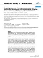
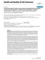
data was band pass filtered at (30-300) Hz. Figure 1 shows
tracings of a typical hippocampal oscillation signal.
Please note that it takes at least 100 msec to see any hip-
pocampal activities in response to the 100 Hz stimulus.
The patterns of hippocampal γ oscillations were irregular
and random at least 100 msec after the stimulus. But,
these patterns noticeably changed between 100 msec and
300 msec and became more regular. Finally, the late (300-
500 msec) segment, the patterns became more irregular
and random. We isolated the γ oscillation segments
(between 150 msec and 250 msec after the stimulus) by
the visual inspection (Figure 1, middle panel) and by
using the spectral components of hippocampal oscilla-
tions in the visually isolated segment (Figure 1, lower
panel) as a reference. Note that the main spectral compo-
nent was around 60 Hz which is considered to be in the
hippocampal gamma oscillation range (30-60 Hz). These
isolated hippocampal γ oscillations were analyzed using
the ApEn method as detailed above.
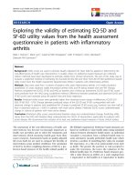
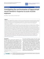
Figure 2 shows tracings of isolated hippocampal γ oscil-
lations in response to the 100 Hz before (control), during
nicotine exposure and after acute nicotine injection
(washout) in the upper panel. The corresponding com-
plexity (approximate entropy) values of these hippocam-
pal oscillations were 0.49, 0.27, and 0.30 respectively, as
shown in the lower panel in Figure 2. The complexity
value was reduced during nicotine exposure, suggesting
the emergence of strong synchronization and regular fir-
ing. During washout period, the complexity value was
increased again by suggesting more irregular pattern like
before nicotine exposure.
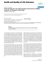
Figure 3 shows the mean complexity values of the hip-
pocampal oscillations during control, nicotine exposure
and washout where n = 14. These values were 0.49 ± 0.01
before (control), 0.42 ± 0.02 during nicotine exposure,
and 0.46 ± 0.02 after acute exposure respectively.
Φ
m
r
m
i
Nm
rCiNm() ln ()/( )=−+
=
−+
∑
1
1
1
(4)
ApEn( , , ) () ()mrN r r
mm
=−
⎡
⎣
⎤
⎦
+
ΦΦ
1
(5)
Figure 1 Shows tracings of a typical hippocampal oscillation sig-
nal (upper panel), the isolated hippocampal γ oscillation seg-
ment between 150 msec and 250 msec after the stimulus (middle
panel) and the corresponding spectral components of the isolat-
ed hippocampal γ oscillation segment (lower panel).
Figure 2 Shows tracings of isolated hippocampal γ oscillations in
response to the 100 Hz before (control), during nicotine exposure
and after acute nicotine injection (washout) in the upper panel
and the corresponding complexity values of these isolated hip-
pocampal γ oscillations in the lower panel.
Control
Washout
Drug
Akkurt et al. Journal of NeuroEngineering and Rehabilitation 2010, 7:31
/>Page 5 of 6
Our results suggest that the complexity values the hip-
pocampal oscillations during control were more or less
the same and high during washout periods. However, the
mean complexity value of the hippocampal oscillations in
response to nicotine exposure were reduced compared to
those of control and washout periods (p < 0.05). These
results furthermore suggest that the hippocampal γ oscil-
lations in response to nicotine exposure are unique and
indicate the emergence of more synchronization of the
hippocampal neural networks since hippocampal neural
firings become regular and deterministic processes in
response to the 100 Hz stimulus.
Discussion and conclusion
It has been widely reported that γ oscillations in the brain
are associated with higher order cognitive function
including selective visual attention, motor task planning,
sensory perception, working memory and dreaming REM
sleep. These oscillations are mainly observed in cortical
regions and also occur in neocortical and subcortical
areas and the hippocampus. Hippocampal neurons play a
critical role in information processing and decision mak-
ing.
Although many components of tobacco smoke are
harmful to the brain, and cardiorespiratory systems, nico-
tine has been found to be useful for the improvement of
cognitive functions including working memory and exec-
utive function. Furthermore, it has been found to be neu-
roprotective [35]. It has been widely reported that acute
exposure to nicotine improves vigilance, selective atten-
tion, memory, and executive function in human and ani-
mals. The activation of the cholinergic system and
subsequent downstream effects on neurotransmitter
release including dopamine and serotonin are responsible
for this [35].
In addition, the nicotine was also proposed for the
treatment of Alzheimer's disease (AD) and schizophre-
nia. The nicotinic-cholinergic system was suggested as a
novel therapeutic mechanism for treatment in schizo-
phrenia. Several cholinergic receptor agonists were pro-
posed to characterize central nervous system cholinergic
function and as potential candidates for the treatment of
schizophrenia as well as dementia of the Alzheimer's
type. In addition, the alpha7 nicotinic receptor agonists
have also been found to be effective on learning and
memory in animals. Thus, these alpha7 nicotinic receptor
agonists appear to be excellent candidates for the treat-
ment of impaired sensory gating, cognitive deficits and
negative symptoms in people with schizophrenia [36-39].
Martin et al discussed a novel model of nicotinic-cho-
linergic dysfunction which may be responsible for the
impairment of sensory gating in schizophrenia and the
related neurobiological and genetic mechanisms as well
as the treatment of this disease with the use of nicotine,
clozapine and the potential place of the new alpha7 nico-
tinic receptor agonists [39,40].
In this paper, we focus only on the influence of nicotine
exposure on the complexity of hippocampal γ oscilla-
tions. We used a nonlinear dynamics approach for data
analysis since the γ oscillations reflect the integrated
properties of the underlying dynamics of the hippocam-
pal neural network and therefore exhibit highly complex/
irregular features. Specifically, the approximate entropy is
a computationally efficient complexity analysis method
that is able to produce accurate estimations of the com-
plexity of the biological signals complexity (irregularity).
Our study suggests that that the sequence of 100 Hz stim-
ulations triggered a response consisting of γ oscillations
(30-120 Hz) 150 msec after the application of the stimu-
lus. The most striking observation was the reduction of
complexity values of isolated hippocampal γ oscillations
in response to acute nicotine exposure. It furthermore
suggests the strong synchronization of hippocampal neu-
ral network in the mid phase (gamma oscillation seg-
ment) in response to acute nicotine exposure. But, the
reorganization of the hippocampal network was a revers-
ible process since all the complexity values were restored
after the washout period.
Competing interests
The authors declare that they have no competing interests.
Authors' contributions
All the authors contributed equally to this work and have read and approved
the final manuscript.
Acknowledgements
The authors wish to thank Dr Jie Wu for his advice and help to build our exper-
imental set-up.
Figure 3 Shows the mean complexity values of the hippocampal
oscillations during control, nicotine exposure and washout
Akkurt et al. Journal of NeuroEngineering and Rehabilitation 2010, 7:31
/>Page 6 of 6
Author Details
1
Harrington Department of Bioengineering, Fulton School of Engineering ASU,
Tempe AZ, USA and
2
Department of Biomedical Engineering, Cullen College of
Engineering, University of Houston, Houston TX, USA
References
1. Brautbar N: Direct effects of nicotine on the brain: evidence for
chemical addiction. Archives of Environmental Health 1995, 1:263.
2. Heishman SC, Taylor RC, Henningfield JE: Nicotine and smoking: a review
of effects on human performance. Exp Clin Psychopharmacol 1994,
2:1-51.
3. Rusted JM, Newhouse PA, Levin ED: Nicotinic treatment for degenerative
neuropsychiatric disorders such as alzheimer's disease and parkinson's
disease. Behav Brain Res 2000, 113:121-129.
4. MacDermott AB, Role LW, Siegelbaum SA: Presynaptic ionotropic
receptors and the control of transmitter release. Nature Reviews
Neuroscience 1999, 22:443-485.
5. Levin E, Wilson W, McEvoy J, Rose J: Nicotine-haloperidol interactions
and cognitive performance in schizophrenics.
Neuropsycopharmacology 1996, 15:429-436.
6. Groenewegen HJ, Uylings HB: The prefrontal cortex and the integration
of sensory, limbic and autonomic information. Prog Brain Res 2000,
126:3-28.
7. Martin SJ, Grimwood PD, Morris RG: Synaptic plasticity and memory: an
evaluation of the hypothesis. Annu Rev Neurosci 2000, 23:649-711.
8. Ji D, Lape R, Dani J: Timing and location of nicotinic activity enhances or
depresses hippocampal synaptic plasticity. Neuron 2001, 31:131-141.
9. Tallon-Baudry C, Bertrand O, Peronnet F, Pernier J: Induced gamma-b and
activity during the delay of a visual short-term memory task in
humans. J Neurosci 1998, 18:4244-4254.
10. Bragin A, Jando G, Nadasdy Z, Hetke J, Wise G, Buzsaki G: Gamma (40-100
hz) oscillations in the hippocampus of the behaving rat. J Neurosci
1995, 15:47-60.
11. Whittington MA, Traub RD, Jefferys JGR: Synchronised oscillations in
interneuron networks driven by metabotropic glutamate receptor
activation. Nature 1995, 373:612-615.
12. Puma C, Deschaux O, Molimard R, Bizot JC: Nicotine improves memory in
an object recognition task in rats. Eur Neuropsychopharmacol 1999,
9:323-327.
13. Levin ED, Weber E, Icenogle L: Baclofen Interactions with Nicotine in
Rats: Effects on Memory. Pharmacol Biochem Behav 2004, 79:343-348.
14. Fries P, Reynolds JH, Rorie AE, Desimone R: Modulation of oscillatory
neuronal synchronization by selective visual attention. Science 2001,
29:1560-1563.
15. Gray R, Rajan AS, Radcliffe KA, Yakehiro M, Dani JA: Hippocampal synaptic
transmission enhanced by low concentrations of nicotine. Nature 1996,
383(6602):713-716.
16. Levin ED: Use of the radial-arm maze to assess learning and memory.
In Methods in Behavioral Pharmacology Edited by: Buccafusco JJ. New York:
CRC; 2000:189-199.
17. Mirza NZ, Bright JL: Nicotine-induced enhancements in the five-choice
serial reaction time task in rats are strain-dependent.
Psychopharmacology (Berl) 2001, 154:8-12.
18. Shoaib M, Schindler CW, Goldberg SR: Nicotine self-administration in
rats: strain and nicotine pre-exposure effects on acquisition.
Psychopharmacology (Berl) 1997, 129:35-43.
19. Kumari V, Gray JA, Ffytche DH, Mitterschiffthaler MT, Das M, Zachariah E,
Vythelingum GN, Williams SC, Simmons A, Sharma T: Cognitive effects of
nicotine in humans: an fMRI study. NeuroImage 2003, 19:1002-1013.
20. Court J, Martin-Ruiz C, Piggott M, Sperden D, Griffiths M, Perry E: Nicotinic
receptor abnormalities in alzheimer's disease. Biol Psychiatry 2001,
49:175-184.
21. Ono K, Hasegawa K, Yamada M, Naiki H: Nicotine breaks down
preformed alzheimer's beta-amyloid fibrils in vitro. Biol Psychiatry 2002,
52:880-886.
22. Harris JG, Kongs S, Allensworth D, Martin L, Tregellas J, Sullivan B, Zerbe G,
Freedman R: Effects of nicotine on cognitive deficits in schizophrenia
.
Neuropsychopharmacology 2004, 29:1378-1385.
23. Allam MF, Campbell MJ, Hofman A, Del Castillo AS, Fernandez-Crehuet
Navajas R: Smoking and parkinson's disease: systematic review of
prospective studies. Mov Disord 2004, 19:614-621.
24. Howson AL, Batth S, Ilivitsky V, Boisjoli A, Jaworski M, Mahoney C, Knott VJ:
Clinical and attentional effects of acute nicotine treatment in tourette's
syndrome. Eur Psychiatry 2004, 19:102-112.
25. Song C, Murray TA, Kimura R, Wakui M, Ellsworth K, Javedan SP, Lukas RJ,
Wu J: Role of alpha7-nicotinic acetylcholine receptors in tetanic
stimulation-induced gamma oscillations in rat hippocampal slices.
Neuropharmacology 2005, 48:869-880.
26. Wang K, Zheng C, Wu C, Gao M, Liu Q, Yang K, Ellsworth K, Xu L, Wu J: α-
chloralose diminishes γ oscillations in rat hippocampal slices.
Neuroscience Letters 2008, 441(1):66-71.
27. Javedan SP, Fisher RS, Eder HG, Smith K, Wu J: Cooling abolishes
neuronal network synchronization in rat hippocampal slices. Epilepsia
2002, 43(6):574-580.
28. Pavlides C, Ogawa S, Kimura A, McEwen BS: Role of adrenal steroid
mineralocorticoid and glucocorticoid receptors in long-term
potentiation in the CA
1
field of hippocampal slices. Brain Research 1996,
738:229-235.
29. Korte M, Carroll P, Wolf E, Brem G, Thoenen H, Bonhoeffer T: Hippocampal
long-term potentiation is impaired in mice lacking brain-derived
neurotrophic factor. Proceedings of the National Academy of Science: USA
1995, 92:8856-8860.
30. Larson J, Wong D, Lynch G: Patterned stimulation at the theta frequency
is optimal for the induction of hippocampal long-term potentiation.
Brain Research 1986, 368:347-350.
31. Pincus SM: Greater signal regularity may indicate increased system
isolation math. Bioscience 1994, 44:122161-81.
32. Pincus SM: Approximate entropy as a measure of system complexity.
Proceedings of the National Academy of Science 1991, 88:2297-2301.
33. Pincus SM, Huang WM: Approximate entropy: statistical properties and
applications. Commun Stat - Theory and Methods 1992, 21(11):3061-3077.
34. Lipsitz LA, Pincus SM, Morin RJ, Tong S, Eberle LP, Gootman PM:
Preliminary evidence for the evolution in complexity of heart rate
dynamics during autonomic maturation in neonatal swine. J Auton
Nerv Syst 1997, 65:1-9.
35. Swan GE, Lessov-Schlaggar CN: The effects of tobacco smoke and
nicotine on cognition and the brain. Neuropsychol Rev 2007, 17:259-273.
36. Levin ED, Simon BB: Nicotinic acetylcholine involvement in cognitive
function in animals. Psychopharmacology 1998, 138:217-230.
37. Levin ED, Rezvani AH: Development of nicotinic drug therapy for
cognitive disorders. Eur J Pharmacol 2000, 393:141-146.
38. Kem WR: The brain alpha7 nicotinic receptor may be an important
therapeutic target for the treatment of alzheimer's disease: studies
with DMXBA (GTS-21). Behav Brain Res 2000, 113:169-181.
39. Nomikos GG, Schilstr B, Hildebrand BE, Panagis G, Grenhoff J, Svensson TH:
Role of alpha7 nicotinic receptors in nicotine dependence and
implications for psychiatric illness. Behav Brain Res 2000, 113:97-103.
40. Martin LF, Kem WR, Freedman R: Alpha-7 nicotinic receptor agonists:
potential new candidates for the treatment of schizophrenia.
Psychopharmacology 2004, 174:54-64.
doi: 10.1186/1743-0003-7-31
Cite this article as: Akkurt et al., Investigating the synchronization of hip-
pocampal neural network in response to acute nicotine exposure Journal of
NeuroEngineering and Rehabilitation 2010, 7:31
Received: 3 April 2009 Accepted: 13 July 2010
Published: 13 July 2010
This article is available from: m/content/7/1/31© 2010 Akkurt et al; licensee BioMed Central Ltd. This is an Open Access article distributed under the terms of the Creative Commons Attribution License ( ), which permits unrestricted use, distribution, and reproduction in any medium, provided the original work is properly cited.Journal of NeuroEn gineerin g and Reha bilitatio n 2010, 7:31