Báo cáo hóa học: " Reduced short term adaptation to robot generated dynamic environment in children affected by Cerebral Palsy" potx
Bạn đang xem bản rút gọn của tài liệu. Xem và tải ngay bản đầy đủ của tài liệu tại đây (7.79 MB, 13 trang )
JNER
JOURNAL OF NEUROENGINEERING
AND REHABILITATION
Reduced short term adaptation to robot
generated dynamic environment in children
affected by Cerebral Palsy
Masia et al.
Masia et al. Journal of NeuroEngineering and Rehabilitation 2011, 8:28
(21 May 2011)
RESEARC H Open Access
Reduced short term adaptation to robot
generated dynamic environment in children
affected by Cerebral Palsy
Lorenzo Masia
1*
, Flaminia Frascarelli
2,5
, Pietro Morasso
1,4
, Giuseppe Di Rosa
2
, Maurizio Petrarca
2
, Enrico Castelli
2
and Paolo Cappa
3
Abstract
Background: It is kno wn that healthy adults can quickly adapt to a novel dynamic environment, generated by a
robotic manipulandum as a structured disturbing force field. We suggest that it may be of clinical in terest to
evaluate to which extent this kind of motor learning capability is impaired in children affected by cerebal palsy.
Methods: We adapted the protocol alread y used with adults, which employs a velocity dependant viscous field,
and compared the performance of a group of subjects affected by Cerebral Palsy (CP group, 7 subjects) with a
Control group of unimpaired age-matched children. The protocol included a familiarization phase (FA), during
which no force was applied, a force field adaptation phase (CF), and a wash-out phase (WO) in which the field was
removed. During the CF phase the field was shut down in a number of randomly selected “catch” trials, which
were used in order to evaluate the “learning index” for each single subject and the two groups. Lateral deviation,
speed and acceleration peaks and average speed were evaluated for each trajectory; a directional analysis was
performed in order to inspect the role of the limb’s inertial anisotropy in the different experimental phases.
Results: During the FA phase the movements of the CP subjects were more curved, displaying greater and
variable directional error; over the course of the CF phase both groups showed a decreasing trend in the lateral
error and an after-effect at the beginning of the wash-out, but the CP group had a non significant adaptation rate
and a lower learning index, suggesting that CP subjects have reduced ability to learn to compensate external force.
Moreover, a directional analysis of trajectories confirms that the control group is able to better predict the force
field by tuning the kinematic features of the movements along different directions in order to account for the
inertial anisotropy of arm.
Conclusions: Spatial abnormalities in children affected by cerebral palsy may be related not only to disturbance in
motor control signals generating weakness and spasticity, but also to an inefficient control strategy which is not
based on a robust knowledge of the dynamical features of their upper limb. This lack of information could be related
to the congenital nature of the brain damage and may contribute to a better delineation of therapeutic intervention.
Background
Cerebral palsy ( CP) is a group of non progressive, but
often changing, motor impairment syndromes secondary
to lesions or anomalies of the brain arising in the early
stages of development [1,2]. Although motor impair-
ment is the leading factor in CP, sensory disorders have
been described [3] and sensorimotor cognitive functions
are probably affected due to the complexity of the
motor impairments implying primary and secondary
deficits [4-6].
The last two decades has brought a tremendous depth of
understanding to the function of central nervous system
(CNS); if on one side molecular biology applied to neuro-
genetics provided unprecedented insight into the patholo-
gic mechanisms of neurologic disorders, on the other side
the use of robotics, as a non-invasive investigating tool of
* Correspondence:
1
Robotics Brain and Cognitive Sciences Dept., Italian Institute of Technology
(IIT), Genoa, Italy
Full list of author information is available at the end of the article
Masia et al. Journal of NeuroEngineering and Rehabilitation 2011, 8:28
/>JNER
JOURNAL OF NEUROENGINEERING
AND REHABILITATION
© 2011 Masia et al; license e BioMed Central Ltd. This is an Open Access article distributed under the terms of the Creative Commons
Attribution License ( which permits unrestricted use, distribution, and reproduction in
any medium, provided the original work is properly cited.
motor recovery and rehabilitation, offered the possibility
to accurately observe and quantify human movements as
the result o f t he response of the CNS to an external
dynamic interaction. In the field of motor control many
paradigms were proposed to study the sensorimotor adap-
tation to force field. Since the seminal work of Shadmehr
and Mussa-Ivaldi [7], multijoint reaching movements
under application of robot generated deviating forces have
been used in order to underst and how the CNS controls
movements in dynamic conditions: it was found that
healthy subjects a re able to gradually tune an internal
model of the arm-environment which allows to perform
almost straig ht trajectories, thus compen sating the effect
of the force field. A key finding of motor adaptation is that
when the external force is unexpectedly removed the sub-
jects move as if the force was still active, making error in
the opposite direction to the one of the generated force.
This motor after-effect [8] demonstrates that the reaction
to an external structured dynamics is the result of an
anticipatory strategy [9,10] gradually developed by the
CNS while experiencing the robot altered dynamic. Adap-
tation and its related after-effects have been demonstrated
for a variety of structured force fields, dependent on differ-
ent kinematic/dynamic features of the movements: posi-
tion [11], acceleration [12], Coriolis force [13], velocity
[14], and skew symmetric “curl” fields [15].
Patton et al. [16] used deviating force field in adult
stroke subjects as error-enhancing robot therapy, and it
was demonstrated that impaired subjects are sti ll able to
learn the force field and the rehabilitation approach was
even more effective than the assistive (error-reducing)one.
The studies on healthy subjects allowed to shed some
light on an another important phenomenon: the magni-
tude of kinematic err or varies as subjects move in differ-
ent directions while experiencing the same structured
force field [17,18]. A computational analysis of the pro-
blem suggested that the directional differences in kine-
matic error may arise from spatial asymmetries in a rm
impedance [19]. Darainy et al. [20] have shown how in
healthy subjects the anisotropic features of the arm impe-
dance play a fundamental role in motor learning and
gen eralization. In general terms an impor tant concept in
motor control is the idea that the CNS uses an internal
model of the motor system and of the surrounding envir-
onment to predict the sensory consequences of com-
mands [21]. In healthy subjects this model allows to
acco unt for the inertial anisotropy of the arm and conse-
quently generate t he right motor commands to counter-
act the external forces. However in impaired subjects,
although able to adapt [22-26], it is not clear whether the
CNS maint ains an intact capability of predicting the arm
dynamics and compensate for the anisotropy or not.
The brain injury resulting in cerebral palsy (CP) occurs
early in neurodevelopment or birth accident, whereas
stroke occurs generally in adult life. There are many scien-
tific results which suggest that plasticity is greater in the
developing brain than in the mature one [27,28] . Despite
evidences that are observable in adults, the ability of CP
subjects to deal with the central planning issues associated
with control of arm is still an open question. The recovery
mechanisms in children are quite different from adults
[29,30], due to their higher plasticity and because the
limbs control ability is age-related. The goals of the cur-
rent study were twofold: 1) to ascertain if impaired chil-
dren affected by CP preserve the ability to adapt to a force
field; 2) to investigate which differences in kinematics
between CP and Control groups lead to an unequal learn-
ing rate. We believe that these findings may suggest new
therapies, as it has been demonstrated in healthy adult
stroke patients [31-33]. Moreover we agree with Papavasi-
liou [2] that the introduction of new therapies facilitates
an individualized management plan and multimodal treat-
ment is optimized with a multidisciplinary team.
Methods
Subjects
Fourteen subjects volunteered to participate to the
experiment (table 1): 7 CP pediatric subjects, mean age
10.14 years (range 7-14) recruited at the Neurorehabilita-
tion Division of the Pediatr ic Hospital Bambino Gesù
(Rome, Italy) and 7 age-matched right-handed healthy
control subjects (mean age 9 years, range 8-14). Research
was approved by the ethical committee of the Hospital
and conforms to the ethical standards laid down in the
1964 Declaration of Helsinki. Before starting the proto-
col, the parents were aske d to sign a consent form. All
CP subjects were in the chronic sta ge and were af fect ed
by hemiparesis with a moderately impaired upper limb
function. The table also reports the score for three rele-
vant clinical scale: 1) the elbow modified Ashworth scale
of spasticity, 2) the arm section of the Fugl Meyer scale,
and 3) the Melbourne scale. The subjects of the CP
group were also characterized by the following exclusion
criteria: 1) bilateral impairment, 2) severe sensory deficit
of the impaired limb, 3) linguistic-cognitive impairment
at a level that would not allow to understand the task
and perform the experiment, 4) use of drugs, as botulin
toxin therapy, that would affect muscular properties.
Experimental apparatus
The subjects grasped a planar manipulandum specifically
designed for rehabilitation and evaluation of motor con-
trol: Inmotion2 Robot ( Interactive Motion Technologies
Inc., Boston, MA, USA). The robot is equipped with abso-
lute encoders, which acquire hand/handle position with a
100 μm accuracy, and with direct-drive motors, which can
transmit to the hand of the subjects force vectors with
controllable amplitude and direction. In addition to the
Masia et al. Journal of NeuroEngineering and Rehabilitation 2011, 8:28
/>Page 2 of 12
hand trajectories, determined by the interplay of the
robot-generated force vectors and the subject-generated
motor commands, we also computed the time course of
the velocity and acceleration of the hand, as well as the
corresponding jerk signal that is considered an indicator
of smoothness of the trajectory formation process. Sub-
jects were comfortably seated so that the center of the
manipulandum workspace was approximately coincident
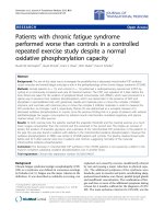
with the center of their reachable workspace (see figure 1).
Trunk strategy compensation was prevented by means of
a seat belt; the elbow was supported in the horizontal
plane by an anatomical or thosis; wrist motion was con-
strained by a thermoplastic cast in order to avoid a com-
pensatory strategy of the wrist [34].
Task
Subjects were instructed to perform reaching move-
men ts from the centre of the workspace to one of eight
peripheral targets and back; each movement has to be
performed in a specified time range of 1.2 ± 0.3 seconds.
The peripheral t argets were distributed uniformly (wit h
a 45 deg spacing) on a circle with a 14 cm radius. A tar-
get-set (TS) consisted of 8 center-out movements plus 8
return movements. The sequence of center-out move-
ments was randomized. Target and hand positions were
presented as circles on a computer screen (visual feed-
back). The criterion for target reaching wa s a position-
ing error less than 5 mm and residual oscillations of the
hand with speed smaller than 1 cm/s. When the criter-
ion was met there was an acoustic feedback and the
next target was shown. The robot was also used in
order to perturb the movements of subjects and to eva l-
uate motor a daptation. The perturbations were charac-
terized by a Curly Viscous Field, i.e. a pattern of force
vectors with amplitude proportional to the instanta-
neous speed of the hand and a direction perpendicular
to the corresponding direction of the velocity vector:
F =
0 λ
−λ 0
˙
x
han
d
(1)
F is the force vector applied by the robot to the handle;
x
hand
is the position vector of the hand; l is the parameter
of the force field. For this parameter we choose the follow-
ing value l = 20 Ns/m. This means that when the hand
reaches a velocity of 1 m/s (typically the peak velocity of a
reach movement) the force generated by the robot, which
pushes the hand laterally, has an amplitude of 20 N. As
shown in figure 1, the force field rotates in the clockwise
direction. The field rotational direction would be inverted
by changing the sign of the l parameter. The value of l =
20 Ns/m was tuned after a pilot test in order to provide a
sufficient deviating force even in case of reduced speed of
the subject’shand.
Protocol
The protocol included a total of 640 pointing move-
ments, distributed in 40 target sets (TSs), and was bro-
ken down into the following experimental phases:
- Familiarization phase (FA): it consisted of 10 TSs,
equivalent to 160 center-out movements. The m ain
Table 1 Patients demographics (CP group)
CP GROUP Age Gender Pathology Brain lesion Affected hand Elbow modified Ashworth Fugl_Meyer Melbourne
S1 7 Male CP Left frontal cortex R 1 45 80
S2 11 Male CP Left frontal cortex R 1+ 43 77,8
S3 10 Male CP basal ganglia bilaterally R 2 30 55
S4 7 Male CP Left frontal cortex R 1+ 41 73,7
S5 8 Male CP Left middle cerebral artery R 1 42 76,7
S6 14 Male CP Left pons and internal capsule R 2 45 74,7
S7 14 Male CP Left subcortical R 1 42 68,8
Figure 1 Experimental setup: the elliptical workspace of the robot
is included in the reachable workspace of the subject. The shoulder
of the subject is aligned with the centre of the workspace of the
robot. The subjects were pre-tested in order to check if they were
able to reach all the targets presented during the experiment. A
clockwise (CW) curl viscous field was used.
Masia et al. Journal of NeuroEngineering and Rehabilitation 2011, 8:28
/>Page 3 of 12
purpose was to allow the subjects to experience the
inertial load of the robot, which is not negligible, in
spite of the fact that the robot is highly backdriveable.
- Curl Viscous Field Adaptation phase (CF):it
included 20 TSs (i.e. a total of 320 movements) with
exposure to a curl viscous field described by equation 1.
During this phase the force field was randomly switched
off five times for each direction (40 catch trials total).
- Wash out phase (WO):10TSs (for a total of 160
center-out movements) witho ut disturbing force, as in
the FA phase.
CP subjects were encouraged to carry out the pointing
movements to the target with an av erage velocity com-
parable to the control subjects, in order to ensure that
the two groups experienced a similar level of force dur-
ing the CF phase.
Data analysis
The two cartesian components of the pointing trajec-
tories were sampled at 200 Hz and sm oothed by using a
6th order Savitzky-Golay filter, with a 170 ms window
(cut-off frequency: ~11 Hz). The same filter was also
used to estimate the time derivatives of the trajectory.
Movement onset and Movement termination were then
evaluated, by detecting when the hand speed curve
crosses a suit able threshold value (0.05 m/s), in order to
isolate each individual reaching movement. The follow-
ing indicators were extracted from the recorded data for
each targeting movement:
-
Lateral deviation (LD): it is defined as the deviation
from the straight line that connects the initial posi-
tion to the target, evaluated at the time of peak velo-
city. Positive and negative errors correspond to
leftward and rightward lateral deviations, respectively.
-
Acceleration peak: it is the highest value of the
acceleration profile. This indicator, if associated with
movement direction, can provide a polar plot that we
expect to be asymmetric, as suggested in a previous
study [21] that analyzes the anisotropy of the inertial
properties of both the human and robot arms.
-
Peak and average speed:aspreviouslynoted,these
indicators were monitored for verifying the substan-
tial equivalence of the field intensity in the two
groups of subjects (CP and Control).
-
Directional analysis: it was performed for each of
the previously defined variables, in order to highlight
the interplay between the effect of the deviating force
field and the effect of the anisotropy of the mechani-
cal impedance. We expect indeed that the CP group
may differently control the inertial anisotropy of the
arm while performing reaching movements. We also
defined an “aniso tropy index” E which measures the
degree of “roundness” of the directional interpolating
ellipses, by considering the major and minor semi
axes of the ellipse (a and b, respectively):
E =
1 −
b
2
a
2
(2)
E = 0/1 for a perfectly round or completely flat ellipse,
respectively.
-
Learning Index: the degree of adaptation to the
force field was measured by means of the following
formula [35] that takes into account the values of
lateral deviations in the force-field and catch trials
(LI
ff
and LI
catch
respectively):
LI =
|
LD
catch
|
|
LD
catch
|
+
LD
ff
(3)
LI
ranges from 0.0 (null adaptation) to 1.0 (complete
adaptation).
We assessed adaptation using a repeated measure
ANOVA with three factors: gr oup (CP vs. Control),
phases (FA vs. FF vs. WO) and time (early vs. late
phase). During the statistical analy sis of the results, all
hypotheses were tested using a significance level of 0.05.
Results
All the subjects were able to perform the assigned task in
the different experimental phases. Moreover, we found
that the control subjects and the CP subjects present simi-
larities in moving their hands in the force field. Figure 2
shows the trajectories (form the center to the peripheral
targets) during the entire experiment for two representa-
tive subjects form the CP and control group. For both sub-
jects the effects to the force field are evident (CF phase,
red trajectories), although the unimpaired subject learns to
compensate the force in a more effective manner perform-
ing straighter trajectories. Catch trials in the CF phase
(blue lines) reveal the presence of an anticipatory strategy
and after effects in the WO phase indicates the compensa-
tion activity of the force field continues after the robot
dynamics is removed.
Thesubject1(S1)fromtheCPgroupismildly
impaired, in fact during the FA phase his trajectories
appear to be comparable with the ones of the unim-
paired subject. An accentuation of lateral error in the
FA phase is anyway visible especially in some directions
(90°N, 270°S, 45°NE) corresponding to those in which
the inertial effect of the arm coupled with the robotic
device is higher. The same effects along the same target
directions are also observable during the CF phase
where the action of the deviating field is in fact less
compensated a nd also the catch trials se em to be char-
acterized by a higher lateral deviation.
Masia et al. Journal of NeuroEngineering and Rehabilitation 2011, 8:28
/>Page 4 of 12
Let us consider first the subjects of the control group
(upper panel of figure 3). Namely, they perform quasi
straight trajectories during the FA phase; the trajectories
are bent in the direction of the force field in the initial
part of the CF phase, as a consequence of the external
disturbance, but the subjects quickly learn to compen-
sate reducing the lateral deviation by the end o f the CF
phase; the feedforward nature of the compensation
mechanism is proved by the catch trials, which show a
bending ( lateral error) in the opposite direction; at the
end o f the beginning of the WO phase an after effect is
clearly noticeable and the original performance is
restored at the end of the WO.
Contrarily the CP subjects (lower panel of figure 3)
clearly perform in a less reliable way, i.e. with a larger
variabili ty, but still they can carry out the task of reach-
ing t he targets in approximately the prescribed time, in
all the directions and in the different phases.
A further insight of the learning capability of the two
groups is provided by the bar plots of the early and late
training in the two groups: figure 4 depicts the comparison
between the average values of the initial and final twenty
trials in the different phases of the experiment. Observing
the performance of the two groups during the different
phases of the experiment we noticed that they both show
a statistical significant reduction of the lateral error
between the early and late phase of the familiar ization [F
(1,12) = 8.2506, p = 0.01402] and early and late phase of
the wash-out [F(1,12) = 31.239, p = 0.00012].
It is anyway crucial to precise that the experiment was
mainly focused on testing if CP subjects were a ble to
develop an a nticipatory strategy to counteract a deviat-
ing force field. Nevertheless, in terms of trajectory cor-
rections it is clear from figure 4 that CP group does not
succeed in significantly decreasing the lateral deviation
over the course of the trials; as depicted in the CF phase
of figure 4 the early and late value of the lateral devia-
tion do not show any significant difference [F(1,12) =
0.04, p = 0 .8384]. Contrarily, control subjects sh ow a
distinct change between the initial and the final part of
force field exposition [F(1,12) = 6.61, p = 0.00051].
In order to evaluate the degree of motor adaptation it is
necessary to inspect the after-effects. When the disturbing
force field is removed at the beginning of the WO phase,
both groups show lateral errors in the opposite direction
of the force field. This error magnitude provides a mea-
sure of how much the subjects developed an anticipatory
strategy of the robo t gener ated dynamics over the course
of the exposition to the field. There is a statistica l signifi-
cance decrease of the lateral error between late familiari-
zation and early wash-out [F(1,12) = 23.5999, p =
0.00039] and also interaction betwe en the two main
effects phases and groups [F(1,12) = 7.2208, p = 0.01977]
showing a lower after effect of the CP group than the one
present in control group. The latter seems to have a bet-
ter adaptation rate to the force field, between the early
and late values of the lateral deviation, than the CP
group, exhibiting a greater after-effect and indicating that
Figure 2 Trajectories during the different experimental phases for two re presentative subjects, from the CP and Control group.The
blue traces correspond to movements with no disturbing field: all trials in the Familiarization phase (FA) and Wash-Out (WO) phase and catch
trials in the Curl Viscous Field Adaptation phase (CF). The red traces correspond to movements affected by the disturbing force during the CF
phase.
Masia et al. Journal of NeuroEngineering and Rehabilitation 2011, 8:28
/>Page 5 of 12
they are more prone to learn the force field and compen-
sate the deviation with a lower lateral error at the end of
the training.
Moreover, a strong indication of the reduced short-
term adapt ation of the C Ps is clearly shown by the
learning index (LI) depicted in figur e 5. The LI corrects
for possible difference in performance due to differences
in the action of the force field; if adaptation occurs, dur-
ing force field the lateral deviation decreases while its
value increases for the trajectories performed during
catch trials due to the higher compensatory action by
the CNS. Figure 5 shows that in the control group LI
grows monotonically, in the initial learning, and this
behavior is followed by an exponential tr end, as
expected for a short term adaptation experiment. In
contrast, in the CP group LI is ch arac terized by a lower
incre asing trend, with an early saturation, which implies
a reduced learning capability.
Movement anisotropy of the human arm is responsi-
ble for directional variability of movement kinematics
Figure 3 Trend of adaptation for the CP group (bott om) and control group (top) over the different ta rget-sets; it refers to all the trials
where black dots are average values of lateral deviation for all the subjects during familiarization and wash-out, while blue and red are referred
to lateral deviation during force field adaptation and catch trials respectively. Error bars for each value refer to standard error over all subjects.
Figure 4 Early Vs Late performance of the two groups. Bar plots of the e arly (initial 20 trials) and late (final 20 trails) exposition the
experiment in the three experimental phases, with standard error for each bar.
Masia et al. Journal of NeuroEngineering and Rehabilitation 2011, 8:28
/>Page 6 of 12
[36], therefore a directional distribution of acceleration
peaks while moving along the targets may help in com-
prehending how inertial anisotropy plays an important
role in the adaptation perform ance to the robot gener-
ated force field. Figure 6 (referred to S1 of the CP
group) shows the polar plots of the lateral deviation and
the acceleration peak, i.e. the distribution along the dif-
ferent target directions of the two indicators. As shown
in figure 6 the kinematic error is distributed towards
those geometrical configurations in which t he arm and
the robot together may result more difficult to control
by the subject. Observing the a verage of acceleration
peaks along the different directions (bottom figure 6),
one can realize how the orientation of the major axes of
the interpolating ellipses seems to be concordant with
the ones interpolating the lateral deviations, over the
three different phases of the experiment; we may
hypothesize that the higher is the lateral deviation over
a certain direction and the higher i s the correspondent
acceleration peak due to the trajectory correction o per-
ated by the subject. The directional distribution of the
kinematic variables seems to shed some light on how
the subjects try to master the interaction with the gener-
ated external environment by taking into account the
anisotropy of the arm.
Under this hypothesis it might be useful to observe
the directional distribution of the acceleration and speed
for both the CP and control groups. Figure 7 shows the
polar plots of the lateral deviation, acceleration peak,
velocity peak and average velocity, respectively, averaged
for the two groups, during the three experimental
phases (FA, CF, WO). Observing the mean speed (bot-
tomofFigure7)itappearsthatthevaluesofthelinear
velocity are comparab le for both CP and control sub-
jects and therefore the curly viscous field is perceived by
the two groups with a similar amount of deviating force.
However, the inspection of the polar plots of the lat-
eral deviation, acceleration and speed peaks depicts
Figure 6 Polar plot of lateral deviation (top) and peak of acceleration (bottom) for a representative subject (subj_1) from CP group.
Red lines along the different directions are the average of the lateral deviation and peak acceleration values. Green lines represent the standard
error and red dot is the centre of the interpolating ellipse. The blue ellipse is an interpolation of the average values over the different directions.
Figure 5 Learning index for CP and Control groups for 5
different target sets (TS). Each learning index value is computed
as the average of eight catch trials, taking into account the
corresponding directions, when the force field is active in the CF
phase. Solid lines indicate mean value and dashed lines indicate
standard error.
Masia et al. Journal of NeuroEngineering and Rehabilitation 2011, 8:28
/>Page 7 of 12
different behaviors of the two groups. In order to ana-
lyze and quantify the di fferences among these kinematic
variables, we computed an eccentricity index of the
interpolating ellipses according to the following formula:
E =
1 −
b
2
a
2
(4)
where b and a are the minor and major semi axes of
the interpolating ellipses. This measure elucidates how
kinematic and the dynamic features of the movements
are directionally varied in response to the applicat ion of
the force field.
Figure 8 shows that the eccentricity of the interpolat-
ing ellipses for control group dramatically increases dur-
ing the CF phase with respect to the FA and WO
phases for acceleration peaks and speed peaks [F(2,18) =
11.89, p < 0.001; F(2,18) = 6.78; p = 0.006]. It means
that unimpaired subjects in order to balance the force
field are able to re-compute and orient the direc tiona l
distribution of the acceleration and velocity, leading to a
better performance while reaching the different targets:
the change in the kinematics over the eight directions,
suggests that the force field is perceived as a disturbance
and in order to perform straight trajectories subjects are
forced to directionally tune the control strategy of the
arm. Observing Figure 8, it is evident that contrarily CP
groups, when the force field is active, do not adjust the
kinematics of the movements; in fact the values of the
eccentricity, in the three phases of the experiment, don ’t
differ for acceleration and speed peaks [F(2,18) = 2.21; p
= 0.1385; F(2,18) = 0.48; p = 0.6264].
Discussion
It is widely accepted from previous studies [37,38] that the
gen eration of coordinated multijoint movement requires
Figure 7 Polar plot of the different performance indicators during the di fferent phases. (FA: familiarization; CF: curl field adaptation; WO:
wash-out) for the two CP and control groups 1) Lateral deviation (first row of panels), 2) Acceleration peak (second row), 3) Speed peak (third
row), 4) Average speed (fourth row). The red, continuous lines link the average values and the green lines link the corresponding standard errors.
The ellipses are derived from an interpolation of the average values and the red point represents its center.
Masia et al. Journal of NeuroEngineering and Rehabilitation 2011, 8:28
/>Page 8 of 12
the CNS to account for joint interaction effects by relying
on suitable “internal models” of the intrinsic arm
dynamics. It is suggested that such models are acquired by
motor learning and are used by the CNS in order to pro-
vide feed-forward control commands, capable to compen-
sate for the anisotropy of the arm and the correspo nding
dynamics [39]. Moreover, haptic robots have been used
for exposing human subjects to novel, extrinsic dynamic
environments [40-43] whose effect is added to the effect of
the intrinsic dynamics. In this way it was possible to inves-
tigate the compensatory strategies adopted for short term
adaptation. It appears that adaptation involves changes in
the cerebellar cortex [44,45], suggesting that internal mod-
els of the external dynamics are stored in the cerebellum
[46,47]. It is also hypothesized that the ability of control-
ling our limbs is acquired early in life and is then continu-
ously updated in order to accommodate gradual
biomechanical, muscular, and neural changes that occur
during development and above all in childhood.
As pointed out by Shadmehr et al [48] two main pro-
blems must be solved by the brain for the acquisition of
efficient internal models: 1) sensory feedback is noisy
and delayed, making movements inaccurate and poten-
tially unstable; 2) the causal relationship between motor
commands and ensuing movement is somehow unpre-
dictable, as the body/environment dynamics is cease-
lessly changing. Forward internal models of the body/
world ensemble can solve such problems by pro viding
predictions of the state of the body as it i nteracts with
the world around it. However, such models are only
useful if they produce unbiased predictions and this
requires, at the same time, that the level of noise in the
system is sufficiently low and the sensorimotor system is
well calibrated.
Another important aspect that must be considered is
that human motor development is the result from a
complex interaction between gene and experience, and
the somatopic organization of the primate motor cortex
(M1) emerges postnatally. As suggested by Stoecke l et
al. [49] an altered motor experience during early motor
development may play a more critical role in the shap-
ing of genetically determined neural networks underly-
ing control of movement.
Thepresentstudydemonstratesthatbothgroupsof
subjects completed the required task without difficulties,
although there is evidence of different pe rformance
between the impaired and unimpaired subjects. In spite
of their young age, which implies shorter arms and
smaller weights than the adults recruited in classical stu-
dies of force field adaptation, the control group behaves
in a very similar way. Therefore, these s ubject are cap-
able not only to lea rn a compensation strategy of the
disturbance (equivalent to the one developed by their
adult counterparts) but also to predict the non-linear
dynamics of the robot which, in relative terms w ith
respect to the intrinsic arm d ynamics, is much more
relevant for the children than the adults.
However there are differences between the two groups
as regards control and adaptation capabilities. The sub-
jects should learn how to master the combined dynamics
of the ro bot and the arm and the inspection of figure 3-4
suggests that the strategy acquired by control subjects
may be more efficient that the one developed by CPs,
having a faster decrease rate of the lateral error in the CF
phase; moreover, during the catch trials (red dots in the
CF phase for the CP group) there is a tendency to
increase the value of the lateral deviation as a result of
the increasing compensatory force exerted by the
Figure 8 Degree of anisot ropy of the interpolating ellipses. The eccentricity was evaluated for the acceleration peak, veloci ty peak, and
average velocity, after interpolation of the CP and Control group in the three experimental phases (FA, CF, WO).
Masia et al. Journal of NeuroEngineering and Rehabilitation 2011, 8:28
/>Page 9 of 12
subjects over the course of the experiment when the
force field was unexpectedly removed. After training, an
after-effect is also visible in the initial trials of the WO
phase, where the kinemat ic errors appear to be shifted
and opposite with respect to those in last trials of the CF
phase.
In this context, the aim of the present study was to
understand t o which extent children affected by conge-
nital hemiparesis have a reduced ability to acquire a pre-
dictive force field compensation strategy because of a
lack of an efficient feed-forward control mechanism. A
crucial point is then the meaning of the after-affects and
catch trials observed during the experiment: is there evi-
dence of anticipatory mechanisms of adaptation,
although degraded, or do CP subjects merely react to
the robot generated force without anticipating sensory
commands? An answer to the question may be found by
looking at the inability of CP subjects to master the arm
anisotropy and to use it depending of the external
dynamics. As reported in previous paper by Gordon et
al. [35] the early portion of hand moveme nt is charac-
terized in term of spatial distribution of the acceleration,
that for healthy subjects result in a systematic direc-
tional variation. This phenomenon can be explained as
an inaccurate account of the inertial anisotropy of the
arm, persisting in adults even when robust information
of the arm is already developed. Previous studies [50]
demonstrated how healthy children adapt to robot gen-
erated force field, but performance is still more variable
than adults, due to movement inconsistency and not
motor adaptation inability . This outcome suggested that
higher movement variability in young children may arise
from higher motor noise and constraining physiological
factors of the developing motor system; in fact computa-
tional processes taking part to internal model formation
are implemented by the CNS early in development and
they need to account for continual control adjustment
in order to compensate for morphological growth dur-
ing the development.
We can hypothesize that in children this updating
process, associated to a higher motor noise, plays a lead-
ing part on motor learning especially in those cases even
weakened due to cerebral palsy, and thus the inertial
anisotropy during movement may strongly influence
subjects’ ability in mastering complex interaction with
the surrounding environment.
We believe these spatial abnormalities in CP children
result from a systematic disturbance in the motor con-
trol signals t o be attributed not only to weakness and
spasticity [51] but above all to a deficient control strat-
egy based on a robust knowledge of their arm dynamics.
It is conceivable indeed that an indirect effect of weak-
ness and spasticity is to degrade the capability of the
brain to calibrate the sensorimotor system, thus making
impossible the acquisition of a reliable internal model of
prediction.
Although in the CF and WO phases the two groups
exhibit comparable trends for catch-trials and after-
effects, it is hard to demonstrate, for the CP group, that
they are due to an anticipatory control strategy acquired
during the exposition to the force field. After effects and
catch trials may indeed be primarily related to a differ-
ent interpretation of the CP subject of the force gener-
ated by the robot rather than a learning effect due to
sensorimotor adaptation. In fact, control subjects inter-
acting with the ro bot are able indeed to adapt to the
force field while changing the directional distribution of
the arm dynamics and kinematics a s shown in figure 7
and 8; in contrast, CP children exhibit high lateral
deviation and non-significant difference between the
early and late phase of the short term a daptation, but
more important they don’ t readjust directionally the
dynamics of their movemen ts as confirmed but the
invariant orientation of the directional ellipse over the
course of the experiment.
But why children aff ected by cerebral palsy, even at a
mild level, do not show same performance as unim-
paired subjects? A possib le answer may come fro m the
distinction proposed by Huang and Krakauer [52]
between motor adaptation and skill learning.Froma
control point of view, skill learning can be seen as a glo-
bal control scheme to solve the task, while motor adap-
tation is the tuning of the skill learning in order to
compensate a change in the operating condition (i.e.
change in environment dynamics). Is the velocity of tun-
ing and therefore the adaptation rate between impaired
and healthy subjects different? Scheidt and Stoeckmann
[53] compared force field adaptation in post-stroke and
healthy subjects. It was found the two groups used the
same compensatory strategy but the influence of the
successive trials was lower in stroke subjects, indicating
that impaired people (stroke) preserve adaptation capa-
city as healthy subjects but they require more practice.
In our experiment short term adaptation was tested for
CP children and control unimpaired group; as expected
the results demonstrate that healthy pediatric subjects
are more prone to adapt to exter nal force field than CP
ones. The adaptation mechanism anyway is quite similar
to that observed between adult stroke and control
group; in fact CP children are characterized by a very
lower adaptation r ate as depicted by the learning index
(see figure 5) and a smaller after affect in comparison to
unimpaired ones. On the basis of these outcomes we
shall believe that an intensive training could lead CP
group to have a better performance but despite all the
training exercises for such kind of pathol ogy should be
oriented towards a protocol which explicitly challenges
the internal model formation: the implementation of
Masia et al. Journal of NeuroEngineering and Rehabilitation 2011, 8:28
/>Page 10 of 12
such rehabilitation therapy might be based on a different
method of observation and evaluation of subject’sper-
formance not only focused on movement accuracy but
based on robust realistic computational models of
motor adaptation which are able to provide insights of
two complementary aspects o f motor control: cortical
reorganization and impedance modulation. Consistent
with this hypothesis it would be possible to compare
muscular co-activity in CP and unimpaired subjects
emphasizing the role of impedance control in motor
adaptation [54,55]. The preliminary results of the pre-
sent study may have possible implications in under-
standing the motor recovery process in cerebral palsy,
offering non-invasiv e and relatively simple tool to study
and quantify motor control disabilities, and to drive
towards a rehabilitation protocol which enhances the
adaptive process in the restoration of motor functions.
Acknowledgements
This research is supported by National Ministry of Health, and Italian Institute
of Technology. The experiments were performed at the Neurorehabilitation
Division of Children Hospital ‘Bambino Gesù’ (Palidoro (Rome), Italy). We
wish to thank all the children and their families who agreed to participate to
the experiment.
Author details
1
Robotics Brain and Cognitive Sciences Dept., Italian Institute of Technology
(IIT), Genoa, Italy.
2
Pediatric Neurorehabilitation Division, IRCCS Children
Hospital ‘Bambino Gesù’, Palidoro Rome, Italy.
3
Mechanics and aeronautics
Dept., ‘Sapienza’ University of Rome, Rome, Italy.
4
Dept. of Informatics,
Systems and Telematics, University of Genoa, Italy.
5
Physical Medicine and
Rehabilitation, ‘Sapienza’ University of Rome, Rome, Italy.
Authors’ contributions
LM conceived, designed the experiment, performed the data analysis and
drafted the manuscript. LM, FF and MP carried out the experiments; PM
participated in the design of the study and manuscript composition; FF and
MP participated in the coordination of the study, assisting the patients
during the robot sessions; EC and PC participated in design and
coordination of the research.
All authors read and approved the final manuscript.
Competing interests
The authors have not competing interests as defined by the BioMed Central
Publishing Group, or other interests that may influence results and
discussion reported in this study.
Received: 25 October 2010 Accepted: 21 May 2011
Published: 21 May 2011
References
1. M Bax, M Goldstein, P Rosenbaum, A Leviton, N Paneth, B Dan, B
Jacobsson, D Damiano, Proposed definition and classification of cerebral
palsy. Dev Med Child Neurol. 47, 571–6 (2005). doi:10.1017/
S001216220500112X
2. AS Papavasiliou, Management of motor problems in cerebral palsy. A
critical update for the clinician. European J of Paediatric Neurology. 13,
387–396 (2009). doi:10.1016/j.ejpn.2008.07.009
3. A Guzzetta, E Mercuri, G Cioni, Visual disorders in children with brain
lesions: 2. Visual impairment associated with cerebral palsy. European J of
Paediatric Neurology. 5, 115 –119 (2001)
4. AA Mallick, FJK O’Callaghan, The epidemiology of childhood stroke.
European J of Paediatric Neurology. 14, 197–205 (2010). doi:10.1016/j.
ejpn.2009.09.006
5. SH Ravn, EM Flachs, P Uldall, Cerebral palsy in eastern Denmark: declining
birth prevalence but increasing numbers of unilateral cerebral palsy in birth
year period 1986-1998. European J of Paediatric Neurology.14, 214–218
6. K Himmelmann, E Beckung, P Uvebrant, Bilateral cerebral palsy - Prevalence
through four decades, motor function and growth. European J of Paediatric
Neurology. 11, 215–222 (2007). doi:10.1016/j.ejpn.2006.12.010
7. R Shadmehr, FA Mussa-Ivaldi, Adaptive representation of dynamics during
learning of a motor task. J Neurosci. 14, 3208–3224 (1994)
8. N Bhushan, R Shadmehr, Evidence for a forward dynamics model in human
adaptive motor control. Adv Neural Inform Proc Systems. 11,3–9 (1999)
9. C Ghez, The control of movement. in Principles of neural science, ed. by
Kandel ER, Scwartz JH, Jessel TM (Appleton & Lange, Nor wolk, 1991), pp.
533–547
10. H Hemami, BT Stokes, Qualitative discussion of mechanisms of feedback
and feedforward control of locomotion. IEEE Trans Biomed Eng. 30,
163–189 (1982)
11. O Bock, Load compensation in human goal-directed arm movements.
Behav Brain Res. 41, 167–177 (1990). doi:10.1016/0166-4328(90)90106-O
12. T Flash, F Gurevitch, Arm movement and stiffness adaptation to external
loads. in Proceedings of the 13th IEEE engineering in medicine and biology
conference, vol. 13. (Orlando, 1992), pp. 885–886
13. JR Lackner, P DiZio, Rapid adaptation to Coriolis force perturbations of arm
trajectories. J Neurophysiol. 72 , 299–313 (1994)
14. MA Conditt, F Gandolfo, FA Mussa-Ivaldi, The motor system does not learn
the dynamics of the arm by rote memorization of past experience. J
Neurophysiol. 78, 554–560 (1997)
15. F Gandolfo, FA Mussa-Ivaldi, E Bizzi, Motor learning by field approximation.
Proc Nat Acad Sci USA. 93, 3843–3846 (1996). doi:10.1073/pnas.93.9.3843
16. JL Patton, ME Phillips-Stoykov, M Stojakovich, FA Mussa-Ivaldi, Evaluation of
robotic training forces that either enhance or reduce error in chronic
hemiparetic stroke survivors. Experimental Brain Research. 168,
368–383
(2006). doi:10.1007/s00221-005-0097-8
17. N Malfait, DM Shiller, DJ Ostry, Transfer of motor learning across arm
configurations. J Neurosci. 22 , 9656–9660 (2002)
18. AA Mattar, DJ Ostry, Modifiability of generalization in dynamics learning. J
Neurophysiol. 98, 3321–3329 (2007). doi:10.1152/jn.00576.2007
19. DW Franklin, G Liaw, TE Milner, R Osu, E Burdet, M Kawato, Endpoint
Stiffness of the Arm Is Directionally Tuned to Instability in the Environment.
Jour of Neur. 27(29):7705–7716 (2007)
20. M Darainy, AA Mattar, DJ Ostry, Effects of human arm impedance on
dynamics learning and generalization. Journal of Neurophysiology. 101,
3158–3168 (2009). doi:10.1152/jn.91336.2008
21. JR Flanagan, S Lolley, The inertial anisotropy of the arm is accurately
predicted during movement planning. Journal of Neuroscience. 21,
1361–1369 (2001)
22. RF Beer, JPA Dewald, WZ Rymer, Deficits in the coordination of multijoint
arm movements in patients with hemiparesis. Exp Brain Res. 131, 305–319
(2000). doi:10.1007/s002219900275
23. RF Beer, JD Given, JPA Dewald, Task-dependent weakness at the elbow
in patients with hemiparesis. Arch Phys Med Rehabil. 80,766–77 2
(1999). doi:10.1016/S0003-9993(99)90225-3
24. HI Krebs, N Hogan, W Hening, SV Adamovich, H Poizner, Procedural motor
learning in Parkinson’s disease. Exp Brain Res. 141, 425–437 (2001).
doi:10.1007/s002210100871
25. M Casadio, V Sanguineti, P Morasso, C Solaro, Abnormal sensorimotor
control, but intact force field adaptiation, in multiple sclerosis subjects with
no clinical disability. Mult Scler. 14, 330 (2008). doi:10.1177/
1352458507085068
26. PH McCrea, JJ Eng, AJ Hodgson, Biomechanics of reaching: clinical
implications for individuals with acquired brain injury. Disabil Rehabil
24(10):534–41 (2002). 10. doi:10.1080/09638280110115393
27. G Berlucchi, HA Buchtel, Neuronal plasticity: historical roots and evolution
of meaning. Exp Brain Res. 192, 307–19 (2009). doi:10.1007/s00221-008-
1611-6
28. K Rosenkranz, A Kacar, JC Rothwell, Differential modulation of motor
cortical plasticity and excitability in early and late phases of human motor
learning. J Neurosci. 27, 12058–66 (2007). doi:10.1523/JNEUROSCI.2663-
07.2007
29. SF Farmer, LM Harrison, DA Ingram, JA Stephens, Plasticity of central motor
pathways in children with hemiplegic cerebral palsy. NEUROLOGY. 41, 1505
(1991)
Masia et al. Journal of NeuroEngineering and Rehabilitation 2011, 8:28
/>Page 11 of 12
30. JA Stephens, Plasticity of central motor pathways in children with
hemiplegic cerebral palsy. Neurology. 41, 1505–10 (1991)
31. ML Aisen, HI Krebs, N Hogan, F McDowell, BT Volpe, The effect of robot-
assisted therapy and rehabilitative training on motor recovery following
stroke. Archives Neurology. 54, 443–446 (1997)
32. L Marchal-Crespo, DJ Reinkensmeyer, Review of control strategies for
robotic movement training after neurologic injury. J NeuroEng & Rehabil. 6,
20 (2009). doi:10.1186/1743-0003-6-20
33. M Casadio, P Giannoni, P Morasso, V Sanguineti, A proof of concept study
for the integration of robot therapy with physiotherapy in the treatment of
stroke patient. Clin Rehab. 23, 217–228 (2009). doi:10.1177/
0269215508096759
34. MC Cirstea, MF Levin, Compensatory strategies for reaching in stroke. Brain.
123(Pt 5):940–53 (2000)
35. EJ Hwang, O Donchin, MA Smith, R Shadmehr, A gain-field encoding of
limb position and velocity in the internal model of arm dynamics. PloS Biol.
1(2):E25 (2003)
36. J Gordon, MF Ghilardi, SE Cooper, C Ghez, Accuracy of planar reaching
movements II. Systematic extent errors resulting from inertial anisotropy.
Exp Brain Res. 99, 112–30 (1994). doi:10.1007/BF00241416
37. MJ Hollerbach, T Flash, Dynamic interactions between limb segments
during planar arm movement. Biol Cybern. 44,67–77 (1982). doi:10.1007/
BF00353957
38. PL Gribble, DJ Ostry, Compensation for interaction torques during single
and multijoint limb movement. J Neurophysiol. 82, 2310–2326 (1999)
39. PN Sabes, The planning and control of reaching movements. Curr Opin
Neurobiol. 10(6):740–6 (2000). doi:10.1016/S0959-4388(00)00149-5
40. DM Wolpert, Z Ghahramani, MI Jordan, An internal model for sensorimotor
integration. Science. 269, 1880–1882 (1995). doi:10.1126/science.7569931
41. JR Flanagan, E Nakano, H Imamizu, R Osu, T Yoshioka, M Kawato,
Composition and decomposition of internal models in motor learning
under altered kinematic and dynamic environments. J azzett RC34(1-5)
(1999). 19
42. M Kawato, Internal models for motor control and trajectory planning. Curr
Opin Neurobiol. 9, 718–727 (1999). doi:10.1016/S0959-4388(99)00028-8
43. JW Krakauer, MF Ghilardi, C Ghez, Independent learning of internal models
for kinematic and dynamic control of reaching. Nat Neurosci. 2, 1026–1031
(1999). doi:10.1038/14826
44. R Shadmehr, HH Holcomb, Neural correlated of motor memory
consolidation. Science. 227, 821–825 (1997)
45. H Imamizu, S Miyauchi, T Tamada, Y Sasaki, R Takino, B Păľtz, T Yoshioka, M
Kawato, Human cerebellar activity reflecting an acquired internal model of
a new tool. Nature. 403, 192–195 (2000). doi:10.1038/35003194
46. RC Miall, DJ Weir, DM Wolpert, JF Stein, Is the cerebellum a Smith
Predictor? J Motor Behav. 25, 203–
216 (1993). doi:10.1080/
00222895.1993.9942050
47. DM Wolpert, RC Miall, M Kawato, Internal models in the cerebellum. Trends
Cogn Science. 2, 338–347 (1998). doi:10.1016/S1364-6613(98)01221-2
48. R Shadmehr, MA Smith, JW Krakauer, Error Correction, Sensory Prediction,
and Adaptation in Motor Control. Annu Rev Neurosci. 33 ,89–108 (2010).
doi:10.1146/annurev-neuro-060909-153135
49. MC Stoeckel, RJ Seitz, CM Buetefisch, Congenitally altered motor experience
alters somatotopic organization of human primary motor cortex. Proc Natl
Acad Sci USA. 106(7):2395–2400 (2009). doi:10.1073/pnas.0803733106
50. CD Takahashi, D Nemet, CM Rose-Gottron, JK Larson, DM Cooper, DJ
Reinkensmeyer, Neuromotor noise limits motor performance, but not motor
adaptation, in children. Journal of Neurophysiology. 90, 703–722 (2003).
doi:10.1152/jn.01173.2002
51. A Boose, J Dichgans, H Topka, Deficits in phasic muscle force generation
explain insufficient compensation for interaction torque in cerebellar
patients. Neurosci Lett. 261,53–56 (1999). doi:10.1016/S0304-3940(98)01013-
1
52. VS Huang, J Krakauer, Robotic neurorehabilitation: a computational motor
learning perspective. Journal of Neuroengineering and Rehabilitation. 6,5
(2009). doi:10.1186/1743-0003-6-5
53. RA Scheidt, T Stoeckmann, Reach adaptation and final position control
amid environmental uncertainty after stroke. J Neurophysiol. 97, 2824–2836
(2007). doi:10.1152/jn.00870.2006
54. DJ Reinkensmeyer, MG Iobbi, LE Kahn, DG Kamper, CD Takahashi, Modeling
reaching impairment after stroke using a population vector model of
movement control that incorporates neural firing-rate variability. Neural
Comput. 15, 2619–2642 (2003). doi:10.1162/089976603322385090
55. CE Han, MA Arbib, N Schweighofer, Stroke rehabilitation reaches a
threshold. PLoS Comput Biol. 4(8):e1000133 (2008). doi:10.1371/journal.
pcbi.1000133
doi:10.1186/1743-0003-8-28
Cite this article as: Masia et al.: Reduced short term adaptation to robot
generated dynamic environment in children affected by Cerebral Palsy.
Journal of NeuroEngineering and Rehabilitation 2011 8:28.
Submit your next manuscript to BioMed Central
and take full advantage of:
• Convenient online submission
• Thorough peer review
• No space constraints or color figure charges
• Immediate publication on acceptance
• Inclusion in PubMed, CAS, Scopus and Google Scholar
• Research which is freely available for redistribution
Submit your manuscript at
www.biomedcentral.com/submit
Masia et al. Journal of NeuroEngineering and Rehabilitation 2011, 8:28
/>Page 12 of 12