Báo cáo hóa học: " Objective assessment of motor fatigue in multiple sclerosis using kinematic gait analysis: a pilot study" docx
Bạn đang xem bản rút gọn của tài liệu. Xem và tải ngay bản đầy đủ của tài liệu tại đây (927.21 KB, 13 trang )
RESEARCH Open Access
Objective assessment of motor fatigue in
multiple sclerosis using kinematic gait analysis:
a pilot study
Aida Sehle
1
, Annegret Mündermann
1,2
, Klaus Starrost
3
, Simon Sailer
3
, Inna Becher
4
, Christian Dettmers
5*
and
Manfred Vieten
1
Abstract
Background: Fatigue is a frequent and serious symptom in patients with Multiple Sclerosis (MS). However, to date
there are only few methods for the objective assessment of fatigue. The aim of this study was to develop a
method for the objective assessment of motor fatigue using kinematic gait analysis based on treadmill walking and
an infrared-guided system.
Patients and methods: Fourteen patients with clinically definite MS participated in this study. Fatigue was defined
according to the Fatigue Scale for Mo tor and Cognition (FSMC). Patients underwent a physic al exertion test
involving walking at their pre-determined patient-specific preferred walking speed until they reached complete
exhaustion. Gait was recorded using a video camera, a three line-scanning camera system with 11 infrared sensors.
Step length, width and height, maximum circumduction with the right and left leg, maximum knee flexion angle
of the right and left leg, and trunk sway were measured and compared using paired t-tests (a = 0.005). In addition,
variability in these parameters during one-minute intervals was examined. The fatigue index was defined as the
number of significant mean and SD changes from the beginning to the end of the exertion test relative to the
total number of gait kinematic parameters.
Results: Clearly, for some patients the mean gait parameters were more affected than the variability of their
movements while other patients had smaller differences in mean gait parameters with greater increases in
variability. Finally, for other patients gait changes with physical exertion manifested both in changes in mean gait
parameters and in altered variability. The variability and fatigue indices correlated significantly with the motoric but
not with the cognitive dimension of the FSMC score (R = -0.602 and R = -0.592, respectively; P < 0.026).
Conclusions: Changes in gait patterns following a physical exertion test in patients with MS suffering from motor
fatigue can be measured objectively. These changes in gait patterns can be described using the motor fatigue
index and represent an objective measure to assess motor fatigue in MS patients. The results of this study have
important implications for the assessments and treatment evaluations of fatigue in MS.
Background
Multiple Sclerosis (MS) is a chronic autoimmune disease
of the central nervous system characterized by inflamma-
tion, demyelization and destruction of axons and neurons,
and by gliosis. MS is the most common neurological disor-
der in younger adults with a prevalence of 30-110 per 100,
000 adults [1,2]. In Germany alone, approximately 130,
000 patients suffer from multiple sclerosis [1]. Multiple
sclerosis comprises a variety of symptoms including cen-
tral paresis, spasticity, paraesthesia, ataxia, dysarthria,
visual impairment, cognitive dysfunction and urinary and
bowel dysf unction [3]. However, the most common and
most debilitating symptom [4-6] experienced by 87-92% of
all persons affected by MS is fatigue, recently termed
‘pathological exhaustion’ [7], which is defined as ‘a subjec-
tive lack of physical or mental energy that is perceived by
* Correspondence:
5
Kliniken Schmieder Konstanz, Konstanz, Germany
Full list of author information is available at the end of the article
Sehle et al. Journal of NeuroEngineering and Rehabilitation 2011, 8:59
/>JNER
JOURNAL OF NEUROENGINEERING
AND REHABILITATION
© 2011 Sehle et al; licensee BioMed Central Ltd. This is an Open Access article distribu ted under the terms of the Creative Commons
Attribution Licens e ( which permits unrestricted use, distribution, and reproduction in
any medium, provided the original work is properly cited.
the individual or caregiver to interfere with activities of
daily living’ [8].
The pathophysiology of fatigue in MS is still poorly
understood and the success rates of available treatments
are low. Fatigue is typically exacerbated by exertion and
by heat, where the latter is known as the Uhthoff phe-
nomenon [9]. Use-dependent cond ucti on block has been
proposed as a likely mechanism of fatigue in MS [10]. It
has been suggested that activity results in axonal hyper-
polarization [11] and that conduction blocks may be
induced by depletion of axonal energy supply or by
inflammatory mediators [12,13]. Other changes asso-
ciated with fatigue in MS patients are increased and
extensive cortical activation (inc luding that of non-motor
cortical areas) and reduced cortical inhibition during
simple moto r tasks [14,15], and white and grey matter
volume loss [16]. Current management of fatigue in MS
includes physical-based options (such as aerobic exercise,
energy conservation strategies, and psychological and
dietary interventions) [17-19 ], cooling [20,21], measures
to ameliorate conduction block [22] and the use of other
pharmacological agents [23,24].
The evaluation of treatment effica cy and a patient’s
ability to better perform occupational tasks require a
valid and reliable assessment of fatigue in MS where
patients may suffer from cognitive or from motor fatigue
of from both. Current clinical methods for the assess-
ment of motor fatigue in MS are self-reported instru-
ments for the assessment of subjective fatigue or the
percep tion that more ef fort is required to perform a task.
These instruments include the Fatigue Severity Scale
(FSS) [25], the Fatigue Impact Scale (FIS) [26], the Fati-
gue Descriptive Scale ( FDS) [2 7], and a Visual Analogue
Scale (VAS) [28]. While mo st of these instruments have
adequate validity and reliability [26,28,29], they all rely
on subjective reporting and are unable to differentiate
between ina bility and reluctanc e to generate or maintain
the required force. While recent tec hnological develop-
ments [30] are promising for measuring fatigue objec-
tively, they do n ot provide information on patient
function.
Clinically, motor fatigue can be defined as a reduction
in maximal walking distance that cannot be explained by
the degree of paresis, ataxia or spasticity. Many patients
with motor fatigue demo nstrate a gait pattern that is
initially close to normal, although angular exertions may
be statistically smaller [31], but d istinctly different from
normal w hen they are exhausted. Patients are generally
able to clearly describe the changes in their gait pattern,
such as, for instance, one of their feet starting to drop,
one leg being dragged or becoming unsteady. Hence,
recording patients’ perception of their function or change
in function provides critical information for assessing a
patient’s status. Interestingly, the maximum walking dis-
tance to exh austion on a tre admill at standardized condi-
tions without prior exertion and after a full night ’srest
appears to be constant for each individual [32] suggesting
a physical cause for t heir perceived exhaustion. Conse-
quently, it is possible that abnormalities will only mani-
fest in a neurological exam following physical exhaustion.
Hence, objective assessment of these fun ctional altera-
tions during an e xertion test may provide insight into
underlying neurological changes associated with MS and
form the foundation for determining limitations of a
patient’s working capacity that may warrant additional or
alternative treatment or early retirement.
The purpose of this study was to develop an objectiv e
tool for the assessment of motor fatigue in MS, the fati-
gue index. It was hypothesized that specific gait para-
meters including step length, width and height, bilateral
circumduction, bilateral knee flexion angle and medio-
lateralswaychangeduringthe exertion test, and that
thevariabilityofthestepcycleisdifferentaftercom-
pared to prior to the exertion test.
Methods
From March to April 2009, fourteen patients with defi-
nite MS were screened in a neurological rehabilitation
clinic for complaints about motor fatigue and having a
limited maximal walking distance. The study was
approved by the Institutional Review Board and was con-
ducted in accordance w ith the Declara tion of Helsinki.
The duration of one data collection session was one hour.
Subjects
Fourteen patients participa ted in t his study afte r giving
informed consent (nine femal es and five males; age: 42 ±
7.6 years; height: 1.71 ± 0.09 m; mass: 76.1 ± 19.2 kg).
Patients’ impairment ranged from minimal to moderate
signs of impairment (Expanded Disability Status Scale
(EDSS): 3.6 ± 1.33; range: 1.0-5.5). Time since onset of
symptoms was 7.5 ± 5.7 years and time since diagnosis
5.0 ± 4.4 years. Maximal walking distance until exhaustion
was 362 ± 439 m (63-1524 m).
Fatigue questionnaire
Fatigue was rated using the self-administered Fatigue Scale
for Motor and Cognition (FSMC). The scale was recently
developed and evaluated [33] and found to be sufficiently
sensitive to discriminate between motor and cognitive fati-
gue. Ten questions relate to motor fatigue and ten to cog-
nitive fatigue. Scores between 22 and 26 points indicate
light motor fatigue, scores between 27 and 31 points indi-
cate mode rate fatigue, and scores of 32 points or h igher
indicate severe fatigue. Corresponding ranges for cognitive
fatigue are 22-27, 28-33 and ≥34 points.
Sehle et al. Journal of NeuroEngineering and Rehabilitation 2011, 8:59
/>Page 2 of 13
Physical Exertion test
Each patient participated in a physical exertion test on a
treadmill. For this test, patients walked on a treadmill
until they experienced complete exhaustion. Patients
were wearing a safety harness to prevent falling. The
speed of the treadmill was set to a subject-specific com-
fortable walking speed and kept constant throughout
the test. During the test, patients were repeatedly asked
to rate their physical exhaustion on a scale from 1 (n ot
exhausted at all) to 10 (unable to continue the test).
The physical exertion test was stopped one minute after
the patient seriously requested to stop or to rest (com-
pletely exhausted; mean exhaustion score: 6.1 ± 2.4).
Gait recording
Gait data was recorded using the wireless AS200 system
(80 Hz; LUKOtron ic, Lutz Mechat ronic Te chnology e.U.,
Innsbruck, Au stria) consisting of a three line- scanning
camera system a nd 11 active infrared markers with a 2-
mm accuracy. The markers are connected by cable to a
unit worn on a belt. The camera unit was positioned pos-
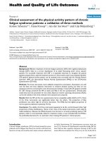
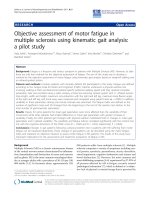
terior of the patient behind the treadmill (F igure 1). The
system was synchronized with a standard video camera
(Digital Ixus 65, Canon Inc., T okyo, Japan). Eleven active
infrared markers were attached to the patient’s body:
bilaterally on the shoes on top of the calcaneus; bilater-
ally on the Achilles tendon at the level of the ankle; bilat-
erally on the posterior aspect of the knee; bilaterally on
the belt at the highest point of the ilium; on the spine at
the level of the sternum; bilaterally centered on Margo
medialis.
After a patient reached comfortable walking speed, three
dimensional marker data and video images were recorded
for one minute at the beginning of the test (t
1
)andforone
minute when patients stated that they could no longer
walk and were completely exhausted (t
2
). Following this
statement, the patient had to walk for one more minute,
and data for this minute was recorded (t
2
). The current
physical exhaustion at each of the recordings was charted
on the physical exhaustion scale (see a bove) before and
Figure 1 Test set-up. Patients wore safety harness during all tests to prevent injury by potential falls. The infrared camera system and the video
camera were positioned posterior of the patient behind the treadmill. The acquisition computer was operated by one tester and placed behind
the cameras to allow for visual observation of all tests.
Sehle et al. Journal of NeuroEngineering and Rehabilitation 2011, 8:59
/>Page 3 of 13
after physical exertion. Processing time of gait data was
one hour per subject.
Pathological diagnostic criteria (gait abnormalities)
Step length, step width, step height, maximum circum-
duction with the right and left leg, maximum knee flex-
ion angl e of the right and left leg , and medi o-later al sway
of the upper body were calculated for each step using the
three-dimensional coordinates of the infrared markers.
Mean and standard deviations for each parameter and
time interval were calculated for each patient and used
for further analysis. Significant changes in the mean and
standard deviations of these parameters were used as
probable indicators of fatigue. It was assumed that a
patient’s gait pattern at the rested state corresponds to
their “normal” gait pattern. Therefore, the changes in gait
parameters after physical exertion can be regarded as
pathological, although the direction of changes was irre-
levant. The fatigue index comprised components of mean
gait changes and changes in variability and was defined as
index
fatigue
=
1
2
·
index
mean
+ index
variability
=
1
2
·
N
significant mean changes
N
gait parameters
+
N
sigificant SD changes
N
gait parameters
where N
significant_mean_changes
was the number of para-
meters that had a significant mean change from t
1
to t
2
,
N
significant_SD_changes
was the number of parameters that
had a significant SD change from t
1
to t
2
and N
gait_para-
meters
was the number of gait parameters. Step length, step
width, step height are global (non-side-specific) measures,
and differences in these parameters can originate from dif-
ferences in the left leg, right leg or both legs. Hence, these
globa l gait parameters were weighted with a factor 2 and
the side-specific parameters right and left circumduction
and right and left knee flexion angle were weighted with a
factor 1. Possible values for the fatigue, mean index and
variability indices are between 0 and 1, respectively.
Statistical Analysis
All statistical tests were performed using S tatFree Ver-
sion 4.4.2.2 (Viet enDynamics) and Stata Version 10.1
(StatCorp LP, College Station, Texas, USA). Descriptive
analyses of numerical parameters included mean, median,
minimum and maximum, and distribution and standard
deviation. All parameters were tested for normal distribu-
tion. Differences in normally distributed parameters
between t
1
and t
2
were detected using Student’st-tests
for paired samples. Differences in no n-normally distribu-
ted parameters between t
1
and t
2
were detected using
Wilcoxon signed-rank tests. Differences in parameter
variability between t
1
and t
2
were detected using the stan-
dard deviation test (SD test). Bonferroni adjustment was
applied to account for multiple comparisons, and the sig-
nificance level for all statistical tests was set a priori to a
= 0.005. Bivariate Pearson correlation coefficients were
used to detect significant associations between the com-
ponents o f the fatigue index, the dimensions of FSMC
and the distance walked during the physical exertion test
(a = 0.05).
Results
The fatigue index for t his patient group ranged from
0.33-0.92, the mean index ra nged from 0.00-0.92 and
the variability index ranged from 0.25-0.92 (Table 1).
Clearly, for some patients the mean gait parameters
were more affected than the variability of their move-
ments while other patients had smaller differences in
mean gait parameters with greater changes in variability.
Finally, for other patients gait changes with physical
exertion manifested in both changes in mean gait para-
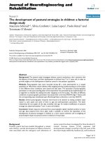
meters and in altered variability. For instance, one
patient (patient 9) showed relatively regular patterns of
circumduction with their right leg at the beginning of
the phy sical exertion test with a shift in circumduction
to smaller values and more variable wave patterns at the
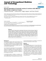
end of the physical exertion test (Figure 2). Another
patient (patient 5) showed similar mean values for their
knee flexion angles during one minute but had clear
irregularities in their pattern manifesting as more irregu-
lar knee extension movements and additional irregulari-
ties close to full knee extension (Figure 3).
The gait parameters that showed significant differences
with fatigue for most patients were step length, width and
height (Figure 4) followed by knee flexion angle (Figure 5)
and circumduction (Figure 6). The gait parameter that
Table 1 Fatigue index with sub-indices mean and
variability for all patients
Patient ID Index
mean
Index
variability
Index
fatigue
1 0.00 0.67 0.33
2 0.83 0.67 0.75
3 0.75 0.58 0.67
4 0.42 0.42 0.42
5 0.58 0.58 0.58
6 0.42 0.25 0.33
7 0.67 0.42 0.54
8 0.58 0.67 0.63
9 0.58 0.50 0.54
10 0.67 0.50 0.58
11 0.75 0.33 0.54
12 0.92 0.92 0.92
13 0.58 0.33 0.46
14 0.50 0.58 0.54
Mean 0.59 0.53 0.56
SD 0.22 0.17 0.16
Sehle et al. Journal of NeuroEngineering and Rehabilitation 2011, 8:59
/>Page 4 of 13
showed significant differences with fatigue for the least
number of subjects was trunk sway (Figure 7).
The variability index and the fatigue index correlated
significantly with the overall FSMC and with the
motoric dimension of the FSMC, respectively (Table 2).
In contrast, the mean index did not correlate signifi-
cantly with any of the FS MC dimensions. Wh ile the
fatigue index correlate d with both the mean index and
Figure 2 Circumduction of the right leg in a 15-sec interval during the first (top graph) and last (bottom graph) minute of the
physical exertion test for patient 9.
Sehle et al. Journal of NeuroEngineering and Rehabilitation 2011, 8:59
/>Page 5 of 13
the variability index, the mean index and t he variability
index did not correlate significantly. None of the com-
ponents of the fatigue index correlated with the dis-
tance walked during the physical exertion test. All
dimensions of the FSMC correlated significantly with
each other. The mean overall, c ognitive and motoric
FSMC scores were 64.3 ± 19.3, 26.6 ± 12.3 and 37.7 ±
8.3 points, respectively (indicating severe global fatigue,
light cognitive fatigue and severe motor fatigue,
respectively).
Figure 3 Knee flexion angle in a 15-sec interval during the first (top graph) and last (bottom graph) minute of the physical exertion
test for patient 5.A–additional variability during knee extension; B–additional variability close to full knee extension.
Sehle et al. Journal of NeuroEngineering and Rehabilitation 2011, 8:59
/>Page 6 of 13
Overall, seven of the eight gait parameters changed
significantly between t
1
and t
2
for this group of patients
(p < 0.001; Table 3). When fatigue d, patients walked on
average with longer step lengths, smaller circumduction
with their right leg, greater circumduction with their left
leg, flexed their knees more and swayed their upper
Figure 4 Mean (1SD) step length, width and height for each patient during one minute of treadmill walking at the beginning and at
the end of the physical exertion test, respectively. * indicates significant differences between mean values at the beginning and end of the
test; † indicates significant differences between the standard deviations at the beginning and end of the test (P < 0.005).
Sehle et al. Journal of NeuroEngineering and Rehabilitation 2011, 8:59
/>Page 7 of 13
bodies more than prior to exertion. The SD-tests
revealed that the variability of steps between t
1
and t
2
increased for seven gait parameters with increasing
exhaustion of the patients (p < 0.003; Table 1). Follow-
ing exertion, the variability of the significant gait para-
meters increased by 9-121% compared to prior to
exertion. On average, the mean index and the variability
index showed comparable values (Table 1).
Discussion
According to guidelines proposed by the MS Council
for Clinical Practice Guidelines in 1998, fatigue is
Figure 5 Mean (1SD) peak knee flexion angle for the right and left leg for each patient during one minute of treadmill walking at
the beginning and at the end of the physical exertion test, respectively. * indicates significant differences between mean values at
the beginning and end of the test; † indicates significant differences between the standard deviations at the beginning and end of the test
(P < 0. 005).
Sehle et al. Journal of NeuroEngineering and Rehabilitation 2011, 8:59
/>Page 8 of 13
defined as „a subjective lack of physical and/or mental
energy that is perceived by the individual or caregivers
to interfere with usual and desired activities” [34].
Within this definition, the term subjective implies that
fatigue is not measurable, may be psychogenic or not
even exist. However, the results of this study clearly
showed–despite pre-determined constant walking
speed–(a) that fatigue in MS patients manifests as
changesingaitpatternsand(b)thatsomechangesin
gait patterns associated with fatigue are consistent
across a group of patients suffering from MS. Hence,
the results of this study provide evidence for the exis-
tence of motor fatigue and suggest that motor fatigue
is a pathophysiological phenomenon.
Figure 6 Mea n ( 1SD) circumd uction for the ri ght and left leg fo r each patient during one minute of treadmill walking at
the beginning and at the end of the physical exertion test, respectively. * indicates significant differences between mean values at
the beginning and end of the test; † indicates significant differences between the standard deviations at the beginning and end of the test
(P < 0. 005).
Sehle et al. Journal of NeuroEngineering and Rehabilitation 2011, 8:59
/>Page 9 of 13
The significant correlations of the fatigue index with
its subcategories mean index and variability index and
the lack of statistical significant correlations between
these two subcategories suggest that both the mean and
variability index described two different phenomena.
Hence, both subcategories are important measures for
motor fatigue in MS. In addition, the significant correla-
tion of the v ariability and fatigue indices wit h the moto-
ric dimension of the FSMC but not with its cognitive
dimension supports the specificity of the fatigue index
for the motoric aspect of fatigue in multiple sclerosis.
Interestingly, the fatigue index correlated negatively with
the FSMC. The FSMC is a self-administered question-
naire, and data obtained with the FSMC may be distorted
by overestimation because of a deficient self-awareness or
underestimation because of depression. Depression is a
well-known confounding fac tor of the FSMC [ 33]. This
discrepancy highlights the urgent need for an objective
marker of fatigue. In additi on, while the F SMC measures
the overall subjective status of a patient, the fatigue index
describes the extent to which a patient’s gait changes with
fatigue. The results of this study suggest that gait patterns
of patients with a p oor overall subjective status will be
affected less by fatigue than those of patients with a better
overall subjective status. It is possible that gait patterns in
patients with a poor overall subjective status are already
compromised at the beginning of the fatigue test. This
result suggests that comparing general gait patterns in MS
patients to those of age-matched healthy subjects may pro-
vide additional objective information about a patient’s
functional status.
Individual results showed cha nges in variability of
movement patterns with fat igue. Greater variability dur-
ing knee extension and close to full extension in one
patient (Figure 2) suggests disrupted motor coordination,
which may be caused by additional activity of the antago-
nists or by insufficient force production by the agonists.
For instance, patients with MS use excessive forces for
daily tasks such as lifting and placing an object [35].
Thus, it is feasible that using excessive muscle force dur-
ing daily activities such as walking may result in addi-
tional fatigue that manifests as increased variability of
movement patterns.
Multiple reasons may be responsible for the changes in
gait patterns observed with fatigue in MS patients.
Patients in this study presented with slightly increased
step length at the end of the physical exertion test,
which–from a clinical perspective–is not typical for
motor fatigue in MS patients. However, t his change
couldbeexplainedbythepresenceofmusclefatigue.
Granacher et al. [3 6] previou sly showed that muscl e fati-
gue generated by isokinetic contraction resulted in
greater stride length in older healthy subjects while
Figure 7 Mean (1SD) medio- lateral trunk sway for each patient
during one minute of treadmill walking at the beginning and at
the end of the physical exertion test, respectively. * indicates
significant differences between mean values at the beginning and
end of the test; † indicates significant differences between the
standard deviations at the beginning and end of the test (P < 0. 005).
Table 2 Cross-correlations (Pearson’s correlation coefficient, P-value) between dimensions of the fatigue index,
dimensions of the Fatigue Scale for Motor and Cognition (FSMC) and distance walked during the physical exertion
test
R
P-value
index
mean
index
variability
index
fatigue
FSMC
overall
FSMC
cognitive
FSMC
motoric
distance walked
index
mean
1
index
variability
0.209
0.473
1
index
fatigue
0.835
< 0.001
0.713
0.004
1
FSMC
overall
-0.209
0.473
-0.560
0.037
-0.465
0.094
1
FSMC
cognitive
-0.092
0.753
-0.473
0.087
-0.331
0.248
0.958
< 0.001
1
FSMC
motoric
-0.350
0.220
-0.602
0.023
-0.592
0.026
0.906
< 0.001
0.747
0.002
1
distance walked 0.366
0.198
0.277
0.338
0.421
0.134
-0.535
0.049
-0.461
0.097
-0.562
0.037
1
Significant correlations (P < 0.05) are shown in bold font.
Sehle et al. Journal of NeuroEngineering and Rehabilitation 2011, 8:59
/>Page 10 of 13
resulting in reduced stride length in younger subjects.
Hence, it is possible that patients with MS suffer from an
ear lier on-set and faster rate of muscle fatigue compared
to healthy control subjects. In addition, MS patients with
greater fatigue have reduced i sometric strength in the
quadriceps muscle [37], w hich may represent compro-
mised capacity to produce sufficiently large muscle
moments about the joints of the lower extremities during
walking.
Interestingly, functional imagi ng studies have reported
increasing evidence that patients with MS experience
greater cerebral activity during performance of motor
and c ognitive tests compared to normal volunteers
[38,39]. Similar observations have been made in patients
after manifestation of their first clinical symptom (clini-
cally isolated syndrome, CIS) [40,41] and in patients
without neurological deficits at the time of the functional
imaging [42]. In addition, patients with a benign course
of MS have shown increased cerebral activity [43] which
may represent some form of compensation. In the late
phase of MS (and with increasing fatigue) this mechan-
ism of compensation is exhausted and compensatory cer-
ebral activity is decreased [44,45]. However, while only
few investigations have investigated a direct relationship
between fatigue and functional i maging [15], sti mulation
studies have found that impaired central motor activation
is involved in MS-fatigue [37]. Other studies [46]
reported an increased central activation during fatiguing
exercis es pro bably reflecting an additional compensatory
central activation. Thus, observed deterioration of gait
parameters in exhausted patients could also reflect a
breakdown of the se compen satory mecha nis ms. In addi-
tion, the fact that patients with a progressive disorder
such as multiple sclerosis show only small improvements
in motor-evoked potential and maximum voluntary con-
traction using functional electrical stimulation [47] sug-
gests compromised plasticity of their motor cortex and
that their impaired motor activation i s presumably asso-
ciated with diminished muscle coordination. Hence, the
gait changes observed following the physical exertion test
in MS patients may stem from the combination of
reduced muscle strength and diminishing coo rdination
reflected in greater variability in movement patterns.
Individual gait changes with fatigue in MS patients are
expected to be asymmetric, that is affecting either the
left or the right side more, b ecause typically dissemi-
nated regions are involved. Indeed, gait compensation
with fatigue in this study population was asymmetric.
However, the sidedness of these effects, that is circum-
duction with their right leg decreased substantially while
circumduction with their left leg increased considerably,
presumably occurred b y chance. It can be assumed that
in a larger study, differences in gait patterns with fatigue
in MS patients would be asymmetric but not side-speci-
fic. In addition, it is possible that different symptomatol-
ogy, such as spastic syndromes or ataxic disturbances,
may be reflected in different changes in gait patterns.
Gait patterns of MS patients differ from those of
healthy persons [31]. Kelleher et al. [31] reported reduced
gait speed, reduced maximum hip and k nee ext ension,
ankle plantarflexion angle and propulsive force for MS
patients compared to healthy persons and that these
Table 3 Results of the t-Test and SD-Test comparing eight gait parameters between t
1
and t
2
(N = 14)
Gait parameters Mean (t
1
) Mean (t
2
) Significance
t-Test
Std. Dev. (t
1
) Std. Dev. (t
2
) Significance
SD-Test
Step width [cm] 15.3 15.5 0.032 3.9 4.4 < 0.001
Step height [cm] 10.1 9.8 n.s. 4.7 5.1 0.002
Step length [cm] 23.6 24.3 0.005 7.4 6.9 n.s.
Circumduction right leg [cm] 6.8 1.6 < 0.001 4.7 10.4 < 0.001
Circumduction left leg [cm] 1.6 5.5 < 0.001 4.9 10.4 < 0.001
Knee flexion angle right leg [°] 12.8 20.5 < 0.001 6.4 11.7 < 0.001
Knee flexion angle left leg [°] 12.7 17.8 < 0.001 7.9 10.8 < 0.001
Sway [cm] 3.4 3.9 < 0.001 5.4 6.0 < 0.001
n.s.–not significant at a = 0.05
Table 4 Differential diagnosis of fatigue or causes of secondary weakness/tiredness in MS
MS related causes for lack of energy Non-MS related causes for lack of energy
Depression Depression
Nocturia Thyroid function
Sleep disturbance Anemia
Spasticity, paresis, uneconomic movement Infection (bladder)
Lack of condition Electrolytes
Side effects of medication (Liuresal, benzodiazepine etc.)
Sehle et al. Journal of NeuroEngineering and Rehabilitation 2011, 8:59
/>Page 11 of 13
changes are more pronounced in more severely affected
patients. Hence, the results of Kelleher et al. and tho se of
this study suggest that fatigue in MS patients appears to
amplify changes in gait patterns already present because
of the disease. While the study sample in this study was
rather small , it is possible that in the general MS popula-
tion the extent of gait changes with fatigue is associated
with the severity of symptoms. For instance, patients
with greater perceived walking limitations have l ess
movement counts from an accelero meter compared to
patients with smaller walking limitations [48]. In addi-
tion, the results of t his study showed that gait patterns
generally become more variable or clumsier with fatigue.
Such changes in gait patterns may generate other pro-
blems such as perception of instability or increased risk
of falling. Thus, the changes in gait patterns observed in
fatigued MS patients likely affect a patient’scompletion
of daily activities.
Therefore, assessing changes in gait patterns using a
physical exert ion test and the fatigue index may be useful
for the objective assessment of functional limitations
ass ociated with fati gue in MS patients and for evaluating
rehabilitation programs aimed at improving patient func-
tion and reducing fatigue. However, the maximum dis-
tance walked during the exertion test should also be
considered in the evaluation of such interventions. In
addition, such an objective t ool may be useful for differ-
entiating between MS related motor fatigue and condi-
tions that are unrelated to MS but may cause lack of
energy (Table 4). Interestingly, only few subjects showed
differences in trunk sway with fatigue, and hence the
inclusion of this parameter in the fatigue index should be
reconsidered. However, it is possible that trunk sway was
restricted by the use of the safety harness in this group of
patients. The inf luence of these factors shoul d be exam-
ined in future studies. While obtaining gait data is more
time-consuming than conventional assessment tools (i.e.
questionnaires [26,27,29,33]) and requires specialized
technical equipment, the information gained in this study
is ob jective–and hence not affected by a patient’scon-
torted self-awareness–and reliable. The latter is the pre-
requisite for obtaining meaningful data on a patient’s
physical status and may be particularly valuable for asses-
sing a patient’s ability to perform occupational tasks and
consequently for determining a patient’s entitlement for
early retirement because of their disease. Comparing gait
patterns in MS patients with and without fatigue and in
healthy volunteers would allow for elucidation of the dif-
ferent dimensio ns, parti cularities and special features in
gait patterns of fatigue in MS patients.
Conclusions
Distinct changes in gait patterns of MS patients were
recorded through two identical tests before and
following physical exertion. These changes in gait p at-
terns can be expressed b y the motor fatigue index and
represent an objective m easure to assess motor fatigue
in MS patients. Assessing gait changes during a physical
exertion test appears to be a useful experimental
method for investigating different dimensions and
pathomechanisms of fatigue in MS. In addition, an
objective tool for assessing motor fatigue in MS is useful
for a more precise diagnosis of motor fatigue in MS, for
the design and evaluation of treatment and rehabilita-
tion programs aimed at improving symptoms and for
evaluating a patient’s ability to perform occupational
tasks.
Author details
1
Division of Sport Science, Universität Konstanz, Konstanz, Germany.
2
School
of Physiotherapy, University of Otago, Dunedin, New Zealand.
3
Kliniken
Schmieder Allensbach, Allensbach, Germany.
4
Department of Politics and
Public Administration, University of Konstanz, Konstanz, Germany.
5
Kliniken
Schmieder Konstanz, Konstanz, Germany.
Authors’ contributions
AS designed the study, collected, processed, analyzed and interpreted the
data and outlined the manuscript. AM participated in data analysis,
interpretation and presentation, and prepared the manuscript. KS and SS
contributed to identifying pathological gait parameters and evaluated
patient’s videos. IB contributed to data processing and analysis. CD
participated in study design, data interpretation and prepared the
manuscript. MV conceived of the study, and participated in its design and
coordination and helped draft the manuscript. All authors read and
approved the final manuscript.
Competing interests
The authors declare that they have no competing interests.
Received: 11 May 2011 Accepted: 26 October 2011
Published: 26 October 2011
References
1. Hein T, Hopfenmuüller W: Estimated prevalence of multiple sklerosis in
Germany [Hochrechnung der Zahl an Multiple Sclerosis erkrankten
Patienten in Deutschland]. Nervenarzt 2000, 71:288-294.
2. Koch-Henriksen N, Sorensen PS: The changing demographic pattern of
multiple sclerosis epidemiology. Lancet Neurology 2010, 9(5):520-532.
3. Stüve O, Oksenberg J: Multiple Sclerosis Overview. In GeneReviews
[Internet]. Edited by: Pagon RA, Bird TC, Dolan CR. Seattle: University of
Washington; 2006:.
4. Krupp LB, Alvarez LA, LaRocca NG, Scheinberg LC: Fatigue in multiple
sclerosis. Archives of Neurology 1988, 45(4) :435-437.
5. Fisk JD, Pontefract A, Ritvo PG, Archibald CJ, Murray TJ: The impact of
fatigue on patients with multiple sclerosis. Canadian Journal of
Neurological Sciences 1994, 21(1):9-14.
6. Krupp LB, Pollina D: Neuroimmune and neuropsychiatric aspects of
chronic fatigue syndrome. Advances in Neuroimmunology 1996,
6(2):155-167.
7. Barnett R: Fatigue. Lancet 2005, 366(9479):21.
8. Kos D, Kerckhofs E, Nagels G, D’Hooghe M B, Ilsbroukx S: Origin of fatigue
in multiple sclerosis: review of the literature. Neurorehabilitation and
Neural Repair 2008, 22(1):91-100.
9. Krupp LB, Alvarez LA, LaRocca NG, Scheinberg LC: Fatigue in multiple
sclerosis. Archives of neurology 1988, 45(4):435-437.
10. McDonald WI, Sears TA: The effects of experimental demyelination on
conduction in the central nervous system. Brain 1970, 93(3):583-598.
11. Vagg R, Mogyoros I, Kiernan MC, Burke D: Activity-dependent
hyperpolarization of human motor axons produced by natural activity.
Journal of Physiology 1998, 507(Pt 3):919-925.
Sehle et al. Journal of NeuroEngineering and Rehabilitation 2011, 8:59
/>Page 12 of 13
12. Bolanos JP, Almeida A, Stewart V, Peuchen S, Land JM, Clark JB, Heales SJ:
Nitric oxide-mediated mitochondrial damage in the brain: mechanisms
and implications for neurodegenerative diseases. Journal of
Neurochemistry 1997, 68(6):2227-2240.
13. Redford EJ, Kapoor R, Smith KJ: Nitric oxide donors reversibly block
axonal conduction: demyelinated axons are especially susceptible. Brain
1997, 120(Pt 12):2149-2157.
14. Leocani L, Colombo B, Magnani G, Martinelli-Boneschi F, Cursi M, Rossi P,
Martinelli V, Comi G: Fatigue in multiple sclerosis is associated with
abnormal cortical activation to voluntary movement–EEG evidence.
Neuroimage 2001, 13(6 Pt 1):1186-1192.
15. Filippi M, Rocca MA, Colombo B, Falini A, Codella M, Scotti G, Comi G:
Functional magnetic resonance imaging correlates of fatigue in multiple
sclerosis. Neuroimage 2002, 15(3):559-567.
16. Tedeschi G, Dinacci D, Lavorgna L, Prinster A, Savettieri G, Quattrone A,
Livrea P, Messina C, Reggio A, Servillo G, et al: Correlation between fatigue
and brain atrophy and lesion load in multiple sclerosis patients
independent of disability. Journal of Neurological Sciences 2007, 263(1-
2):15-19.
17. Rasova K, Havrdova E, Brandejsky P, Zalisova M, Foubikova B, Martinkova P:
Comparison of the influence of different rehabilitation programmes on
clinical, spirometric and spiroergometric parameters in patients with
multiple sclerosis. Multiple Sclerosis 2006, 12(2):227-234.
18. Romberg A, Virtanen A, Ruutiainen J, Aunola S, Karppi SL, Vaara M,
Surakka J, Pohjolainen T, Seppanen A: Effects of a 6-month exercise
program on patients with multiple sclerosis: a randomized study.
Neurology 2004, 63(11):2034-2038.
19. Surakka J, Romberg A, Ruutiainen J, Aunola S, Virtanen A, Karppi SL,
Maentaka K: Effects of aerobic and strength exercise on motor fatigue in
men and women with multiple sclerosis: a randomized controlled trial.
Clinical Rehabilitation 2004, 18(7):737-746.
20. Beenakker EA, Oparina TI, Hartgring A, Teelken A, Arutjunyan AV, De
Keyser J: Cooling garment treatment in MS: clinical improvement and
decrease in leukocyte NO production. Neurology 2001, 57(5):892-894.
21. Schwid SR, Petrie MD, Murray R, Leitch J, Bowen J, Alquist A, Pelligrino R,
Roberts A, Harper-Bennie J, Milan MD, et al: A randomized controlled
study of the acute and chronic effects of cooling therapy for MS.
Neurology 2003, 60(12):1955-1960.
22. Goodman AD, Brown TR, Krupp LB, Schapiro RT, Schwid SR, Cohen R,
Marinucci LN, Blight AR: Sustained-release oral fampridine in multiple
sclerosis: a randomised, double-blind, controlled trial. Lancet 2009,
373(9665):732-738.
23. Tomassini V, Pozzilli C, Onesti E, Pasqualetti P, Marinelli F, Pisani A, Fieschi C:
Comparison of the effects of acetyl L-carnitine and amantadine for the
treatment of fatigue in multiple sclerosis: results of a pilot, randomised,
double-blind, crossover trial. Journal of Neurological Sciences 2004, 218(1-
2):103-108.
24. Krupp LB, Coyle PK, Doscher C, Miller A, Cross AH, Jandorf L, Halper J,
Johnson B, Morgante L, Grimson R: Fatigue therapy in multiple sclerosis:
results of a double-blind, randomized, parallel trial of amantadine,
pemoline, and placebo. Neurology 1995, 45(11):1956-1961.
25. Krupp LB, LaRocca NG, Muir-Nash J, Steinberg AD: The fatigue severity
scale. Application to patients with multiple sclerosis and systemic lupus
erythematosus. Archives of Neurology 1989, 46(10):1121-1123.
26. Mathiowetz V: Test-retest reliability and convergent validity of the
Fatigue Impact Scale for persons with multiple sclerosis. American
Journal of Occupational Therapy 2003, 57(4):389-395.
27. Iriarte J, Katsamakis G, de Castro P: The Fatigue Descriptive Scale (FDS): a
useful tool to evaluate fatigue in multiple sclerosis. Multiple Sclerosis 1999,
5(1):10-16.
28. Kos D, Nagels G, D ’Hooghe MB, Duportail M, Kerckhofs E: A rapid
screening tool for fatigue impact in multiple sclerosis. BMC Neurology
2006, 6:27.
29. Flensner G, Ek AC, Soderhamn O: Reliability and validity of the Swedish
version of the Fatigue Impact Scale (FIS). Scandinavian Journal of
Occupational Therapy 2005, 12(4):170-180.
30. Kim E, Lovera J, Schaben L, Melara J, Bourdette D, Whitham R: Novel
method for measurement of fatigue in multiple sclerosis: Real-Time
Digital Fatigue Score. Journal of Rehabilitation Research and Development
2010, 47(5):477-484.
31. Kelleher KJ, Spence W, Solomonidis S, Apatsidis D: The characterisation of
gait patterns of people with multiple sclerosis. Disability and
Rehabilitation 2010, 32(15):1242-1250.
32. Dettmers C, Sulzmann M, Ruchay-Plossl A, Gutler R, Vieten M: Endurance
exercise improves walking distance in MS patients with fatigue. Acta
Neurologica Scandinavica 2009, 120(4):251-257.
33. Penner IK, Raselli C, Stocklin M, Opwis K, Kappos L, Calabrese P: The
Fatigue Scale for Motor and Cognitive Functions (FSMC): validation of a
new instrument to assess multiple sclerosis-related fatigue. Multiple
Sclerosis 2009, 15(12):1509-1517.
34. Guidelines MSCfCP: Fatigue and Multiple Sclerosis: evidence based
management strategies for fatigue in multiple sclerosis. Multiple Sclerosis
Council for Clinical Practice Guidelines; 1998.
35. Iyengar V, Santos MJ, Ko M, Aruin AS: Grip force control in individuals
with multiple sclerosis. Neurorehabilitation and neural repair 2009,
23(8):855-861.
36. Granacher U, Wolf I, Wehrle A, Bridenbaugh S, Kressig RW: Effects of
muscle fatigue on gait characteristics under single and dual-task
conditions in young and older adults. J Neuroeng Rehabil 2010, 7:56.
37. Andreasen AK, Jakobsen J, Petersen T, Andersen H: Fatigued patients with
multiple sclerosis have impaired central muscle activation. Multiple
Sclerosis 2009,
15(7):818-827.
38. Buckle GJ: Functional magnetic resonance imaging and multiple
sclerosis: the evidence for neuronal plasticity. Journal of Neuroimaging
2005, 15(4 Suppl):82S-93S.
39. Mainero C, Pantano P, Caramia F, Pozzilli C: Brain reorganization during
attention and memory tasks in multiple sclerosis: insights from
functional MRI studies. J Neurol Sci 2006, 245(1-2):93-98.
40. Filippi M, Rocca MA, Mezzapesa DM, Ghezzi A, Falini A, Martinelli V, Scotti G,
Comi G: Simple and complex movement-associated functional MRI
changes in patients at presentation with clinically isolated syndromes
suggestive of multiple sclerosis. Hum Brain Mapp 2004, 21(2):108-117.
41. Pantano P, Iannetti GD, Caramia F, Mainero C, Di Legge S, Bozzao L,
Pozzilli C, Lenzi GL: Cortical motor reorganization after a single clinical
attack of multiple sclerosis. Brain 2002, 125(Pt 7):1607-1615.
42. Filippi M, Rocca MA, Falini A, Caputo D, Ghezzi A, Colombo B, Scotti G,
Comi G: Correlations between structural CNS damage and functional
MRI changes in primary progressive MS. Neuroimage 2002, 15(3):537-546.
43. Rocca MA, Valsasina P, Ceccarelli A, Absinta M, Ghezzi A, Riccitelli G,
Pagani E, Falini A, Comi G, Scotti G, et al: Structural and functional MRI
correlates of Stroop control in benign MS. Hum Brain Mapp 2009,
30(1):276-290.
44. Penner IK, Kappos L, Rausch M, Opwis K, Radu EW: Therapy-induced
plasticity of cognitive functions in MS patients: insights from fMRI. J
Physiol Paris 2006, 99(4-6):455-462.
45. Penner IK, Opwis K, Kappos L: Relation between functional brain imaging,
cognitive impairment and cognitive rehabilitation in patients with
multiple sclerosis. J Neurol 2007, 254(Suppl 2):II53-57.
46. Thickbroom GW, Sacco P, Faulkner DL, Kermode AG, Mastaglia FL:
Enhanced corticomotor excitability with dynamic fatiguing exercise of
the lower limb in multiple sclerosis. Journal of neurology 2008,
255(7):1001-1005.
47. Everaert DG, Thompson AK, Chong SL, Stein RB: Does functional electrical
stimulation for foot drop strengthen corticospinal connections?
Neurorehabilitation and neural repair 2010, 24(2) :168-177.
48. Motl RW, Dlugonski D, Suh Y, Weikert M, Fernhall B, Goldman M:
Accelerometry and its association with objective markers of walking
limitations in ambulatory adults with multiple sclerosis. Arch Phys Med
Rehabil 2010, 91(12):1942-1947.
doi:10.1186/1743-0003-8-59
Cite this article as: Sehle et al.: Objective assessment of motor fatigue
in multiple sclerosis using kinematic gait analysis: a pilot study. Journal
of NeuroEngineering and Rehabilitation 2011 8:59.
Sehle et al. Journal of NeuroEngineering and Rehabilitation 2011, 8:59
/>Page 13 of 13