báo cáo hóa học:" Emerging applications of fluorescence spectroscopy in medical microbiology field" potx
Bạn đang xem bản rút gọn của tài liệu. Xem và tải ngay bản đầy đủ của tài liệu tại đây (354.01 KB, 6 trang )
BioMed Central
Page 1 of 6
(page number not for citation purposes)
Journal of Translational Medicine
Open Access
Review
Emerging applications of fluorescence spectroscopy in medical
microbiology field
Aamir Shahzad*
1
, Gottfried Köhler
1
, Martin Knapp
2
, Erwin Gaubitzer
2
,
Martin Puchinger
1
and Michael Edetsberger
2
Address:
1
Max F. Perutz Laboratories, Department of Structural Biology and Biomolecular Chemistry, University of Vienna, Vienna, Austria and
2
OnkoTec GmbH. Waidhofen/Thaya, Vienna, Austria
Email: Aamir Shahzad* - ; Gottfried Köhler - ;
Martin Knapp - ; Erwin Gaubitzer - ; Martin Puchinger - ;
Michael Edetsberger -
* Corresponding author
Abstract
There are many diagnostic techniques and methods available for diagnosis of medically important
microorganisms like bacteria, viruses, fungi and parasites. But, almost all these techniques and
methods have some limitations or inconvenience. Most of these techniques are laborious, time
consuming and with chances of false positive or false negative results. It warrants the need of a
diagnostic technique which can overcome these limitations and problems. At present, there is
emerging trend to use Fluorescence spectroscopy as a diagnostic as well as research tool in many
fields of medical sciences. Here, we will critically discuss research studies which propose that
Fluorescence spectroscopy may be an excellent diagnostic as well as excellent research tool in
medical microbiology field with high sensitivity and specificity.
Discussion
Limitations/Drawback of current diagnostic Tools
Infectious diseases are caused by microorganisms such as
bacteria, viruses, fungi and parasites. Infectious diseases
are major killer around the world especially in developing
countries. Infectious diseases were responsible for 14.7
million deaths around the world in 2002 [1] major por-
tion of health care budget are allocated for diagnosis and
treatment of infectious diseases. There are many diagnos-
tic methods and techniques available for microorganisms
associated diseases. These include morphological exami-
nation by microscopy, culture examination, biochemical
tests, and histopathology approach. There are modern
sophisticated methods are also available like PCR, ELISA,
molecular DNA analysis. But, there are many limitations
and drawbacks associated with these diagnostic tech-
niques. These techniques are time consuming, laborious
and require many reagents [2]. Also, some techniques lack
high sensitivity and specificity which warrants the need
for a new diagnostic technique with high sensitivity and
specificity.
Current traditional diagnostic techniques and methods
for diagnosis of microorganisms like bacteria take nor-
mally at least one day. Also, Antibiotic sensitivity testing
is also required by physicians to choose specific antibiotic
for treating infection. This sensitivity testing usually takes
one more day. Bacteria are cultured for at least one day
and then diagnosis is made. This causes delay in start of
specific treatment. As a result physicians usually prescribe
Published: 26 November 2009
Journal of Translational Medicine 2009, 7:99 doi:10.1186/1479-5876-7-99
Received: 16 September 2009
Accepted: 26 November 2009
This article is available from: />© 2009 Shahzad et al; licensee BioMed Central Ltd.
This is an Open Access article distributed under the terms of the Creative Commons Attribution License ( />),
which permits unrestricted use, distribution, and reproduction in any medium, provided the original work is properly cited.
Journal of Translational Medicine 2009, 7:99 />Page 2 of 6
(page number not for citation purposes)
broad spectrum antibiotics which are unnecessary and
very expensive for patients. Also, microorganisms have
unique mechanisms to develop resistance for antimicro-
bial treatment. It justify for fast diagnosis of microorgan-
isms and start of specific treatment as soon as possible.
Fluorescence spectroscopy
Fluorescence spectroscopy seems to be promising diag-
nostic technique with fast and rapid diagnosis ability.
Studies indicate high sensitivity and specificity rate which
makes Fluorescence spectroscopy an ideal diagnostic tool
for medical microbiology field. But, there is need for fur-
ther studies and clinical trials to validate this new diagnos-
tic technique.
At present, Fluorescence spectroscopy is being applied in
medical microbiology field for various purposes. There
are many studies which indicate that Fluorescence spec-
troscopy is promising diagnostic technique with high sen-
sitivity and specificity for microorganisms associated
diseases diagnosis with the help of spectroscopic finger-
prints. Also, Fluorescence spectroscopy and Fluorescence
correlation spectroscopy (FCS) may be applied to under-
stand various pathophysiological steps of various micro-
organisms [3,4].
Fluorescence spectroscopy is a type of electromagnetic
spectroscopy which analyzes fluorescence from a sample.
The sample is excited by using a beam of light which
results in emission of light of a lower energy resulting in
an emission spectrum which is used to interpret results
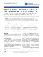
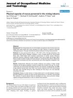
[5]. Fluorescence correlation spectroscopy (FCS), a tech-
nique basically used for spatial and temporal analysis of
molecular interactions of extremely low concentrated bio-
molecules in solution. (Figure 1) FCS measures both the
average number of molecules in the detection volume and
the diffusion time of the molecules through the open
detection volume [6]. As the diffusion speed is directly
correlated with the molecular mass and shape of the fluo-
rescent molecule, it is possible to study the complex for-
mation between a small fluorescent labeled and a big
unlabelled molecule [7].
Fluorescence correlation spectroscopy (FCS)
Fluorescence correlation spectroscopy (FCS) use the basic
principle that a fluorescing molecule shows a specific free
diffusion velocity which is directly correlated with its size.
So, bigger the molecule, slower it will diffuse through a
given spherical volume. This basic phenomenon of mole-
cules is used in FCS to study protein-protein interactions,
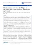
attachment and many more. (Figure 1) Fluorescence Cor-
relation Spectroscopy (FCS) uses statistical deviations of
the fluctuations in fluorescence in order to study dynamic
molecular events, such as diffusion or conformational
fluctuations of bio molecules or artificial particles. (Figure
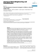
2) Mainly, the auto correlation function (ACF) is used to
extract the number and diffusion coefficient of fluorescent
particles diffusing through the focus volume. (Figure 3)
These all properties of FCS make it an excellent diagnostic
and research tool for many medically important diseases.
Various properties of FCS make it an ideal tool for under-
standing various pathophysiological processes involved
with microbial infectious diseases. An excellent advantage
of FCS is that it requires very low concentrations and
amounts of samples, as compared to routinely used tech-
niques which require high concentration of diagnostic
sample.
Tryptophan which is fluorophore in UV is present in both
viruses and host bacterial protein. Indole group of tryp-
tophan residues are major source of UV absorbance and
emission in proteins. Tryptophan in pure water emits at
353 nm [8]. Tryptophan emission is strongly associated
with its local environment. Many phenomena such as
protein-protein association result in spectral shifts in tryp-
tophan emission [8]. It is proposed that emission and
excitation spectral differences may be due to presence of
different environments of tryptophan residues in specific
proteins of microorganism's cells [9].
Diagnostic Applications
At present, many studies reported successful application
of Fluorescence spectroscopy as a diagnostic tool for dif-
ferent bacteria at genus, species and group level by use of
spectral fingerprints [10-12]. Spectral studies for black
pigmented bacilli which are a group of oral bacteria
showed significant difference in spectral signatures of
each bacterium [12]. Fluorescent profiles of Bacteria
which are responsible for otitis Media in children: S. pneu-
moniae, S. aureus, M. catarrhalis, and H. influenzae have
been studied. These studies proved that each bacterium
produce a different specific Fluorescence profile. The data
indicate that it may be an excellent non invasive fluores-
cence based diagnostic technique for otitis media [12]. In
another study; three different bacterial species (Escherichia
coli, EC, Enterococcus faecalis, EF and Staphylococcus aureus,
SA) were rapidly identified by autofluorescence spectrum
differences coupled with Principal Components Analysis
(PCA) technique. These studies proposed that bacteria can
be rapidly diagnosed with sensitivity and specificity
higher then 90% [13].
Bacterial taxonomy
Fluorescence spectroscopy was utilized for pseudomonad
taxonomic purpose at species and genus level [14]. Results
proved that Fluorescence spectroscopy may be an excel-
lent tool in polyphasic approach to pseudomonad taxon-
omy. This approach provide more information as
compared to rRNA and DNA bacterial homology group-
ing as they provide more information about strain related-
Journal of Translational Medicine 2009, 7:99 />Page 3 of 6
(page number not for citation purposes)
ness and good differentiation between strains which are
difficult to differentiate on PCR and API 20NE identifica-
tion methods [14].
Fungal applications
Fungal infections are common in many diseases like dia-
betes, many types of cancers, endocrinopathies, and
patients on prolonged antibiotics or immunosuppressive
drugs. Diagnosis of fungal infection is made either by
morphological examination of fungi or by biochemical
and molecular biology techniques [15]. These techniques
may not differentiate between different types of yeast.
There are studies which have utilized spectroscopic finger-
prints method for rapid diagnosis of different fungi such
as yeast, Microsporum gypseum, Microsporum canis, Tricho-
phyton schoenleinii, Trichophyton rubrum, Epidermophyton
floccosum and Fusarium solani [9,16].
Viral Applications
Studies indicate that Fluorescence spectroscopy may be a
novel diagnostic tool to detect viruses. Also viral infec-
tions of cells can be monitored by Fluorescence spectros-
copy [3]. These studies were carried out on viruses from
cystovirus family and pseudomonad host cells. Tryp-
tophan which is fluorophore in UV is present in both
viruses and host bacterial protein. Within proteins, tryp-
tophan structural environment is not same and this struc-
tural difference is responsible for specific spectroscopic
signatures [3]. This property can be used to monitor viral
attachment process and to study the release of progeny
FCS instrumentation for use in living cellsFigure 1
FCS instrumentation for use in living cells. On the left hand, there is schematic FCS setup including laser excitation fil-
ters, emission filters, confocal pin hole and single photon detector (APD). To use this setup, the laser beam is positioned inside
the cell (A). The exact position of the focus is established by performing a Z scan (B). The pin hole cuts out a defined focal ele-
ment from the laser focus (C). The Fluorescence signals from fluorescenct entities moving through the focal element are
recorded by the single photon detector, resulting in a Fluorescence trace (D). (Source: Shahzad A, Edetsberger M, Köhler G.
Fluorescence Spectroscopy: An emerging excellent diagnostic tool in Medical Sciences. Applied Spectroscopy Reviews J (In
press).
Journal of Translational Medicine 2009, 7:99 />Page 4 of 6
(page number not for citation purposes)
virus particles by analysis of tryptophan emission spectra
during infection process.
In author's Lab, Fluorescence correlation spectroscopy
(FCS) has been applied successfully to understand human
rhino virus-receptor interaction [17]. These experiments
provide informative data for understanding virus-receptor
interactions. Fluorescence correlation spectroscopy (FCS)
studies revealed different binding modes for an icosahe-
dral virus along the five-fold symmetry axis. We proposed
that Fluorescence correlation spectroscopy (FCS) may be
a valuable technique to study various receptor binding
affinities of viruses.
Future Research
Spectroscopic technique may be automatized which can
then process many diagnostic samples at the same time.
Also, fiber optic systems may be integrated with this spec-
troscopic technique to diagnose microorganisms in vivo.
By this modification, infections in many body parts can be
detected with ease. Further research is required to estab-
lish flexible and portable spectroscopic devices which can
be integrated in daily medical practice.
There is need for reference libraries for spectral signatures
of individual microorganism. This will be very helpful for
comparison with spectral signatures from an unknown
microorganism sample. But, there are many questions
which remain to be answered like if biological sample
contains more than one microorganism, then how it will
affect the spectral signature appearance and how to inter-
pret these spectral for making definite diagnosis. Also,
microorganisms like bacteria have many chemicals which
are same like in human cells and in extracellular space,
thus body fluids samples may contain same chemicals as
found in microorganisms. As a result, it may interfere with
spectroscopic spectral analysis and may be a hurdle to
reach on definite diagnosis. This justifies the need for
studies which can enable to make distinction between
microorganism and human cells. Also, future studies
should be directed to determine the specific spectral
regions which will be suitable for identification of specific
microorganisms. It will help to design invasive and non
invasive techniques for microorganism's diagnosis inside
the body cavities by use of fiber optic devices.
Conclusion
At present, nearly all the diagnostic techniques and meth-
ods used for microorganism's diagnosis are not perfect
and have some limitations. There is great need for a diag-
nostic technique which can overcome limitations and
drawbacks of commonly used microbiological techniques
and methods. Studies indicate that Fluorescence spectros-
copy have great potential to become an excellent and per-
fect diagnostic technique for microorganisms. In many
research studies, fluorescence emission spectra derived
from autofluorescence property of many medically
important bacteria make it possible to distinguish
between various bacterial species and also enable to clas-
sify the bacteria into genus, species and groups. Recent
research studies indicate that virus particles can be moni-
tored inside cells and various processes of viral infections
can be detected by means of Fluorescence spectroscopy.
Difference between fungal microorganisms like yeast can
be made easily by use of spectroscopic fingerprinting.
Future clinical trials on large scale should be performed to
validate Fluorescence spectroscopy as a diagnostic tool for
microorganisms. Flexible and portable spectroscopic
devices should be design which can be integrated in rou-
tine medical practice.
Fluorescence fluctuations measured by FCSFigure 2
Fluorescence fluctuations measured by FCS. (Source: Shahzad A, Edetsberger M, Köhler G. Fluorescence Spectroscopy:
An emerging excellent diagnostic tool in Medical Sciences. Applied Spectroscopy Reviews J (In press).
Journal of Translational Medicine 2009, 7:99 />Page 5 of 6
(page number not for citation purposes)
Overall, emerging research studies and data points that
Fluorescence spectroscopy is a potential diagnostic tool
for microorganisms. Based on these data and research
studies, we expect that in near future, Fluorescence spec-
troscopy will be available as a routine diagnostic tool for
microorganisms in daily medical practice. Ultimately,
Patients will benefit from its low cost, fast processing and
high sensitivity properties. In the long term, spectroscopy
fingerprinting may become an excellent tool to classify
microorganisms into their respective groups, genus and
species level. This will be very promising system with high
sensitivity and high specificity for microorganisms classi-
fication.
Competing interests
The authors declare that they have no competing interests.
Authors' contributions
All authors participated in the preparation of the manu-
script, and read and approved the final manuscript.
Acknowledgements
The authors acknowledge "The Vienna Science and Technology Fund"
(WWTF), Vienna, Austria, for the generous funding of Mathematic call
und 2007 project. The authors also acknowledge "OnkoTec GmbH.
Waidhofen/Thaya", Vienna. Austria, for their kind technical support.
References
1. The World Health Report - changing history. 2004.
2. Barnett JA, Payne RW, Yarrow D: 2nd edition. Yeasts: Characteristics
and Identification, Cambridge University Press, Cambridge; 1990.
3. Alexandra , et al.: Virus Particles Monitored by Fluorescence
Spectroscopy: A Potential Detection Assay for Macromo-
lecular Assembly. Photochemistry and Photobiology 2004,
80(1):41-46.
4. Alexandra , et al.: Virus Particles and Receptor Interaction
Monitored by Fluorescence Spectroscopy. Photochemistry and
Photobiology 2005, 1(4):879-883.
5. Ramanujam N, Mitchell MF, Mahadevan A, Thomsen S, Silva E, Rich-
ards-Kortum R: luorescence spectroscopy: A diagnostic tool
Autocorrelation function generated from fluorescence fluctuations (Fig. 2) This function is used to determine the average diffu-sion time of the particles inside the FCS focus during measurement timeFigure 3
Autocorrelation function generated from fluorescence fluctuations (Fig. 2) This function is used to determine
the average diffusion time of the particles inside the FCS focus during measurement time. (Source: Shahzad A,
Edetsberger M, Köhler G. Fluorescence Spectroscopy: An emerging excellent diagnostic tool in Medical Sciences. Applied
Spectroscopy Reviews J (In press).
Publish with Bio Med Central and every
scientist can read your work free of charge
"BioMed Central will be the most significant development for
disseminating the results of biomedical research in our lifetime."
Sir Paul Nurse, Cancer Research UK
Your research papers will be:
available free of charge to the entire biomedical community
peer reviewed and published immediately upon acceptance
cited in PubMed and archived on PubMed Central
yours — you keep the copyright
Submit your manuscript here:
/>BioMedcentral
Journal of Translational Medicine 2009, 7:99 />Page 6 of 6
(page number not for citation purposes)
for cervical intraepithelial neoplasia. Gynecol Oncol 1994,
52:31-8.
6. Pack CG, Nishimura G, Tamura M, Aoki K, Taguchi H, Yoshida M,
Kinjo M: Analysis ofinteraction between chaperonin GroEL
and its substrate using fluorescence correlation spectros-
copy. Cytometry 1999, 6:247-253.
7. Klinger J, Friedrich T: Site-specific interaction of thrombin and
inhibitors observed by fluorescence correlation spectros-
copy. Biophys J 1997, 3:2195-2200.
8. Lakowicz JR: Principles of Fluorescence Spectroscopy 2nd edition. Kluwer
Academic/Plenum Publishers, New York; 1999.
9. Bhatta H, Goldys EM, Learmonth RP: Use of fluorescence spec-
troscopy to differentiate yeast and bacterial cells. Appl Micro-
biol Biotechnol 2006, 1:121-126.
10. Leblanc Ludovic, Dufour Eric: Monitoring the identity of bacteria
using their intrinsic fluorescence. FEMS Microbiology Letters 2002,
11:147-153.
11. Sarkissian Ani: Fiber optic fluorescence microprobe for endo-
dontic diagnosis. J Dent Educ 2005, 9(6):633-8.
12. Sorrell Matthew J, et al.: Bacteria identification of otitis media
with fluorescence spectroscopy. Lasers in Surgery and Medicine
2005, 2:155-163.
13. Giana HE, et al.: Rapid Identification of Bacterial Species by
Fluorescence Spectroscopy and Classification Through Prin-
cipal Components Analysis. Journal of Fluorescence 2003,
13(5):489-493.
14. Tourkya , et al.: Fluorescence Spectroscopy as a Promising
Tool for a Polyphasic Approach to Pseudomonad Taxon-
omy. Current Microbiology 2009, 58(8):39-46.
15. Rippon JW: Medical Mycology: The Pathogenic Fungi and the Pathogenic
Actinomycetes
3rd edition. W.B. Saunders Co.; 1988:808.
16. Rativa Diego, et al.: Perspectives on in vitro fungal diagnosis
with UV light. Revista Brasileira de Engenharia Biomédica 2007,
23:25-30.
17. Köhler , et al.: Attachment of VLDL Receptors to an Icosahe-
dral Virus along the 5-fold Symmetry Axis: Multiple Binding
Modes Evidenced by Fluorescence Correlation Spectros-
copy. Biochemistry 2007, 46:6331-6339.